Haptics-based virtual reality periodontal training simulator
Upload
nottinghamCategory
view
8download
0
Regulation of Periodontal Ligament CellBehavior by Cyclic Mechanical Loadingand Substrate NanotextureNa Yu,* Ljupcho Prodanov,* Joost te Riet,† Fang Yang,* X. Frank Walboomers,*and John A. Jansen*
Background: Periodontal ligament (PDL) cells play animportant role in regulating osseous remodeling and liga-ment formation. Mechanical loading and the specific cellu-lar environment are involved in these processes, regulatingcell behavior. However, most in vitro experimental setupsinvestigate mechanical loading or substrate texture sepa-rately and thus do not fully represent the PDL microenvi-ronment. Therefore, the authors investigated the influenceof combined mechano-topographical stimuli on PDL cellmorphology, proliferation, and osteogenic and ligament dif-ferentiation.
Methods: Human PDL cells were subjected to nanometricsubstrate patterning and cyclic tensile stress for 2 days. Cellmorphology was assessed by fluorescent staining. Further,DNA content and messenger RNA expression of osteogenic(Runx2, OCN) and ligament-related (scleraxis transcriptionfactor (SCXA), ELN) genes were determined.
Results: PDL cells adapted to the topography of nano-metric groove patterns, aligning parallel with the texture.When subjected to mechanical stress, cells lost their initialorientation to the nanopattern. When subjected to dual stim-uli, total DNA amounts were increased at 3 days of culture.Moreover, a significant synergistic effect on upregulation ofRunx2 was observed in the combined group. For ligament-related markers, SCXA and elastin expression increasedwith mechanical loading and decreased on nanopatternedsurfaces.
Conclusion: These results suggest that mechanical stim-ulation is crucial in regulating periodontal cell behavior,through modulation of osteogenic and ligament gene activ-ity, while extracellular matrix–resembling structures inducedifferent responses from PDL cells in morphology and geneexpression. J Periodontol 2013;84:1504-1513.
KEY WORDS
Cells; mechanical processes; nanostructures; periodontalligament; topography, medical.
The cells within the periodontal re-gion form a heterogeneous pop-ulation containing osteoblasts,
fibroblasts, mesenchymal progenitorcells, epithelial cells from the rests ofMalassez, cementoblasts, and peri-odontal ligament (PDL) stem cells.1 It isknown that under appropriate condi-tions, osteoblasts and cementoblasts inthe periodontal region can synthesizehard tissue, i.e., alveolar bone and toothcementum. This biologic reaction playsa major role in regulating the osseousremodeling that occurs during physio-logic and orthodontic tooth move-ment.2 On the other hand, as the majorcell type, the fibroblasts within the PDLproduce collagens and proteoglycans,which form the main fiber bundles in-serted into the tooth cement and alveo-lar bone. It is such hierarchical structurethat provides these tissues with the nec-essary tensile properties to withstandconstant loading from mastication. Be-cause regeneration of periodontal tissueis a complex process that includes thesimultaneous regeneration of both hardand soft tissues, it is crucial to know howPDL cell differentiation is regulated andperhaps even can be controlled. Toachieve such control, it will be essentialthat cell culture models advance to in-corporate physiologic cues, which arenatural to the PDL environment.
First, cells are capable of sensing envi-ronmental signals, including biophysical
* Department of Biomaterials, Radboud University Nijmegen Medical Center, Nijmegen,The Netherlands.
† Department of Tumor Immunology, Nijmegen Centre for Molecular Life Sciences,Radboud University Nijmegen Medical Centre, Nijmegen, The Netherlands.
doi: 10.1902/jop.2012.120513
Volume 84 • Number 10
1504
and biochemical factors.3,4 Many of these factors areprovided by the extracellular matrix (ECM), whichacts as a cellular scaffold.5 In histologic observation, itis evident that the PDL is a highly oriented tissue,containing parallel nanoscale collagen fibers. Sucha nanofibrous niche has been shown to play an im-portant role in regulating tissue function, cellularperformance, and differentiation through its struc-tural and molecular composition.6-9 By transformingthose fibrous niches into controlled cell-culture sub-stratum features, the function of topographic cues canbe studied and interpreted, independent of bio-chemical influence. Previous cell culture studies haveshown that osteoblasts10 and mesenchymal stemcells11,12 are affected through ‘‘contact guidance’’ bymicron- and nanoscale substrate architectures, suchas regular collagen-like groove patterns. However,the effect of nanotextured topography on PDL cellbehavior has not been studied in detail. Therefore,the authors postulate that nanotexture substratescan be used as a tool to control PDL cell behaviorand differentiation.
A second mechanism of PDL cell control couldbe focused on biomechanical cues. One majorfunction of PDL is to absorb mechanical forces frommastication. It has been shown that the PDL un-dergoes atrophic changes when the tooth losesocclusal function.13 In clinical orthodontics, it isapparent that under appropriate mechanical force,hard tissues on the compression side will be ab-sorbed, while new periodontal tissue will be formedon the tension side. Such hard and soft tissuechanges are due to the alteration of PDL cell ac-tivity on mechanical forces, which makes possiblethe therapeutically stable movement of teeth. In-deed, it has been reported that mechanical forcescan influence PDL cells toward osteogenic geneexpression and alkaline phosphatase (ALP) activity.Other studies have shown that cellular responses tomechanical strain are interactively affected by typeof stretching and cell-culture substrate texture.14,15
Therefore, it is likely that the nature of the mechanicalforces and the ECM topography interaction will affectregulation of the PDL cell. However, thus far there hasbeen no report on the correlation between me-chanical loading and a nanotextured environment asan influence on PDL cell activity. Thus, the objectiveof the present study is to clarify the influence of sucha multifactorial system, focusing on PDL cell mor-phology as well as gene expression levels.
MATERIALS AND METHODS
CellsAll procedures were performed according to na-tional guidelines16 for working with human materials.Impacted third molars without caries or other dental
problems were obtained from two female patients(aged 18 to 22) with informed oral consents. Afterextraction, the teeth were washed three times for 10minutes in phosphate-buffered saline (PBS) with 100units/mL penicillin and streptomycin. The primaryculture procedure of PDL cells was adapted fromBrunette et al.17 Briefly, using a sterile scalpel, thePDL tissues were scraped from the middle third ofthe roots, avoiding contamination of epithelial orpulpal cells. The freed portions of the PDL wereminced and transferred to a T-25 flask containing4 mL a-minimal essential medium (a-MEM) with10% fetal calf serum (FCS) and 100 units/mLpenicillin and streptomycin.‡ Cells were cultured at37�C in a humidified atmosphere of 95% air and 5%CO2. Medium was replaced every 2 to 3 days. Uponsubconfluence, cells were released with 0.25% trypsin/EDTA, subcultured for two passages, and frozen inliquid nitrogen.
After defrosting, an ALP activity test was per-formed as in a previous in vitro study18 to confirmthe PDL cell phenotype, in comparison to humanosteoblasts harvested from alveolar bone (positivecontrol) and gingival fibroblasts (negative control).Cells were cultured for 8 days with an initial seedingdensity of 1 · 104 cells/cm2 in osteogenic mediumas described below.
Silicone Dishes and Nanotexture SubstratesCulture dishes were prepared from a two-componentpolydimethylsiloxane (PDMS) silicone rubber.§ Thisrubber has a tensile strength of 7.0 N/mm2. The twocomponents of elastomer were mixed, poured intodish molds, allowed to sit at room temperature for 1hour, and cured at 60�C overnight.
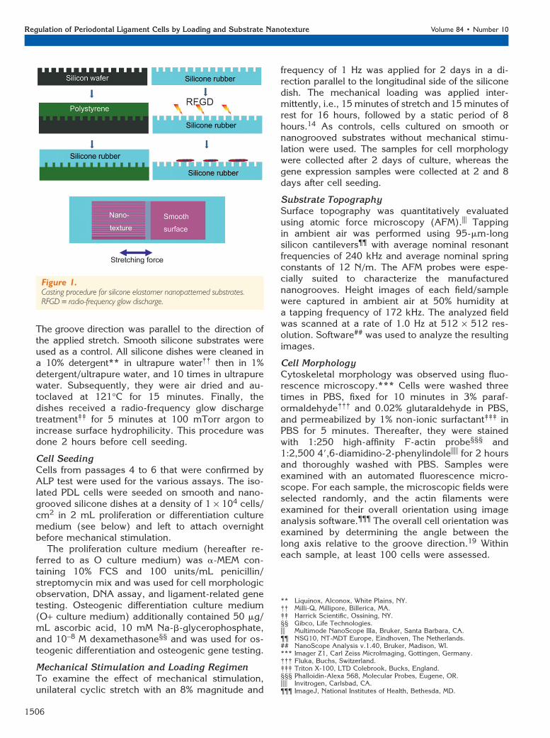
To produce nanotextured surfaces, a silicon wafertemplate was generated using laser interference li-thography as described previously.19 This patternedwafer contained parallel grooves 300 nm wide (600nm pitch) with a depth of 150 nm. The procedure ofmaking the silicone substrates is shown in Figure 1.First, polystyrene replicates of the templates weremade by solvent casting. The casting solution wasmadebydissolvingpieces of tissue culturepolystyrenei
at 25 g/150 mL in chloroform¶ and stirring gently for24 hours. After casting this solution on the templates,the chloroform evaporated overnight in a laminarflow hood. The replicates were removed from thetemplates and replicated a second time in siliconerubber, using the same material as for the siliconedish. Subsequently, the grooved rubber replicas wereglued into the silicone dish with silicone adhesive.#
‡ Gibco, Life Technologies, Breda, The Netherlands.§ Elastosil RT 601, Wacker Chemie, Munchen, Germany.i Greiner, Kremsmunster, Austria.¶ Labscan, Dublin, Ireland.# RTV silicone adhesive, NuSil Technology, Carpinteria, CA.
J Periodontol • October 2013 Yu, Prodanov, te Riet, Yang, Walboomers, Jansen
1505
The groove direction was parallel to the direction ofthe applied stretch. Smooth silicone substrates wereused as a control. All silicone dishes were cleaned ina 10% detergent** in ultrapure water†† then in 1%detergent/ultrapure water, and 10 times in ultrapurewater. Subsequently, they were air dried and au-toclaved at 121�C for 15 minutes. Finally, thedishes received a radio-frequency glow dischargetreatment‡‡ for 5 minutes at 100 mTorr argon toincrease surface hydrophilicity. This procedure wasdone 2 hours before cell seeding.
Cell SeedingCells from passages 4 to 6 that were confirmed byALP test were used for the various assays. The iso-lated PDL cells were seeded on smooth and nano-grooved silicone dishes at a density of 1 · 104 cells/cm2 in 2 mL proliferation or differentiation culturemedium (see below) and left to attach overnightbefore mechanical stimulation.
The proliferation culture medium (hereafter re-ferred to as O culture medium) was a-MEM con-taining 10% FCS and 100 units/mL penicillin/streptomycin mix and was used for cell morphologicobservation, DNA assay, and ligament-related genetesting. Osteogenic differentiation culture medium(O+ culture medium) additionally contained 50 mg/mL ascorbic acid, 10 mM Na-b-glycerophosphate,and 10-8 M dexamethasone§§ and was used for os-teogenic differentiation and osteogenic gene testing.
Mechanical Stimulation and Loading RegimenTo examine the effect of mechanical stimulation,unilateral cyclic stretch with an 8% magnitude and
frequency of 1 Hz was applied for 2 days in a di-rection parallel to the longitudinal side of the siliconedish. The mechanical loading was applied inter-mittently, i.e., 15 minutes of stretch and 15minutes ofrest for 16 hours, followed by a static period of 8hours.14 As controls, cells cultured on smooth ornanogrooved substrates without mechanical stimu-lation were used. The samples for cell morphologywere collected after 2 days of culture, whereas thegene expression samples were collected at 2 and 8days after cell seeding.
Substrate TopographySurface topography was quantitatively evaluatedusing atomic force microscopy (AFM).ii Tappingin ambient air was performed using 95-mm-longsilicon cantilevers¶¶ with average nominal resonantfrequencies of 240 kHz and average nominal springconstants of 12 N/m. The AFM probes were espe-cially suited to characterize the manufacturednanogrooves. Height images of each field/samplewere captured in ambient air at 50% humidity ata tapping frequency of 172 kHz. The analyzed fieldwas scanned at a rate of 1.0 Hz at 512 · 512 res-olution. Software## was used to analyze the resultingimages.
Cell MorphologyCytoskeletal morphology was observed using fluo-rescence microscopy.*** Cells were washed threetimes in PBS, fixed for 10 minutes in 3% paraf-ormaldehyde††† and 0.02% glutaraldehyde in PBS,and permeabilized by 1% non-ionic surfactant‡‡‡ inPBS for 5 minutes. Thereafter, they were stainedwith 1:250 high-affinity F-actin probe§§§ and1:2,500 49,6-diamidino-2-phenylindoleiii for 2 hoursand thoroughly washed with PBS. Samples wereexamined with an automated fluorescence micro-scope. For each sample, the microscopic fields wereselected randomly, and the actin filaments wereexamined for their overall orientation using imageanalysis software.¶¶¶ The overall cell orientation wasexamined by determining the angle between thelong axis relative to the groove direction.19 Withineach sample, at least 100 cells were assessed.
Figure 1.Casting procedure for silicone elastomer nanopatterned substrates.RFGD = radio-frequency glow discharge.
** Liquinox, Alconox, White Plains, NY.†† Milli-Q, Millipore, Billerica, MA.‡‡ Harrick Scientific, Ossining, NY.§§ Gibco, Life Technologies.ii Multimode NanoScope IIIa, Bruker, Santa Barbara, CA.¶¶ NSG10, NT-MDT Europe, Eindhoven, The Netherlands.## NanoScope Analysis v.1.40, Bruker, Madison, WI.*** Imager Z1, Carl Zeiss MicroImaging, Gottingen, Germany.††† Fluka, Buchs, Switzerland.‡‡‡ Triton X-100, LTD Colebrook, Bucks, England.§§§ Phalloidin-Alexa 568, Molecular Probes, Eugene, OR.iii Invitrogen, Carlsbad, CA.¶¶¶ ImageJ, National Institutes of Health, Bethesda, MD.
Regulation of Periodontal Ligament Cells by Loading and Substrate Nanotexture Volume 84 • Number 10
1506
DNA ContentUltrapure water was used to lyse the cells. After twofreeze–thaw cycles, the supernatants were stored at-80�C for further analysis. For the DNA assay,a kit### was used according to manufacturer’s in-structions. Briefly, 100 mL sample solution wasadded to 100 mL working solution.**** After 2 to 5minutes of incubation in the dark, the amount ofDNA was measured using a fluorescence microplatereader†††† with a 485-nm excitation filter and a 530-nm emission filter. The DNA content in the experi-mental samples was determined from a standardcurve with known amounts of DNA.
Real-Time Polymerase Chain ReactionGene expression was studied after 2 days (i.e., im-mediately after mechanical loading) and 8 days ofcell culture. The cell layers were collected, and totalRNA was isolated using guanidinium thiocyanate-phenol-chloroform extraction reagent‡‡‡‡ accordingto the manufacturer’s instructions. The RNA con-centration was measured with spectrophotometry.§§§§
After obtaining messenger RNA (mRNA), first-strandreverse-transcription polymerase chain reaction (RT-PCR) was performed using a kitiiii according to themanufacturer’s protocol. The complementary DNA(cDNA) was then amplified, and specific gene ex-pression was quantified in real-time PCR. For eachreaction, 12.5 mL master mix, 2 mL DNA, 3 mLprimer mix (1.5 mL forward primer and 1.5 mL re-verse primer), and 7.5 mL diethylpyrocarbonatewere added to the reaction system. Subsequently,PCR was performed in a real-time PCR reaction ap-paratus with the desired temperatures. Primers weredesigned so as to avoid amplification of genomicDNA, and therefore each amplicon spans at least oneintron (Table 1). The genes studied in the real-timequantitative PCR included two bone differentiationmarkers, Runx2 and OCN, and two ligament differ-entiation markers, scleraxis transcription factor (SCXA)and ELN. Relative gene expression was normalized tothe expression of household gene GAPDH. The ex-pression of the tested genes was calculated via the
2-DDCt method15 relative to the smooth, static-culturegroup. Each sample was tested in duplicate, and therelative gene expression values were based on threesamples from each group and expressed as mean –SD. The RNA-negative control consisted of ultrapurewater that was reverse transcribed and amplified inparallel with the cell samples.
Statistical AnalysesSamples were triplicate for each test. The completeexperiment was performed twice to ensure re-producibility. The outcome of the two experimentswas alike, and thus only the first experiment isrepresented. Statistical analysis for cell morphologywas performed using a non-parametric Wilcoxonsigned rank test.20 Other data were analyzed usingone-way analysis of variance (ANOVA) with Dunnettpost hoc test or two-way ANOVA for synergy testing.Calculations were performed using statisticalsoftware.¶¶¶¶
RESULTS
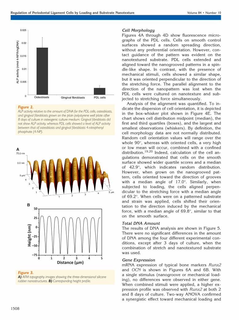
PDL Cell Phenotype IdentificationALP activity was confirmed in the PDL cell culture asa whole after 8 days in O+ culture medium (Fig. 2).The ALP activity in PDL cells was between osteo-blasts from alveolar bone (positive control) andgingival fibroblasts (negative control) cultured in thesame conditions.
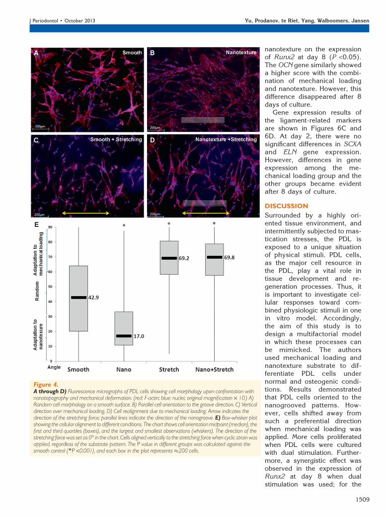
Substrate TopographyNanotextured substrates were routinely checked byAFM (Fig. 3). Topography height profiles of siliconesubstrates showed groove dimensions with a pitch of572 – 10 nm and depth of 97.9 – 5.1 nm.
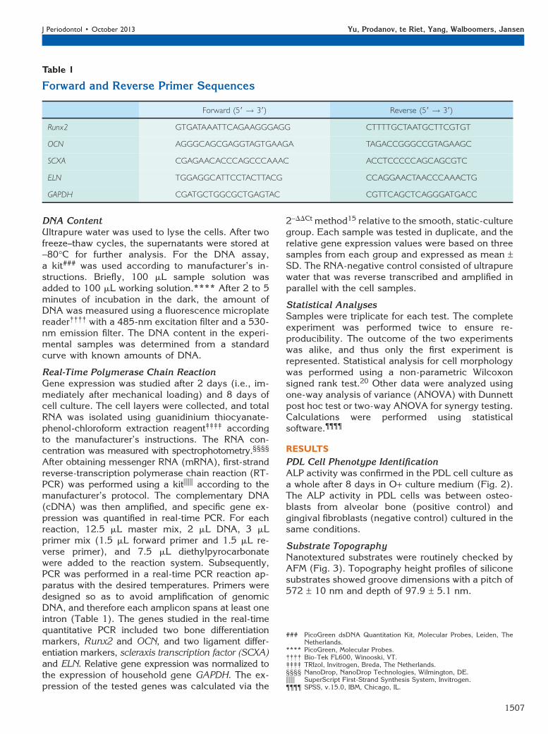
Table 1
Forward and Reverse Primer Sequences
Forward (59 ! 39) Reverse (59 ! 39)
Runx2 GTGATAAATTCAGAAGGGAGG CTTTTGCTAATGCTTCGTGT
OCN AGGGCAGCGAGGTAGTGAAGA TAGACCGGGCCGTAGAAGC
SCXA CGAGAACACCCAGCCCAAAC ACCTCCCCCAGCAGCGTC
ELN TGGAGGCATTCCTACTTACG CCAGGAACTAACCCAAACTG
GAPDH CGATGCTGGCGCTGAGTAC CGTTCAGCTCAGGGATGACC
### PicoGreen dsDNA Quantitation Kit, Molecular Probes, Leiden, TheNetherlands.
**** PicoGreen, Molecular Probes.†††† Bio-Tek FL600, Winooski, VT.‡‡‡‡ TRIzol, Invitrogen, Breda, The Netherlands.§§§§ NanoDrop, NanoDrop Technologies, Wilmington, DE.iiii SuperScript First-Strand Synthesis System, Invitrogen.¶¶¶¶ SPSS, v.15.0, IBM, Chicago, IL.
J Periodontol • October 2013 Yu, Prodanov, te Riet, Yang, Walboomers, Jansen
1507
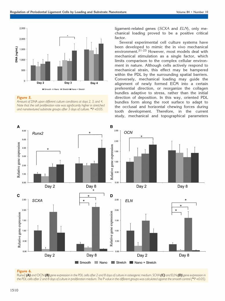
Cell MorphologyFigures 4A through 4D show fluorescence micro-graphs of the PDL cells. Cells on smooth controlsurfaces showed a random spreading direction,without any preferential orientation. However, con-tact guidance of the pattern was evident on thenanotextured substrate. PDL cells extended andaligned toward the nanogrooved patterns in a spin-dle-like shape. In contrast, with the presence ofmechanical stimuli, cells showed a similar shape,but it was oriented perpendicular to the direction ofthe stretching force. The parallel alignment to thedirection of the nanopattern was lost when thePDL cells were cultured on nanotexture and sub-jected to stretching force simultaneously.
Analysis of the alignment was quantified. To in-dicate the dispersion of cell orientation, it is depictedin the box-whisker plot shown in Figure 4E. Thechart shows cell distribution midpoint (median), thefirst and third quartiles (boxes), and the largest andsmallest observations (whiskers). By definition, thecell morphology data are not normally distributed.Random cell orientation values will range over thewhole 90�, whereas with oriented cells, a very highor low mean will occur, combined with a confineddistribution.19,20 Indeed, calculation of the cell an-gulations demonstrated that cells on the smoothsurface showed wider quartile scores and a medianof 42.9�, which indicates random distribution.However, when grown on the nanogrooved pat-tern, cells oriented toward the direction of grooveswith a median angle of 17.0�. Similarly, whensubjected to loading, the cells aligned perpen-dicular to the stretching force with a median angleof 69.2�. When cells were on a patterned substrateand strain was applied, cells shifted their orien-tation to the direction induced by the mechanicalforce, with a median angle of 69.8�, similar to thaton the smooth surface.
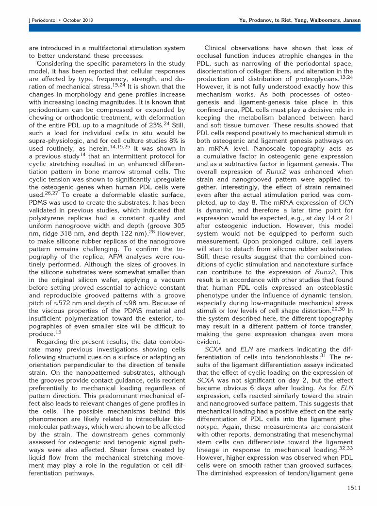
Total DNA AmountThe results of DNA analysis are shown in Figure 5.There were no significant differences in the amountof DNA among the four different experimental con-ditions, except after 3 days of culture, when thecombination of stretch and nanotextured substratewas used.
Gene ExpressionmRNA expression of typical bone markers Runx2and OCN is shown in Figures 6A and 6B. Witha single stimulus (nanogroove or mechanical load-ing), no differences were observed in either gene.When combined stimuli were applied, a higher ex-pression profile was observed with Runx2 at both 2and 8 days of culture. Two-way ANOVA confirmeda synergistic effect toward mechanical loading and
Figure 3.A) AFM topography images showing the three-dimensional siliconerubber nanostructures. B) Corresponding height profile.
Figure 2.ALP activity relative to the amount of DNA for the PDL cells, osteoblasts,and gingival fibroblasts grown on the plain polystyrene well plate after8 days of culture in osteogenic culture medium. Gingival fibroblasts didnot show ALP activity, whereas PDL cells showed a level of ALP activitybetween that of osteoblasts and gingival fibroblasts 4-nitrophenylphosphate (4-NP).
Regulation of Periodontal Ligament Cells by Loading and Substrate Nanotexture Volume 84 • Number 10
1508
nanotexture on the expressionof Runx2 at day 8 (P <0.05).The OCN gene similarly showeda higher score with the combi-nation of mechanical loadingand nanotexture. However, thisdifference disappeared after 8days of culture.
Gene expression results ofthe ligament-related markersare shown in Figures 6C and6D. At day 2, there were nosignificant differences in SCXAand ELN gene expression.However, differences in geneexpression among the me-chanical loading group and theother groups became evidentafter 8 days of culture.
DISCUSSION
Surrounded by a highly ori-ented tissue environment, andintermittently subjected to mas-tication stresses, the PDL isexposed to a unique situationof physical stimuli. PDL cells,as the major cell resource inthe PDL, play a vital role intissue development and re-generation processes. Thus, itis important to investigate cel-lular responses toward com-bined physiologic stimuli in onein vitro model. Accordingly,the aim of this study is todesign a multifactorial modelin which these processes canbe mimicked. The authorsused mechanical loading andnanotexture substrate to dif-ferentiate PDL cells undernormal and osteogenic condi-tions. Results demonstratedthat PDL cells oriented to thenanogrooved patterns. How-ever, cells shifted away fromsuch a preferential directionwhen mechanical loading wasapplied. More cells proliferatedwhen PDL cells were culturedwith dual stimulation. Further-more, a synergistic effect wasobserved in the expression ofRunx2 at day 8 when dualstimulation was used; for the
Figure 4.A through D) Fluorescence micrographs of PDL cells showing cell morphology upon confrontation withnanotopography and mechanical deformation. (red: F-actin; blue: nuclei; original magnification · 10.) A)Random cell morphology on a smooth surface. B) Parallel cell orientation to the groove direction. C) Verticaldirection over mechanical loading. D) Cell realignment due to mechanical loading. Arrow indicates thedirection of the stretching force; parallel lines indicate the direction of the nanogroove. E) Box-whisker plotshowing the cellular alignment to different conditions. The chart shows cell orientationmidpoint (median), thefirst and third quartiles (boxes), and the largest and smallest observations (whiskers). The direction of thestretching forcewas set as 0� in the chart. Cells aligned vertically to the stretching forcewhen cyclic strain wasapplied, regardless of the substrate pattern. The P value in different groups was calculated against thesmooth control (*P <0.001), and each box in the plot represents �200 cells.
J Periodontol • October 2013 Yu, Prodanov, te Riet, Yang, Walboomers, Jansen
1509
ligament-related genes (SCXA and ELN), only me-chanical loading proved to be a positive criticalfactor.
Several experimental cell culture systems havebeen developed to mimic the in vivo mechanicalenvironment.21-23 However, most models deal withmechanical stimulation as a single factor, whichlimits comparison to the complex cellular environ-ment in nature. Although cells actively respond tomechanical strain, this effect may be hamperedwithin the PDL by the surrounding spatial barriers.Conversely, mechanical loading may guide thealignment of newly formed ECM into a certainpreferential direction, or reorganize the collagenbundles adaptive to stress, rather than the initialdirection of deposition. In this way, oriented PDLbundles form along the root surface to adapt tothe occlusal and horizontal chewing forces duringtooth development. Therefore, in the currentstudy, mechanical and topographical parameters
Figure 5.Amount of DNA upon different culture conditions at days 2, 3, and 4.Note that the cell proliferation rate was significantly higher in stretchedand nanotextured substrate groups after 3 days of culture. *P <0.05.
Figure 6.Runx2 (A) andOCN (B) gene expression in the PDL cells after 2 and 8 days of culture in osteogenic medium. SCXA (C) and ELN (D) gene expression inthe PDL cells after 2 and 8 days of culture in proliferationmedium. The P value in the different groups was calculated against the smooth control (*P <0.05).
Regulation of Periodontal Ligament Cells by Loading and Substrate Nanotexture Volume 84 • Number 10
1510
are introduced in a multifactorial stimulation systemto better understand these processes.
Considering the specific parameters in the studymodel, it has been reported that cellular responsesare affected by type, frequency, strength, and du-ration of mechanical stress.15,24 It is shown that thechanges in morphology and gene profiles increasewith increasing loading magnitudes. It is known thatperiodontium can be compressed or expanded bychewing or orthodontic treatment, with deformationof the entire PDL up to a magnitude of 23%.24 Still,such a load for individual cells in situ would besupra-physiologic, and for cell culture studies 8% isused routinely, as herein.14,15,25 It was shown ina previous study14 that an intermittent protocol forcyclic stretching resulted in an enhanced differen-tiation pattern in bone marrow stromal cells. Thecyclic tension was shown to significantly upregulatethe osteogenic genes when human PDL cells wereused.26,27 To create a deformable elastic surface,PDMS was used to create the substrates. It has beenvalidated in previous studies, which indicated thatpolystyrene replicas had a constant quality anduniform nanogroove width and depth (groove 305nm, ridge 318 nm, and depth 122 nm).28 However,to make silicone rubber replicas of the nanogroovepattern remains challenging. To confirm the to-pography of the replica, AFM analyses were rou-tinely performed. Although the sizes of grooves inthe silicone substrates were somewhat smaller thanin the original silicon wafer, applying a vacuumbefore setting proved essential to achieve constantand reproducible grooved patterns with a groovepitch of �572 nm and depth of �98 nm. Because ofthe viscous properties of the PDMS material andinsufficient polymerization toward the exterior, to-pographies of even smaller size will be difficult toproduce.15
Regarding the present results, the data corrobo-rate many previous investigations showing cellsfollowing structural cues on a surface or adapting anorientation perpendicular to the direction of tensilestrain. On the nanopatterned substrates, althoughthe grooves provide contact guidance, cells reorientpreferentially to mechanical loading regardless ofpattern direction. This predominant mechanical ef-fect also leads to relevant changes of gene profiles inthe cells. The possible mechanisms behind thisphenomenon are likely related to intracellular bio-molecular pathways, which were shown to be affectedby the strain. The downstream genes commonlyassessed for osteogenic and tenogenic signal path-ways were also affected. Shear forces created byliquid flow from the mechanical stretching move-ment may play a role in the regulation of cell dif-ferentiation pathways.
Clinical observations have shown that loss ofocclusal function induces atrophic changes in thePDL, such as narrowing of the periodontal space,disorientation of collagen fibers, and alteration in theproduction and distribution of proteoglycans.13,24
However, it is not fully understood exactly how thismechanism works. As both processes of osteo-genesis and ligament-genesis take place in thisconfined area, PDL cells must play a decisive role inkeeping the metabolism balanced between hardand soft tissue turnover. These results showed thatPDL cells respond positively to mechanical stimuli inboth osteogenic and ligament genesis pathways onan mRNA level. Nanoscale topography acts asa cumulative factor in osteogenic gene expressionand as a subtractive factor in ligament genesis. Theoverall expression of Runx2 was enhanced whenstrain and nanogrooved pattern were applied to-gether. Interestingly, the effect of strain remainedeven after the actual stimulation period was com-pleted, up to day 8. The mRNA expression of OCNis dynamic, and therefore a later time point forexpression would be expected, e.g., at day 14 or 21after osteogenic induction. However, this modelsystem would not be equipped to perform suchmeasurement. Upon prolonged culture, cell layerswill start to detach from silicone rubber substrates.Still, these results suggest that the combined con-ditions of cyclic stimulation and nanotexture surfacecan contribute to the expression of Runx2. Thisresult is in accordance with other studies that foundthat human PDL cells expressed an osteoblasticphenotype under the influence of dynamic tension,especially during low-magnitude mechanical stressstimuli or low levels of cell shape distortion.29,30 Inthe system described here, the different topographymay result in a different pattern of force transfer,making the gene expression changes even moreevident.
SCXA and ELN are markers indicating the dif-ferentiation of cells into tendonoblasts.31 The re-sults of the ligament differentiation assays indicatedthat the effect of cyclic loading on the expression ofSCXA was not significant on day 2, but the effectbecame obvious 6 days after loading. As for ELNexpression, cells reacted similarly toward the strainand nanogrooved surface pattern. This suggests thatmechanical loading had a positive effect on the earlydifferentiation of PDL cells into the ligament phe-notype. Again, these measurements are consistentwith other reports, demonstrating that mesenchymalstem cells can differentiate toward the ligamentlineage in response to mechanical loading.32,33
However, higher expression was observed when PDLcells were on smooth rather than grooved surfaces.The diminished expression of tendon/ligament gene
J Periodontol • October 2013 Yu, Prodanov, te Riet, Yang, Walboomers, Jansen
1511
expression in the nanopatterned condition may bedue to the rearrangement of cells, a general cellularresponse for resisting external stimulation and/orexcitation.
CONCLUSIONS
In summary, the present findings suggest that me-chanical stimulation is a crucial factor in regulatingPDL cell behavior through modulation of the levels ofosteogenic and ligament-forming activities. More-over, ECM-resembling surface structures inducedifferent responses from PDL cells in terms of mor-phology and gene expression. Multifactorial modelsseem useful to provide a physiologic analog for in-vivo studies.
ACKNOWLEDGMENTS
NY is financially supported by the China ScholarshipCouncils (no. 2008627114). JtR is supported bya personal VENI grant (680-47-421) of the Nether-lands Organisation for Scientific Research (NWO).The authors of this study have no financial relation-ships related to any products involved in this study.The authors report no conflicts of interest relatedto this study.
REFERENCES1. Seo BM, Miura M, Gronthos S, et al. Investigation of
multipotent postnatal stem cells from human peri-odontal ligament. Lancet 2004;364:149-155.
2. Howard PS, Kucich U, Taliwal R, Korostoff JM.Mechanical forces alter extracellular matrix synthesisby human periodontal ligament fibroblasts. J Periodon-tal Res 1998;33:500-508.
3. Nikkhah M, Edalat F, Manoucheri S, Khademhosseini A.Engineering microscale topographies to control thecell-substrate interface. Biomaterials 2012;33:5230-5246.
4. Chan MW, Hinz B, McCulloch CA. Mechanical in-duction of gene expression in connective tissue cells.Methods Cell Biol 2010;98:178-205.
5. Hynes RO. The extracellular matrix: Not just prettyfibrils. Science 2009;326:1216-1219.
6. Dalby MJ, McCloy D, Robertson M, et al. Osteoproge-nitor response to semi-ordered and random nano-topographies. Biomaterials 2006;27:2980-2987.
7. Recknor JB, Sakaguchi DS, Mallapragada SK. Di-rected growth and selective differentiation of neuralprogenitor cells on micropatterned polymer sub-strates. Biomaterials 2006;27:4098-4108.
8. Guilak F, Cohen DM, Estes BT, Gimble JM, Liedtke W,Chen CS. Control of stem cell fate by physical in-teractions with the extracellular matrix. Cell Stem Cell2009;5:17-26.
9. Ivanovski S. Periodontal regeneration. Aust DentJ 2009;54(Suppl. 1):S118-S128.
10. Lenhert S, Meier MB, Meyer U, Chi L, Wiesmann HP.Osteoblast alignment, elongation and migrationon grooved polystyrene surfaces patterned byLangmuir-Blodgett lithography. Biomaterials 2005;26:563-570.
11. Biggs MJ, Richards RG, McFarlane S, Wilkinson CD,Oreffo RO, Dalby MJ. Adhesion formation of primaryhuman osteoblasts and the functional response ofmesenchymal stem cells to 330nm deep micro-grooves. J R Soc Interface 2008;5:1231-1242.
12. Fujita S, Ohshima M, Iwata H. Time-lapse observationof cell alignment on nanogrooved patterns. J R SocInterface 2009;6(Suppl. 3):S269-S277.
13. Kaneko S, Ohashi K, Soma K, Yanagishita M. Occlusalhypofunction causes changes of proteoglycan contentin the rat periodontal ligament. J Periodontal Res2001;36:9-17.
14. Winter LC, Walboomers XF, Bumgardner JD, JansenJA. Intermittent versus continuous stretching effects onosteoblast-like cells in vitro. J Biomed Mater ResA 2003;67:1269-1275.
15. Prodanov L, te Riet J, Lamers E, et al. The interactionbetween nanoscale surface features and mechanicalloading and its effect on osteoblast-like cells behav-ior. Biomaterials 2010;31:7758-7765.
16. Dutch federation of biomedical scientific societies.Human tissue and medical research: code of conductfor responsible use. Available at: http://www.federa.org/. Accessed August 16, 2013.
17. Brunette DM, Melcher AH, Moe HK. Culture and originof epithelium-like and fibroblast-like cells from porcineperiodontal ligament explants and cell suspensions.Arch Oral Biol 1976;21:393-400.
18. Oortgiesen DA, Yu N, Bronckers AL, Yang F,Walboomers XF, Jansen JA. A three-dimensional cellculture model to study the mechano-biological behav-ior in periodontal ligament regeneration. Tissue EngPart C Methods 2012;18:81-89.
19. Lamers E, Walboomers XF, Domanski M, et al. Theinfluence of nanoscale grooved substrates on osteo-blast behavior and extracellular matrix deposition.Biomaterials 2010;31:3307-3316.
20. Lamers E, van Horssen R, te Riet J, et al. The influenceof nanoscale topographical cues on initial osteoblastmorphology and migration. Eur Cell Mater 2010;20:329-343.
21. Klein-Nulend J, Veldhuijzen JP, de Jong M, BurgerEH. Increased bone formation and decreased boneresorption in fetal mouse calvaria as a result ofintermittent compressive force in vitro. Bone Miner1987;2:441-448.
22. Yousefian J, Firouzian F, Shanfeld J, Ngan P, Lanese R,Davidovitch Z. A new experimental model for studyingthe response of periodontal ligament cells to hydro-static pressure. Am J Orthod Dentofacial Orthop 1995;108:402-409.
23. Yamaguchi R, Kohtoh K. Sinusoidal variation of wallshear stress in daughter tube through 45 deg branchmodel in laminar flow. J Biomech Eng 1994;116:119-126.
24. Ozaki S, Kaneko S, Podyma-Inoue KA, Yanagishita M,Soma K. Modulation of extracellular matrix synthesisand alkaline phosphatase activity of periodontal liga-ment cells by mechanical stress. J Periodontal Res2005;40:110-117.
25. Neidlinger-Wilke C, Wilke HJ, Claes L. Cyclic stretch-ing of human osteoblasts affects proliferation andmetabolism: A new experimental method and itsapplication. J Orthop Res 1994;12:70-78.
26. Pinkerton MN, Wescott DC, Gaffey BJ, Beggs KT,Milne TJ, Meikle MC. Cultured human periodontalligament cells constitutively express multiple osteotropic
Regulation of Periodontal Ligament Cells by Loading and Substrate Nanotexture Volume 84 • Number 10
1512
cytokines and growth factors, several of which are re-sponsive to mechanical deformation. J PeriodontalRes 2008;43:343-351.
27. Wescott DC, Pinkerton MN, Gaffey BJ, BeggsKT, Milne TJ, Meikle MC. Osteogenic geneexpression by human periodontal ligament cellsunder cyclic tension. J Dent Res 2007;86:1212-1216.
28. Lamers E, Walboomers XF, Domanski M, et al. In vitroand in vivo evaluation of the inflammatory responseto nanoscale grooved substrates. Nanomedicine 2012;8:308-317.
29. Yang YQ, Li XT, Rabie AB, Fu MK, Zhang D. Humanperiodontal ligament cells express osteoblastic phe-notypes under intermittent force loading in vitro.Front Biosci 2006;11:776-781.
30. Zhao Y, Wang C, Li S, et al. Expression of Osterix inmechanical stress-induced osteogenic differentiation
of periodontal ligament cells in vitro. Eur J Oral Sci2008;116:199-206.
31. Benhardt HA, Cosgriff-Hernandez EM. The role ofmechanical loading in ligament tissue engineering.Tissue Eng Part B Rev 2009;15:467-475.
32. Altman GH, Horan RL, Martin I, et al. Cell differentiationby mechanical stress. FASEB J 2002;16:270-272.
33. Noth U, Schupp K, Heymer A, et al. Anterior cruciateligament constructs fabricated from human mesen-chymal stem cells in a collagen type I hydrogel.Cytotherapy 2005;7:447-455.
Correspondence: Prof. John A. Jansen, Department ofBiomaterials (309), Radboud University Nijmegen Med-ical Centre, Nijmegen, the Netherlands. Fax: +31-24-3614657; e-mail: [email protected].
Submitted August 20, 2012; accepted for publicationOctober 28, 2012.
J Periodontol • October 2013 Yu, Prodanov, te Riet, Yang, Walboomers, Jansen
1513
Copyright © 2022 FDOKUMEN