Comparative Gene Expression Analysis of the Human Periodontal Ligament in Deciduous and Permanent...
10
Comparative Gene Expression Analysis of the Human Periodontal Ligament in Deciduous and Permanent Teeth Je Seon Song 1,2. , Dong Hwan Hwang 1. , Seong-Oh Kim 1,2 , Mijeong Jeon 2 , Byung-Jai Choi 1,2 , Han- Sung Jung 3 , Seok Jun Moon 4 , Wonse Park 5 , Hyung-Jun Choi 1,2 * 1 Department of Pediatric Dentistry, College of Dentistry, Yonsei University, Seoul, Korea, 2 Oral Science Research Center, College of Dentistry, Yonsei University, Seoul, Korea, 3 Division in Anatomy & Developmental Biology, Department of Oral Biology, College of Dentistry, Yonsei University, Seoul, Korea, 4 Division in Pharmacology, Department of Oral Biology, College of Dentistry, Yonsei University, Seoul, Korea, 5 Department of General Dentistry, College of Dentistry, Yonsei University, Seoul, Korea Abstract There are histological and functional differences between human deciduous and permanent periodontal ligament (PDL) tissues. The aim of this study was to determine the differences between these two types of tissue at the molecular level by comparing their gene expression patterns. PDL samples were obtained from permanent premolars (n = 38) and anterior deciduous teeth (n = 31) extracted from 40 healthy persons. Comparative cDNA microarray analysis revealed several differences in gene expression between the deciduous and permanent PDL tissues. These findings were verified by qRT-PCR (quantitative reverse-transcription–polymerase chain reaction) analysis, and the areas where genes are expressed were revealed by immunohistochemical staining. The expressions of 21 genes were up-regulated in deciduous relative to PDL tissues, and those of 30 genes were up-regulated in permanent relative to deciduous PDL tissues. The genes that were up- regulated in deciduous PDL tissues were those involved in the formation of the extracellular matrix (LAMC2, LAMB3, and COMP), tissue development (IGF2BP, MAB21L2, and PAX3), and inflammatory or immune reactions leading to tissue degradation (IL1A, CCL21, and CCL18). The up-regulated genes in permanent PDL tissues were related to tissue degradation (IL6 and ADAMTS18), myocontraction (PDE3B, CASQ2, and MYH10), and neurological responses (FOS, NCAM2, SYT1, SLC22A3, DOCK3, LRRTM1, LRRTM3, PRSS12, and ARPP21). The analysis of differential gene expressions between deciduous and permanent PDL tissues aids our understanding of histological and functional differences between them at the molecular level. Citation: Song JS, Hwang DH, Kim S-O, Jeon M, Choi B-J, et al. (2013) Comparative Gene Expression Analysis of the Human Periodontal Ligament in Deciduous and Permanent Teeth. PLoS ONE 8(4): e61231. doi:10.1371/journal.pone.0061231 Editor: Baochuan Lin, Naval Research Laboratory, United States of America Received November 1, 2012; Accepted March 7, 2013; Published April 8, 2013 Copyright: ß 2013 Song et al. This is an open-access article distributed under the terms of the Creative Commons Attribution License, which permits unrestricted use, distribution, and reproduction in any medium, provided the original author and source are credited. Funding: This research was supported by Basic Science Research Program through the National Research Foundation of Korea funded by the Ministry of Education, Science and Technology (2011-0022160 and 2012R1A1A2041910). The funders had no role in study design, data collection and analysis, decision to publish, or preparation of the manuscript. Competing Interests: The authors have declared that no competing interests exist. * E-mail: [email protected] . These authors contributed equally to this work. Introduction The periodontal ligament (PDL) tissues are components of the dental apparatus that connect the tooth to the alveolar jaw bone in the area surrounding the root surfaces. Similar to tendons, collagen type I is a predominant component of PDL tissue, but collagen types III, V, VI, and XII, and proteoglycans, which are known to regulate collagen fibril formation, are also found in this tissue [1,2]. The PDL tissues comprise various cells such as PDL fibroblasts, epithelial cell rests of Malassez, osteoblasts, cemento- blasts, vascular cells, and sensory nerve cells. There are many anatomical, embryological, functional, and structural differences between human deciduous and permanent teeth. For example, deciduous and permanent teeth exhibit different responses to external stimuli, and the sensory nerve endings are fewer and looser in permanent than in deciduous teeth [3,4]. Because of the different responses of dental pulp tissues, the modalities of pulp therapy should differ between deciduous and permanent teeth [5]. Besides, in vitro and in vivo experiments have shown that cells originating from deciduous teeth behave differently to those from permanent teeth [6,7]. Since deciduous teeth are resorbed and exfoliated in association with the eruption of successive permanent teeth, the periodontal tissues of the former are more easily resorbed than those of the latter [8,9]. To explain this at the molecular level, some investigators reported that the periodontal tissues of deciduous teeth contain more bone sialoprotein and osteopontin with the Arg-Gly-Asp (RGD) sequence, to which odontoclasts bind [10,11]. Others reported that PDL cells obtained from areas being resorbed in deciduous teeth express more of the receptor activator of the nuclear factor k-light-chain-enhancer of activated B cells ligand (RANKL) gene, which is known to be associated with osteoclastogenesis [12,13]. In addition, extracellular-matrix-de- grading enzymes such as collagenase [14], metalloproteinases [15,16], and mucopolysaccharidase [17] were found to be up- regulated upon the resorption of deciduous periodontium. PLOS ONE | www.plosone.org 1 April 2013 | Volume 8 | Issue 4 | e61231
-
Upload
independent -
Category
Documents
-
view
1 -
download
0
Transcript of Comparative Gene Expression Analysis of the Human Periodontal Ligament in Deciduous and Permanent...

Comparative Gene Expression Analysis of the HumanPeriodontal Ligament in Deciduous and PermanentTeethJe Seon Song1,2., Dong Hwan Hwang1., Seong-Oh Kim1,2, Mijeong Jeon2, Byung-Jai Choi1,2, Han-
Sung Jung3, Seok Jun Moon4, Wonse Park5, Hyung-Jun Choi1,2*
1 Department of Pediatric Dentistry, College of Dentistry, Yonsei University, Seoul, Korea, 2 Oral Science Research Center, College of Dentistry, Yonsei University, Seoul,
Korea, 3 Division in Anatomy & Developmental Biology, Department of Oral Biology, College of Dentistry, Yonsei University, Seoul, Korea, 4 Division in Pharmacology,
Department of Oral Biology, College of Dentistry, Yonsei University, Seoul, Korea, 5 Department of General Dentistry, College of Dentistry, Yonsei University, Seoul, Korea
Abstract
There are histological and functional differences between human deciduous and permanent periodontal ligament (PDL)tissues. The aim of this study was to determine the differences between these two types of tissue at the molecular level bycomparing their gene expression patterns. PDL samples were obtained from permanent premolars (n = 38) and anteriordeciduous teeth (n = 31) extracted from 40 healthy persons. Comparative cDNA microarray analysis revealed severaldifferences in gene expression between the deciduous and permanent PDL tissues. These findings were verified by qRT-PCR(quantitative reverse-transcription–polymerase chain reaction) analysis, and the areas where genes are expressed wererevealed by immunohistochemical staining. The expressions of 21 genes were up-regulated in deciduous relative to PDLtissues, and those of 30 genes were up-regulated in permanent relative to deciduous PDL tissues. The genes that were up-regulated in deciduous PDL tissues were those involved in the formation of the extracellular matrix (LAMC2, LAMB3, andCOMP), tissue development (IGF2BP, MAB21L2, and PAX3), and inflammatory or immune reactions leading to tissuedegradation (IL1A, CCL21, and CCL18). The up-regulated genes in permanent PDL tissues were related to tissue degradation(IL6 and ADAMTS18), myocontraction (PDE3B, CASQ2, and MYH10), and neurological responses (FOS, NCAM2, SYT1,SLC22A3, DOCK3, LRRTM1, LRRTM3, PRSS12, and ARPP21). The analysis of differential gene expressions between deciduousand permanent PDL tissues aids our understanding of histological and functional differences between them at themolecular level.
Citation: Song JS, Hwang DH, Kim S-O, Jeon M, Choi B-J, et al. (2013) Comparative Gene Expression Analysis of the Human Periodontal Ligament in Deciduousand Permanent Teeth. PLoS ONE 8(4): e61231. doi:10.1371/journal.pone.0061231
Editor: Baochuan Lin, Naval Research Laboratory, United States of America
Received November 1, 2012; Accepted March 7, 2013; Published April 8, 2013
Copyright: � 2013 Song et al. This is an open-access article distributed under the terms of the Creative Commons Attribution License, which permitsunrestricted use, distribution, and reproduction in any medium, provided the original author and source are credited.
Funding: This research was supported by Basic Science Research Program through the National Research Foundation of Korea funded by the Ministry ofEducation, Science and Technology (2011-0022160 and 2012R1A1A2041910). The funders had no role in study design, data collection and analysis, decision topublish, or preparation of the manuscript.
Competing Interests: The authors have declared that no competing interests exist.
* E-mail: [email protected]
. These authors contributed equally to this work.
Introduction
The periodontal ligament (PDL) tissues are components of the
dental apparatus that connect the tooth to the alveolar jaw bone in
the area surrounding the root surfaces. Similar to tendons,
collagen type I is a predominant component of PDL tissue, but
collagen types III, V, VI, and XII, and proteoglycans, which are
known to regulate collagen fibril formation, are also found in this
tissue [1,2]. The PDL tissues comprise various cells such as PDL
fibroblasts, epithelial cell rests of Malassez, osteoblasts, cemento-
blasts, vascular cells, and sensory nerve cells.
There are many anatomical, embryological, functional, and
structural differences between human deciduous and permanent
teeth. For example, deciduous and permanent teeth exhibit
different responses to external stimuli, and the sensory nerve
endings are fewer and looser in permanent than in deciduous teeth
[3,4]. Because of the different responses of dental pulp tissues, the
modalities of pulp therapy should differ between deciduous and
permanent teeth [5]. Besides, in vitro and in vivo experiments
have shown that cells originating from deciduous teeth behave
differently to those from permanent teeth [6,7].
Since deciduous teeth are resorbed and exfoliated in association
with the eruption of successive permanent teeth, the periodontal
tissues of the former are more easily resorbed than those of the
latter [8,9]. To explain this at the molecular level, some
investigators reported that the periodontal tissues of deciduous
teeth contain more bone sialoprotein and osteopontin with the
Arg-Gly-Asp (RGD) sequence, to which odontoclasts bind [10,11].
Others reported that PDL cells obtained from areas being
resorbed in deciduous teeth express more of the receptor activator
of the nuclear factor k-light-chain-enhancer of activated B cells
ligand (RANKL) gene, which is known to be associated with
osteoclastogenesis [12,13]. In addition, extracellular-matrix-de-
grading enzymes such as collagenase [14], metalloproteinases
[15,16], and mucopolysaccharidase [17] were found to be up-
regulated upon the resorption of deciduous periodontium.
PLOS ONE | www.plosone.org 1 April 2013 | Volume 8 | Issue 4 | e61231

However, these findings are not sufficient to explain the differences
in the normal-functioning periodontium of deciduous and
permanent teeth.
The recent development of microarray analysis allows evalua-
tion of the expressions of large numbers of genes simultaneously,
and has been used to investigate periodontal tissues [18] and
periodontal cell cultures [19,20]. Given the anatomical and
functional differences between the periodontal tissues of deciduous
and permanent teeth, it is reasonable to assume that there are also
differences in the gene expression patterns of the cells within those
tissues. Therefore, the aims of the present study were to identify
and compare the gene expression patterns of human deciduous
and permanent periodontal tissues in order to enhance our
understanding of the molecular basis of the observed functional
differences between these two tissue types.
Materials and Methods
PDL samplesThe experimental protocol was approved by the Institutional
Review Board of the Yonsei University Dental Hospital, and
informed consent to participate was obtained from all of the
subjects and their parents (#2-2011-0009). PDL samples were
obtained from healthy permanent premolars (n = 38; from 4 males
and 10 females, aged 10–19 years) extracted for orthodontic
reasons and from anterior deciduous teeth (n = 31; from 14 males
and 12 females aged 5–13 years) extracted for space management
in 40 healthy persons. For RNA isolation, each of the extracted
teeth (36 permanent teeth and 29 deciduous teeth) was immedi-
ately frozen and stored in liquid nitrogen. All of the teeth were
subsequently thawed at room temperature, and the PDL tissues
were obtained carefully using sterile curettes from the middle-third
in permanent teeth or from the nonresorbed root surface in
deciduous teeth. We mixed the PDL tissues and divided them into
three equal groups, and then immediately submerged them in a
RNA stabilizing reagent (RNAlater, Qiagen, Valencia, CA, USA).
The remaining four teeth (two permanent teeth and two deciduous
teeth) were used for immunohistochemical (IHC) staining.
RNA isolationThe PDL tissues were homogenized using a homogenizer
(Bullet Blender, Next Advance, NY, USA). The RNeasy Fibrous
Mini kit (Qiagen) was then used to extract total RNA from the
PDL tissues, according to the manufacturer’s instructions. The
extracted RNA was eluted in 25 ml of RNase-free sterile water
(provided with the kit). The quality and concentration of RNA
were determined by measuring the absorbance at a wavelength of
260 nm with the aid of a spectrophotometer (NanoDrop ND-
1000, Thermo Scientific Inc, Rockford, IL, USA). The RNA
samples used in this study had 260/280 nm ratios of $1.8. Half of
isolated RNA was used for complementary DNA (cDNA)
microarray experiments, and the other half was used for gene
expression analysis with the quantitative reverse-transcription–
polymerase chain reaction (qRT-PCR).
cDNA microarrayGlobal gene expression analyses were conducted using oligo-
nucleotide arrays (GeneChip Human Gene 1.0 ST, Affymetrix,
Santa Clara, CA, USA). The samples were prepared according to
the instructions and recommendations provided by the manufac-
turer. The quality and quantity of RNA were assessed using a
bioanalyzer (model 2100 with RNA 6000 Nano Chips, Agilent
Technologies, Amstelveen, The Netherlands).
As recommended by the manufacturer’s protocol, 300-ng
samples were used. In brief, 300 ng of total RNA from each
sample was converted to double-stranded cDNA. Amplified RNA
(cRNA) was generated from the double-stranded cDNA template
using a random hexamer incorporating a T7 promoter, via in-
vitro transcription, and purified with the Affymetrix sample
cleanup module. cDNA was regenerated through a random-
primed reverse transcription (RT) using a dNTP mix containing
dUTP. The cDNA was then fragmented by UDG and APE1
restriction endonucleases and end-labeled by a terminal transfer-
ase reaction incorporating a biotinylated dideoxynucleotide. The
fragmented end-labeled cDNA was hybridized to the GeneChip
Human Gene 1.0 ST arrays for 16 hours at 45uC and 60 rpm, as
per the GeneChip Whole Transcript Sense Target Labeling Assay
Manual (Affymetrix). The chips were then stained and washed in a
GeneChip Fluidics Station 450 (Affymetrix) and scanned using a
GeneChip Array scanner (3000 G7, Affymetrix). The image data
were extracted using Affymetrix Command Console software
(version 1.1, Affymetrix). The raw file generated using this
procedure provided the expression intensity data that was used
for the next step.
Analysis of microarray dataThe generated expression data were normalized using the
Robust MultiAverage (RMA) algorithm in the Affymetrix Expres-
sion Console software. Whether genes were differentially expressed
between the three groups was determined by subjecting the RMA
expression data to one-way ANOVA. Multiple testing correction
was applied to the p values of the F-statistics to adjust for the false
discovery rate. Genes with adjusted F-statistic p values of ,0.05
were extracted. Strongly expressed genes in each test group that
were up-regulated by over twofold compared to the signal value
were selected for further study. The coexpression gene group, with
similar expression patterns, was classified using hierarchical
clustering and k-means clustering with Multi Experiment Viewer
software (version 4.4, www.tm4.org, Dana-Farber Cancer Insti-
tute, Boston, MA, USA). The Web-based tool Database for
Annotation, Visualization, and Integrated Discovery was used to
interpret the biological implications of the differentially expressed
genes. These genes were then classified based on the gene function
information provided in the Gene Ontology (GO), Kyoto
Encyclopedia of Genes and Genomes Pathway database (http://
david.abcc.ncifcrf.gov/home.jsp).
qRT-PCRThe single-stranded cDNA required in the polymerase chain
reaction (PCR) analysis was produced using 500 ng of extracted
total RNA as a templates for RT (Superscript III Reverse
Transcriptase and random primer, Invitrogen, Renfrew, UK). The
RT reaction was performed at 65uC for 5 minutes, followed by
25uC for 5 minutes, 50uC for 1 hour, and 70uC for 15 minutes to
inactivate the activity of the reverse transcriptase. The synthesized
cDNA was diluted 10:1 in distilled water and used as a template
for qRT-PCR, which was performed using the ABI 7300 RT-PCR
system (Applied Biosystems, Warrington, UK). Samples of 25 ml
containing 16Universal TaqMan Master Mix (4369016, Applied
Biosystems), PCR primers at a concentration of 0.9 mM, and the
diluted cDNA were prepared in triplicate. The amplification
conditions were 50uC for 2 minutes and 95uC for 10 minutes,
followed by 40 cycles of 95uC for 15 seconds and 60uC for
1 minute. The following TaqMan gene expression assay primers
(Applied Biosystems) were used: chemokine (C-C motif) ligand 21
(CCL21), laminin subunit c-2 (LAMC2), insulin-like growth factor 2
mRNA-binding protein 1 (IGF2BP1), interleukin (IL)-6 (IL6),
Gene Expressions in the Periodontal Ligaments
PLOS ONE | www.plosone.org 2 April 2013 | Volume 8 | Issue 4 | e61231
ADAM metalloproteinase with thrombospondin motifs-like 3
(ADAMTSL3), and leucine-rich repeat transmembrane neuronal
protein 1 (LRRTM1). 18S rRNA which is known to be a reliable
housekeeping gene, was used as an internal control [21–23]. ABI
7300 SDS 1.3.1 software (Applied Biosystems) recorded the
fluorescence intensity of the reporter and quencher dyes; the
results are plotted versus time, quantified as the cycle number. A
precise quantification of the initial target was obtained by
examining the amplification plots during the early log phase of
product accumulation above background [the threshold cycle (Ct)
number]. Ct values were subsequently used to determine DCt
values (DCt = Ct of the gene minus Ct of the 18S rRNA control),
and differences in Ct values were used to quantify the relative
amount of PCR product, expressed as the relative change by
applying the equation 22DCt. The specific primer assay ID and
product sizes for each gene are listed in Table 1.
IHC stainingIHC staining was performed in order to determine where the
genes were expressed. In preparation for IHC staining, deciduous
and permanent teeth were fixed in 10% buffered formalin (Sigma,
St. Louis, MO, USA) for 1 day, decalcified with 10% EDTA
(pH 7.4; Fisher Scientific, Houston, TX, USA) for 8 weeks,
embedded in paraffin, and then sectioned at a thickness of 3 mm.
The sections were subjected to IHC staining with antihuman
IGF2BP1 (rabbit polyclonal, diluted 1:100; Ab82968, Abcam,
Cambridge, UK), antihuman IL6 (rabbit polyclonal, diluted
1:1600; Ab6672, Abcam), antihuman LRRTM1 (rabbit polyclon-
al, diluted 1:50; Ab102968, Abcam), and antihuman ADAMTSL3
(rabbit polyclonal, diluted 1:50; NBP1-81426, Novus Biologicals,
CO, USA). Endogenous peroxidase activity was quenched with
3% hydrogen peroxide. The sections were then incubated in 5%
bovine serum albumin (Sigma) to block nonspecific binding and
then incubated overnight with primary antibodies, which had been
diluted to give optimal staining. After incubation, EnVision+System-HRP-labeled polymer anti-rabbit (K4003, Dako North
America, Carpinteria, CA, USA; ready to use) was applied for
20 minutes or Vectastain Elite ABC Kit (PK-6105, Vector
Laboratories, Burlingame, CA, USA; goat IgG, diluted 1:200)
was applied for 30 minutes. Labeled streptavidin biotin kits (Dako)
were used for color development according to the manufacturer’s
instructions. The sections were counterstained with Gill’s hema-
toxylin (Sigma). Control sections were treated in the same manner
but without primary antibodies.
Results
Gene expression profiles of deciduous and permanentPDL tissues
The distribution and frequency of all of the data were first
confirmed using density and box plots. The cDNA microarray
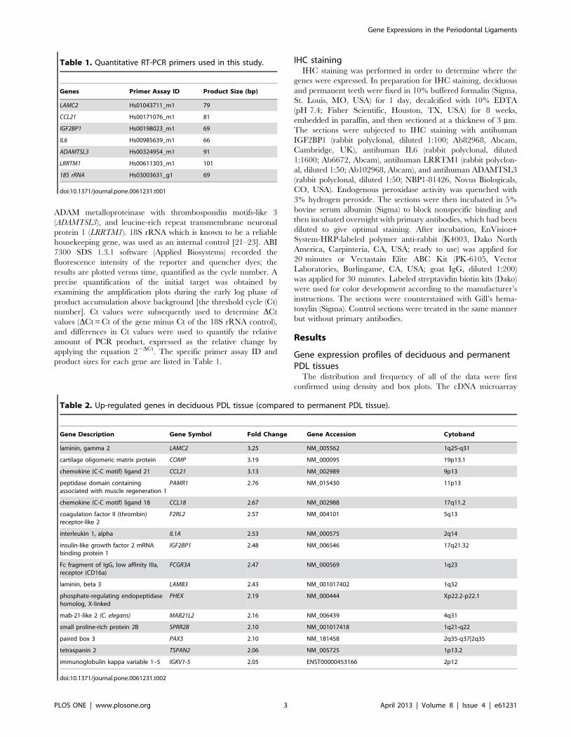
Table 1. Quantitative RT-PCR primers used in this study.
Genes Primer Assay ID Product Size (bp)
LAMC2 Hs01043711_m1 79
CCL21 Hs00171076_m1 81
IGF2BP1 Hs00198023_m1 69
IL6 Hs00985639_m1 66
ADAMTSL3 Hs00324954_m1 91
LRRTM1 Hs00611303_m1 101
18S rRNA Hs03003631_g1 69
doi:10.1371/journal.pone.0061231.t001
Table 2. Up-regulated genes in deciduous PDL tissue (compared to permanent PDL tissue).
Gene Description Gene Symbol Fold Change Gene Accession Cytoband
laminin, gamma 2 LAMC2 3.25 NM_005562 1q25-q31
cartilage oligomeric matrix protein COMP 3.19 NM_000095 19p13.1
chemokine (C-C motif) ligand 21 CCL21 3.13 NM_002989 9p13
peptidase domain containingassociated with muscle regeneration 1
PAMR1 2.76 NM_015430 11p13
chemokine (C-C motif) ligand 18 CCL18 2.67 NM_002988 17q11.2
coagulation factor II (thrombin)receptor-like 2
F2RL2 2.57 NM_004101 5q13
interleukin 1, alpha IL1A 2.53 NM_000575 2q14
insulin-like growth factor 2 mRNAbinding protein 1
IGF2BP1 2.48 NM_006546 17q21.32
Fc fragment of IgG, low affinity IIIa,receptor (CD16a)
FCGR3A 2.47 NM_000569 1q23
laminin, beta 3 LAMB3 2.43 NM_001017402 1q32
phosphate-regulating endopeptidasehomolog, X-linked
PHEX 2.19 NM_000444 Xp22.2-p22.1
mab-21-like 2 (C. elegans) MAB21L2 2.16 NM_006439 4q31
small proline-rich protein 2B SPRR2B 2.10 NM_001017418 1q21-q22
paired box 3 PAX3 2.10 NM_181458 2q35-q37|2q35
tetraspanin 2 TSPAN2 2.06 NM_005725 1p13.2
immunoglobulin kappa variable 1–5 IGKV1-5 2.05 ENST00000453166 2p12
doi:10.1371/journal.pone.0061231.t002
Gene Expressions in the Periodontal Ligaments
PLOS ONE | www.plosone.org 3 April 2013 | Volume 8 | Issue 4 | e61231

results, presented as plots of the standardized log intensity ratio
(M) to the average intensity (A) in three deciduous PDL tissues and
three permanent PDL tissues (i.e., ‘‘M-A’’ plots), are shown in
Figure S1. M-A plots are used to visualize intensity-dependent
ratios of raw microarray data, and provide an easy-to-understand
overview of the data distribution. These plots confirmed the
normalization and standardization of the distributions of the data
yielded in this study.
The results demonstrate that the expressions of 51 genes were
up-regulated by twofold or more in one tissue type relative to the
other. Seven genes (five in deciduous PDL tissue and two in
permanent PDL tissue) were not identified well. Ultimately, 44
genes were analyzed further. In deciduous PDL tissues, the
expressions of 16 genes were up-regulated by twofold or more
compared to permanent PDL tissue, especially LAMC2, cartilage
oligomeric matrix protein (COMP), and CCL21 were expressed
more than threefold (Table 2). Whereas, those of 28 genes were
up-regulated by twofold in permanent compared to deciduous
PDL tissue, especially IL6, SH3 and cysteine-rich domain (STAC),
and FBJ murine osteosarcoma viral oncogene homolog B (FOSB)
were found to be up-regulated by more than fourfold (Table 3).
GO analysisGO classes with an F-statistic p value of ,0.05 following
analysis on the basis of their biological processes are shown in
Figure 1. Those GO-class processes found more frequently in the
PDL tissues of permanent teeth include neurological system
processes, cognition, and response to organic substances. In
contrast, the GO-class processes found more frequently in the
PDL tissues of deciduous teeth included positive regulation of
cellular biosynthetic processes and response to wounding.
The GO classes with an F-statistic p value of ,0.05 following
analysis on the basis of their molecular functions are shown in
Figure 2. These classes did not differ noticeably between the PDL
tissues of deciduous teeth and those of permanent teeth.
qRT-PCRqRT-PCR analysis was performed to verify the differential
expression levels determined via cDNA microarray analysis.
Among interesting genes, we selected three genes that are
expressed more in the deciduous PDL tissues and three genes
that are expressed more in the permanent PDL tissues. The six
genes (i.e., LAMC2, CCL21, IGF2BP1, IL6, LRRTM1, and
Table 3. Up-regulated genes in permanent PDL tissue (compared to deciduous PDL tissue).
Gene Description Gene Symbol Fold Change Gene Accession Cytoband
interleukin 6 (interferon, beta 2) IL6 4.95 NM_000600 7p21
SH3 and cysteine rich domain STAC 4.19 NM_003149 3p22.3
FBJ murine osteosarcoma viral oncogene homolog B FOSB 4.02 NM_006732 19q13.32
cellular retinoic acid binding protein 1 CRABP1 3.74 NM_004378 15q24
ADAMTS-like 3 ADAMTSL3 3.51 NM_207517 15q25.2
FBJ murine osteosarcoma viral oncogene homolog FOS 2.97 NM_005252 14q24.3
dedicator of cytokinesis 3 DOCK3 2.91 NM_004947 3p21.2
phosphodiesterase 3B, cGMP-inhibited PDE3B 2.81 NM_000922 11p15.1
leucine rich repeat transmembrane neuronal 1 LRRTM1 2.71 NM_178839 2p12
ADAM metallopeptidase with thrombospondin type 1motif, 18
ADAMTS18 2.64 NM_199355 16q23
corin, serine peptidase CORIN 2.57 NM_006587 4p13-p12
cAMP-regulated phosphoprotein, 21 kDa ARPP21 2.55 NM_016300 3p22.3
hypothetical LOC644714 LOC644714 2.50 BC047037 3p21.31
V-kit Hardy-Zuckerman 4 feline sarcoma viral oncogenehomolog
KIT 2.33 NM_000222 4q11-q12
neural cell adhesion molecule 2 NCAM2 2.29 NM_004540 21q21.1
sestrin 3 SESN3 2.27 NM_144665 11q21
leucine rich repeat transmembrane neuronal 3 LRRTM3 2.26 NM_178011 10q21.3
protease, serine, 12 (neurotrypsin, motopsin) PRSS12 2.24 NM_003619 4q28.1
calsequestrin 2 (cardiac muscle) CASQ2 2.21 NM_001232 1p13.3-p11
myosin, heavy chain 10, non-muscle MYH10 2.17 NM_005964 17p13
ribosomal protein L21 RPL21 2.16 NM_000982 13q12.2
secreted frizzled-related protein 1 SFRP1 2.13 NM_003012 8p11.21
synaptotagmin I SYT1 2.12 NM_005639 12cen-q21
solute carrier family 22, member 3 SLC22A3 2.12 NM_021977 6q25.3
cytochrome P450, family 1, subfamily B, polypeptide 1 CYP1B1 2.11 NM_000104 2p21
jun proto-oncogene JUN 2.08 NM_002228 1p32-p31
small nucleolar RNA, H/ACA box 4 SNORA4 2.07 NR_002588 3q27
adenylate cyclase 10 pseudogene LOC221442 2.05 NR_026938 6p21.1
doi:10.1371/journal.pone.0061231.t003
Gene Expressions in the Periodontal Ligaments
PLOS ONE | www.plosone.org 4 April 2013 | Volume 8 | Issue 4 | e61231

ADAMTSL3) that were selected for this verification procedure
exhibited an increase of at least fourfold in the gene expression
level compared to the other tissue types (Table 4). The results are
consistent with those of the cDNA microarray analysis.
IHC stainingThe following four proteins were the targets of the IHC study:
IGF2BP1, IL6, ADAMTLS3, and LRRTM1 (Figure 3). IGF2BP1
was broadly stained in the deciduous PDL tissues, but only weakly
in permanent PDL tissues, with the exception of the epithelial cell
rests of Malassez. ADAMTLS3 and LRRTM1 were stained more
strongly across the board in permanent compared to deciduous
PDL tissues. IL6 was located mainly around blood vessels, and its
staining was more prominent in the permanent PDL tissues.
Discussion
The periodontal tissues used in this study were located at the
surface of the extracted teeth. PDLs usually tear across their
middle during tooth extraction, and they contain not only PDL
fibroblasts, epithelial cell rests of Malassez, cementoblasts, vascular
cells, and sensory nerve cells, but also osteoblasts, which make and
maintain the alveolar bone. However, since there are no reports
about differences in cell populations between the PDLs of
permanent and deciduous teeth, the obtained RNAs must have
originated from these cell populations.
Just 51 of the known .20,000 human genes [24] were
differentially expressed by more than twofold between the two
PDL tissue types. This indicates that while differences do exist
between the tissues, their compositions and functions are very
similar. There were similar quantities of genes predominantly or
specific expressed in PDLs (periostin, scleraxis, and collagen XII)
[25–27] and cementoblasts [cementum protein-23 (CP-23) and
cementum attachment protein (CAP)] [28,29]. In addition,
inhibitors of PDL mineralization (aspirin) [30], matrix metallo-
proteinases (MMPs) [31], which are involved with turnover of the
PDL matrix, and factors associated with osteoclast differentiation
[RANKL and osteoprotegerin (OPG)] [32] were expressed to similar
degrees in the two tissue types. This similarity probably explains
why histological differences between permanent PDLs and
deciduous PDLs have seldom been reported previously.
The changes that the PDL undergoes during the root resorption
of deciduous teeth have been investigated previously. Fukushima
et al. [12] insisted that cells obtained from the root-resorbing area
Figure 1. Main categories of genes expressed specifically in deciduous PDL tissues (de-PDL) and permanent PDL tissues (pe-PDL)on the basis of their biological processes. Numbers of involved genes are listed in x-axis, F-statistic p,0.05.doi:10.1371/journal.pone.0061231.g001
Gene Expressions in the Periodontal Ligaments
PLOS ONE | www.plosone.org 5 April 2013 | Volume 8 | Issue 4 | e61231

of deciduous PDLs expressed more RANKL (encoding a factor
that enhances osteoclastogenesis) and less OPG (encoding a factor
that suppresses osteoclastogenesis), and that cells isolated from
either nonresorbing deciduous teeth or permanent teeth expressed
equal ratios of RANKL and OPG, in line with our present
findings. Furthermore, one group reported that the mucopoly-
saccharidase and collagenolytic activities of deciduous PDL were
higher in a resorbing root than in nonresorbing roots and
permanent PDLs [17,33]. Another study showed that the IHC
signals of bone sialoprotein and osteopontin, which contain the
osteoclast-binding RGD sequence, were higher in the root-
resorbing area than in normal deciduous PDL and permanent
PDL areas [10]. Our finding of no differences in those genes
between the two types of PDL tissues is probably due to the PDL
in the root-resorbing area not being included in the analysis.
However, although several of the molecular and cellular events
that occur during the resorption of deciduous roots have been
revealed, further investigation is required to determine which
factor awakens silent deciduous PDLs to become involved in the
root-resorbing process.
Several of the genes that were up-regulated in deciduous PDL
tissues were associated with the formation of the extracellular
matrix and organ development. For example, LAMC2 and laminin
beta 3 (LAMB3) encode the laminin family proteins, which are
components of the basement membrane. Laminin is known to be
involved in a wide variety of biological processes such as cell
adhesion, differentiation, migration, signaling, neurite outgrowth,
and metastasis [34], while cartilage oligomeric matrix protein
(COMP) is known to be a type of noncollagenous extracellular
matrix protein, a marker of cartilage turnover [35], and an
abundant component of tendon [36]. IGF2BP1 plays an important
role in control dental development [37], and regulates the growth
factor IGF2. Phosphate-regulating endopeptidase homolog, X-
linked (PHEX) is involved in bone and dentin mineralization and
renal phosphate reabsorption [38]. It is thought that Mab-21-like
2 (C. elegans) (MAB21L2) is involved in neural development [39].
Paired box 3 (PAX3) has been identified in association with fetal
development [40]. The present study is the first to demonstrate
that these genes are expressed more strongly in deciduous PDL
than permanent PDL, but their precise functions in periodontal
tissues remain to be elucidated.
Figure 2. Main categories of genes expressed specifically in de-PDL and pe-PDL on the basis of their molecular functions. Numbersof involved genes are listed in the pie charts, F-statistic p,0.05.doi:10.1371/journal.pone.0061231.g002
Table 4. The relative gene expressions in the deciduous andpermanent PDL tissues.
Gene Relative Gene Expression (mean±SE)
Deciduous PDL Tissues Permanent PDL Tissues
LAMC2 4.9060.20 1
CCL21 6.0260.03 1
IGF2BP1 43.7860.00 1
IL6 1 34.3860.02
ADAMTSL3 1 4.0260.06
LRRTM1 1 12.6760.05
SE; standard error.doi:10.1371/journal.pone.0061231.t004
Gene Expressions in the Periodontal Ligaments
PLOS ONE | www.plosone.org 6 April 2013 | Volume 8 | Issue 4 | e61231

Interestingly, in spite of their origin in the nonresorbed area, the
deciduous PDL tissues expressed more genes associated with
inflammation or immune reaction than did the permanent PDL
tissues. The biological function of CCL21 and chemokine (C-C
motif) ligand 18 (CCL18) is chemotaxis for lymphocytes, the
inflammatory response, and the immune response [41,42]. In
addition, these chemoattractants exhibit a slight chemotactic
activity for monocytes [43,44], which are precursors of osteoclasts
or odontoclasts. It is therefore suggested that these chemokines are
involved in the degradation of deciduous PDL tissues and roots.
Interleukin 1, alpha (IL1A) is involved in the immune response,
inflammatory processes, and apoptosis [45,46], and can enhance
the biosynthesis of prostaglandin [47] and some kinds of MMPs
[48]. In addition, it up-regulates RANKL and down-regulates OPG
in PDL cells [49], and positively affects the survival and
differentiation of osteoclasts or odontoclasts and consecutive bone
or tooth resorption [50–52]. Therefore, deciduous PDL tissues
appear to be prone to resorption because of the relatively strong
expressions of genes associated with tissue destruction even in the
normal functioning state.
Several of the up-regulated genes in permanent PDL tissues
seem to be associated with degradation of the extracellular matrix.
IL6 functions as a proinflammatory cytokine and plays a role in
orthodontic movement and lipopolysaccharide-induced inflamma-
tory reactions [53–55]. ADAMTS18 belongs to the ADAMTS
family of extracellular proteases, which are zinc-dependent
metalloproteinases that play an important role in both normal
and pathological events, and especially in connective tissue
organization, coagulation, inflammation, arthritis, angiogenesis,
and cell migration [56]. It has been reported that PDL cells and
cementoblasts regulate the extracellular accumulation of a large
extracellular matrix proteoglycan using the degrading enzymes
ADAMTS1, -4, and -5, which are members of ADAMTS family
[57]. Interestingly, stimulation of IL6 up-regulates the expressions
of ADAMTS4 and ADAMTS-5 [58]. Although it is unknown
whether IL6 and ADAMTS18 are related, genes that were shown
in the present study to be up-regulated in permanent PDL tissues
are likely to play a part in the turnover of the extracellular matrix
in permanent PDL tissues.
On the other hand, several of the genes that are relatively
strongly expressed in permanent PDL tissues are associated with
myocontraction. In the present study, IL6 was found mainly
around the blood vessels, and it is known that IL6 induces the
expression of vascular endothelial growth factor and is associated
with angiogenesis [59]. Phosphodiesterase 3B, cGMP-inhibited
(PDE3B) is expressed in vascular smooth muscle cells and is
thought to modulate contraction [60,61]. Calsequestrin 2 (CASQ2)
is a calcium-binding protein of the sarcoplasmic reticulum in
Figure 3. Immunohistochemical (IHC) staining of permanent and deciduous PDL tissues (A–P). (A, E) IHC staining for IGF2BP1 inpermanent and (I, M) deciduous PDL tissues. (B, F) IHC staining for IL6 in permanent and (J, N) deciduous PDL tissues. (C, G) IHC staining for LRRTM1in permanent and (K, O) deciduous PDL tissues. (D, H) IHC staining for ADAMTSL3 in permanent PDL tissues and (L, P) deciduous PDL tissues.Abbreviations: pe-PDL, permanent PDL tissues; de-PDL, deciduous PDL tissues. Scale bars: 100 mm in A–D and M–P; 20 mm in E–H and I–L.doi:10.1371/journal.pone.0061231.g003
Gene Expressions in the Periodontal Ligaments
PLOS ONE | www.plosone.org 7 April 2013 | Volume 8 | Issue 4 | e61231

cardiac and slow skeletal muscle; the release of calsequestrin-
bound calcium triggers muscle contraction [62]. Myosin, heavy
chain 10, non-muscle (MYH10) regulates cell adhesion and
migration [63]. While it is not certain that these aforementioned
genes are related to the presence of abundant blood vessels or
abundant myofibroblasts, which are generally present in organs
with a high remodeling capacity such as the PDL [64],
unfortunately there are no reports of quantitative differences in
blood vessels or myofibroblasts between permanent and deciduous
PDL tissues.
Some of the genes that were relatively strongly expressed in
permanent PDL tissues are associated with neural function.
According to the GO groupings identified for biological processes
and molecular functions, the genes involved in neurological system
processes were expressed much more strongly in permanent than
in deciduous PDL tissues. Only PAX3 plays a role in this function
in deciduous PDL tissues, but in permanent PDL tissues, eight
genes were identified: FOS, V-kit Hardy-Zuckerman 4 feline
sarcoma viral oncogene homolog (KIT), neural cell adhesion
molecule 2 (NCAM2), MYH10, synaptotagmin I (SYT1), solute
carrier family 22, member 3 (SLC22A3), cytochrome P450, family
1, subfamily B, polypeptide 1 (CYP1B1), and jun proto-oncogene
(JUN). Among these genes, FOS is known to be an indirect marker
of neuronal activity because it is often expressed when neurons fire
action potentials [65], NCAM2 is a type I membrane protein and is
thought to induce neurite outgrowth [66,67], SYT1 functions as a
calcium regulator of neurotransmitter release [68], and SLC22A3 is
an extraneuronal monoamine transporter and is involved in
neurotransmission [69]. Furthermore, among the genes that are
relatively strongly expressed in permanent PDL tissues, dedicator
of cytokinesis 3 (DOCK3), LRRTM1, leucine-rich repeat trans-
membrane neuronal 3 (LRRTM3), protease, serine 12 (PRSS12),
and cAMP-regulated phosphoprotein, 21 kDa (ARPP21) are genes
known to be associated with neuronal signaling systems. DOCK3 is
specifically expressed in neurons, especially in the central nervous
system, and induces axonal outgrowth [70]. LRRTM1 and
LRRTM3 are transmembrane proteins that contain many
leucine-rich repeats; they are found in neurons and are associated
with synaptic function [71,72]. PRSS12 is a mosaic serine protease
that is expressed preferentially in motor neurons, and defects of
this gene are correlated with mental retardation [73]. ARPP21 is
enriched in the caudate nucleus and cerebellar cortex, and
regulates the effects of dopamine [74]. Although we were unable to
determine the precise function of these genes, the up-regulation of
genes related to neurological system processes in permanent PDL
tissues is consistent with the degree of innervation being lower for
deciduous incisors and canines than for permanent premolars
[3,75,76].
In conclusion, the genes in deciduous PDL tissues that are up-
regulated relative to their permanent counterparts are involved in
the formation of the extracellular matrix and inflammation or
immune reactions, leading to PDL degradation and root
resorption. Those that are up-regulated in permanent compared
to deciduous PDL tissues are related to the regulation of
extracellular components, myocontraction, and neurological
responses. Although only the RNA from the entire periodontal
tissue of permanent and deciduous teeth was investigated in this
study, and not from the individual diverse types of cells that
constitute these tissues, our results provide clues as to their
respective functions and support the histologic findings for
permanent and deciduous PDL tissues.
Supporting Information
Figure S1 M-A plot comparing three deciduous PDLtissue samples and three permanent PDL tissue sam-ples. In each plot the x-axis (A) is 0.56(log2(case)+log2(control))
and the y-axis (M) is log2(case/control). M and A correspond to the
difference between the log intensities and the average log intensity,
respectively. The data of all plots were normally distributed.
(TIF)
Author Contributions
Conceived and designed the experiments: JSS SOK. Performed the
experiments: DHH JSS SOK MJ. Analyzed the data: DHH JSS SOK MJ
HSJ SJM BJC HJC. Contributed reagents/materials/analysis tools: DHH
JSS SOK HJC WP. Wrote the paper: JSS DHH. Approved the paper
finally: HJC.
References
1. Lukinmaa PL Waltimo WJ (1992) Immunohistochemical localization of types I,
V, and VI collagen in human permanent teeth and periodontal ligament. J Dent
Res 71: 391–397.
2. MacNeil RL, Berry JE, Strayhorn CL, Shigeyama Y, Somerman MJ (1998)
Expression of type I and XII collagen during development of the periodontal
ligament in the mouse. Arch Oral Biol 43: 779–787.
3. Itoh K (1976) The distribution of nerves in human deciduous and permanent
teeth. Arch Histol Jpn 39: 379–399.
4. Johnsen D, Johns S (1978) Quantitation of nerve fibres in the primary and
permanent canine and incisor teeth in man. Arch Oral Biol 23: 825–829.
5. Ranly D, Garcia-Godoy F (2000) Current and potential pulp therapies for
primary and young permanent teeth. J Dent 28: 153–161.
6. Miura M, Gronthos S, Zhao M, Lu B, Fisher LW, et al. (2003) SHED: stem cells
from human exfoliated deciduous teeth. Proc Natl Acad Sci U S A 100: 5807–
5812.
7. Song JS, Kim SO, Kim SH, Choi HJ, Son HK, et al. (2012) In vitro and in vivo
characteristics of stem cells derived from the periodontal ligament of human
deciduous and permanent teeth. Tissue Eng Part A 18: 2040–2051.
8. Harokopakis-Hajishengallis E (2007) Physiologic root resorption in primary
teeth: molecular and histological events. J Oral Sci 49: 1–12.
9. Davies KR, Schneider GB, Southard TE, Hillis SL, Wertz PW, et al. (2001)
Deciduous canine and permanent lateral incisor differential root resorption.
Am J Orthod Dentofacial Orthop 120: 339–347.
10. Lee A, Schneider G, Finkelstein M, Southard T (2004) Root resorption: the
possible role of extracellular matrix proteins. Am J Orthod Dentofacial Orthop
126: 173–177.
11. Bosshardt DD, Degen T, Lang NP (2005) Sequence of protein expression of
bone sialoprotein and osteopontin at the developing interface between repair
cementum and dentin in human deciduous teeth. Cell Tissue Res 320: 399–407.
12. Fukushima H, Kajiya H, Takada K, Okamoto F, Okabe K (2003) Expression
and role of RANKL in periodontal ligament cells during physiological root-
resorption in human deciduous teeth. Eur J Oral Sci 111: 346–352.
13. Lossdorfer S, Gotz W, Jager A (2002) Immunohistochemical localization of
receptor activator of nuclear factor kappaB (RANK) and its ligand (RANKL) in
human deciduous teeth. Calcif Tissue Int 71: 45–52.
14. Alexander SA, Swerdloff M (1980) Collagenolytic activity in traumatized human
primary teeth undergoing accelerated resorption. Pediatr Dent 2: 287–290.
15. Wu YM, Richards DW, Rowe DJ (1999) Production of matrix-degrading
enzymes and inhibition of osteoclast-like cell differentiation by fibroblast-like
cells from the periodontal ligament of human primary teeth. J Dent Res 78: 681–
689.
16. Linsuwanont B, Takagi Y, Ohya K, Shimokawa H (2002) Expression of matrix
metalloproteinase-9 mRNA and protein during deciduous tooth resorption in
bovine odontoclasts. Bone 31: 472–478.
17. Alexander SA, Swerdloff M (1979) Mucopolysaccharidase activity during human
deciduous root resorption. Arch Oral Biol 24: 735–738.
18. Suda N (2008) Comprehensive gene expression analysis in human periodontal
ligaments of the mandibular third molars performing vertical movement and the
maxillary second premolars with occlusal contact. Orthod Craniofac Res 11: 1–
7.
19. Lee YH, Nahm DS, Jung YK, Choi JY, Kim SG, et al. (2007) Differential gene
expression of periodontal ligament cells after loading of static compressive force.
J Periodontol 78: 446–452.
20. Kim SH, Kim YS, Lee SY, Kim KH, Lee YM, et al. (2011) Gene expression
profile in mesenchymal stem cells derived from dental tissues and bone marrow.
J Periodontal Implant Sci 41: 192–200.
Gene Expressions in the Periodontal Ligaments
PLOS ONE | www.plosone.org 8 April 2013 | Volume 8 | Issue 4 | e61231

21. Zhang Y, Kim SO, Opsahl-Vital S, Ho SP, Souron JB, et al. (2011) Multiple
effects of the cellular prion protein on tooth development. Int J Dev Biol 55:
953–960.
22. Goidin D, Mamessier A, Staquet M-J, Schmitt D, Berthier-Vergnes O (2001)
Ribosomal 18S RNA prevails over glyceraldehyde-3-phosphate dehydrogenase
and b-actin genes as internal standard for quantitative comparison of mRNA
levels in invasive and noninvasive human melanoma cell subpopulations. Anal
Biochem 295: 17–21.
23. Schmittgen TD, Zakrajsek BA (2000) Effect of experimental treatment on
housekeeping gene expression: validation by real-time, quantitative RT-PCR.
J Biochem Biophys Methods 46: 69–81.
24. Pertea M, Salzberg SL (2010) Between a chicken and a grape: estimating the
number of human genes. Genome Biol 11: 206.
25. Seo BM, Miura M, Gronthos S, Mark Bartold P, Batouli S, et al. (2004)
Investigation of multipotent postnatal stem cells from human periodontal
ligament. Lancet 364: 149–155.
26. Horiuchi K, Amizuka N, Takeshita S, Takamatsu H, Katsuura M, et al. (1999)
Identification and characterization of a novel protein, periostin, with restricted
expression to periosteum and periodontal ligament and increased expression by
transforming growth factor b. J Bone Miner Res 14: 1239–1249.
27. Karimbux N, Nishimura I (1995) Temporal and spatial expressions of type XII
collagen in the remodeling periodontal ligament during experimental tooth
movement. J Dent Res 74: 313–318.
28. Alvarez-Perez MA, Narayanan S, Zeichner-David M, Rodrıguez Carmona B,
Arzate H (2006) Molecular cloning, expression and immunolocalization of a
novel human cementum-derived protein (CP-23). Bone 38: 409–419.
29. Wu D, Ikezawa K, Parker T, Saito M, Narayanan AS (1996) Characterization of
a collagenous cementum-derived attachment protein. J Bone Miner Res 11:
686–692.
30. Yamada S, Tomoeda M, Ozawa Y, Yoneda S, Terashima Y, et al. (2007) PLAP-
1/asporin, a novel negative regulator of periodontal ligament mineralization.
J Biol Chem 282: 23070–23080.
31. Takahashi I, Nishimura M, Onodera K, Bae JW, Mitani H, et al. (2003)
Expression of MMP-8 and MMP-13 genes in the periodontal ligament during
tooth movement in rats. J Dent Res 82: 646–651.
32. Kanzaki H, Chiba M, Shimizu Y, Mitani H (2001) Dual regulation of osteoclast
differentiation by periodontal ligament cells through RANKL stimulation and
OPG inhibition. J Dent Res 80: 887–891.
33. Alexander SA, Swerdloff M (1980) Mucopolysaccharidase activity and
glycosaminoglycan content in traumatized resorbing deciduous teeth. J Dent
Res 59: 766–770.
34. Kallunki P, Sainio K, Eddy R, Byers M, Kallunki T, et al. (1992) A truncated
laminin chain homologous to the B2 chain: structure, spatial expression, and
chromosomal assignment. J Cell Biol 119: 679–693.
35. Paulsson M, Heinegard D (1981) Purification and structural characterization of
a cartilage matrix protein. Biochem J 197: 367–375.
36. DiCesare P, Hauser N, Lehman D, Pasumarti S, Paulsson M (1994) Cartilage
oligomeric matrix protein (COMP) is an abundant component of tendon. FEBS
Lett 354: 237–240.
37. Kim KM, Lim J, Choi Y, Kim JY, Shin HI, et al. (2012) Gene expression
profiling of oral epithelium during tooth development. Arch Oral Biol 57: 1100–
1107.
38. Quarles LD (2003) FGF23, PHEX, and MEPE regulation of phosphate
homeostasis and skeletal mineralization. Am J Physiol Endocrinol Metab 285:
E1–E9.
39. Mariani M, Baldessari D, Francisconi S, Viggiano L, Rocchi M, et al. (1999)
Two murine and human homologs of mab-21, a cell fate determination gene
involved in Caenorhabditis elegans neural development. Hum Mol Genet 8:
2397–2406.
40. Lang D, Powell SK, Plummer RS, Young KP, Ruggeri BA (2007) PAX genes:
roles in development, pathophysiology, and cancer. Biochem Pharmacol 73: 1–
14.
41. Schutyser E, Richmond A, Van Damme J (2005) Involvement of CC chemokine
ligand 18 (CCL18) in normal and pathological processes. J Leukoc Biol 78: 14–
26.
42. Marsland BJ, Battig P, Bauer M, Ruedl C, Lassing U, et al. (2005) CCL19 and
CCL21 induce a potent proinflammatory differentiation program in licensed
dendritic cells. Immunity 22: 493–505.
43. Cravens PD, Hayashida K, Davis LS, Nanki T, Lipsky PE (2007) Human
peripheral blood dendritic cells and monocyte subsets display similar chemokine
receptor expression profiles with differential migratory responses.
Scand J Immunol 65: 514–524.
44. Schraufstatter I, Takamori H, Sikora L, Sriramarao P, DiScipio RG (2004)
Eosinophils and monocytes produce pulmonary and activation-regulated
chemokine, which activates cultured monocytes/macrophages. Am J Physiol
Lung Cell Mol Physiol 286: L494–L501.
45. Nesic O, Xu GY, McAdoo D, Westlund High K, Hulsebosch C, et al. (2001) IL-
1 receptor antagonist prevents apoptosis and caspase-3 activation after spinal
cord injury. J Neurotrauma 18: 947–956.
46. Parker KP, Benjamin WR, Kaffka KL, Kilian PL (1989) Presence of IL-1
receptors on human and murine neutrophils. Relevance to IL-1-mediated effects
in inflammation. J Immunol 142: 537–542.
47. Romero R, Durum S, Dinarello C, Oyarzun E, Hobbins J, et al. (1989)Interlukin-1 stimulates prostaglandin biosynthesis by human amnion. Prosta-
glandins 37: 13–22.
48. Shi J, Schmitt-Talbot E, DiMattia D, Dullea R (2004) The differential effects ofIL-1 and TNF-a on proinflammatory cytokine and matrix metalloproteinase
expression in human chondrosarcoma cells. Inflamm Res 53: 377–389.
49. Fukushima H, Jimi E, Okamoto F, Motokawa W, Okabe K (2005) IL-1-inducedreceptor activator of NF-kB ligand in human periodontal ligament cells involves
ERK-dependent PGE 2 production. Bone 36: 267–275.
50. Tani-Ishii N, Tsunoda A, Teranaka T, Umemoto T (1999) Autocrine regulationof osteoclast formation and bone resorption by IL-1a and TNFa. J Dent Res 78:
1617–1623.
51. Lee ZH, Lee SE, Kim CW, Lee SH, Kim SW, et al. (2002) IL-1a stimulation ofosteoclast survival through the PI 3-kinase/Akt and ERK pathways. J Biochem
131: 161–166.
52. Zhang D, Goetz W, Braumann B, Bourauel C, Jaeger A (2003) Effect of solublereceptors to interleukin-1 and tumor necrosis factor alpha on experimentally
induced root resorption in rats. J Periodontal Res 38: 324–332.
53. Kiecolt-Glaser JK, Preacher KJ, MacCallum RC, Atkinson C, Malarkey WB, etal. (2003) Chronic stress and age-related increases in the proinflammatory
cytokine IL-6. Proc Natl Acad Sci U S A 100: 9090–9095.
54. Yamamoto T, Kita M, Kimura I, Oseko F, Terauchi R, et al. (2006) Mechanical
stress induces expression of cytokines in human periodontal ligament cells. Oral
Dis 12: 171–175.
55. Yamaji Y, Kubota T, Sasaguri K, Sato S, Suzuki Y, et al. (1995) Inflammatory
cytokine gene expression in human periodontal ligament fibroblasts stimulated
with bacterial lipopolysaccharides. Infect Immun 63: 3576–3581.
56. Tang BL (2001) ADAMTS: a novel family of extracellular matrix proteases.
Int J Biochem Cell Biol 33: 33–44.
57. Sone S, Nakamura M, Maruya Y, Takahashi I, Mizoguchi I, et al. (2005)Expression of versican and ADAMTS during rat tooth eruption. J Mol Histol 36:
281–288.
58. Mimata Y KA, Oikawa S, Murakami K, Uzuki M, Shimamura T, et al. (2012)Interleukin-6 upregulates expression of ADAMTS-4 in fibroblast-like synovio-
cytes from patients with rheumatoid arthritis. Int J Rheum Dis 15: 36–44.
59. Motro B, Itin A, Sachs L, Keshet E (1990) Pattern of interleukin 6 geneexpression in vivo suggests a role for this cytokine in angiogenesis. Proc Natl
Acad Sci U S A 87: 3092–3096.
60. Rybalkin SD, Yan C, Bornfeldt KE, Beavo JA (2003) Cyclic GMPphosphodiesterases and regulation of smooth muscle function. Circ Res 93:
280–291.
61. Polson JB, Strada SJ (1996) Cyclic nucleotide phosphodiesterases and vascular
smooth muscle. Annu Rev Pharmacol Toxicol 36: 403–427.
62. Terentyev D, Kubalova Z, Valle G, Nori A, Vedamoorthyrao S, et al. (2008)Modulation of SR Ca release by luminal Ca and calsequestrin in cardiac
myocytes: effects of CASQ2 mutations linked to sudden cardiac death. Biophys J
95: 2037–2048.
63. Vicente-Manzanares M, Ma X, Adelstein RS, Horwitz AR (2009) Non-muscle
myosin II takes centre stage in cell adhesion and migration. Nat Rev Mol Cell
Biol 10: 778–790.
64. Tomasek JJ, Gabbiani G, Hinz B, Chaponnier C, Brown RA (2002)
Myofibroblasts and mechano-regulation of connective tissue remodelling. NatRev Mol Cell Biol 3: 349–363.
65. VanElzakker M, Fevurly RD, Breindel T, Spencer RL (2008) Environmental
novelty is associated with a selective increase in Fos expression in the outputelements of the hippocampal formation and the perirhinal cortex. Learn Mem
15: 899–908.
66. Kulahin N, Walmod PS (2010) The neural cell adhesion molecule NCAM2/OCAM/RNCAM, a close relative to NCAM. Adv Exp Med Biol 663: 403–420.
67. Winther M, Berezin V, Walmod PS (2012) NCAM2/OCAM/RNCAM: Cell
adhesion molecule with a role in neuronal compartmentalization. Int J BiochemCell Biol 44: 441–446.
68. Fernandez-Chacon R, Konigstorfer A, Gerber SH, Garcia J, Matos MF, et al.
(2001) Synaptotagmin I functions as a calcium regulator of release probability.Nature 410: 41–49.
69. Mooney JJ, Samson JA, Hennen J, Pappalardo K, McHale N, et al. (2008)
Enhanced norepinephrine output during long-term desipramine treatment: apossible role for the extraneuronal monoamine transporter (SLC22A3).
J Psychiatr Res 42: 605–611.
70. Namekata K, Harada C, Taya C, Guo X, Kimura H, et al. (2010) Dock3induces axonal outgrowth by stimulating membrane recruitment of the WAVE
complex. Proc Natl Acad Sci U S A 107: 7586–7591.
71. Lauren J AM, Saarma M, Timmusk T (2003) A novel gene family encodingleucine-rich repeat transmembrane proteins differentially expressed in the
nervous system. Genomics 81: 411–421.
72. Linhoff MW, Lauren J, Cassidy RM, Dobie FA, Takahashi H, et al. (2009) An
unbiased expression screen for synaptogenic proteins identifies the LRRTM
protein family as synaptic organizers. Neuron 61: 734–749.
73. Mitsui S, Yamaguchi N, Osako Y, Yuri K (2007) Enzymatic properties and
localization of motopsin (PRSS12), a protease whose absence causes mental
retardation. Brain Res 1136: 1–12.
74. Ouimet C, Hemmings Jr H, Greengard P (1989) ARPP-21, a cyclic AMP-
regulated phosphoprotein enriched in dopamine-innervated brain regions. II.
Immunocytochemical localization in rat brain. J Neurosci 9: 865–875.
Gene Expressions in the Periodontal Ligaments
PLOS ONE | www.plosone.org 9 April 2013 | Volume 8 | Issue 4 | e61231

75. Johnsen DC (1985) Innervation of teeth: qualitative, quantitative, and
developmental assessment. J Dent Res 64 Spec No: 555–563.
76. Rapp R, Avery JK, Strachan DS (1967) The distribution of nerves in human
primary teeth. Anat Rec 159: 89–103.
Gene Expressions in the Periodontal Ligaments
PLOS ONE | www.plosone.org 10 April 2013 | Volume 8 | Issue 4 | e61231