The Chairside Periodontal Diagnostic Toolkit - MDPI
23
diagnostics Review The Chairside Periodontal Diagnostic Toolkit: Past, Present, and Future Tae-Jun Ko , Kevin M. Byrd and Shin Ae Kim * Citation: Ko, T.-J.; Byrd, K.M.; Kim, S.A. The Chairside Periodontal Diagnostic Toolkit: Past, Present, and Future. Diagnostics 2021, 11, 932. https://doi.org/10.3390/diagnostics 11060932 Academic Editor: Timo Sorsa Received: 10 March 2021 Accepted: 19 May 2021 Published: 22 May 2021 Publisher’s Note: MDPI stays neutral with regard to jurisdictional claims in published maps and institutional affil- iations. Copyright: © 2021 by the authors. Licensee MDPI, Basel, Switzerland. This article is an open access article distributed under the terms and conditions of the Creative Commons Attribution (CC BY) license (https:// creativecommons.org/licenses/by/ 4.0/). ADA Science & Research Institute, American Dental Association, Gaithersburg, MD 20879, USA; [email protected] (T.-J.K.); [email protected] (K.M.B.) * Correspondence: [email protected]; Tel.: +1-301-975-6805 Abstract: Periodontal diseases comprise a group of globally prevalent, chronic oral inflammatory conditions caused by microbial dysbiosis and the host immune response. These diseases specifically affect the tooth-supporting tissues (i.e., the periodontium) but are also known to contribute to systemic inflammation. If left untreated, periodontal diseases can ultimately progress to tooth loss, lead to compromised oral function, and negatively impact the overall quality of life. Therefore, it is important for the clinician to accurately diagnose these diseases both early and accurately chairside. Currently, the staging and grading of periodontal diseases are based on recording medical and dental histories, thorough oral examination, and multiple clinical and radiographic analyses of the periodontium. There have been numerous attempts to improve, automate, and digitize the collection of this information with varied success. Recent studies focused on the subgingival microbiome and the host immune response suggest there is an untapped potential for non-invasive oral sampling to assist clinicians in the chairside diagnosis and, potentially, prognosis. Here, we review the available toolkit available for diagnosing periodontal diseases, discuss commercially available options, and highlight the need for collaborative research initiatives and state-of-the-art technology development across disciplines to overcome the challenges of rapid periodontal disease diagnosis. Keywords: periodontal diseases; oral diagnosis; dental equipment; periodontal probe; diagnostic imaging; biomarkers 1. Introduction Periodontal diseases are a diverse group of chronic oral inflammatory conditions caused by microbial dysbiosis and the host immune response. In a recent study of global disease burden, periodontal diseases rank 11th of prevalent diseases in the world [1]. According to the new criteria presented in the 2017 World Workshop, periodontal diseases can be classified into dental biofilm-induced gingivitis, non-dental biofilm-induced gingival disease, necrotizing periodontal disease, periodontal manifestations of systemic disease, and periodontitis [2–4]. For periodontitis, the disease severity, extent, and progression are further classified based on multi-dimensional staging and grading systems [5]. Due to the heterogeneous clinical presentation of periodontal diseases, clinicians conduct multiple diagnostic analyses at the chairside—often tooth-by-tooth—to make an accurate diagnosis of both stage and grade. Several risk factors that may influence the condition, e.g., smoking, diabetes, obesity, stress, and genetic susceptibility, are also systematically examined for a more comprehensive diagnosis. Clinical diagnostic analyses are generally based on the signs and symptoms of gingival inflammation and periodontal tissue destruction. The major diagnostic tools commonly used in the clinics are clinical observation (exam, photography), periodontal probing, and radiography. The tooth-supporting tissues, like the gums (i.e., gingiva), are observed for color changes, pain, edema, and positional changes, whereas the teeth themselves are examined manually for loosening and other direct signs of damage [6]. Moreover, the presence of plaque and calculus on teeth are examined. The use of the periodontal Diagnostics 2021, 11, 932. https://doi.org/10.3390/diagnostics11060932 https://www.mdpi.com/journal/diagnostics
-
Upload
khangminh22 -
Category
Documents
-
view
1 -
download
0
Transcript of The Chairside Periodontal Diagnostic Toolkit - MDPI

diagnostics
Review
The Chairside Periodontal Diagnostic Toolkit: Past, Present,and Future
Tae-Jun Ko , Kevin M. Byrd and Shin Ae Kim *
�����������������
Citation: Ko, T.-J.; Byrd, K.M.; Kim,
S.A. The Chairside Periodontal
Diagnostic Toolkit: Past, Present, and
Future. Diagnostics 2021, 11, 932.
https://doi.org/10.3390/diagnostics
11060932
Academic Editor: Timo Sorsa
Received: 10 March 2021
Accepted: 19 May 2021
Published: 22 May 2021
Publisher’s Note: MDPI stays neutral
with regard to jurisdictional claims in
published maps and institutional affil-
iations.
Copyright: © 2021 by the authors.
Licensee MDPI, Basel, Switzerland.
This article is an open access article
distributed under the terms and
conditions of the Creative Commons
Attribution (CC BY) license (https://
creativecommons.org/licenses/by/
4.0/).
ADA Science & Research Institute, American Dental Association, Gaithersburg, MD 20879, USA;[email protected] (T.-J.K.); [email protected] (K.M.B.)* Correspondence: [email protected]; Tel.: +1-301-975-6805
Abstract: Periodontal diseases comprise a group of globally prevalent, chronic oral inflammatoryconditions caused by microbial dysbiosis and the host immune response. These diseases specificallyaffect the tooth-supporting tissues (i.e., the periodontium) but are also known to contribute tosystemic inflammation. If left untreated, periodontal diseases can ultimately progress to tooth loss,lead to compromised oral function, and negatively impact the overall quality of life. Therefore, it isimportant for the clinician to accurately diagnose these diseases both early and accurately chairside.Currently, the staging and grading of periodontal diseases are based on recording medical anddental histories, thorough oral examination, and multiple clinical and radiographic analyses of theperiodontium. There have been numerous attempts to improve, automate, and digitize the collectionof this information with varied success. Recent studies focused on the subgingival microbiome andthe host immune response suggest there is an untapped potential for non-invasive oral sampling toassist clinicians in the chairside diagnosis and, potentially, prognosis. Here, we review the availabletoolkit available for diagnosing periodontal diseases, discuss commercially available options, andhighlight the need for collaborative research initiatives and state-of-the-art technology developmentacross disciplines to overcome the challenges of rapid periodontal disease diagnosis.
Keywords: periodontal diseases; oral diagnosis; dental equipment; periodontal probe; diagnosticimaging; biomarkers
1. Introduction
Periodontal diseases are a diverse group of chronic oral inflammatory conditionscaused by microbial dysbiosis and the host immune response. In a recent study of globaldisease burden, periodontal diseases rank 11th of prevalent diseases in the world [1].According to the new criteria presented in the 2017 World Workshop, periodontal diseasescan be classified into dental biofilm-induced gingivitis, non-dental biofilm-induced gingivaldisease, necrotizing periodontal disease, periodontal manifestations of systemic disease,and periodontitis [2–4]. For periodontitis, the disease severity, extent, and progression arefurther classified based on multi-dimensional staging and grading systems [5]. Due to theheterogeneous clinical presentation of periodontal diseases, clinicians conduct multiplediagnostic analyses at the chairside—often tooth-by-tooth—to make an accurate diagnosisof both stage and grade. Several risk factors that may influence the condition, e.g., smoking,diabetes, obesity, stress, and genetic susceptibility, are also systematically examined for amore comprehensive diagnosis.
Clinical diagnostic analyses are generally based on the signs and symptoms of gingivalinflammation and periodontal tissue destruction. The major diagnostic tools commonlyused in the clinics are clinical observation (exam, photography), periodontal probing, andradiography. The tooth-supporting tissues, like the gums (i.e., gingiva), are observedfor color changes, pain, edema, and positional changes, whereas the teeth themselvesare examined manually for loosening and other direct signs of damage [6]. Moreover,the presence of plaque and calculus on teeth are examined. The use of the periodontal
Diagnostics 2021, 11, 932. https://doi.org/10.3390/diagnostics11060932 https://www.mdpi.com/journal/diagnostics

Diagnostics 2021, 11, 932 2 of 23
probe in the periodontal pockets around teeth provides information on pocket depth,periodontal tissue loss (clinical attachment loss), and presence of bleeding upon probing(i.e., a stimulating event). When these clinical observations and measurements with theprobe are combined with radiographs, the pattern and extent of alveolar bone loss can beaccurately evaluated. Only after collectively analyzing these data can the clinician ascribethe disease’s stage and grade, which aids in planning treatment [7].
Although the clinical diagnostic analyses inform both periodontal disease diagnosisand treatment, they are also inherently limited in that they can only assess disease historybut not disease activity [8]. This is because once periodontal disease initiates, it does notfollow a linear progression and can be characterized by periods of activity and remis-sion [9,10]. To compensate for this limitation, many oral biomarkers from either oral fluid(saliva, gingival crevicular fluid (GCF)) or oral rinse have been developed for periodontaldiagnosis, including host proteins, bacteria/bacterial products, ions, volatile compounds,and additional genotypic/phenotypic markers [11–15]. This remains a nascent field, butthe incorporation of these biomarkers is expected to provide current disease activity andrisk factors associated with the disease, allowing early and accurate diagnosis [16]. Inthis review article, (1) we summarize the current chairside periodontal diagnostic toolkitfocusing first on the commercially available probing tools for the advanced clinical exami-nation of the gingival sulcus, (2) we present advances in dental imaging technology (fromtwo-dimensional to three-dimensional) for measurement of periodontal bone and tissues,(3) we briefly introduce the chairside biomarker technology currently on the market, and(4) we lastly summarize the future of chairside diagnosis and propose future research.
2. Diagnosis with the Available Chairside Probing Tools2.1. Pocket Depth and Clinical Attachment Loss
Since chronic periodontal inflammation can lead to a loss of the supporting periodon-tium, measuring the loss of this attachment has been a key criterion for classification ofthe disease stage and grade and a strong predictor for future tissue destruction and thusdisease progression [17]. Generally, a periodontal probe is used to measure both clinicalattachment loss (CAL) and pocket depth (PD) (Figure 1a). CAL is defined as the distancefrom the base of the pocket (coronal end of junctional epithelium) to the cementoenameljunction (CEJ) of the tooth (hard tissue reference). In contrast, PD is the distance from thebase of the pocket to the gingival margin (soft tissue reference). Both CAL and PD havebeen used as the defining periodontal disease analysis because each can be used to recordchanges in the periodontal condition over time. However, the PD quantifies the tissueloss without accounting for gingival margin level changes observed in gingival recessionor overgrowth, and while more time-intensive to measure, CAL is a considered betterdiagnostic parameter to quantify the loss of periodontal attachment. Despite this, PD hasbeen commonly used and is still recorded by general dentists and periodontists [6,18]. The2017 World Workshop has now standardized that CAL between teeth (the interdental CAL)determines the periodontal disease classification [5].
There are many types of commercially available periodontal probes; however, manualprobes remain the gold standard in dental clinics for measuring CAL and PD. The manualprobes consist of a handgrip and probe tip. The tip design can range in length (12–16.5 mmin length), thin (0.4~0.6 mm diameter), and shape of the working end (spherical end,domed end, and a flat end with rounded corners) that can be inserted into the base of thepocket. In addition, various types and colors of graduated scales are displayed on the probetip surface to help the manual reading of CAL and PD in millimeters (Figure 1b,c). Forstandardization of the probe, a general requirement for manual stainless-steel periodontalprobes was specified by the International Organization for Standardization (ISO 21672-1and -2:2012) [19,20]. However, even with these standardizations and specialized training,manual periodontal probe measurements can vary significantly due to inter-examinerdifferences in the probing force, probing angle, reading variations when reading man-

Diagnostics 2021, 11, 932 3 of 23
ual scales, clinician proficiency, and the unpredictable anatomy of periodontal pockets,especially as the disease progresses [21].
Diagnostics 2021, 11, 932 3 of 24
Figure 1. (a) Schematic presenting the measurement of PD between the tooth and gingiva using a periodontal probe with distance markings. (b) Various types of commonly used manual periodon-tal probes and (c) specifications [19].
There are many types of commercially available periodontal probes; however, man-ual probes remain the gold standard in dental clinics for measuring CAL and PD. The manual probes consist of a handgrip and probe tip. The tip design can range in length (12–16.5 mm in length), thin (0.4~0.6 mm diameter), and shape of the working end (spher-ical end, domed end, and a flat end with rounded corners) that can be inserted into the base of the pocket. In addition, various types and colors of graduated scales are displayed on the probe tip surface to help the manual reading of CAL and PD in millimeters (Figure 1b,c). For standardization of the probe, a general requirement for manual stainless-steel periodontal probes was specified by the International Organization for Standardization
(ISO 21672-1 and -2:2012) [19,20]. However, even with these standardizations and specialized training, manual periodontal probe measurements can vary significantly due to inter-examiner differences in the probing force, probing angle, reading variations when reading manual scales, clinician proficiency, and the unpredictable anatomy of periodon-tal pockets, especially as the disease progresses [21].
Several attempts have been made to correct these inconsistencies. For example, force-controlled probes were designed that minimize the variation in probing force between clinicians have been developed. The force-controlled probes typically apply a force of 0.20 N to 0.25 N, allowing accurate diagnostic readings while minimizing patient discomfort [22,23]. These include True Pressure Sensitive probe (TPS probe, Ivoclar Vivadent, Schaan, Liechtenstein) and Pro-DenRx® Sensor Probe (Den-Mat Holdings, Lompoc, CA, USA). These force-controlled probes are still operated manually but maintain constant probing force without electronic control. For example, the TPS probe has a sliding scale for visual guidance where two indicator lines meet at a specified force of 0.20 N. The visual-guided probing allows the clinicians to apply a constant force through the end of the probe. In addition to the visual guidance technique, electronic force-controlled probes were also introduced by Polson in 1980 [22]. Polson’s probe allowed an electronically controlled probing force that was constantly controlled at 0.25 N with an audio signal. This probe was further developed into the Yeaple probe, which is now used for dentinal hypersensi-tivity testing [24]. Despite the development of probing force control technology, measure-ment variation still exists due to the inter-examiner differences, and it is time-consuming to manually record and analyze a large amount of data accumulated in many sites (6 sites per tooth).
To reduce measurement variations caused by reading errors by eyes and save diag-nostic time, automated digital readouts and computer-aided recording and analysis tech-niques have been applied. The Florida Probe® (Florida Probe Corp., Gainesville, FL, USA) is one of the well-known computerized periodontal probing systems. The Florida Probe® consists of a probe handpiece and sleeve, a displacement transducer, a footswitch, and a
Figure 1. (a) Schematic presenting the measurement of PD between the tooth and gingiva using aperiodontal probe with distance markings. (b) Various types of commonly used manual periodontalprobes and (c) specifications [19].
Several attempts have been made to correct these inconsistencies. For example, force-controlled probes were designed that minimize the variation in probing force between clin-icians have been developed. The force-controlled probes typically apply a force of 0.20 N to0.25 N, allowing accurate diagnostic readings while minimizing patient discomfort [22,23].These include True Pressure Sensitive probe (TPS probe, Ivoclar Vivadent, Schaan, Liecht-enstein) and Pro-DenRx® Sensor Probe (Den-Mat Holdings, Lompoc, CA, USA). Theseforce-controlled probes are still operated manually but maintain constant probing forcewithout electronic control. For example, the TPS probe has a sliding scale for visual guid-ance where two indicator lines meet at a specified force of 0.20 N. The visual-guidedprobing allows the clinicians to apply a constant force through the end of the probe. Inaddition to the visual guidance technique, electronic force-controlled probes were alsointroduced by Polson in 1980 [22]. Polson’s probe allowed an electronically controlledprobing force that was constantly controlled at 0.25 N with an audio signal. This probe wasfurther developed into the Yeaple probe, which is now used for dentinal hypersensitivitytesting [24]. Despite the development of probing force control technology, measurementvariation still exists due to the inter-examiner differences, and it is time-consuming tomanually record and analyze a large amount of data accumulated in many sites (6 sites pertooth).
To reduce measurement variations caused by reading errors by eyes and save diagnos-tic time, automated digital readouts and computer-aided recording and analysis techniqueshave been applied. The Florida Probe® (Florida Probe Corp., Gainesville, FL, USA) is oneof the well-known computerized periodontal probing systems. The Florida Probe® consistsof a probe handpiece and sleeve, a displacement transducer, a footswitch, and a computerinterface with automatic charting. Through the coil springs inside the handpiece, the probeprovides a constant probing force of 0.15 N. Based on the constant probing force, the PDvalues are electronically measured with 0.2 mm precision and automatically transferredto the computer chart through the footswitch control or voice-activated software. Similarelectronic systems that can measure the PD include the InterProbe™ (Dental Probe Inc.,Ashland, VA, USA) and the pa-on Parometer® (Orangedental GmbH & Co., Biberach, Ger-many). The InterProbe™ is designed to reduce probing pain with a flexible probe tip, andthe pa-on Parometer® is designed to be easier to use with graphical and acoustic feedbackand an ergonomic wireless design. These advanced electronic probing systems eliminatereadout and recording errors and save diagnostic time for PD measurements. However,despite the integration of computer-aided technologies and systemic automation, some

Diagnostics 2021, 11, 932 4 of 23
clinical studies have not found significant differences in accuracy or measurement vari-ability between the electronic and manual probes when measuring the PD or CAL [25–28].Moreover, the additional training and costs limit the clinical use of these electronic probingsystems.
Although several electronic periodontal probes have been successfully commercial-ized for automatic PD measurements, measuring CAL has been even more challenging toautomate. The Foster-Miller probe (Foster-Miller, Inc., Waltham, MA, USA) was proposedto control the probing force and electronically detect the position of CEJ for CAL measure-ment. A ball tip of the probe is used to slide along the tooth’s surface at a controlled speed.Once the tip reaches CEJ, the speed of the tip rapidly changes then moves to the base of theperiodontal pocket until it reaches the preset force. The CAL value can be electronicallycalculated and recorded based on the controlled speed, preset force, slide time, and acceler-ation time history. However, the prototype still requires further development, and it is notcommercially available yet.
Overall, incorporating new techniques (e.g., force-control, digitalization, and automa-tion) has allowed the development of various types of periodontal probes, but only a fewhave been accepted in clinics due to their complexity in operation, low cost-effectiveness,and no significant improvements in accuracy nor reproducibility. The stainless-steel man-ual probes are still the most commonly used diagnostic tools for CAL and PD. To achievebetter diagnostic accuracy and reproducibility, more innovation is still required. Onesuch example can be developing multifunctional probes using nanotechnology and mi-crofabrication in probe design and fabrication. Another innovation could be attemptedin developing new probing protocols. Because the current probing protocol is done byprobing six sites per tooth sequentially, it is time-consuming and uncomfortable for patients.Thus, more innovation and incorporation of new ideas will be the driving force for thefuture development of periodontal probes.
2.2. Bleeding on Probing
Gingivitis and periodontitis weaken the gums (i.e., pocket ulceration), so bleeding canoccur during the probing of disease sites. For that reason, bleeding on probing (BOP) hasbeen considered as a sign of periodontal disease and is evaluated as a numerical indicatorcalled a BOP score [5,29]. The BOP score is assessed as a proportion of bleeding siteswithin six tested sites on all present teeth when stimulated by a standardized probe witha controlled force (0.2–0.25 N) to the bottom of the pocket. While the presence of BOPcan be a poor predictor of periodontal disease activity, the absence of BOP is an excellentindicator of periodontal stability [30,31]. In the 2017 World Workshop, the BOP score wasalso recognized as a basic parameter that set thresholds for the diagnosis of gingivitis andassures the state of a healthy (BOP score < 10%) or gingivitis (localized: 10% ≤ BOP score≤ 30%, generalized: BOP score > 30%) [32]. This was the first time that gingival healthhad been defined, serving as an important benchmark for future diagnostic and prognosticwork.
To assess BOP, a binary readout (presence or absence) is performed, and manualperiodontal probes are most commonly used. To test for BOP, clinicians use a manualperiodontal probe to gently stimulate the tissue at the base of the periodontal pocket.Once bleeding occurs at the probing sites, the number of bleeding sites is quantified as aproportion of the total evaluated sites. When assessing gingivitis, dental floss or woodeninterdental cleaners can also be utilized to assess BOP as an initial evaluation of a patient’speriodontal health [33,34]. However, some factors may influence BOP assessments, sothey should be carefully considered [35]. For example, BOP can be less noticeable insmokers compared to non-smokers since smoking can cause gingival tissue keratinizationand vasoconstriction [7]. Additionally, the manual probing force also affects BOP sincethe difference can lead to low reproducibility. In this case, force-controlled probes can beutilized for BOP assessment to possibly increase reproducibility [34]. So far, no electronicprobe which automatically measures BOP score has yet to be commercialized. It is recently

Diagnostics 2021, 11, 932 5 of 23
reported that hemoglobin (Hb) presence in GCF suggests slight tissue damage, even inhealthy sites defined as an absence of BOP [36]. Therefore, the emergence of new probescapable of detecting the bleeding (or Hb) within the periodontal pocket in a quantitativeway may help improve the utility of the analysis and increase the diagnostic sensitivity ofperiodontal diseases.
2.3. Tooth Mobility
Tooth mobility is an important physical feature of periodontal diseases. When peri-odontal diseases occur, it can cause bone resorption and damage to the supporting softtissues. As a result, the structure that holds the teeth firmly in place is lost, and the teethbecome mobile [37]. Tooth mobility is determined clinically by putting directional pressureon the tooth and observing its movement. The Miller index is the most commonly usedmanual method in which the tooth is held firmly between two instruments and movedback and forth [38]. For a more accurate and reproducible evaluation of the degree oftooth mobility, numerous techniques have been studied and tested. These include devicesof electronic registration [39], microperiodontometer [40,41], dental holographic interfer-ometry [42,43], laser vibrometer [44], piezoelectric transducer [45], resonance frequencyanalysis (RFA) [46], and non-contact vibration device [47,48].
Among various devices, the Periotest®M (Medizintechnik Gulden, Modautal, Ger-many) is an electronic wireless device for assessing periodontal disease parameters (includ-ing mobility) of teeth and osseointegration of dental implant [49,50]. The basic principleof the device is based on measuring the response reaction from a reproducible impact,which is applied to the center of the tooth surface. It measures contact duration per impactbetween the rod and the tooth while an electrically controlled rod percusses the tooth andthen recoils. If there is any periodontal structural deviation in bone and/or soft tissue thatis caused by any periodontal disease, it will be reflected in the contact duration. Anothersimilar device, Implomates (Bio Tech One Inc., Taipei, Taiwan), percusses the tooth with ametal rod driven by an electromagnetic field and records the induced vibrations using amicrophone with 50 Hz resolution. However, the output of these percussion tests lacksreproducibility since they undergo significant changes as recording position and angulationof the test device vary [51]. As a non-contact method, the Osstell IDx (Osstell AB, Gothen-burg, Sweden) measures the implant stability (osseointegration) with non-invasive andnon-destructive techniques. This product utilizes RFA with resonance frequency rangingfrom 3000 to 8500 Hz for an implant stability quotient of 0–100. The device consists of atransducer, a computerized analysis module, and an excitation source [52,53]. Althoughthe Osstell IDx is optimized for osseointegration instead of natural tooth mobility test,the device enhances patient comfort and increases reproducibility [51]. Therefore, it isexpected that the tools of tooth mobility test are also developed based on a non-contact vi-bration technique to minimize the variations depending on the test position and operatingcondition of the device.
2.4. Plaque and Calculus
Over 700 species of oral microbiome inhabit the human oral cavity, and about 400species are found in the subgingival plaque [54]. Periodontal disease is driven by dysbiotic,subgingival biofilms in susceptible hosts. These subgingival species secrete various com-pounds that can cause tooth decay and periodontal tissue inflammation [55,56]. Moreover,this irritation causes an inflammatory reaction that can lead to biofilm-induced gingivitisand periodontal disease [4,32]. When the plaque is not removed effectively, calcium phos-phate mineral salts in the saliva combine with the plaque between and within remnants offormerly viable micro-organisms to develop calculus [57]. Calculus (colloquially referredto as “tartar”) is also well known as a biofilm-retentive factor. Thus, teeth with calculusshow a significantly higher risk of attachment loss than teeth without calculus [58].
To examine the presence of plaque and calculus, sharp explorers or manual periodontalprobes have been typically used. Like with BOP, clinicians binarily assess the presence

Diagnostics 2021, 11, 932 6 of 23
or absence of this accumulated material. There are indices used in research to gradeplaque on more nuanced scales; however, these are not widely implemented in the clinic.However, the traditional tactile assessment of the subgingival root surface without visualaccessibility lacks accuracy, specificity, and reproducibility. Since the average percentage ofaccurate detections of clinically identifiable calculus depends on the clinical expertise, thesubgingival debridement may lead to varying degrees of residual calculus, removal of rootcementum, or both [59,60].
To overcome these shortcomings in identifying the calculus, several different technolo-gies have been incorporated into the probe platform. The DetecTar (NEKS TechnologiesInc., Quebec, Canada) identifies subgingival calculus by evaluating the characteristic op-tical signals on root surfaces and detecting spectro-optical differences between calculusand the tooth surface. When the subgingival calculus is irradiated with a specific wave-length light, it results in the production of a characteristic spectral signature caused byabsorption, reflection, and diffraction. These spectral signals are sensed by an optical fiberand converted into an electrical signal for computer analysis. The shape and dimension ofthe DetecTar probe tip are similar (0.45 mm diameter) to those of conventional periodon-tal probes. The system is also available as a portable cordless handpiece with a curvedperiodontal probe with millimeter scales to measure CAL and PD. As another tool, thePerioscopy (Zest Dental Solutions, Carlsbad, CA, USA), which is a miniature periodontalendoscope, is inserted into the periodontal pocket for subgingival visualization of the rootsurface with tens of magnifications (maximum 48× magnification). This endoscope-basedsystem optically helps to identify the residual calculus spots during the examination. TheDiagnodent (KaVo Kerr, Brea, CA, USA) is a pen-like probe that sends a safe, painlesslaser beam into the tooth to detect the autofluorescent signal from calculus lesions. TheDiagnodent can measure a wide range of fluorescence intensities that are displayed on adigital display as a series of relative calculus detection values.
Furthermore, some products have combined the diagnosis and treatment functions forclinical convenience. The PerioScan (Dentsply Sirona, York, PA, USA) provides a diagnosismode to detect calculus deposits and a treatment mode for the conventional ultrasonicdebridement with different power levels. When the ultrasonic tip detects calculus onthe tooth surface, blue light and an acoustic signal simultaneously are displayed on boththe handpiece and the display to facilitate diagnosis. The Key Laser 3+ (KaVo Kerr,Brea, CA, USA) is another product that can conduct calculus detection and removal in afeedback-controlled manner. The automated device contains a 655-nm InGaAs diode laserfor calculus detection and a 2940-nm solid-state erbium-doped yttrium aluminum garnet(Er:YAG) laser for calculus removal. The Er:YAG laser is activated or inactivated dependingon the detected calculus level based on the feedback-controlled system. According to someliterature, there are no statistically significant differences between the feedback-controlledlaser debridement and the ultrasonic treatment [61,62]. The advantage of both systemsis that the diagnostic and treatment modes can be used continuously on the same toothsurface without changing tools, but the specificity of the calculus detection still needs tobe improved. Especially, false detection, in which irregularities on the root surface areerroneously recognized as calculus, remains a challenge to be solved.
2.5. Other Parameters with Diagnostic Potential
In addition to traditional clinical examinations, sensor-integrated probes also havebeen used to measure additional disease-related parameters. Here, we introduce twoexamples of chairside sensor-integrated probes based on auxiliary parameters: temperatureand sulfide concentration.
In general, the gingival temperature is increased by elevated blood flow and cel-lular/metabolic reaction as a host-response to inflammation. The vascular changes areaccompanied during the inflammatory process (i.e., vasodilatation, increased vascular per-meability, and increased blood flow), and the increased fluid transport in the inflamed siteraises the temperature. Therefore, temperature evaluation in the specific gingival area can

Diagnostics 2021, 11, 932 7 of 23
be clinically supported as an inflammation indicator [63–67]. Compared to a healthy site, atemperature rise of 0.7–3.0 ◦C was reported in the gingival sulcus (or gingival surface) withperiodontal disease [63–69]. The PerioTemp® probe (Abiodent Inc., Danvers, MA, USA)has been used in many clinical studies for subgingival temperature measurement [67,68,70].Key benefits of using this probe include a rapid response time (<1sec), high accuracy (±0.1◦C), and high reproducibility. Its physical shape and dimensions, similar to conventionalperiodontal probes, allow the measurement of CAL, PD, and BOP as well as temperature.It also has a computerized thermometer that displays actual subgingival temperature(optional) and a risk level with two-color light indicators [21,67]. It has been reportedthat this probe can help in the early diagnosis of periodontal disease and detect diseaseactivity by detecting subgingival temperature related to inflammatory changes [66,67]. Inaddition, the performance of the PerioTemp® was reported to be comparable to that of aninfrared thermometer (Thermoscan® IRT 3520, Braun, Kronberg, Germany) with only about0.18 ◦C in the mean difference for the measured gingival temperature [71]. Although manyclinical trials evaluate periodontal disease progress or activity, the subgingival temperaturechange assessment has not been perceived as a periodontal disease evaluation method [68].Determining the normal temperature distribution can be very complicated as there is alarge variation between the patient, the location of the examination site, and the environ-mental conditions (e.g., respiratory airflow and ambient temperature). For the subgingivaltemperature to be recognized as a diagnostic parameter, further investigation is needed,including the development of a new material showing better reliability and reproducibility,the design of a new temperature probe that comprehensively considers various factors,the standardization of a reliable measurement procedure, and the accumulation of datashowing clinical relevance.
It is well known that gram-negative bacteria are plaque-induced bacteria that generatesulfur-related substances as by-products from their metabolism. These substances areknown to be volatile sulfur compounds (VSC), including hydrogen sulfide (H2S), methylmercaptan (CH3SH), and dimethyl sulfide (CH3SCH3) [72]. When gram-negative bacteriainvade the underlying connective tissue of the periodontium, an inflammatory reactioncan begin and result in a tissue disruption [72]. Several clinical studies have reportedthat sulfide by-products are associated with periodontal diseases, especially includingplaque-induced gingivitis [72–77]. Many sulfide detection tools have also been introduced,e.g., Halimeter® (Interscan Corp., Simi Valley, CA, USA), Oral Chroma™ (Nissha FIS,Osaka, Japan), and Breathtron® (Yoshida, Tokyo, Japan), but they are designed for halitosissensing and unavailable for diagnosing of PDs in the gingiva sulcus and periodontalpockets. The Diamond Probe®/Perio 2000® System (Diamond General DevelopmentCorp, Ann Arbor, MI, USA) is designed to detect sulfide levels in the gingival sulcus andperiodontal pockets in real-time for gram-negative bacteria monitoring. In this system, amicroscale sulfide sensor was incorporated into a modified Michigan O-type periodontalprobe to measure CAL, PD, BOP, and sulfide levels. When the sensor-integrated probe tipencounters sulfides in the GCF, the system provides information in three ways: a four-colorlight bar, an audible tone, and a sulfide level [75]. According to multiple clinical studies,this sulfide probe demonstrated that the intra-pocket sulfide level is positively correlatedwith the progression and severity of periodontal disease and was higher in untreatedsubjects than maintenance subjects [72,74,78,79]. However, the sulfide probing system isnot commonly implemented in the clinic. The key reason is that the newly introducedprobe is still in the conceptual stage, and their cost-effectiveness does not offer a significantadvantage for periodontal disease diagnosis.
Since periodontal diseases are associated with systemic diseases, fast and accuratediagnosis is becoming more important. Periodontal probe systems have made variousefforts to determine the PD and CAL with high accuracy and reproducibility. Additionally,to provide additional evidence in periodontal disease diagnosis, various sensors (e.g.,mobility sensor, calculus sensor, temperature sensor, sulfide sensor) have been integratedinto the probing tools. However, the probing tools commercialized so far are still at a very

Diagnostics 2021, 11, 932 8 of 23
early stage of development. Therefore, ongoing developments of probing tools, innovativeapproaches, and collecting verifiable evidence will help in the early and accurate detectionof the risk of periodontal diseases, disease onset, and monitoring of oral health conditionsand systemic diseases (e.g., cardiovascular disease, diabetes, and inflammatory boweldiseases) [80].
3. Diagnosis with the Imaging Tools3.1. 2-Dimensional Imaging with Radiography
Conventional two-dimensional oral radiography is the most widely used imagingmethod for diagnosing periodontal disease. The radiography minimizes patient discomfortand pain in a non-invasive manner [81,82]. Conventional radiography provides a two-dimensional (2D) image from a three-dimensional (3D) structure in the oral cavity andreflects the anatomy of the hard tissues, including bone, cementum, dentin, enamel, andcalculus. The primary purpose of radiograph for periodontal disease assessment is tomeasure the level of alveolar bone together with the observation of the factors includingbone level, bone destruction pattern, marginal contour, and extent of bone loss.
The common types of dental 2D radiography are panoramic and intraoral X-ray. Thepanoramic X-ray captures the entire mouth with an overall view (a half-circle from earto ear) in a single image (Figure 2a). Since it visualizes the entire area in a single film,including maxilla, mandible, and temporomandibular joints, it is useful for treatmentplanning of extractions, implants, and dentures. Also, it is a reliable tool in observationof alveolar bone loss caused by periodontal disease; however, this method is not suitableto diagnose the activity of periodontal disease since it hardly provides information aboutperiodontal soft tissues [83].
Diagnostics 2021, 11, 932 9 of 24
Figure 2. (a–c) Conventional 2D radiography. (a) Panoramic X-ray for capturing the entire mouth with an overall view. Reprinted with permission from [84]. (b) Periapical X-ray and (c) bitewing X-ray for evaluation of periodontal disease. Reprinted with permission from [85]. (d) Digital subtraction radiography for bone-regeneration assessment; (I) baseline radiograph, (II) after 12-month radiograph, and (III) subtraction of (II) from (I). Reprinted with permission from [86]. (e) Deep learning-based periodontal bone-loss diagnosis; (I) panoramic radiograph, (II) bone-loss lesion annotated by clini-cians, and (III) bone-loss class activation map highlighted by the deep-learning-based system. Reprinted with permission from [87]. (f) CBCT software interface including pan-map (top-right), horizontal section (top-left), vertical sections (bot-tom-right) and 3D reconstructed model (bottom-left). Reprinted with permission from [88]. (g) The depth and volumetric measurement of the periodontal pockets using CBCT. Reprinted with permission from [89]. (h) 3D volumetric reconstruc-tive CBCT image obtained from a patient with aggressive periodontitis. Reprinted with permission from [90].
Intraoral X-rays take an image with a radiographic film or a detector placed inside the mouth and are used in periapical views to provide precise and detailed information about each tooth and partial bone with various views. (Figure 2b). On the other hand, bitewing radiographs differ from periapical radiographs in that they are usually limited to capturing the image of the 3–4 upper and lower teeth in one area of the mouth. A hori-zontal bitewing radiograph is primarily used to detect bone height measurements along the tooth root, while vertical bitewings are used to evaluate bone loss (Figure 2c) [91]. The change of intraoral radiograph is used as important evidence of periodontitis progression. In addition, observation of furcation involvement based on intraoral radiographs can be utilized to evaluate the amount of bone loss and tissue destruction [82].
Since digital radiography was first introduced to dentistry in 1987, it has rapidly ex-panded to clinics by overcoming the shortcomings of traditional film-based radiography, such as time and space constraints for printing [92,93]. Moreover, radiography obtained with a digital detector can reduce the amount of radiation dose to the patient up to 90% compared with film-based radiography due to the high sensitivity of the digital imaging detectors [94]. Digital radiographs can be instantly displayed, stored, printed, and sent to other electronics by use of a digital image capture device and a computer. Based on the digital radiographs, the changes in bone density or volume can be easily recognized by the contrast difference, i.e., lighter area refers to large bone density, and darker area refers to bone loss. Furthermore, computer-aided image processing software enables high pre-cision analysis allowing easy assessment of disease severity and progression. Digital sub-traction radiography (DSR) is one of the representative technologies that use digitized radiographs. DSR can record and superimpose two images of the same object obtained at different time points, allowing for a visualized direct comparison (Figure 2d). An algo-rithm can then subtract the image intensities from the identical pixel and automatically highlight the area that has any differential. This technique allows the clinicians to easily diagnose tissue or bone loss in a specific area by fading out of unchanged areas. One form of subtraction radiography widely utilized in the research and clinic is computer-assisted
Figure 2. (a–c) Conventional 2D radiography. (a) Panoramic X-ray for capturing the entire mouth with an overall view.Reprinted with permission from [84]. (b) Periapical X-ray and (c) bitewing X-ray for evaluation of periodontal disease.Reprinted with permission from [85]. (d) Digital subtraction radiography for bone-regeneration assessment; (I) baselineradiograph, (II) after 12-month radiograph, and (III) subtraction of (II) from (I). Reprinted with permission from [86]. (e)Deep learning-based periodontal bone-loss diagnosis; (I) panoramic radiograph, (II) bone-loss lesion annotated by clinicians,and (III) bone-loss class activation map highlighted by the deep-learning-based system. Reprinted with permission from [87].(f) CBCT software interface including pan-map (top-right), horizontal section (top-left), vertical sections (bottom-right) and3D reconstructed model (bottom-left). Reprinted with permission from [88]. (g) The depth and volumetric measurement ofthe periodontal pockets using CBCT. Reprinted with permission from [89]. (h) 3D volumetric reconstructive CBCT imageobtained from a patient with aggressive periodontitis. Reprinted with permission from [90].
Intraoral X-rays take an image with a radiographic film or a detector placed insidethe mouth and are used in periapical views to provide precise and detailed informationabout each tooth and partial bone with various views. (Figure 2b). On the other hand,

Diagnostics 2021, 11, 932 9 of 23
bitewing radiographs differ from periapical radiographs in that they are usually limited tocapturing the image of the 3–4 upper and lower teeth in one area of the mouth. A horizontalbitewing radiograph is primarily used to detect bone height measurements along the toothroot, while vertical bitewings are used to evaluate bone loss (Figure 2c) [91]. The changeof intraoral radiograph is used as important evidence of periodontitis progression. Inaddition, observation of furcation involvement based on intraoral radiographs can beutilized to evaluate the amount of bone loss and tissue destruction [82].
Since digital radiography was first introduced to dentistry in 1987, it has rapidlyexpanded to clinics by overcoming the shortcomings of traditional film-based radiography,such as time and space constraints for printing [92,93]. Moreover, radiography obtainedwith a digital detector can reduce the amount of radiation dose to the patient up to 90%compared with film-based radiography due to the high sensitivity of the digital imagingdetectors [94]. Digital radiographs can be instantly displayed, stored, printed, and sent toother electronics by use of a digital image capture device and a computer. Based on thedigital radiographs, the changes in bone density or volume can be easily recognized by thecontrast difference, i.e., lighter area refers to large bone density, and darker area refers tobone loss. Furthermore, computer-aided image processing software enables high precisionanalysis allowing easy assessment of disease severity and progression. Digital subtractionradiography (DSR) is one of the representative technologies that use digitized radiographs.DSR can record and superimpose two images of the same object obtained at different timepoints, allowing for a visualized direct comparison (Figure 2d). An algorithm can thensubtract the image intensities from the identical pixel and automatically highlight the areathat has any differential. This technique allows the clinicians to easily diagnose tissue orbone loss in a specific area by fading out of unchanged areas. One form of subtractionradiography widely utilized in the research and clinic is computer-assisted densitometricimage analysis (CADIA). It uses a computerized video camera and an image analyzingprocessor to measure the light that is transmitted through the radiographs. The light signalsthat are converted to greyscale images can be mathematically processed. The quantitativeinformation results in two radiographs of the same anatomical location compared viasuperimposition [95]. This then allows comparison and highlights any area with changes indensity possibly caused by bone density change or the existence of furcation involvement.The CADIA system also evaluates bone regeneration in the extraction socket or the bonedensity changes in a furcation [96,97]. Furthermore, in a recent study, deep learningtechnology using artificial intelligence has been applied to automatically detect periodontalbone loss in a 2D dental radiography (Figure 2e) [98,99].
Even though 2D imaging has been widely accepted as a standard imaging techniquefor an oral health assessment, it contains some inevitable limitations while the 3D structuralinformation is presented in a 2D plane image by superimposition. Generally, imagedistortion and blurring occur by the superimposition, hindering the accurate assessment ofdelicate bone structures. For instance, the 2D imaging-based diagnosis of bone destructioncaused by periodontal disease tends to underestimate the actual severity [100]. To overcomethese limitations, various 3D-based imaging techniques that can reveal complex bonestructures have been introduced. These efforts help to determine a more accurate diagnosisand plan for periodontal disease treatment.
3.2. 3-Dimensional Imaging with Computed Tomography
Computed tomography (CT), invented in 1972, has been used in oral health assess-ment as an extraoral radiography method [82]. CT can build 3D image models withoutsuperimposition of structures by assembling 2D cross-sectional images obtained from allplanes of interest. A study demonstrated that the alveolar bone height and intrabonypockets could be precisely evaluated using CT assessment [101]. Although CT offers high-quality 3D images with improved accuracy, increased radiation exposure and high cost areconcerns in adopting this periodontal disease diagnosis technique in dental clinics [102].

Diagnostics 2021, 11, 932 10 of 23
As an alternative option to conventional CT technology, a cone-beam computedtomography (CBCT) has been introduced. The dental CBCT system allows a conical X-ray beam to capture data from rotating around the patient with a ten-fold reduction inradiation than conventional CT [103]. These data are then used to reconstruct 3D images ofthe patient’s dental and maxillofacial areas (transverse section, sagittal section, and buccalsection) using software (Figure 2f). These reconstructed 3D models help the clinician clearlyunderstand the relationship between the lesion’s size and the surrounding anatomicalstructures. It is also used to evaluate the progress of the disease and establish a treatmentplan. The 3D dental CBCT technology is more advantageous in bone and periodontalpocket assessment than the conventional 2D radiography (Figure 2g,h) [89,90,104]. Theadvantages and specifications of CBCT compared to the conventional 2D radiography aresummarized in Table 1. Up to date, various CBCT equipment have been commerciallyavailable, including CS 9300 (Carestream Health, Rochester, NY, USA), Orthophos SL3D (Dentsply Sirona, York, PA, USA), i-CAT FLX (KaVo Kerr, Brea, CA, USA), ProMax®
3D (Planmeca, Helsinki, Finland), PaX-i3D (Vatech, Hwaseong, South Korea), Prexion3D(Prexion, San Jose, CA, USA).
Table 1. Comparison between 2D dental radiography and CBCT.
2D Dental Radiography Techniques 3D CBCT2D Intraoral X-ray 2D Panoramic X-ray
~20 lp/mm (Film) [105]6–15 lp/mm
(Digital) [105]1–4 lp/mm [106] Resolution
(lp/mm) 0.6–2.8 lp/mm [107]
0.005 mSv(Bitewing) [108]
0.035–0.171 mSv (Full-mouthseries) [108]
0.003–0.024 mSv [108] Effective radiation dose
0.011–0.674 mSv (DentoalveolarCBCT with small and medium field
view) [108]0.030–1.073 mSv (Maxillofacial
CBCT with large field of view) [108]
Periapical view-Visualize the root apex, assess
severe bone lossBitewing-Evaluate bone
height, assess moderate tosevere bone loss
Provides overall view(bird’s-eye view) of theperiodontium with the
minimized radiationexposure.
Features
Provides axial, coronal, and sagittalmultiplanar images with
reconstructedform without magnification.
Pre-surgical bone qualityassessment for
osteotomy and implant insertion[109]
Assessing crater defects andfurcation involvements [110]
2D imagingSuperpositions, distortion, and magnification Imaging method
3D imagingCross-sectional and volumetric
modelsNo image deformation
Lower cost, lower radiation dose, relatively small device AdvantagesHigh accuracy for detecting
periodontalbone defects [111,112]
For a series of axial cross-sectional images, tuned aperture computed tomography(TACT) has also been utilized. It is designed to produce holographic images with 3D viewsof teeth and pathology [113]. This equipment effectively detects the location of periodontalbone gain or loss, alveolar contour, and even recurrent caries [113,114]. Cone-beam volu-metric tomography (CBVT) is another CT-based technique that offers high resolution andaccuracy for assessment after regenerative therapy (e.g., bone fill or defect resolution) [115].Quantitative computed tomography (QCT) offers more detailed bone mineral density infor-mation with precise 3D anatomic localization of bone density assessment [116]. Moreover,micro-focus CT can provide adequate information about the alveolar bone structure and

Diagnostics 2021, 11, 932 11 of 23
tooth morphology based on its fine spatial resolution (<10 µm). This technique enablesfaster 3D image reconstruction and allows minimal side effects. In addition, 3D imaging ofinterface in bone-implant has been reported using micro-focus CT [117].
Although radiation doses from advanced CT are generally lower than conventionalCT, 3D imaging with CT typically delivers more radiation than 2D radiography. Therefore,the American Dental Association (ADA) and the US Food and Drug Administration(FDA) recommend that clinicians perform dental radiography, including dental CBCT,only when necessary to diagnose or treat the disease [104]. Efforts to reduce radiationexposure and increase image resolution will continue in the future. Table 1 summarizesthe characteristics of 2D radiography and 3D CBCT, including effective radiation doses forradiographic examination.
3.3. Ultrasonography
Diagnostic imaging with non-ionizing radiation can non-invasively evaluate thestructure and function of the human body without the risks associated with ionizingradiation. Ultrasonography is a diagnostic imaging technique that exhibits the internaltissue structure using reflections or echoes of ultrasound signals and is thus, non-ionizing.The ultrasonic image uses a small probe to send ultrasound pulses (1~20 MHz in medicaldiagnosis) to the tissue and displays the acoustic impedance of a 2D cross-section of tissuebased on the reflective properties of each tissue.
In dentistry, ultrasound was first used for diagnosis in 1963 [118]. Baum et al. triedto observe the tooth’s internal structure using a 15 MHz transducer, but the image clarityand quality were too poor to confirm a detailed structure. Afterward, based on advancedimaging technology and software development, ultrasound devices for intraoral diagno-sis were developed, including Krupp SDM® (Krupp Medizintechnik, Essen, Germany),SonoTouch (Ultrasonix Medical Corporation, Richmond, Canada), IO3-12 (Alpinion, Seoul,South Korea), and UltraSonographic Probe (US Probe, Visual Programs Inc., Ashland, VA,USA). These instruments have been clinically applied to measure gingival lesions, toothfractures, soft tissue lesions, maxillofacial fractures, alveolar bone defects, and gingivalthickness [82,88,119–122]. In particular, the US Probe adopts the periodontal probe platformwith the tapered tip, which produces a narrow ultrasonic beam profile (~0.5 mm) usinga 1–20 MHz transducer. These ultrasonic waves are carried through an area created by asmall water stream into the periodontal pocket. The US probe can provide PD informationwithout probing pain as well as gingival tissue images with sufficient signal strength andpenetration depth along the gingival line [123].
So far, the use of ultrasound has not been widely adopted for disease diagnosis indentistry as an alternative method of radiography. However, recent studies demonstratedthat high-resolution ultrasonography clearly shows the cross-sectional morphologicalimages of the periodontal tissues, suggesting the possibility of applying ultrasound forperiodontal imaging. For example, Nguyen et al. reported that the alveolar crest level, theCEJ location, and the alveolar crest’s thickness measured from ultrasonography presentedless than 10% of difference compared with those obtained from CBCT, proving ultrasoundcan provide clinically reliable data [124]. Although ultrasound application in dentistry isstill in its infancy, it is believed to have massive potential as a diagnostic tool due to itsvarious benefits, e.g., portability, cost-effectiveness, free-of-ionizing radiation, and real-timeobservation.
3.4. Magnetic Resonance Imaging
Based on its excellence for soft tissue imaging, magnetic resonance imaging (MRI) hasbeen considered a promising diagnostic tool in dentistry since 1981 [125]. MRI has exhibitedoutstanding ability in soft tissue imaging among non-invasive diagnostic techniques,allowing the MRI to have expanded its application for disease diagnosis in various medicalfields. MRI forms an image by measuring a pattern in which a hydrogen atom nucleusabsorbs and emits an electromagnetic wave in a magnetic field in the 0.2 to 7 Tesla (T)

Diagnostics 2021, 11, 932 12 of 23
range [126]. Since hydrogen is found in abundance as a form of water in soft tissue, MRIcan provide high contrast sensitivity to soft tissue, in contrast with CBCT that is specializedin hard tissue imaging. A study conducted by Assaf et al. reported the visibility of osseousand tooth-related structures using MRI. The result demonstrated that the region withsoft tissue (e.g., pulp chamber, apical foramen, mandibular nerve canal) presented goodvisibility, while the enamel-dentin junction, cementum-dentin junction, and periodontalspace were challenging to visualize [127].
In recent years, MRI has been utilized for temporomandibular joint or jaw lesionobservation, pulp vitality evaluation, as well as endodontic treatment and implant plan-ning [128,129]. In contrast, 2D radiography or CBCT specialized in the depiction of hardtissue structures such as alveolar or mandible bone, MRI is adequate in visualization ordifferentiation of the soft tissues. Therefore, it is capable of detecting the histopathologicalchange that occurs in the gingiva during the early stage of periodontal disease [130–132].A recent comparison study between MRI and CBCT demonstrated that MRI is superiorto CBCT in the visualization of periodontal space and cortical/trabecular bone. Primarily,MRI provided significantly better images for periodontal structures like lamina dura aswell as bone structure (e.g., cortical and trabecular bone), suggesting the high potentialcapability of MRI in periodontal disease detection and periapical lesion observation [132].Geibel et al. also reported that MRI could be more advantageous for accurate observationof apical periodontal lesions compared to CBCT imaging in terms of characterization andidentification of periapical cysts and granuloma [133,134]. MRI also has been employed forfurcation involvement observation. The furcation involvement is one of the symptoms ofperiodontal disease and occurs with bone resorption into the furcation area of a tooth root.It has been typically evaluated using a curved periodontal probe in clinical assessment, butit is challenging to accurately evaluate the severity due to complex root morphology andlimited accessibility of the probe [135]. In a study conducted by Alexander et al., MRI wasproven to demonstrate higher accuracy and reliability in imaging of furcation involvementin maxillary molars compared to CBCT, allowing the accurate classification of furcationinvolvement [135]. Based on several advantages in soft tissue imaging, MRI remains apromising imaging tool for the diagnosis and treatment planning for periodontal disease.
Even though MRI devices were successfully applied for soft tissue imaging, the imag-ing of hard tissues such as teeth, dentin, and enamel, which have low water content, isinevitably restricted [136]. This poor visibility for hard tissue has been regarded as one ofthe obstacles to the clinical implementation of MRI. However, a recent study suggestedovercoming the limitation of hard tissue imaging using MRI and demonstrating that usingMRI can be effective for both soft tissue and hard tissue observation. Algarin et al. designeda special-purpose MRI scanner (DentMRI-Gen I) capable of producing high-quality com-bined images of soft and hard biological tissues at sub-Tesla fields (260 mT) [137]. However,simultaneous imaging of soft and hard tissues using MRI requires more clinical validation,so there are remains opportunities for technology development. Some challenges that re-main include the accessibility of equipment due to its high cost as well as the discomfort ofits use during long scanning time. The magnetic field may also cause metal-based implants(e.g., hearing aid, cardiac pacemaker, or electrical stimulator) to malfunction and possiblyresult in an injury. Lastly, higher technological expertise is required for MRI utilizationthan for other imaging tools, which should be addressed to expand MRI application.
3.5. Digital Dental Photography
Digital dental photography is a type of macro-photography that provides informationon the surface of the oral cavity as part of the patient workup. Recently, there have beenreports of using digital photographs for examination, diagnosis, and treatment planningof oral diseases [138]. With the advent of digital single-lens reflex (DSLR) cameras, dig-ital photography has become photographic documentation to accurately record clinicalmanifestations of the oral cavity. Correct color rendition with accurate exposure controland sufficient resolution is essential to record salient features of both soft and hard tissue

Diagnostics 2021, 11, 932 13 of 23
details [139]. In specific, the correct color rendition of the photographs is an excellentmethod for distinguishing between healthy and diseased soft tissue, including whitepatches, inflammation, ulceration, carcinoma. In addition, digital photographs of sufficientresolution can distinguish between healthy and diseased tissue by providing morphologi-cal information such as gingival clefts and recession. Although digital photographs havenot yet been widely used for diagnosing periodontal diseases, their use in virtual patientcare is foreseen [140]. In addition, advances in the digital camera, optimization of imageprocessing, and automation of machine learning are expected to further expand the use ofdigital dental photography in the future.
3.6. Intraoral Scanners
Intraoral scanners were developed to replace conventional dental impressions usedin prosthodontics, orthodontics, and restorative dentistry [141]. With the advancementin computer-aided design and digital photography technologies, the digital intraoralscanner has been successfully expanded the application based on its several advantages. Itprojects a light source onto the object to be scanned and sends morphological informationto a connected computer system to render the 3D model with a digitized form. Whileconventional dental impressions require complicated steps, the intraoral scanner can reducethe time to obtain the information, which relieves patients’ pain and discomfort as wellas clinicians’ burden. Also, it is capable of real-time scanning and visualization, allowingrapid diagnosis and communication based on the digitized result without concern ofpotential deformation [142].
A few recent studies suggested that the digital assessment method using the intraoralscanner is possible to apply in gingiva health assessment [143]. For example, oral scannerswere used to measure periodontal defects and evaluate tooth mobility, one of the parametersevaluating periodontal disease [144,145]. Zhang et al. also reported that gingival volumechange in patients with periodontitis after therapy could be recorded using an intraoralscanner. The results were positively correlated with other parameters, including PD,bleeding index, and keratinized gingival width [143]. Intraoral scanning can also assessgingival health or tissue regeneration by identifying the color difference in soft tissue.Considering that there is a color difference between keratinized and nonkeratinized tissues,it is possible to evaluate the keratinized mucosa’s dimensions [146]. In a study by Leeet al., they used a 3D intraoral scanner with LED as a light source that is capable of colorrecognition and claimed that the digital scanning could more accurately measure thekeratinized tissue width than using a periodontal probe [147]. This digital oral scanningtechnology enabling color recognition is expected to expand its application as a simple andpromising tool for gingival health assessment and sulcus detection in the future [148]. Todate, several intraoral scanning devices have been commercially available, such as TRIOS 4(3Shape, Copenhagen, Denmark), iTero Element® (Align Technology, San Jose, CA, USA),CEREC Omnicam (Dentsply Sirona, York, PA, USA), and Emerald™ (Planmeca, Helsinki,Finland).
3.7. Endoscopic Capillaroscopy
Another imaging technique for periodontal health assessment is endoscopic capil-laroscopy. This technology can image and record the microvasculature of the periodontalpocket and gingival crevice in vivo by inserting a sub-millimeter-sized optical fiber intothe periodontal pocket. It uses a green light with a wavelength of 520 nm for illuminationabsorbed by both oxygenated and deoxygenated blood. The blood vessels with red bloodcells appear dark on a green background, allowing the high-resolution imaging of theperiodontal pocket microcirculation [149]. Although no cases have been reported for actualperiodontal disease evaluation, it is expected that the combination of capillary examinationand optical fiber technology can be used to observe the size change of microvessels causedby periodontal disease.
Diagnostics 2021, 11, 932 14 of 23
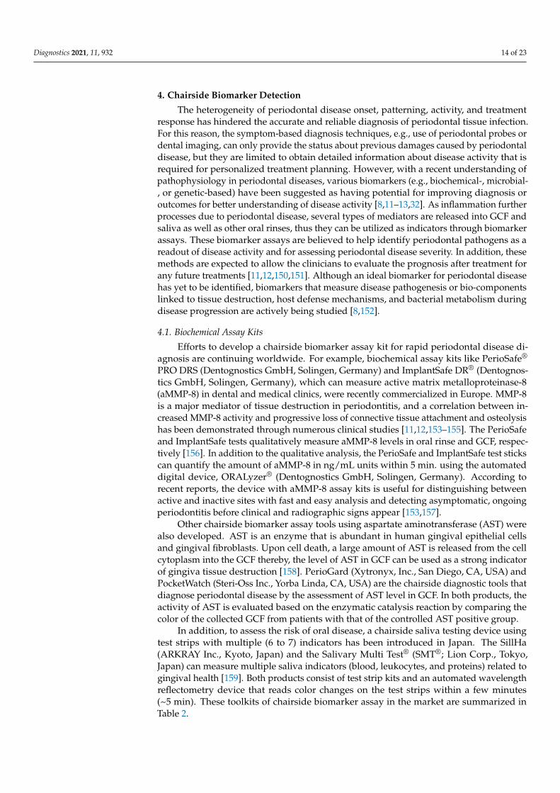
4. Chairside Biomarker Detection
The heterogeneity of periodontal disease onset, patterning, activity, and treatmentresponse has hindered the accurate and reliable diagnosis of periodontal tissue infection.For this reason, the symptom-based diagnosis techniques, e.g., use of periodontal probes ordental imaging, can only provide the status about previous damages caused by periodontaldisease, but they are limited to obtain detailed information about disease activity that isrequired for personalized treatment planning. However, with a recent understanding ofpathophysiology in periodontal diseases, various biomarkers (e.g., biochemical-, microbial-, or genetic-based) have been suggested as having potential for improving diagnosis oroutcomes for better understanding of disease activity [8,11–13,32]. As inflammation furtherprocesses due to periodontal disease, several types of mediators are released into GCF andsaliva as well as other oral rinses, thus they can be utilized as indicators through biomarkerassays. These biomarker assays are believed to help identify periodontal pathogens as areadout of disease activity and for assessing periodontal disease severity. In addition, thesemethods are expected to allow the clinicians to evaluate the prognosis after treatment forany future treatments [11,12,150,151]. Although an ideal biomarker for periodontal diseasehas yet to be identified, biomarkers that measure disease pathogenesis or bio-componentslinked to tissue destruction, host defense mechanisms, and bacterial metabolism duringdisease progression are actively being studied [8,152].
4.1. Biochemical Assay Kits
Efforts to develop a chairside biomarker assay kit for rapid periodontal disease di-agnosis are continuing worldwide. For example, biochemical assay kits like PerioSafe®
PRO DRS (Dentognostics GmbH, Solingen, Germany) and ImplantSafe DR® (Dentognos-tics GmbH, Solingen, Germany), which can measure active matrix metalloproteinase-8(aMMP-8) in dental and medical clinics, were recently commercialized in Europe. MMP-8is a major mediator of tissue destruction in periodontitis, and a correlation between in-creased MMP-8 activity and progressive loss of connective tissue attachment and osteolysishas been demonstrated through numerous clinical studies [11,12,153–155]. The PerioSafeand ImplantSafe tests qualitatively measure aMMP-8 levels in oral rinse and GCF, respec-tively [156]. In addition to the qualitative analysis, the PerioSafe and ImplantSafe test stickscan quantify the amount of aMMP-8 in ng/mL units within 5 min. using the automateddigital device, ORALyzer® (Dentognostics GmbH, Solingen, Germany). According torecent reports, the device with aMMP-8 assay kits is useful for distinguishing betweenactive and inactive sites with fast and easy analysis and detecting asymptomatic, ongoingperiodontitis before clinical and radiographic signs appear [153,157].
Other chairside biomarker assay tools using aspartate aminotransferase (AST) werealso developed. AST is an enzyme that is abundant in human gingival epithelial cellsand gingival fibroblasts. Upon cell death, a large amount of AST is released from the cellcytoplasm into the GCF thereby, the level of AST in GCF can be used as a strong indicatorof gingiva tissue destruction [158]. PerioGard (Xytronyx, Inc., San Diego, CA, USA) andPocketWatch (Steri-Oss Inc., Yorba Linda, CA, USA) are the chairside diagnostic tools thatdiagnose periodontal disease by the assessment of AST level in GCF. In both products, theactivity of AST is evaluated based on the enzymatic catalysis reaction by comparing thecolor of the collected GCF from patients with that of the controlled AST positive group.
In addition, to assess the risk of oral disease, a chairside saliva testing device usingtest strips with multiple (6 to 7) indicators has been introduced in Japan. The SillHa(ARKRAY Inc., Kyoto, Japan) and the Salivary Multi Test® (SMT®; Lion Corp., Tokyo,Japan) can measure multiple saliva indicators (blood, leukocytes, and proteins) related togingival health [159]. Both products consist of test strip kits and an automated wavelengthreflectometry device that reads color changes on the test strips within a few minutes(~5 min). These toolkits of chairside biomarker assay in the market are summarized inTable 2.

Diagnostics 2021, 11, 932 15 of 23
Table 2. Examples of biomarker assay kits in the market.
BiomarkerClassification Sampling From Product Name Detecting Target Detecting Principle Analyzing in
Biochemicalassay
GCF Periocheck Neutral proteasesEnzymatic digestion
reaction(Colorimetric assays)
Chairside
GCF PocketWatch ASTEnzymatic catalysis
reaction(Colorimetric assays)
GCF PerioGard ASTEnzymatic catalysis
reaction(Colorimetric assays)
Oral rinse PerioSafeaMMP-8
Lateral flow test withdigital reader (OraLyzer®)GCF ImplantSafe
Oral rinse SillHa ST-4910 Blood, leukocytes, andprotein
Lateral flow test withdual-wavelength
reflectometry
Microbiologicalassay
Subgingival plaque Evalusite Aa, Pg, PiSandwich enzyme
immunoassay(Colorimetric assays) Chairside
Subgingival plaque BANA-Enzymatictest kit Pg, Td, Tf BANA hydrolysis reaction
(Colorimetric assays)
Gums and plaque OMNIgene ORAL/OMR-110
Characterization ofvirus species of all
genome type including Aa,Pg, Pt, Fn, Td, Ec
DNA hybridization
Company orresearch
laboratory
Saliva OMNIgene ORAL/OM-501, 505
Subgingival plaque Carpegen® PerioDiagnostik
Aa, Pg, Tf, Td, Fn, Pi Real-time qPCR
Oral rinse MyPerioPath® Aa, Pg, Td, Tf, En, Fn, Pi, Cr,Pm, Ec, Cs DNA hybridization
Microbiologicalsamples/subgingival
plaqueiai Pado Test Aa, Pg, Pi, Td, Tf, Fa DNA hybridization
Subgingival plaque micro-IDent®plus11Aa, Pg, Pi, Tf, Td, Pm, Fn, Cr,
En, Ec, Cs DNA hybridization
Genetic assay
Cheek swab PerioPredict™ genes for IL-1 DNA hybridizationCompanylaboratoryOral rinse MyPerioID® IL-6 or
IL-1genes for IL-6 or IL-1 Genetic polymorphisms
detection
GCF: Gingival crevicular fluid, AST: Aspartate aminotransferase, aMMP: active Matrix metalloproteinase, Aa: Aggregatibacter actino-mycetemcomitans, Pg: Porphyromonas gingivalis, Pi: Prevotella intermedia, Td: Treponema denticola, Tf: Tannerella forsythia, Fn:Fusobacterium nucleatum, Ec: Eikenella corrodens, En: Eubacterium nodatum, Fn: Fusobacterium nucleatum/periodonticum, Cr: Campy-lobacter rectus, Pm: Peptostreptococcus (Micromonas) micros, Cs: Capnocytophaga species (gingivalis, ochracea, sputigena), Fa: Filifactoralocis, IL: Interleukin, qPCR: quantitative polymerase chain reaction.
4.2. Microbiological Assay Kits
Microbiological diagnostic tools also have been widely studied. It has been foundthat over 700 species of oral microbiome related to oral disease, and some are significantlyassociated with periodontal disease and can be used as microbiological biomarkers for thedisease diagnosis [54]. To date, the onset of periodontitis is related to pathogens includingPorphyromonas gingivalis (Pg), Treponema denticola (Td), Tannerella forsythia (Tf), Actinobacillusactinomycetemcomitans (Aa), Prevotella intermedia (Pi), Fusobacterium nucleatum (Fn), andFilifactor alocis (Fa) [158]. These bacteria generally co-locate in periodontal pockets with awide distribution and increased numbers suggesting that these can be potential biomarkers.Some periodontal pathogens, including Pg, Td, Tf, and some Capnocytophaga specie, producebacterial trypsin-like proteases by utilizing the hydrolysis reaction of BANA (N-BenzoylD-L Arginine -2 Naphalamide) in the biofilm. Thus, based on this hydrolysis reaction, theBANA-Enzymatic test™ kit (Ora Tec Corporation, Manassas, USA) is developed as rapid

Diagnostics 2021, 11, 932 16 of 23
and reliable chairside diagnostic tests. The Evalusite (Eastman Kodak company, Rochester,NY, USA) is a rapid microbiological assay kit that detects three recognized pathogens: Aa,Pg, and Pi. By collecting a subgingival plaque, the kit detects the presence of the pathogensbased on an antibody-bounded sandwich-type enzyme-linked immunosorbent within10 min. As a chairside diagnostic platform, OMNIgene (DNAgenotekTM, Ottawa, ON,Canada), iai PadoTest (IAI AG, Zuchwil, Switzerland), MyPerioPath® (OralDNA Labs,Eden Prairie, MN, USA), micro-IDent®plus11 (Hain Lifescience GmbH, Nehren, Germany),and Carpegen® Perio Diagnostik (Carpegen GmbH, Münster, Germany) have also beenintroduced. They detect several periodontal disease-related pathogens in collected saliva,oral rinse, or plaque based on nucleic acid-based assays. However, many of the microbialassay kits are available in laboratories with some expensive equipment.
4.3. Genetic Assay Kits
Analyzing the genetic “susceptibilities” may also help identify or anticipate the po-tential risk of periodontal disease initiation and progression. It has been known that thepolymorphism in the interleukin-1 (IL-1) gene has been shown to be proinflammation-causing periodontal disease [7]. To identify the genetic risk of periodontal disease, multipletest kits have been introduced in the market, including PerioPredict™ (Interleukin Genetics,Inc., Waltham, MA, USA), and MyPerioID® (IL-6 and IL-1; OralDNA Labs, Eden Prairie,MN, USA). However, these tests require significant laboratory equipment or additionaltime to deliver the sample to the manufacturer’s laboratories for data analysis. Thus, thesekits cannot be truly recognized as chairside diagnostics.
Despite several efforts on finding biomarkers, biomarker-based detection has been oc-casionally applied in dental offices. One reason for the low practical use of biomarker-basedtechniques is the lack of standardized assays. In addition, there is still no FDA-approvedsaliva diagnostic test or point-of-care technology for clinical diagnosis of periodontal dis-eases in the United States (as of September 2020). The good news is that the practicalapplication of biomarkers in periodontal disease diagnosis starts to be discussed in dentalresearch society. In the 2017 World Workshop, the introduction of biomarkers was stronglyencouraged as a supportive indicator to identify periodontal disease and to estimate itsstage and grade [5]. So far, the specific biomarkers and their thresholds have not been estab-lished yet, but we expect they will be incorporated and used in periodontal disease gradeassessment as evidence will become available soon. Efforts to accelerate the developmentof chairside periodontal assay kits or automated biosensors by combining clinically rele-vant biomarkers with lab-on-a-chip or point-of-care technologies are still active [8,11,158].Compared to conventional labor-intensive and time-consuming laboratory procedures,the automated chairside periodontal assay methods are believed to provide immediateanalysis results related to the disease. Moreover, technologies will evolve toward improv-ing diagnostic sensitivity and accuracy by analyzing multiple analytes simultaneously,although the size of the biosensor is reduced. Furthermore, the ability of chairside analysisof biomarkers for accurate diagnosis and prognosis of the disease will be an importantadvantage in preventing irreversible damage to periodontal bones and tissues.
5. Future Directions
Manual periodontal probing and 2D radiography have been the two major diagnostictools for periodontal disease. Over the years, various new technologies have been incorpo-rated into these two diagnostic tools in attempts to improve their accuracy, reproducibility,speed, and patient comfort (Figure 3). For periodontal probing in clinical examination,early attempts at improvements were derived from the incorporation of advanced mechan-ical and electrical technologies that enabled accurate and automated assessment. Varioussensors (e.g., calculus, temperature, sulfide, and pressure) are being integrated into theperiodontal probe platform to provide new information inside the periodontal pocket for acomprehensive analysis. With the development of microfabrication and nanotechnologyin the coming years, these sensors are further expected to be miniaturized and integrated

Diagnostics 2021, 11, 932 17 of 23
into the probe for multifunctional analysis. Furthermore, the approach of non-invasivetechnology that can quickly and accurately provide 3D information of the diseased area isexpected to reduce patient pain and discomfort.
Diagnostics 2021, 11, 932 18 of 24
observation, and based on this, it started to gain considerable interest from the periodontal disease research community [137,160]. Lastly, with the integration of state-of-the-art im-age processing algorithms and artificial intelligence technology, higher accuracy in diag-nosis and better prediction in prognosis are expected [98,99,161–163].
Figure 3. Advancement of chairside periodontal diagnostic tools and their future directions.
Although the current standard in the diagnosis of periodontal diseases is still mainly based on clinical examination and diagnostic imaging, recent advances in biomarkers pro-pose a new possibility in early-stage detection and rapid diagnosis. To date, large-scale laboratory assays and many clinical trials have been conducted to identify candidate bi-omarkers. Moreover, some promising biomarkers have been reported for simultaneous multi-analyte sensing to promote diagnosis accuracy. Based on these developments, it is plausible to believe that automated chairside diagnostic protocols with effective bi-omarkers will soon be available [158]. Overall, the diagnosis methods for periodontal dis-eases have continuously advanced with the incorporation of various technologies. It will be no surprise that this trend will continue as we experience more technological advance-ment and pathological/biological understanding. By embracing new technological devel-opments, clinicians may expand their chairside toolkits to identify, treat, and manage per-iodontal diseases.
Funding: This research was funded by ADA Foundation, grant number 97700140.
Acknowledgments: The authors would like to thank the ADASRI colleagues for their administra-tive and technical support.
Conflicts of Interest: The authors declare no conflict of interest.
References 1. Vos, T.; Abajobir, A.A.; Abate, K.H.; Abbafati, C.; Abbas, K.M.; Abd-Allah, F.; Abdulkader, R.S.; Abdulle, A.M.; Abebo, T.A.; Abera,
S.F.; et al. Global, regional, and national incidence, prevalence, and years lived with disability for 328 diseases and injuries for 195 countries, 1990–2016: A systematic analysis for the Global Burden of Disease Study 2016. Lancet 2017, 390, 1211–1259, doi:10.1016/S0140-6736(17)32154-2.
2. Herrera, D.; Retamal-Valdes, B.; Alonso, B.; Feres, M. Acute periodontal lesions (periodontal abscesses and necrotizing periodontal diseases) and endo-periodontal lesions. J. Clin. Periodontol. 2018, 45, S78–S94, doi:10.1111/jcpe.12941.
3. Jepsen, S.; Caton, J.G.; Albandar, J.M.; Bissada, N.F.; Bouchard, P.; Cortellini, P.; Demirel, K.; de Sanctis, M.; Ercoli, C.; Fan, J. Periodontal manifestations of systemic diseases and developmental and acquired conditions: Consensus report of workgroup
Figure 3. Advancement of chairside periodontal diagnostic tools and their future directions.
As radiography technology is becoming more developed, the radiograph imagingof the periodontal lesions has gained more significance in periodontal health assessment.In particular, the development of 3D imaging technology like CBCT allowed accuratevisualization of bone destruction, which then enabled the precise diagnosis of diseaseseverity and progress. Moreover, since 3D imaging technology is reported to successfullyvisualize bone and soft tissue using various radio-opaque agents, an accurate 3D mapof the periodontal pocket morphology is expected to be possible without painful tactileexamination for patients in the future [93]. However, diagnostic radiography exposes apatient to doses of ionizing radiation. As an alternative to radiography, other imagingtechnologies without ionizing radiation (e.g., ultrasonography or MRI) are being activelystudied. It was recently found that a method using MRI can be used for both soft tissueand hard tissue observation, and based on this, it started to gain considerable interestfrom the periodontal disease research community [137,160]. Lastly, with the integration ofstate-of-the-art image processing algorithms and artificial intelligence technology, higheraccuracy in diagnosis and better prediction in prognosis are expected [98,99,161–163].
Although the current standard in the diagnosis of periodontal diseases is still mainlybased on clinical examination and diagnostic imaging, recent advances in biomarkerspropose a new possibility in early-stage detection and rapid diagnosis. To date, large-scale laboratory assays and many clinical trials have been conducted to identify candidatebiomarkers. Moreover, some promising biomarkers have been reported for simultaneousmulti-analyte sensing to promote diagnosis accuracy. Based on these developments, it isplausible to believe that automated chairside diagnostic protocols with effective biomarkerswill soon be available [158]. Overall, the diagnosis methods for periodontal diseaseshave continuously advanced with the incorporation of various technologies. It will be nosurprise that this trend will continue as we experience more technological advancement andpathological/biological understanding. By embracing new technological developments,clinicians may expand their chairside toolkits to identify, treat, and manage periodontaldiseases.
Funding: This research was funded by ADA Foundation, grant number 97700140.

Diagnostics 2021, 11, 932 18 of 23
Acknowledgments: The authors would like to thank the ADASRI colleagues for their administrativeand technical support.
Conflicts of Interest: The authors declare no conflict of interest.
References1. Vos, T.; Abajobir, A.A.; Abate, K.H.; Abbafati, C.; Abbas, K.M.; Abd-Allah, F.; Abdulkader, R.S.; Abdulle, A.M.; Abebo, T.A.;
Abera, S.F.; et al. Global, regional, and national incidence, prevalence, and years lived with disability for 328 diseases and injuriesfor 195 countries, 1990–2016: A systematic analysis for the Global Burden of Disease Study 2016. Lancet 2017, 390, 1211–1259.[CrossRef]
2. Herrera, D.; Retamal-Valdes, B.; Alonso, B.; Feres, M. Acute periodontal lesions (periodontal abscesses and necrotizing periodontaldiseases) and endo-periodontal lesions. J. Clin. Periodontol. 2018, 45, S78–S94. [CrossRef] [PubMed]
3. Jepsen, S.; Caton, J.G.; Albandar, J.M.; Bissada, N.F.; Bouchard, P.; Cortellini, P.; Demirel, K.; de Sanctis, M.; Ercoli, C.; Fan, J.Periodontal manifestations of systemic diseases and developmental and acquired conditions: Consensus report of workgroup 3of the 2017 World Workshop on the Classification of Periodontal and Peri-Implant Diseases and Conditions. J. Clin. Periodontol.2018, 45, S219–S229. [CrossRef] [PubMed]
4. Caton, J.G.; Armitage, G.; Berglundh, T.; Chapple, I.L.; Jepsen, S.; Kornman, K.S.; Mealey, B.L.; Papapanou, P.N.; Sanz, M.; Tonetti,M.S. A new classification scheme for periodontal and peri-implant diseases and conditions—Introduction and key changes fromthe 1999 classification. J. Periodontol. 2018, 89, S1–S8. [CrossRef]
5. Tonetti, M.S.; Greenwell, H.; Kornman, K.S. Staging and grading of periodontitis: Framework and proposal of a new classificationand case definition. J. Periodontol. 2018, 89, S159–S172. [CrossRef] [PubMed]
6. Page, R.C.; Eke, P.I. Case Definitions for Use in Population-Based Surveillance of Periodontitis. J. Periodontol. 2007, 78, 1387–1399.[CrossRef]
7. Kinane, D.F.; Stathopoulou, P.G.; Papapanou, P.N. Periodontal diseases. Nat. Rev. Dis. Primers 2017, 3, 17038. [CrossRef][PubMed]
8. Gul, S.S.; Abdulkareem, A.A.; Sha, A.M.; Rawlinson, A. Diagnostic Accuracy of Oral Fluids Biomarker Profile to Determine theCurrent and Future Status of Periodontal and Peri-Implant Diseases. Diagnostics 2020, 10, 838. [CrossRef]
9. Socransky, S.S.; Haffajee, A.D.; Goodson, J.M.; Lindhe, J. New concepts of destructive periodontal disease. J. Clin. Periodontol.1984, 11, 21–32. [CrossRef] [PubMed]
10. Nomura, Y.; Morozumi, T.; Nakagawa, T.; Sugaya, T.; Kawanami, M.; Suzuki, F.; Takahashi, K.; Abe, Y.; Sato, S.; Makino-Oi, A.;et al. Site-level progression of periodontal disease during a follow-up period. PLoS ONE 2017, 12, e0188670. [CrossRef] [PubMed]
11. Steigmann, L.; Maekawa, S.; Sima, C.; Travan, S.; Wang, C.-W.; Giannobile, W.V. Biosensor and Lab-on-a-chip Biomarker-identifying Technologies for Oral and Periodontal Diseases. Front. Pharmacol. 2020, 11, 588480. [CrossRef]
12. Ghallab, N.A. Diagnostic potential and future directions of biomarkers in gingival crevicular fluid and saliva of periodontaldiseases: Review of the current evidence. Arch. Oral Biol. 2018, 87, 115–124. [CrossRef] [PubMed]
13. Taba, M., Jr.; Kinney, J.; Kim, A.S.; Giannobile, W.V. Diagnostic biomarkers for oral and periodontal diseases. Dent. Clin. N. Am.2005, 49, 551–571. [CrossRef]
14. Kostelc, J.G.; Zelson, P.R.; Preti, G.; Tonzetich, J. Quantitative differences in volatiles from healthy mouths and mouths withperiodontitis. Clin. Chem. 1981, 27, 842–845. [CrossRef]
15. Galgut, P. The relevance of pH to gingivitis and periodontitis. J. Int. Acad. Periodontol. 2001, 3, 61–67. [PubMed]16. Chapple, I.L.C. Periodontal diagnosis and treatment—Where does the future lie? Periodontol. 2000 2009, 51, 9–24. [CrossRef]17. Van der Velden, U.; Abbas, F.; Armand, S.; Loos, B.G.; Timmerman, M.F.; Van der Weijden, G.A.; Van Winkelhoff, A.J.; Winkel, E.G.
Java project on periodontal diseases. The natural development of periodontitis: Risk factors, risk predictors and risk determinants.J. Clin. Periodontol. 2006, 33, 540–548. [CrossRef] [PubMed]
18. Mdala, I.; Olsen, I.; Haffajee, A.D.; Socransky, S.S.; Thoresen, M.; de Blasio, B.F. Comparing clinical attachment level and pocketdepth for predicting periodontal disease progression in healthy sites of patients with chronic periodontitis using multi-stateMarkov models. J. Clin. Periodontol. 2014, 41, 837–845. [CrossRef] [PubMed]
19. International Organization for Standardization. Dentistry—Periodontal Probes—Part 1: General Requirements. Available online:https://www.iso.org/standard/53649.html (accessed on 24 December 2020).
20. International Organization for Standardization. Dentistry—Periodontal Probes—Part 2: Designation. Available online: https://www.iso.org/standard/53650.html (accessed on 24 December 2020).
21. Ramachandra, S.S.; Mehta, D.S.; Sandesh, N.; Baliga, V.; Amarnath, J. Periodontal probing systems: A review of availableequipment. Compend. Contin. Educ. Dent. 2011, 32, 71–77. [PubMed]
22. Polson, A.M.; Caton, J.G.; Yeaple, R.N.; Zander, H.A. Histological determination of probe tip penetration into gingival sulcus ofhumans using an electronic pressure-sensitive probe. J. Clin. Periodontol. 1980, 7, 479–488. [CrossRef]
23. Garnick, J.; Keagle, J.; Searle, J.; King, G.; Thompson, W. Gingival Resistance to Probing Forces: II. The Effect of Inflammation andPressure on Probe Displacement in Beagle Dog Gingivitis. J. Periodontol. 1989, 60, 498–505. [CrossRef]
24. Kleinberg, I.; Kaufman, H.W.; Wolff, M. Measurement of tooth hypersensitivity and oral factors involved in its development.Arch. Oral Biol. 1994, 39, S63–S71. [CrossRef]

Diagnostics 2021, 11, 932 19 of 23
25. Kour, A.; Kumar, A.; Puri, K.; Khatri, M.; Bansal, M.; Gupta, G. Comparative evaluation of probing depth and clinical attachmentlevel using a manual probe and Florida probe. J. Indian Soc. Periodontol. 2016, 20, 299–306. [CrossRef]
26. Renatus, A.; Trentzsch, L.; Schönfelder, A.; Schwarzenberger, F.; Jentsch, H. Evaluation of an Electronic Periodontal Probe Versusa Manual Probe. J. Clin. Diagn. Res. 2016, 10, ZH03–ZH07. [CrossRef] [PubMed]
27. Osborn, J.B.; Stoltenberg, J.L.; Huso, B.A.; Aeppli, D.M.; Pihlstrom, B.L. Comparison of measurement variability in subjects withmoderate periodontitis using a conventional and constant force periodontal probe. J. Periodontol. 1992, 63, 283–289. [CrossRef][PubMed]
28. Reddy, M.S.; Palcanis, K.G.; Geurs, N.C. A comparison of manual and controlled-force attachment-level measurements. J. Clin.Periodontol. 1997, 24, 920–926. [CrossRef]
29. Goodson, J.M. Diagnosis of Periodontitis by Physical Measurement: Interpretation From Episodic Disease Hypothesis. J.Periodontol. 1992, 63, 373–382. [CrossRef]
30. Lang, N.P.; Adler, R.; Joss, A.; Nyman, S. Absence of bleeding on probing An indicator of periodontal stability. J. Clin. Periodontol.1990, 17, 714–721. [CrossRef]
31. Joss, A.; Adler, R.; Lang, N.P. Bleeding on probing. A parameter for monitoring periodontal conditions in clinical practice. J. Clin.Periodontol. 1994, 21, 402–408. [CrossRef]
32. Trombelli, L.; Farina, R.; Silva, C.O.; Tatakis, D.N. Plaque-induced gingivitis: Case definition and diagnostic considerations. J.Clin. Periodontol. 2018, 45, S44–S67. [CrossRef]
33. Aldredge, W.A. Bleeding on probing defined. Dimens. Dent. Hyg. 2012, 10, 23–26.34. Newbrun, E. Indices to Measure Gingival Bleeding. J. Periodontol. 1996, 67, 555–561. [CrossRef] [PubMed]35. Räisänen, I.T.; Sorsa, T.; Tervahartiala, T.; Raivisto, T.; Heikkinen, A.M. Low association between bleeding on probing propensity
and the salivary aMMP-8 levels in adolescents with gingivitis and stage I periodontitis. J. Periodontal Res. 2021, 56, 289–297.[CrossRef] [PubMed]
36. Ito, H.; Numabe, Y.; Hashimoto, S.; Uehara, S.; Wu, Y.-H.; Ogawa, T. Usefulness of hemoglobin examination in gingival crevicularfluid during supportive periodontal therapy to diagnose the pre-symptomatic state in periodontal disease. Clin. Oral Investig.2021, 25, 487–495. [CrossRef]
37. Scipioni, A.; Bruschi, G.B.; Giargia, M.; Berglundh, T.; Lindhe, J. Healing at implants with and without primary bone contact. Clin.Oral Implant. Res. 1997, 8, 39–47. [CrossRef]
38. Laster, L.; Laudenbach, K.W.; Stoller, N.H. An evaluation of clinical tooth mobility measurements. J. Periodontol. 1975, 46, 603–607.[CrossRef]
39. Körber, K.H. Electronic registration of tooth movements. Int. Dent. J. 1971, 21, 466–477. [PubMed]40. Mühlemann, H.R. Tooth Mobility: The Measuring Method. Initial and Secondary Tooth Mobility. J. Periodontol. 1954, 25, 22–29.
[CrossRef]41. O’Leary, T.J. An instrument for measuring horizontal tooth mobility. Periodontics 1963, 1, 249.42. Wedendal, P.R.; Bjelkhagen, H.I. Dental holographic interferometry in vivo utilizing a ruby laser system: I. Introduction and
development of methods for precision measurements on the functional dynamics of human teeth and prosthodontic appliances.Acta Odontol. Scand. 1974, 32, 131–145. [CrossRef]
43. Wedendal, P.R.; Bjelkhagen, H.I. Dental holographic interferometry in vivo utilizing a ruby laser system II. Clinical applications.Acta Odontol. Scand. 1974, 32, 345–356. [CrossRef]
44. Castellini, P.; Revel, G.M.; Scalise, L.; De Andrade, R.M. Experimental and numerical investigation on structural effects of laserpulses for modal parameter measurement. Opt. Lasers Eng. 1999, 32, 565–581. [CrossRef]
45. Oka, H.; Shimizu, Y.; Saratani, K.; Shi, S.-G.; Kawazoe, T. Bender-type Tooth-Movement Transducer. IEEJ Trans. Sens. Micromach.1998, 118, 22–27. [CrossRef]
46. Hayashi, M.; Kobayashi, C.; Ogata, H.; Yamaoka, M.; Ogiso, B. A no-contact vibration device for measuring implant stability.Clin. Oral Implant. Res. 2010, 21, 931–936. [CrossRef]
47. Yamane, M.; Yamaoka, M.; Hayashi, M.; Furutoyo, I.; Komori, N.; Ogiso, B. Measuring tooth mobility with a no-contact vibrationdevice. J. Periodontal Res. 2008, 43, 84–89. [CrossRef]
48. Kobayashi, H.; Hayashi, M.; Yamaoka, M.; Yasukawa, T.; Ibi, H.; Ogiso, B. Evaluation of Qualitative Changes in SimulatedPeriodontal Ligament and Alveolar Bone Using a Noncontact Electromagnetic Vibration Device with a Laser DisplacementSensor. BioMed Res. Int. 2016, 2016, 9636513. [CrossRef] [PubMed]
49. Schulte, W.; D’Hoedt, B.; Lukas, D.; Maunz, M.; Steppeler, M. Periotest for measuring periodontal characteristics–Correlationwith periodontal bone loss. J. Periodontal Res. 1992, 27, 184–190. [CrossRef]
50. Schulte, W.; Lukas, D. The Periotest method. Int. Dent. J. 1992, 42, 433–440.51. Zanetti, E.M.; Pascoletti, G.; Calì, M.; Bignardi, C.; Franceschini, G. Clinical Assessment of Dental Implant Stability During
Follow-Up: What Is Actually Measured, and Perspectives. Biosensors 2018, 8, 68. [CrossRef]52. Huang, H.-M.; Chiu, C.-L.; Yeh, C.-Y.; Lin, C.-T.; Lin, L.-H.; Lee, S.-Y. Early detection of implant healing process using resonance
frequency analysis. Clin. Oral Implant. Res. 2003, 14, 437–443. [CrossRef]53. Park, J.-C.; Lee, J.-W.; Kim, S.-M.; Lee, J.-H. Implant Stability-Measuring Devices and Randomized Clinical Trial for ISQ Value
Change Pattern Measured from Two Different Directions by Magnetic RFA. In Implant Dentistry—A Rapidly Evolving Practice;Turkyilmaz, I., Ed.; IntechOpen: London, UK, 2011; pp. 112–128, ISBN 978-953-307-658-4.

Diagnostics 2021, 11, 932 20 of 23
54. The Forsyth Institute. Expanded Human Oral Microbiome Database. Available online: http://www.homd.org/index.php(accessed on 15 January 2021).
55. Paster, B.J.; Boches, S.K.; Galvin, J.L.; Ericson, R.E.; Lau, C.N.; Levanos, V.A.; Sahasrabudhe, A.; Dewhirst, F.E. Bacterial diversityin human subgingival plaque. J. Bacteriol. 2001, 183, 3770–3783. [CrossRef]
56. Loesche, W.J. Microbiology of Dental Decay and Periodontal Disease. In Medical Microbiology; Baron, S., Ed.; The University ofTexas Medical Branch: Galveston, TX, USA, 1996; ISBN 0-9631172-1-1.
57. White, D.J. Dental calculus: Recent insights into occurrence, formation, prevention, removal and oral health effects of supragingi-val and subgingival deposits. Eur. J. Oral Sci. 1997, 105, 508–522. [CrossRef] [PubMed]
58. Kingman, A.; Löe, H.; Ånerud, Å.; Boysen, H. Errors in Measuring Parameters Associated With Periodontal Health and Disease.J. Periodontol. 1991, 62, 477–486. [CrossRef]
59. Sherman, P.R.; Hutchens, L.H., Jr.; Jewson, L.G. The Effectiveness of Subgingival Scaling and Root Planing II. Clinical ResponsesRelated to Residual Calculus. J. Periodontol. 1990, 61, 9–15. [CrossRef] [PubMed]
60. Archana, V. Calculus detection technologies: Where do we stand now? J. Med. Life 2014, 7, 18–23.61. Sculean, A.; Schwarz, F.; Berakdar, M.; Romanos, G.E.; Arweiler, N.B.; Becker, J. Periodontal Treatment With an Er:YAG Laser
Compared to UltrasonicInstrumentation: A Pilot Study. J. Periodontol. 2004, 75, 966–973. [CrossRef] [PubMed]62. Tomasi, C.; Schander, K.; Dahlén, G.; Wennström, J.L. Short-Term Clinical and Microbiologic Effects of Pocket Debridement With
an Er:YAG Laser During Periodontal Maintenance. J. Periodontol. 2006, 77, 111–118. [CrossRef] [PubMed]63. Kung, R.T.V.; Ochs, B.; Goodson, J.M. Temperature as a periodontal diagnostic. J. Clin. Periodontol. 1990, 17, 557–563. [CrossRef]
[PubMed]64. Fedi, P.F., Jr.; Killoy, W.J. Temperature Differences at Periodontal Sites in Health and Disease. J. Periodontol. 1992, 63, 24–27.
[CrossRef]65. Gunupati, S.; Sappiti, H.; Nagarakanti, S.; Reddy, B.R.; Chava, V.K. Validating gingival surface temperature as an alternative tool
in the diagnosis of periodontal disease activity: An observational clinical trial. J. Dent. Res. Dent. Clin. Dent. Prospect. 2019, 13,123–127. [CrossRef]
66. Haffajee, A.D.; Socransky, S.S.; Goodson, J.M. Subgingival temperature (I). Relation to baseline clinical parameters. J. Clin.Periodontol. 1992, 19, 401–408. [CrossRef] [PubMed]
67. Niederman, R.; Naleway, C.; Lu, B.-Y.; Buyle-Bodin, Y.; Robinson, P. Subgingival temperature as a gingival inflammatory indicator.J. Clin. Periodontol. 1995, 22, 804–809. [CrossRef]
68. Singh, D.K.; Kumar, G. Comparison of the subgingival temperature of smokers and nonsmokers in healthy and diseased sites ofgingiva in association with sublingual body temperature. J. Fam. Med. Prim. Care 2019, 8, 3166–3172. [CrossRef] [PubMed]
69. Allam, P.; Vandana, K.L.; Khatri, M. A comparative assessment of subgingival temperature in bleeding and non-bleeding sitesbefore and after periodontal treatment. Indian J. Dent. Res. 2001, 12, 167–173. [PubMed]
70. Maiden, M.F.J.; Tanner, A.C.R.; Macuch, P.J.; Murray, L.; Kent, R.L., Jr. Subgingival temperature and microbiota in initialperiodontitis. J. Clin. Periodontol. 1998, 25, 786–793. [CrossRef]
71. Benson, P.E.; Khan, K.A.M. PerioTemp®vs. Infrared Thermometer for Measuring Gingival Temperature. In Proceedings of theInternational Association for Dental Research (IADR) Southeast Asian Division Meeting, Malacca, Malaysia, 5 September 2004.
72. Pavolotskaya, A.; McCombs, G.; Darby, M.; Marinak, K.; Dayanand, N.N. Sulcular Sulfide Monitoring: An Indicator of EarlyDental Plaque-Induced Gingival Disease. J. Dent. Hyg. 2006, 80, 11–22. [PubMed]
73. Abdullah, M.A.; Alasqah, M.; Sanaa, M.S.; Gufran, K. The Relationship between Volatile Sulfur Compounds and the Severity ofChronic Periodontitis: A Cross-sectional Study. J. Pharm. Bioallied Sci. 2020, 12, S268–S273. [CrossRef]
74. Morita, M.; Wang, H.-L. Relationship of Sulcular Sulfide Level to Severity of Periodontal Disease and BANA Test. J. Periodontol.2001, 72, 74–78. [CrossRef] [PubMed]
75. Zhou, H.; McCombs, G.; Darby, M.; Marinak, K. Sulphur by-product: The relationship between volatile sulphur compounds anddental plaque-induced gingivitis. J. Contemp. Dent. Pract. 2004, 5, 27–39. [CrossRef]
76. Tsai, C.-C.; Chou, H.-H.; Wu, T.-L.; Yang, Y.-H.; Ho, K.-Y.; Wu, Y.-M.; Ho, Y.-P. The levels of volatile sulfur compounds in mouthair from patients with chronic periodontitis. J. Periodontal Res. 2008, 43, 186–193. [CrossRef]
77. Makino, Y.; Yamaga, T.; Yoshihara, A.; Nohno, K.; Miyazaki, H. Association between Volatile Sulfur Compounds and PeriodontalDisease Progression in Elderly Non-Smokers. J. Periodontol. 2012, 83, 635–643. [CrossRef] [PubMed]
78. Khaira, N.; Palmer, R.; Wilson, R.; Scott, D.; Wade, W. Production of volatile sulphur compounds in diseased periodontal pocketsis significantly increased in smokers. Oral Dis. 2000, 6, 371–375. [CrossRef]
79. Torresyap, G.; Haffajee, A.; Uzel, N.; Socransky, S. Relationship between periodontal pocket sulfide levels and subgingival species.J. Clin. Periodontol. 2003, 30, 1003–1010. [CrossRef] [PubMed]
80. Beck, J.D.; Papapanou, P.N.; Philips, K.H.; Offenbacher, S. Periodontal Medicine: 100 Years of Progress. J. Dent. Res. 2019, 98,1053–1062. [CrossRef]
81. Shah, N.; Bansal, N.; Logani, A. Recent advances in imaging technologies in dentistry. World J. Radiol. 2014, 6, 794–807. [CrossRef][PubMed]
82. Kripal, K.; Dileep, A. Role of Radiographic Evolution: An Aid to Diagnose Periodontal Disease. In Periodontal Disease—Diagnosticand Adjunctive Non-Surgical Considerations; Yussif, N., Ed.; IntechOpen: London, UK, 2020; pp. 1–17, ISBN 978-1-78984-461-0.

Diagnostics 2021, 11, 932 21 of 23
83. Machado, V.; Proença, L.; Morgado, M.; Mendes, J.J.; Botelho, J. Accuracy of Panoramic Radiograph for Diagnosing PeriodontitisComparing to Clinical Examination. J. Clin. Med. 2020, 9, 2313. [CrossRef] [PubMed]
84. Le, L.H.; Nguyen, K.-C.T.; Kaipatur, N.R.; Major, P.W. Ultrasound for Periodontal Imaging. In Dental Ultrasound in Periodontologyand Implantology: Examination, Diagnosis and Treatment Outcome Evaluation; Chan, H.-L., Kripfgans, O.D., Eds.; Springer: Cham,Switzerland, 2021; pp. 115–129, ISBN 978-3-030-51288-0.
85. Zhang, W.; Rajani, S.; Wang, B.-Y. Comparison of periodontal evaluation by cone-beam computed tomography, and clinical andintraoral radiographic examinations. Oral Radiol. 2018, 34, 208–218. [CrossRef]
86. Górski, B.; Jalowski, S.; Górska, R.; Zaremba, M. Treatment of intrabony defects with modified perforated membranes inaggressive periodontitis: Subtraction radiography outcomes, prognostic variables, and patient morbidity. Clin. Oral Investig.2019, 23, 3005–3020. [CrossRef]
87. Kim, J.; Lee, H.-S.; Song, I.-S.; Jung, K.-H. DeNTNet: Deep Neural Transfer Network for the detection of periodontal bone lossusing panoramic dental radiographs. Sci. Rep. 2019, 9, 17615. [CrossRef]
88. Corbet, E.; Ho, D.; Lai, S. Radiographs in periodontal disease diagnosis and management. Aust. Dent. J. 2009, 54, S27–S43.[CrossRef]
89. Elashiry, M.; Meghil, M.M.; Kalathingal, S.; Buchanan, A.; Elrefai, R.; Looney, S.; Rajendran, M.; Ochieng, M.; Young, N.; Elawady,A.; et al. Application of radiopaque micro-particle fillers for 3-D imaging of periodontal pocket analogues using cone beam CT.Dent. Mater. 2018, 34, 619–628. [CrossRef]
90. Mohan, R.; Mark, R.; Sing, I.; Jain, A. Diagnostic Accuracy of CBCT for Aggressive Periodontitis. J. Clin. Imaging Sci. 2014, 4,2:1–2:5. [CrossRef]
91. Chakrapani, S.; Sirisha, K.; Srilalitha, A.; Srinivas, M. Choice of diagnostic and therapeutic imaging in periodontics andimplantology. J. Indian Soc. Periodontol. 2013, 17, 711–718. [CrossRef] [PubMed]
92. Jayachandran, S. Digital Imaging in Dentistry: A Review. Contemp. Clin. Dent. 2017, 8, 193–194. [CrossRef] [PubMed]93. McCann, D.; Fisch, S. Dental technology: Knocking at high-tech’s door. J. Am. Dent. Assoc. 1989, 118, 285–294. [CrossRef]
[PubMed]94. Iannucci, J.M.; Howerton, L.J. Dental Radiography: Principles and Techniques, 5th ed.; Elsevier: Amsterdam, The Netherlands, 2016;
ISBN 978-0-3232-9742-4.95. Brägger, U.; Pasquali, L.; Rylander, H.; Carnes, D.; Kornman, K.S. Computer-assisted densitometric image analysis in periodontal
radiography. J. Clin. Periodontol. 1988, 15, 27–37. [CrossRef]96. Bräegger, U.; Pasquali, L.; Weber, H.; Kornman, K.S. Computer-assisted densitometric image analysis (CADIA) for the assessment
of alveolar bone density changes in furcations. J. Clin. Periodontol. 1989, 16, 46–52. [CrossRef]97. Khalid, I.; Kumar, Y.; Rao, S. Use of Computer-Assisted Densitometric Image Analysis (CADIA) in Assessing Bone Density
Changes in Extraction Socket. Indian J. Stomatol. 2011, 2, 168–171.98. Krois, J.; Ekert, T.; Meinhold, L.; Golla, T.; Kharbot, B.; Wittemeier, A.; Dörfer, C.; Schwendicke, F. Deep Learning for the
Radiographic Detection of Periodontal Bone Loss. Sci. Rep. 2019, 9, 8495. [CrossRef]99. Lee, J.-H.; Kim, D.; Jeong, S.-N.; Choi, S.-H. Diagnosis and prediction of periodontally compromised teeth using a deep
learning-based convolutional neural network algorithm. J. Periodontal. Implant. Sci. 2018, 48, 114–123. [CrossRef]100. Eickholz, P.; Kim, T.-S.; Benn, D.K.; Staehle, H.J. Validity of radiographic measurement of interproximal bone loss. Oral Surg. Oral
Med. Oral Pathol. Oral Radiol. Endod. 1998, 85, 99–106. [CrossRef]101. Fuhrmann, R.A.W.; Bücker, A.; Diedrich, P.R. Assessment of alveolar bone loss with high resolution computed tomography. J.
Periodontal Res. 1995, 30, 258–263. [CrossRef]102. Ngan, D.; Kharbanda, O.P.; Geenty, J.P.; Darendeliler, M. Comparison of radiation levels from computed tomography and
conventional dental radiographs. Aust. Orthod. J. 2003, 19, 67–75.103. Suomalainen, A.; Kiljunen, T.; Käser, Y.; Peltola, J.; Kortesniemi, M. Dosimetry and image quality of four dental cone beam
computed tomography scanners compared with multislice computed tomography scanners. Dentomaxillofac. Radiol. 2009, 38,367–378. [CrossRef]
104. Vandenberghe, B.; Jacobs, R.; Bosmans, H. Modern dental imaging: A review of the current technology and clinical applicationsin dental practice. Eur. Radiol. 2010, 20, 2637–2655. [CrossRef]
105. Udupa, H.; Mah, P.; Dove, S.B.; McDavid, W.D. Evaluation of image quality parameters of representative intraoral digitalradiographic systems. Oral Surg. Oral Med. Oral Pathol. Oral Radiol. 2013, 116, 774–783. [CrossRef]
106. Choi, D.-H.; Choi, B.-R.; Choi, J.-W.; Huh, K.-H.; Yi, W.-J.; Heo, M.-S.; Choi, S.-C.; Lee, S.-S. Reference line-pair values of panoramicradiographs using an arch-form phantom stand to assess clinical image quality. Imaging Sci. Dent. 2013, 43, 7–15. [CrossRef][PubMed]
107. Brüllmann, D.; Schulze, R.K.W. Spatial resolution in CBCT machines for dental/maxillofacial applications-what do we knowtoday? Dentomaxillofac. Radiol. 2015, 44, 20140204. [CrossRef]
108. American Dental Association Council on Scientific Affairs. The use of cone-beam computed tomography in dentistry: Anadvisory statement from the American Dental Association Council on Scientific Affairs. J. Am. Dent. Assoc. 2012, 143, 899–902.[CrossRef] [PubMed]
109. Lee, S.; Gantes, B.; Riggs, M.; Crigger, M. Bone density assessments of dental implant sites: 3. Bone quality evaluation duringosteotomy and implant placement. Int. J. Oral Maxillofac. Implant. 2007, 22, 208–212.

Diagnostics 2021, 11, 932 22 of 23
110. Vandenberghe, B.; Jacobs, R.; Yang, J. Diagnostic validity (or acuity) of 2D CCD versus 3D CBCT-images for assessing periodontalbreakdown. Oral Surg. Oral Med. Oral Pathol. Oral Radiol. Endodontol. 2007, 104, 395–401. [CrossRef] [PubMed]
111. Mengel, R.; Candir, M.; Shiratori, K.; Flores-de-Jacoby, L. Digital Volume Tomography in the Diagnosis of Periodontal Defects: AnIn Vitro Study on Native Pig and Human Mandibles. J. Periodontol. 2005, 76, 665–673. [CrossRef] [PubMed]
112. Noujeim, M.; Prihoda, T.; Langlais, R.; Nummikoski, P. Evaluation of high-resolution cone beam computed tomography in thedetection of simulated interradicular bone lesions. Dentomaxillofac. Radiol. 2009, 38, 156–162. [CrossRef]
113. Webber, R.L.; Horton, R.A.; Tyndall, D.A.; Ludlow, J.B. Tuned-aperture computed tomography (TACT). Theory and applicationfor three-dimensional dento-alveolar imaging. Dentomaxillofac. Radiol. 1997, 26, 53–62. [CrossRef] [PubMed]
114. Nair, M.K.; Seyedain, A.; Agarwal, S.; Webber, R.L.; Nair, U.P.; Piesco, N.P.; Mooney, M.P.; Grondahl, H.G. Tuned ApertureComputed Tomography to Evaluate Osseous Healing. J. Dent. Res. 2001, 80, 1621–1624. [CrossRef]
115. Tyndall, D.; Kohltfarber, H. Application of cone beam volumetric tomography in endodontics. Aust. Dent. J. 2012, 57, 72–81.[CrossRef]
116. Cann, C.E. Quantitative CT for determination of bone mineral density: A review. Radiology 1988, 166, 509–522. [CrossRef]117. Irie, M.S.; Rabelo, G.D.; Spin-Neto, R.; Dechichi, P.; Borges, J.S.; Soares, P.B.F. Use of Micro-Computed Tomography for Bone
Evaluation in Dentistry. Braz. Dent. J. 2018, 29, 227–238. [CrossRef]118. Baum, G.; Greenwood, I.; Slawski, S.; Smirnow, R. Observation of Internal Structures of Teeth by Ultrasonography. Science 1963,
139, 495–496. [CrossRef] [PubMed]119. Mahmoud, A.M.; Ngan, P.; Crout, R.; Mukdadi, O.M. High-Resolution 3D Ultrasound Jawbone Surface Imaging for Diagnosis of
Periodontal Bony Defects: An In Vitro Study. Ann. Biomed. Eng. 2010, 38, 3409–3422. [CrossRef]120. Parmar, B.J.; Longsine, W.; Sabonghy, E.P.; Han, A.; Tasciotti, E.; Weiner, B.K.; Ferrari, M.; Righetti, R. Characterization of
controlled bone defects using 2D and 3D ultrasound imaging techniques. Phys. Med. Biol. 2010, 55, 4839–4859. [CrossRef][PubMed]
121. Zimbran, A.; Dudea, S.M.; Dudea, D. Evaluation of Periodontal Tissues Using 40MHz Ultrasonography. Preliminary report. Med.Ultrason. 2013, 15, 6–9. [CrossRef]
122. Izzetti, R.; Vitali, S.; Aringhieri, G.; Caramella, D.; Nisi, M.; Oranges, T.; Dini, V.; Graziani, F.; Gabriele, M. The efficacy ofUltra-High Frequency Ultrasonography in the diagnosis of intraoral lesions. Oral Surg. Oral Med. Oral Pathol. Oral Radiol. 2020,129, 401–410. [CrossRef]
123. Lynch, J.E.; Hinders, M.K.; McCombs, G.B. Clinical comparison of an ultrasonographic periodontal probe to manual andcontrolled-force probing. Measurement 2006, 39, 429–439. [CrossRef]
124. Nguyen, K.-C.T.; Le, L.H.; Kaipatur, N.R.; Zheng, R.; Lou, E.H.; Major, P.W. High-Resolution Ultrasonic Imaging of Dento-Periodontal Tissues Using a Multi-Element Phased Array System. Ann. Biomed. Eng. 2016, 44, 2874–2886. [CrossRef]
125. Van Luijk, J.A. NMR: Dental imaging without X-rays? Oral Surg. Oral Med. Oral Pathol. 1981, 52, 321–324. [CrossRef]126. International Commission on Non-Ionizing Radiation Protection. ICNIRP Statement on Diagnostic Devices Using Non-ionizing
Radiation: Existing Regulations and Potential Health Risks. Health Phys. 2017, 112, 305–321. [CrossRef]127. Assaf, A.T.; Zrnc, T.A.; Remus, C.C.; Schönfeld, M.; Habermann, C.R.; Riecke, B.; Friedrich, R.E.; Fiehler, J.; Heiland, M.; Sedlacik, J.
Evaluation of four different optimized magnetic-resonance-imaging sequences for visualization of dental and maxillo-mandibularstructures at 3 T. J. Craniomaxillofac. Surg. 2014, 42, 1356–1363. [CrossRef] [PubMed]
128. Niraj, L.K.; Patthi, B.; Singla, A.; Gupta, R.; Ali, I.; Dhama, K.; Kumar, J.K.; Prasad, M. MRI in Dentistry—A Future towardsRadiation Free Imaging—Systematic Review. J. Clin. Diagn. Res. 2016, 10, ZE14–ZE19. [CrossRef]
129. Prager, M.; Heiland, S.; Gareis, D.; Hilgenfeld, T.; Bendszus, M.; Gaudino, C. Dental MRI using a dedicated RF-coil at 3 Tesla. J.Cranio Maxillofac. Surg. 2015, 43, 2175–2182. [CrossRef]
130. Schara, R.; Sersa, I.; Skaleric, U. T1 relaxation time and magnetic resonance imaging of inflamed gingival tissue. Dentomaxillofac.Radiol. 2009, 38, 216–223. [CrossRef]
131. Ruetters, M.; Juerchott, A.; El Sayed, N.; Heiland, S.; Bendszus, M.; Kim, T.S. Dental magnetic resonance imaging for periodontalindication—A new approach of imaging residual periodontal bone support. Acta Odontol. Scand. 2019, 77, 49–54. [CrossRef][PubMed]
132. Gaudino, C.; Cosgarea, R.; Heiland, S.; Csernus, R.; Beomonte Zobel, B.; Pham, M.; Kim, T.-S.; Bendszus, M.; Rohde, S. MR-Imaging of teeth and periodontal apparatus: An experimental study comparing high-resolution MRI with MDCT and CBCT. Eur.Radiol. 2011, 21, 2575–2583. [CrossRef] [PubMed]
133. Geibel, M.A.; Schreiber, E.S.; Bracher, A.K.; Hell, E.; Ulrici, J.; Sailer, L.K.; Ozpeynirci, Y.; Rasche, V. Assessment of ApicalPeriodontitis by MRI: A Feasibility Study. Rofo 2015, 187, 269–275. [CrossRef]
134. Geibel, M.A.; Schreiber, E.; Bracher, A.K.; Hell, E.; Ulrici, J.; Sailer, L.K.; Rasche, V. Characterisation of apical bone lesions:Comparison of MRI and CBCT with histological findings—A case series. Eur. J. Oral. Implantol. 2017, 10, 197–211.
135. Juerchott, A.; Sohani, M.; Schwindling, F.S.; Jende, J.M.E.; Kurz, F.T.; Rammelsberg, P.; Heiland, S.; Bendszus, M.; Hilgenfeld, T.In vivo accuracy of dental magnetic resonance imaging in assessing maxillary molar furcation involvement: A feasibility study inhumans. J. Clin. Periodontol. 2020, 47, 809–815. [CrossRef]
136. Hilgenfeld, T.; Juerchott, A.; Jende, J.M.E.; Rammelsberg, P.; Heiland, S.; Bendszus, M.; Schwindling, F.S. Use of dental MRI forradiation-free guided dental implant planning: A prospective, in vivo study of accuracy and reliability. Eur. Radiol. 2020, 30,6392–6401. [CrossRef]

Diagnostics 2021, 11, 932 23 of 23
137. Algarín, J.M.; Díaz-Caballero, E.; Borreguero, J.; Galve, F.; Grau-Ruiz, D.; Rigla, J.P.; Bosch, R.; González, J.M.; Pallás, E.; Corberán,M.; et al. Simultaneous imaging of hard and soft biological tissues in a low-field dental MRI scanner. Sci. Rep. 2020, 10, 21470.[CrossRef]
138. Ahmad, I. Digital dental photography. Part 2: Purposes and uses. Br. Dent. J. 2009, 206, 459–464. [CrossRef]139. Ahmad, I. Digital dental photography. Part 1: An overview. Br. Dent. J. 2009, 206, 403–407. [CrossRef]140. Mackenzie, L.; Sharland, M. Dental photography: A practical guide. Dent. Update 2020, 47, 802–811. [CrossRef]141. Ting-shu, S.; Jian, S. Intraoral Digital Impression Technique: A Review. J. Prosthodont. 2015, 24, 313–321. [CrossRef] [PubMed]142. Suese, K. Progress in digital dentistry: The practical use of intraoral scanners. Dent. Mater. J. 2020, 39, 52–56. [CrossRef] [PubMed]143. Zhang, J.; Huang, Z.; Cai, Y.; Luan, Q. Digital assessment of gingiva morphological changes and related factors after initial
periodontal therapy. J. Oral Sci. 2021, 63, 59–64. [CrossRef]144. Icen, M.; Orhan, K.; Seker, Ç.; Geduk, G.; Özlü, F.C.; Cengiz, M.I. Comparison of CBCT with different voxel sizes and intraoral
scanner for detection of periodontal defects: An in vitro study. Dentomaxillofac. Radiol. 2020, 49, 20190197. [CrossRef]145. Meirelles, L.; Siqueira, R.; Garaicoa-Pazmino, C.; Yu, S.-H.; Chan, H.-L.; Wang, H.-L. Quantitative tooth mobility evaluation based
on intraoral scanner measurements. J. Periodontol. 2020, 91, 202–208. [CrossRef] [PubMed]146. Deferm, J.T.; Schreurs, R.; Baan, F.; Bruggink, R.; Merkx, M.A.W.; Xi, T.; Bergé, S.J.; Maal, T.J.J. Validation of 3D documentation
of palatal soft tissue shape, color, and irregularity with intraoral scanning. Clin. Oral Investig. 2018, 22, 1303–1309. [CrossRef][PubMed]
147. Lee, J.-S.; Jeon, Y.-S.; Strauss, F.-J.; Jung, H.-I.; Gruber, R. Digital scanning is more accurate than using a periodontal probe tomeasure the keratinized tissue width. Sci. Rep. 2020, 10, 3665. [CrossRef]
148. Nedelcu, R.; Olsson, P.; Nyström, I.; Rydén, J.; Thor, A. Accuracy and precision of 3 intraoral scanners and accuracy of conventionalimpressions: A novel in vivo analysis method. J. Dent. 2018, 69, 110–118. [CrossRef]
149. Townsend, D.; Francesco, D. Periodontal Capillary Imaging in vivo by Endoscopic Capillaroscopy. J. Med. Biol. Eng. 2010, 30,119–123.
150. Buduneli, N.; Kinane, D.F. Host-derived diagnostic markers related to soft tissue destruction and bone degradation in periodontitis.J. Clin. Periodontol. 2011, 38, 85–105. [CrossRef]
151. Slots, J. Periodontology: Past, present, perspectives. Periodontol. 2000 2013, 62, 7–19. [CrossRef]152. Taylor, J.J. Protein biomarkers of periodontitis in saliva. ISRN Inflamm. 2014, 2014, 593151. [CrossRef]153. Alassy, H.; Parachuru, P.; Wolff, L. Peri-Implantitis Diagnosis and Prognosis Using Biomarkers in Peri-Implant Crevicular Fluid:
A Narrative Review. Diagnostics 2019, 9, 214. [CrossRef] [PubMed]154. Nwhator, S.O.; Ayanbadejo, P.O.; Umeizudike, K.A.; Opeodu, O.I.; Agbelusi, G.A.; Olamijulo, J.A.; Arowojolu, M.O.; Sorsa, T.;
Babajide, B.S.; Opedun, D.O. Clinical Correlates of a Lateral-Flow Immunoassay Oral Risk Indicator. J. Periodontol. 2014, 85,188–194. [CrossRef]
155. Heikkinen, A.M.; Nwhator, S.O.; Rathnayake, N.; Mäntylä, P.; Vatanen, P.; Sorsa, T. Pilot Study on Oral Health Status as Assessedby an Active Matrix Metalloproteinase-8 Chairside Mouthrinse Test in Adolescents. J. Periodontol. 2016, 87, 36–40. [CrossRef]
156. Sorsa, T.; Alassiri, S.; Grigoriadis, A.; Räisänen, I.T.; Pärnänen, P.; Nwhator, S.O.; Gieselmann, D.-R.; Sakellari, D. Active MMP-8(aMMP-8) as a grading and staging biomarker in the periodontitis classification. Diagnostics 2020, 10, 61. [CrossRef] [PubMed]
157. Al-Majid, A.; Alassiri, S.; Rathnayake, N.; Tervahartiala, T.; Gieselmann, D.-R.; Sorsa, T. Matrix metalloproteinase-8 as aninflammatory and prevention biomarker in periodontal and peri-implant diseases. Int. J. Dent. 2018, 2018, 7891323. [CrossRef]
158. He, W.; You, M.; Wan, W.; Xu, F.; Li, F.; Li, A. Point-of-Care Periodontitis Testing: Biomarkers, Current Technologies, andPerspectives. Trends Biotechnol. 2018, 36, 1127–1144. [CrossRef] [PubMed]
159. Iwasaki, M.; Usui, M.; Ariyoshi, W.; Nakashima, K.; Nagai-Yoshioka, Y.; Inoue, M.; Nishihara, T. A Preliminary Study on theAbility of the Trypsin-Like Peptidase Activity Assay Kit to Detect Periodontitis. Dent. J. 2020, 8, 98. [CrossRef]
160. Flügge, T.; Hövener, J.B.; Ludwig, U.; Eisenbeiss, A.K.; Spittau, B.; Hennig, J.; Schmelzeisen, R.; Nelson, K. Magnetic resonanceimaging of intraoral hard and soft tissues using an intraoral coil and FLASH sequences. Eur. Radiol. 2016, 26, 4616–4623.[CrossRef] [PubMed]
161. Lee, J.-H.; Kim, D.-H.; Jeong, S.-N.; Choi, S.-H. Detection and diagnosis of dental caries using a deep learning-based convolutionalneural network algorithm. J. Dent. 2018, 77, 106–111. [CrossRef] [PubMed]
162. Joo, J.; Jeong, S.; Jin, H.; Lee, U.; Yoon, J.Y.; Kim, S.C. Periodontal Disease Detection Using Convolutional Neural Networks. InProceedings of the International Conference on Artificial Intelligence in Information and Communication (ICAIIC), Okinawa,Japan, 11–13 February 2019.
163. Lakshmi, T.K.; Dheeba, J. Digitalization in Dental problem diagnosis, Prediction and Analysis: A Machine Learning Perspectiveof Periodontitis. Int. J. Recent Technol. Eng. 2020, 67–74. [CrossRef]