Reduced Cystathionine γ-Lyase and Increased miR-21 Expression Are Associated with Increased...
11
VASCULAR BIOLOGY, ATHEROSCLEROSIS, AND ENDOTHELIUM BIOLOGY Reduced Cystathionine g-Lyase and Increased miR-21 Expression Are Associated with Increased Vascular Resistance in Growth-Restricted Pregnancies Hydrogen Sulfide as a Placental Vasodilator Tereza Cindrova-Davies,* Emilio A. Herrera,* y Youguo Niu,* John Kingdom, z Dino A. Giussani,* and Graham J. Burton* From the Department of Physiology, Development, and Neuroscience* and the Centre for Trophoblast Research, University of Cambridge, Cambridge, United Kingdom; the Physiopathology Program, y Faculty of Medicine, University of Chile, Santiago, Chile; and the Department of Obstetrics and Gynaecology, z Mount Sinai Hospital, University of Toronto, Toronto, Ontario, Canada Accepted for publication January 3, 2013. Address correspondence to Graham J. Burton, DSc, Department of Physiology, Development, and Neurosci- ence, University of Cambridge, Downing St., Cambridge, CB2 3EG, UK. E-mail: [email protected]. Increased vascular impedance in the fetoplacental circulation is associated with fetal hypoxia and growth restriction. We sought to investigate the role of hydrogen sulfide (H 2 S) in regulating vasomotor tone in the fetoplacental vasculature. H 2 S is produced endogenously by catalytic activity of cystathionine b-synthase and cystathionine g-lyase (CSE). Immunohistochemical analysis localized CSE to smooth muscle cells encircling arteries in stem villi. Immunoreactivity was reduced in placentas from pregnancies with severe early-onset growth-restriction and preeclampsia displaying abnormal umbilical artery Doppler waveforms compared with preeclamptic placentas with normal waveforms and controls. These findings were confirmed at the protein and mRNA levels. MicroRNA-21, which negatively regulates CSE expression, was increased in placentas with abnormal Doppler waveforms. Exposure of villus explants to hypoxia-reoxygenation significantly reduced CSE protein and mRNA and increased microRNA-21 expression. No changes were observed in cystathionine b-synthase expression, immunolocalized principally to the trophoblast, in pathologic placentas or in vitro. Finally, perfusion of normal placentas with an H 2 S donor, after pre- constriction with a thromboxane mimetic, resulted in dose-dependent vasorelaxation. Glibenclamide and N G -nitro-L-arginine methyl ester partially blocked the effect, indicating that H 2 S acts through ATP-sensitive K þ channels and nitric oxide synthesis. These results demonstrate that H 2 S is a powerful vasodilator of the placental vasculature and that expression of CSE is reduced in placentas associated with increased vascular resistance. (Am J Pathol 2013, 182: 1448e1458; http://dx.doi.org/10.1016/ j.ajpath.2013.01.001) One of the principal functions of the placenta is to act as a gas exchanger, which it achieves by bringing the maternal and fetal circulations into close approximation over a large surface area. The flow in these circulations is now routinely assessed noninvasively in vivo in high-risk pregnancies using Doppler ultrasound. 1 Umbilical artery Doppler velocimetry provides an indirect measure of downstream vascular resistance in the fetoplacental vasculature. 2 Pregnancies complicated by severe early-onset forms of preeclampsia (PE) 3 and/or intra- uterine growth restriction (IUGR) 4 typically exhibit increased resistance in the umbilical circulation, with absent or even reversed end-diastolic flow velocity. These highly abnormal Doppler flow patterns are associated with a poor perinatal prognosis, 5 in part because they identify a subset of preg- nancies with chronic fetal hypoxia, metabolic acidosis, and reduced circulating glucose and amino acid levels. 6e8 In the human placenta, the umbilical arteries branch into a series of resistance arteries contained in the distributing stem villi. These arteries eventually supply the capillary network in the terminal villi that are the principal sites of gaseous Supported by the Wellcome Trust (084804/2/08/Z). E.A.H. was partially funded by the U-Apoya Program, Universidad de Chile. T.C-D. and E.A.H. contributed equally to this work. Copyright ª 2013 American Society for Investigative Pathology. Published by Elsevier Inc. All rights reserved. http://dx.doi.org/10.1016/j.ajpath.2013.01.001 ajp.amjpathol.org The American Journal of Pathology, Vol. 182, No. 4, April 2013
Transcript of Reduced Cystathionine γ-Lyase and Increased miR-21 Expression Are Associated with Increased...

The American Journal of Pathology, Vol. 182, No. 4, April 2013
ajp.amjpathol.org
VASCULAR BIOLOGY, ATHEROSCLEROSIS, AND ENDOTHELIUM BIOLOGY
Reduced Cystathionine g-Lyase and Increased miR-21Expression Are Associated with Increased VascularResistance in Growth-Restricted Pregnancies
Hydrogen Sulfide as a Placental VasodilatorTereza Cindrova-Davies,* Emilio A. Herrera,*y Youguo Niu,* John Kingdom,z Dino A. Giussani,* and Graham J. Burton*
From the Department of Physiology, Development, and Neuroscience* and the Centre for Trophoblast Research, University of Cambridge, Cambridge, UnitedKingdom; the Physiopathology Program,y Faculty of Medicine, University of Chile, Santiago, Chile; and the Department of Obstetrics and Gynaecology,z
Mount Sinai Hospital, University of Toronto, Toronto, Ontario, Canada
Accepted for publication
C
P
h
January 3, 2013.
Address correspondence toGraham J. Burton, DSc,Department of Physiology,Development, and Neurosci-ence, University of Cambridge,Downing St., Cambridge,CB2 3EG, UK. E-mail:[email protected].
opyright ª 2013 American Society for Inve
ublished by Elsevier Inc. All rights reserved
ttp://dx.doi.org/10.1016/j.ajpath.2013.01.001
Increased vascular impedance in the fetoplacental circulation is associated with fetal hypoxia and growthrestriction. We sought to investigate the role of hydrogen sulfide (H2S) in regulating vasomotor tone in thefetoplacental vasculature. H2S is produced endogenously by catalytic activity of cystathionine b-synthaseand cystathionine g-lyase (CSE). Immunohistochemical analysis localized CSE to smooth muscle cellsencircling arteries in stem villi. Immunoreactivity was reduced in placentas from pregnancies with severeearly-onset growth-restriction and preeclampsia displaying abnormal umbilical artery Doppler waveformscomparedwith preeclamptic placentas with normal waveforms and controls. These findingswere confirmedat the protein and mRNA levels. MicroRNA-21, which negatively regulates CSE expression, was increased inplacentas with abnormal Doppler waveforms. Exposure of villus explants to hypoxia-reoxygenationsignificantly reduced CSE protein and mRNA and increased microRNA-21 expression. No changes wereobserved in cystathionine b-synthase expression, immunolocalized principally to the trophoblast, inpathologic placentas or in vitro. Finally, perfusion of normal placentas with an H2S donor, after pre-constriction with a thromboxane mimetic, resulted in dose-dependent vasorelaxation. Glibenclamide andNG-nitro-L-arginine methyl ester partially blocked the effect, indicating that H2S acts throughATP-sensitive Kþ channels and nitric oxide synthesis. These results demonstrate that H2S is a powerfulvasodilator of the placental vasculature and that expression of CSE is reduced in placentas associatedwith increased vascular resistance. (Am J Pathol 2013, 182: 1448e1458; http://dx.doi.org/10.1016/j.ajpath.2013.01.001)
Supported by the Wellcome Trust (084804/2/08/Z). E.A.H. was partiallyfunded by the U-Apoya Program, Universidad de Chile.T.C-D. and E.A.H. contributed equally to this work.
One of the principal functions of the placenta is to act as a gasexchanger, which it achieves by bringing the maternal andfetal circulations into close approximation over a large surfacearea. The flow in these circulations is now routinely assessednoninvasively in vivo in high-risk pregnancies using Dopplerultrasound.1 Umbilical artery Doppler velocimetry providesan indirect measure of downstream vascular resistance inthe fetoplacental vasculature.2 Pregnancies complicated bysevere early-onset forms of preeclampsia (PE)3 and/or intra-uterine growth restriction (IUGR)4 typically exhibit increasedresistance in the umbilical circulation, with absent or evenreversed end-diastolic flow velocity. These highly abnormal
stigative Pathology.
.
Doppler flow patterns are associated with a poor perinatalprognosis,5 in part because they identify a subset of preg-nancies with chronic fetal hypoxia, metabolic acidosis, andreduced circulating glucose and amino acid levels.6e8
In the human placenta, the umbilical arteries branch intoa series of resistance arteries contained in the distributing stemvilli. These arteries eventually supply the capillary network inthe terminal villi that are the principal sites of gaseous

Hydrogen Sulfide and Human Placenta
exchange. In the absence of nerves in the placenta, vasomotorcontrol of the resistance arteries is performed by locallyproduced vasoreactive molecules. To date, nitric oxide (NO)9
and carbonmonoxide (CO)10 have been shown to have dilatoractions in vitro. Accumulating studies suggest that hydrogensulfide (H2S) also exerts significant effects in the adultcardiovascular system, mainly via endothelium-independentvasodilation.11 However, its potential role in the placentalcirculation has not been investigated. We, therefore, investi-gated expression of the enzymes generatingH2S in pathologicplacentas with and without increased vascular resistance.
Endogenous H2S production is catalyzed by cystathionineb-synthase (CBS) and cystathionineg-lyase (CSE).12,13 CSE isthe dominant enzyme for synthesis ofH2S in the cardiovascularsystem, and reduced expression/activity and decreased H2Sexpression contribute to the pathogenesis of pulmonaryhypertension in rodents.14 Although CBS is not presentin vascular tissues, CBS deficiency leads to secondaryhypertension and endothelial dysfunction through hyper-homocysteinemia.15,16 The most compelling evidence for therole of H2S in the cardiovascular system comes from studiesusing CSE single and double knockout mice, where signifi-cantly higher systolic arterial blood pressure was reported inCSE�/� and, to a lesser extent,CSEþ/�- comparedwithCSEþ/þ
mice. In addition, CSE�/� mice are highly sensitive to H2S-mediated vasodilatation compared with wild-type mice.17 Themechanism of action of H2S has been best described in thenervous system, where ATP-sensitive Kþ (KATP) channels
18
and N-methyl-D-aspartate receptors19 have been identified asthe prime targets. Similarly, KATP channels present in vascularsmooth muscle cells (SMCs) were identified as the targetprotein of H2S, mediating its vascular effect.20 In addition, thevascular effect ofH2S can be potentiated byNO.
20,21 In viewofthese findings, we tested the effect of the KATP channel blockerglibenclamide and the NO synthesis blocker NG-nitro-L-arginine methyl ester (L-NAME) when applied with sodiumhydrosulfide (NaHS) in perfused human placentas.
In intrauterine tissues, Patel et al22 confirmed endogenousH2S placental production and release and the presence ofplacental CSE and CBS in rats and humans. Holwerdaet al23 showed decreased placental mRNA expression ofCBS in early-onset PE, and You et al24 showed down-regulation of CBS and CSE mRNA and of CBS proteinexpression in the myometrium during labor. However, thevasoactive effects of H2S in the fetoplacental vascular bedremain unexplored in any species.
We hypothesized that placentas from pregnanciescomplicated by the severe forms of PE and IUGR withabnormal umbilical artery Doppler waveforms show impairedendogenous H2S production secondary to a reduction in theH2S-producing enzymes. To test this hypothesis, the first aimwas to determine the expression of CSE and CBS incesarean-delivered human placentas from healthy pregnan-cies and from pregnancies complicated by IUGR or PE. Totest the relationship between local H2S production andumbilical artery Doppler waveforms in high-risk pregnancies,
The American Journal of Pathology - ajp.amjpathol.org
we additionally split the PE placentas into two groups withrespect to umbilical artery Doppler findings, ie, PE withabnormal umbilical artery Doppler findings (PE-AD) and PEwith normal umbilical artery Doppler findings and, thus,normal resistance (PE-ND). Because CSE protein and mRNAexpression were significantly reduced in IUGR and PE-ADsamples, we further explored its regulation by examiningthe expression of microRNA-21 (miR-21). Recent evidencesuggests that miR-21 overexpression participates in CSE/H2S-mediated SMC differentiation by repressing the proteinexpression of CSE and SP1, inhibiting H2S production,stimulating SMC proliferation, and reducing SMC differen-tiation.25 Second, because oxidative stress due to placentalischemia-reperfusion injury is a major component of theplacental pathology associated with PE and IUGR,26,27 weexamined the effects of hypoxia-reoxygenation (H/R) on thegene and protein expression of CSE and CBS and that ofmiR-21 in human placental explants in vitro. The final aimwas to test whether H2S plays a role in dilating the humanplacenta by perfusing preconstricted healthy human cesarean-delivered placentas with an H2S donor. The mechanism ofaction of H2S in the fetoplacental circulation was determinedusing the KATP channel blocker glibenclamide and the NOsynthesis blocker L-NAME.
Materials and Methods
Placental Collection and Ethical Approval
IUGR was diagnosed ultrasonically when abdominalcircumference measurements were below the 10th percen-tile28 and was confirmed at birth by a neonatal weight belowthe 10th percentile.29 Of the six IUGR cases, three hadnormal umbilical artery Doppler velocimetry findings, twohad abnormal umbilical arterial Doppler velocimetry find-ings, and one had absent end diastolic blood flow of theumbilical artery (Table 1). PE was defined as the onset ofgestational hypertension and proteinuria after 20 weeks ofgestation, and the cases examined herein were early-onsetPE. We additionally divided the PE category into twogroups based on the results of Doppler velocimetry, ie,PE-AD (n Z 7) and PE-ND (n Z 7). Hypertension wasdefined as two or more recordings of a diastolic bloodpressure of �90 mm Hg taken �4 hours apart (PE-AD:means � SEM systolic blood pressure, 171 � 14 mm Hg,and means� SEM diastolic blood pressure, 109� 7 mm Hg;PE-ND: means � SEM systolic blood pressure, 172 �15 mm Hg, and means � SEM diastolic blood pressure,102 � 4 mm Hg). Proteinuria was defined as the excretionof �300 mg of protein over 24 hours. The PE-AD groupincluded all the criteria for IUGR, and birth weight wassignificantly higher in the PE-ND group (10th to 50thpercentile) (Table 1).
Human placentas for perfusion and explant culture werecollected with Cambridge Local Ethical Committee approvaland informed written patient consent on an anonymous basis.
1449

Table 1 Clinical Information for Term Healthy and Pathologic Placentas
Characteristic
Group
Healthy IUGR PE-AD PE-ND
Gestational age (weeks) 39 � 0.6 32 � 3.5* 28.5 � 1.3* 29.5 � 1.5*Fetal weight (g) 3615 � 417 1045 � 465* 928 � 159* 1255 � 226*Umbilical artery Doppler H Z 6 NH Z 3
PEDF Z 2AREDF Z 1
PEDF Z 2AREDF Z 5
H Z 7
Data are presented as means � SD.*P < 0.001.AREDF, absent or reversed end-diastolic blood flow; H, healthy; PEDF, abnormal but present end-diastolic blood flow.
Cindrova-Davies et al
Gestational age was calculated from the last menstrual periodand was confirmed by routine ultrasonography at 11 to 12weeks of gestation. All the placentas were delivered at termby elective cesarean birth from nonlaboring normotensivehealthy singleton pregnancies, with no history of cigarettesmoking, diabetes, autoimmune diseases, or thrombophilicconditions.
Culture of Placental Explants
Placental tissue was collected from cesarean-delivered sin-gleton pregnancies within 10 minutes of delivery. Villoussamples were taken midway between the chorionic and basalplates from the periphery of three lobules free of visibleinfarction, calcification, hematoma, or tears on a randomsystematic basis. Samples were briefly rinsed in cold PBSand placed in ice-cold transport medium (TCS large-vesselendothelial cell basal medium; TCS Cellworks, MiltonKeynes, UK) containing 2% fetal bovine serum, heparin,hydrocortisone, human epidermal growth factor, humanbasic fibroblast growth factor, 25 mg/mL of gentamicin and50 ng/mL of amphotericin B, 1 mmol/L vitamin C, and1 mmol/L Trolox that had been equilibrated with 5%O2/90%N2/5% CO2.
After transport to the laboratory on ice for approximately1 hour, placental samples were further dissected into smallpieces approximately 5 mm in diameter in ice-cold culturemedium in a glove box under 10% O2/85% N2/5% CO2, aspreviously described.30 Briefly, 5-mm3 placental explantswere cultured on individual Costar Netwell inserts (CorningInc., Lowell, MA) (24-mm diameter, 500-mm mesh) in 4 mLof the large-vessel endothelial cell basal medium containing2% fetal bovine serum, heparin, hydrocortisone, humanepidermal growth factor, human basic fibroblast growthfactor, 25 mg/mL of gentamicin, and 50 ng/mL of ampho-tericin B, equilibrated with 5% O2/90% N2/5% CO2 per wellin 6-well plates for 24 hours. Approximately 6 to 10 pieceswere added to each well, depending on the experimentalrequirements. Placental explants were incubated in pre-gassed medium under normoxic conditions (controls) (10%O2/85% N2/5% CO2) or were subjected to hypoxia (0.5%O2/94.5% N2/5% CO2) for 3 hours and subsequent reox-ygenation at normoxia (10% O2/85% N2/5% CO2) for the
1450
following 20 hours (N Z 4) (H/R treatment). All the insertswere transferred into pre-gassed medium after 1 hour ofincubation. At the end of the 24-hour culture period, someplacental explants for each condition were flash frozen inliquid nitrogen and stored at �80�C until further processingfor mRNA or protein, and a few were fixed in 4% para-formaldehyde overnight for immunohistochemical analysis.
Western Blot Analysis
Frozen villous samples were homogenized in ice-cold lysisbuffer (1 mL of buffer per 100 mg of tissue) containing20 mmol/L Tris, pH 7.4, 1 mmol/L EGTA, 0.01% TritonX-100, 1 mmol/L sodium pyrophosphate, 1 mmol/L sodiumorthovanadate, 10 mmol/L b-glycerol phosphate, 50 mmol/Lsodium fluoride, and a complete mini protease inhibitorcocktail (Roche Diagnostics, East Sussex, UK). Tissuehomogenates were centrifuged at 15,000 � g, 4�C for 20minutes to remove tissue debris and the clear supernatants.Protein concentrations were determined on the supernatantusing a bicinchoninic acid protein assay kit (Sigma-Aldrich,Poole, UK). Lysates were mixed with 3� SDS-PAGEsample buffer, boiled for 5 minutes, and allowed to returnto room temperature. Equal amounts of protein (20 to 30 mg)were separated by SDS-PAGE using 10% polyacrylamideresolving gels and were transferred onto nitrocellulosemembrane (Invitrogen, Paisley, UK) and subjected to im-munoblot analysis. Membranes were blocked for 1 hour at25�C in 5% milk diluted in Tris-buffered saline and 0.1%Tween 20 and incubated with the following primary anti-bodies overnight at 4�C: anti-CSE (diluted 1:500; Abnova,Novus Biologicals, Cambridge, UK) and anti-CBS (diluted1:500; Sigma-Aldrich). After washing and incubating withsecondary antibodies, immunoreactive proteins werevisualized by the ECL Plus chemiluminescence system(Amersham Biosciences, Buckinghamshire, UK) followingthe manufacturer’s instructions. Protein bands were quan-tified using ImageJ software version 1.46r (NIH, Bethesda,MD). Protein loading was normalized against Ponceau Sstaining. We used Ponceau S as a loading control becauseit has been validated and found to be comparable withb-actin staining.31 The values are expressed as percentagesof the control lysate (100%) for each experiment.
ajp.amjpathol.org - The American Journal of Pathology

Hydrogen Sulfide and Human Placenta
RNA Isolation and Quantitative Real-Time RT-PCRAnalysis
Total RNA was isolated from human placentas using anRNeasy micro kit (Qiagen, Hilden, Germany). The RNA wasquantified by spectrophotometry (Nanodrop Technologies,Wilmington, DE). In brief, 20 mg of total RNA from eachsample was reverse transcribed using a master mix containingSuperScript II Reverse Transcriptase (Invitrogen). The ABIPRISM 7700 sequence detection system (ABI, Warrington,UK) was used to perform real-time PCR according to themanufacturer’s protocols. CT values for each transcript (ie,CSE, CBS) were compared with those for 18S rRNA. RNU48was used as an endogenous control for real-time PCR quanti-fication of miR21. All the primers and probes were obtainedfrom Applied Biosystems Assays-on-Demand (ABI) and useda 50 FAMreporter and a 30 nonfluorescentminor groovebinder.
Colorimetric Immunohistochemical Analysis
Paraformaldehyde-fixed tissues embedded in paraffin waxwere sectioned at 7 mm. After dewaxing and blocking ofendogenous peroxidases by incubation with 3% H2O2 for30 minutes, the sections were incubated with nonimmuneserum for 20 minutes. Primary antibodies [anti-CSE (diluted1:300; Abnova) and anti-CBS (diluted 1:200; Sigma-Aldrich)] were applied overnight at 4�C, and binding wasdetected using Vectastain Elite ABC kits (Vector Labora-tories, Burlingame, CA) and SigmaFast diaminobenzidine(Sigma-Aldrich) according to the manufacturers’ instruc-tions. Sections were lightly counterstained with hematox-ylin. When necessary, antigen retrieval was performedbefore blocking using 1 mmol/L Trise0.1 mmol/L EDTAbuffer, pH 9.0, in a pressure cooker for 2 minutes. Negativecontrols were performed by replacement of primary anti-body with equal concentrations of nonimmune serum.
Placental Perfusion
Perfusion pressure and flow (Transonic Systems Inc., Ithaca,NY)weremeasured in eight healthy cesarean-delivered humanplacentas. The placental perfusion protocol was modified fromthat of Brownbill and Sibley.32 Briefly, a single lobe from eachplacenta was perfused with equilibrated (95%O2/5% CO2, pH7.4) Earle’s bicarbonate buffer containing dextran 70 (mol. wt.70 to 80 kDa) (31390; Sigma-Aldrich), 1% bovine serumalbumin, 0.402 mmol/L L-arginine, and 10 UI/mL of heparin(Wockhardt UK Ltd., Wrexham, UK) by cannulating thechorionic artery and vein of the lobe. The basal pressure wassettled at 30 to 40 mm Hg, with a constant flow for �20minutes. The flow to achieve this perfusion pressure wasregulated between 5 and 8mL.min�1 (depending on the lobulesize), with a peristaltic pump giving a fixed pulsatility of 160bpm. The experiments were performed at a constant flow andpulsatility (rate). Placentas that did not stabilize pressure after20 minutes of a constant flow and rate were thus rejected.
The American Journal of Pathology - ajp.amjpathol.org
The catheters implanted in the chorionic artery were connectedto sterile pressure transducers set at the placental chorionic platelevel (COBE; Argon Division, MaxximMedical, Athens, TX),and these pressures were recorded continuously by a custom-built data acquisition system (Maastricht-ProgrammableAcQuisition system; Maastricht Instruments, Maastricht,The Netherlands) at a 500-Hz sample rate. The Gilson Min-ipuls 2 peristaltic pumps (Anachem, Luton, UK) used wereappropriately set for each placenta to maintain 30- to 40-mmHg arterial pressure (simulating the in vivo physiologicumbilical arterial pressure). To fix flow without significantlyaltering the pulsatility, we used different inside diameters forthe tubing in the peristaltic pump.
After preconstriction with the thromboxane mimeticU46619 (10�7 mol/L; Cayman Chemical, Ann Arbor, MI),increasing doses of NaHS solution (10�12 to 10�6 mol/L)were infused. Glibenclamide (10�5 mol/L) was adminis-tered to block KATP channels and L-NAME (10�5 mol/L) toinhibit endogenous NO synthesis in a continuous mannerin the perfusate before preconstriction with U46619. Theplacenta reached steady state constriction after 5 to 10minutes of the U46619 infusion. After 3 minutes of steadyplateau, the increasing doses of NaHS were given. TheL-NAME and glibenclamide blockades were given in theinfusion (perfusate), and the NaHS was administered in 5-mL boluses, replacing the perfusate infusion (therefore,the concentration in the lumen is that infused during the 40to 60 seconds that last the bolus, depending on the flowrate). Between boluses, a waiting period of 3 to 5 minuteswas introduced (with the perfusate) until reaching a plateauin the vasodilator response.
Flow and pressure were continuously recorded by a 2Rflow probe connected to a T106 monitor (Transonic SystemsInc.) and the Maastricht-Programmable AcQuisition system.The flow was compared with the venous output, confirmingthe integrity of the vessel network at the beginning and endof the experiment. Doses of U46619, glibenclamide, andL-NAME were optimized in pilot experiments.
Statistical Analysis
Data are expressed asmeans� SEM. Comparisons weremadeusing a 2-tailed t-test or analysis of variance with a StudentNewman-Keuls post hoc test or Wilcoxon nonparametric testwhere appropriate. Differences were considered to be signifi-cant at P � 0.05.
Results
Localization and Expression of CBS and CSE in Healthyand Pathologic Human Placentas
CBSwas localized only to the syncytiotrophoblast (Figure1A).In contrast, CSE was immunolocalized principally to theSMCs surrounding the large placental stem villus arteries(Figure 1B). We investigated whether the potential for
1451

Figure 1 Placental immunostaining. Localiza-tion of CBS (A) and CSE (B) in human placentasfrom healthy cesarean-delivered (CS), IUGR, PE-AD, and PE-ND pregnancies and localization ofCSE in term human placental explants cultured atdifferent oxygen concentrations [ie, normoxia(10% O2 for 20 hours) and H/R (0.5% O2/10% O2for 20 hours)] (C). D: Negative control stained byreplacing primary antibody with nonimmuneserum. Brown color signifies positive staining. CBSlocalized in the syncytiotrophoblast, whereas CSEwas predominantly expressed by SMCs of largeplacental arteries. Scale bars Z 100 mm.
Cindrova-Davies et al
synthesis of H2S was deficient in placentas from pregnanciescomplicated by PE and IUGR. Protein (Figure 2) and mRNA(Figure 3) expression levels of CSE were significantlydecreased in IUGR and PE-AD placentas compared withhealthy placentas delivered by cesarean birth. However, thiswas not the case in PE-ND samples, which had unalteredmRNA CSE expression (Figure 3) and even marginallyincreased CSE protein expression (Figure 2C). Reducedplacental expression of CSE thus associated with increasedplacental vascular resistance, as assessed by umbilical Dopplervelocimetry. There were no differences in protein (Figure 2) ormRNA (Figure 3) levels of CBS among the four differentgroups. The protein expression ofCBS andCSEwas examinedby Western blot analysis, and bands of approximately 43 kDacorresponding to CSE and approximately 61 kDa corre-sponding to CBS were recognized. Immunohistochemicalanalysis results matched the protein findings (Figure 1, A andB). We used a semiquantitative scale from 0 to 3 to analyze sixrandomly selected areas per four slides per group at �40magnification: 0Z no staining, 1Zweak intensity of staining,2 Z moderate staining, and 3 Z strong staining. Randomlyselected slides showed that controls exhibited a staining scoreof 2.6; PE-AD, 1.7; and PE-ND, 2.7.
1452
We used term cesarean-delivered healthy placentas ascontrols for these experiments. Significant differences inmeans � SEM gestational ages among control (39 � 0.6weeks), IUGR (32 � 3.5 weeks), PE-AD (28.5 � 1.3weeks), and PE-ND (29.5 � 1.5 weeks) samples (Table 1)reflect the inability to obtain cesarean-delivered healthypreterm controls to match the gestational age of severeIUGR and PE samples that were delivered prematurely forclinical reasons. However, no differences in CBS mRNAexpression were reported between first-trimester and termhuman placentas.33 It is, thus, unlikely that the reduction inCSE expression in pathologic pregnancies is due to differ-ences in gestational age. Both proteins are present in earlyplacental samples and display the same localization as interm samples (data not shown).
miR-21 as a Potential Regulator of CSE Expression
Because recent evidence suggests that SMC CSE expressioncould be regulated by miR-21,25 we quantified miR-21expression using quantitative real-time-PCR in PE andIUGR placentas compared with controls and found signifi-cant up-regulation of miR-21 expression in IUGR and
ajp.amjpathol.org - The American Journal of Pathology
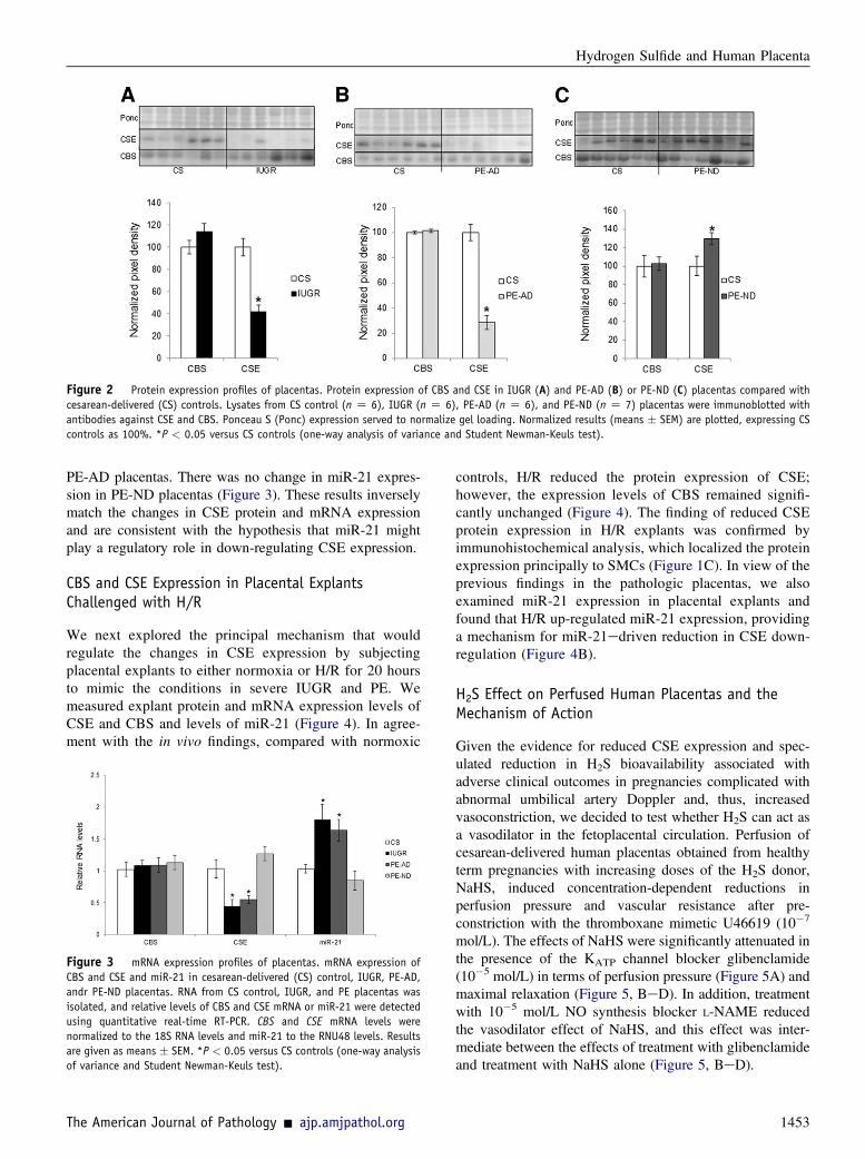
Figure 2 Protein expression profiles of placentas. Protein expression of CBS and CSE in IUGR (A) and PE-AD (B) or PE-ND (C) placentas compared withcesarean-delivered (CS) controls. Lysates from CS control (n Z 6), IUGR (n Z 6), PE-AD (n Z 6), and PE-ND (n Z 7) placentas were immunoblotted withantibodies against CSE and CBS. Ponceau S (Ponc) expression served to normalize gel loading. Normalized results (means � SEM) are plotted, expressing CScontrols as 100%. *P < 0.05 versus CS controls (one-way analysis of variance and Student Newman-Keuls test).
Hydrogen Sulfide and Human Placenta
PE-AD placentas. There was no change in miR-21 expres-sion in PE-ND placentas (Figure 3). These results inverselymatch the changes in CSE protein and mRNA expressionand are consistent with the hypothesis that miR-21 mightplay a regulatory role in down-regulating CSE expression.
CBS and CSE Expression in Placental ExplantsChallenged with H/R
We next explored the principal mechanism that wouldregulate the changes in CSE expression by subjectingplacental explants to either normoxia or H/R for 20 hoursto mimic the conditions in severe IUGR and PE. Wemeasured explant protein and mRNA expression levels ofCSE and CBS and levels of miR-21 (Figure 4). In agree-ment with the in vivo findings, compared with normoxic
Figure 3 mRNA expression profiles of placentas. mRNA expression ofCBS and CSE and miR-21 in cesarean-delivered (CS) control, IUGR, PE-AD,andr PE-ND placentas. RNA from CS control, IUGR, and PE placentas wasisolated, and relative levels of CBS and CSE mRNA or miR-21 were detectedusing quantitative real-time RT-PCR. CBS and CSE mRNA levels werenormalized to the 18S RNA levels and miR-21 to the RNU48 levels. Resultsare given as means � SEM. *P < 0.05 versus CS controls (one-way analysisof variance and Student Newman-Keuls test).
The American Journal of Pathology - ajp.amjpathol.org
controls, H/R reduced the protein expression of CSE;however, the expression levels of CBS remained signifi-cantly unchanged (Figure 4). The finding of reduced CSEprotein expression in H/R explants was confirmed byimmunohistochemical analysis, which localized the proteinexpression principally to SMCs (Figure 1C). In view of theprevious findings in the pathologic placentas, we alsoexamined miR-21 expression in placental explants andfound that H/R up-regulated miR-21 expression, providinga mechanism for miR-21edriven reduction in CSE down-regulation (Figure 4B).
H2S Effect on Perfused Human Placentas and theMechanism of Action
Given the evidence for reduced CSE expression and spec-ulated reduction in H2S bioavailability associated withadverse clinical outcomes in pregnancies complicated withabnormal umbilical artery Doppler and, thus, increasedvasoconstriction, we decided to test whether H2S can act asa vasodilator in the fetoplacental circulation. Perfusion ofcesarean-delivered human placentas obtained from healthyterm pregnancies with increasing doses of the H2S donor,NaHS, induced concentration-dependent reductions inperfusion pressure and vascular resistance after pre-constriction with the thromboxane mimetic U46619 (10�7
mol/L). The effects of NaHS were significantly attenuated inthe presence of the KATP channel blocker glibenclamide(10�5 mol/L) in terms of perfusion pressure (Figure 5A) andmaximal relaxation (Figure 5, BeD). In addition, treatmentwith 10�5 mol/L NO synthesis blocker L-NAME reducedthe vasodilator effect of NaHS, and this effect was inter-mediate between the effects of treatment with glibenclamideand treatment with NaHS alone (Figure 5, BeD).
1453

Figure 4 Protein and mRNA expression profiles in placental explants.Protein (A) and mRNA (B) expression of CBS and CSE and miR-21 expression(B) in term placental explants cultured at different oxygen concentrations.A: Lysates from term placental explants (nZ 4), which were cultured undernormoxia (10% O2) or H/R (1% to 10% O2) for 20 hours, were immuno-blotted with antibodies against CSE and CBS. Ponceau S (Ponc) expressionserved to normalize gel loading. Normalized results (means � SEM) areplotted, expressing cesarean-delivered (CS) controls as 100%. *P < 0.05versus CS controls (analysis of variance and the Protected Least SignificantDifference test with P < 0.05). B: RNA was isolated and relative levels ofCBS and CSE mRNA or miR-21 were detected using quantitative real-timeRT-PCR. CBS and CSE mRNA levels (means � SEM) were normalized to the18S RNA levels and miR-21 to the RNU48 levels. *P < 0.05 H/R versusnormoxic controls (Wilcoxon nonparametric test).
Cindrova-Davies et al
Discussion
These studies demonstrate that reduced placental CSEexpression at the mRNA and protein levels is associatedwith abnormal umbilical artery Doppler waveforms in high-risk pregnancies. These are novel observations that need tobe further confirmed. They allude to the potential role ofdecreased H2S availability in the pathogenesis of increasedumbilical arterial resistance in cases of severe pretermIUGR. We suggest that these changes are epigeneticallyregulated because the expression of miR-21, which has beenreported to play a role in CSE down-regulation,25 is elevatedin these abnormal placentas. We mimicked the changes inCSE and miR-21 expression in normal placental explantssubjected to H/R, providing a mechanism to integrate thesefindings with the known underlying pathology of the ute-roplacental circulation.34 CSE protein and mRNA expres-sion were unaffected in placentas from PE-ND pregnancies,
1454
implying that H2S acts as a local fetoplacental vasodilatoragent. This was confirmed by administering increasingdoses of the H2S donor, NaHS, which decreased vascularresistance in a dose-dependent manner. We found the effectsof NaHS to be significantly attenuated in the presence of theKATP channel blocker glibenclamide in terms of perfusionpressure and maximal relaxation; however, the effect oftreatment with the NO synthesis blocker L-NAME wasintermediate between the effects of treatment with gliben-clamide and treatment with NaHS alone. These observationsimply that the primary dilator actions of H2S in the humanplacenta are mediated via KATP channels and that there is anadditional interaction between H2S and NO that functions tomodulate vascular tone. There are potential interactionsbetween NO and H2S for NO donor treatmenteenhancedendogenous production of H2S from rat aortic tissues.NO can increase CSE activity in vascular tissues viacGMP-dependent protein kinases and by regulating CSEexpression.20
Patel et al22 confirmed endogenous H2S production andthe presence of CSE and CBS in rat and human intrauterinetissues. These authors additionally reported endogenousH2S production in the human placenta but did not identifythe cellular source. Herein, we immunolocalized CSE to theSMCs of large placental vessels and to the syncytio-trophoblast; however, CBS expression was confined to theouter syncytiotrophoblast layer of placental villi. Thispattern matched reports of different tissue localizations ofthe two enzymes in other systems. CSE is expressed invascular SMCs but not in the endothelium,17,20,35 whereasCBS is predominantly found in the brain. CBS proteinlocalizes in most areas of the brain but is predominantlyexpressed in the cell bodies and neuronal processes ofPurkinje cells and Ammon’s horn neurons,36,37 and it doesnot play an important role in cardiovascular tissues.38
We investigated whether the potential for synthesis ofH2S was deficient in placentas from pathologic pregnancies.PE and IUGR are the most serious perinatal complicationsof human pregnancy that adversely affect maternal and fetalhealth. They are associated with deficient uteroplacentalvascular conversion, leading to maternal placental malper-fusion.26,27 In the more severe cases, there is evidence ofassociated increased resistance in the umbilical circulation,resulting in reduced, or even reversed, end-diastolic flow.1
Therefore, it was of interest to determine the localizationof the two H2S-generating enzymes in these placentas. Wefound that the protein and RNA expression levels of CSEwere significantly decreased in IUGR placentas and in PE-AD placentas, which also had a trajectory for IUGR,compared with cesarean-delivered healthy placentas or PE-ND placentas. These are striking results and suggest thatreduced H2S production could contribute to fetoplacentalvasoconstriction in these growth-restricted placentas. Therewere no differences in CBS expression among the fourdifferent groups. In the human placenta, the terminal villiare the principal sites of gaseous exchange. The capillary
ajp.amjpathol.org - The American Journal of Pathology

Figure 5 Perfusion pressure and vasodilatorresponse in human placentas. A: Perfusion pres-sure (percentage of U46619 Michaelis constantKm) during the experimental protocol for twohealthy human placentas. Top arrows showpreconstriction with the thromboxane mimeticU46619. Bottom arrows show logarithmic scale ofNaHS concentration. Different line colors representdifferent placentas. Set of curves: basal response(untreated) (left panel); responses in the pres-ence of the NO synthase blocker L-NAME (middlepanel); and responses in the presence of the KATPblocker glibenclamide (right panel). B: Vasodi-lator response to NaHS in human placentas. Valuesare means � SEM for the concentration-responsecurves (B), the area above the curve (C), and themaximal relaxation (%Rmax) (D) to NaHS. Groupsare untreated placentas (C; n Z 6), placentastreated with the NO synthase blocker L-NAME (LN;n Z 6), and placentas treated with the KATPblocker glibenclamide (Glib; n Z 6). *P < 0.05versus the untreated group; yP < 0.05 versus theL-NAME group (one-way analysis of variance andStudent Newman-Keuls test).
Hydrogen Sulfide and Human Placenta
network within them is supplied by the umbilical arteriesthat branch into a series of resistance arteries contained inthe distributing stem villi. As there is no nerve supply in theplacenta, vasomotor control of the resistance arteries isperformed by locally produced vasoactive molecules. Thesyncytiotrophoblast expresses endothelial NO synthase,heme oxygenase, and CBS and, as such, produces NO, CO,and H2S locally. The half-life of these gaseous molecules isseconds (NO) to minutes (CO and H2S). In addition, NOand CO are produced by vascular endothelial cells, whereasH2S is produced by vascular SMCs. These vasodilators arelikely to interact, mediate the vascular tone locally, and actas signaling molecules.39
We then investigated a potential mechanism that controlsCSE suppression. Yang et al25 recently reported the in-volvement of miR-21 in down-regulation of the SMCexpression of CSE. miRNAs are endogenous small single-strand noncoding RNAs of approximately 22 nucleotidesthat control gene expression by targeting the 30 untransla-tional regions of mRNAs for degradation and/or translationalrepression.40e42 miR-21, an oncogenic antiapoptotic miRNAthat can affect proliferation, migration, and invasion,43,44 hasbeen shown to be aberrantly overexpressed in some cardio-vascular conditions, such as cardiac hypertrophy induced byaortic banding, which could be reversed by miR-21 down-
The American Journal of Pathology - ajp.amjpathol.org
regulation,45 or the heart tissue of patients with end-stageheart failure.46 Angiotensin II can increase miR-21 expres-sion in neonatal rat cardiomyocytes in vitro.47 These datasuggest that miR-21 could be a new pathologic factor andpotentially a therapeutic target in cardiovascular disorders,including complicated pregnancies with increased vascularresistance.
Most of the pathologic placentas were associated withabnormal umbilical Doppler velocimetry. Altered vascularreactivity in IUGR could be a consequence of reducedgeneration of vasodilators, increased expression of constric-tors, modified oxygen tension, and/or oxidative stress. Thesefindings suggest that reduced CSE expression and conse-quent reduced H2S bioavailability contribute to the IUGRpathology by increasing resistance in the umbilical circula-tion, which manifests as reduced or even reversed end-diastolic blood flow. The study design did not allow us totest this hypothesis directly. Such experiments would requirethe administration of an enzyme substrate to placentas fromhealthy and complicated pregnancies, the worthy subject ofa future investigation.
Note that in the perfusion protocol, the perfusion mediumwas gassed with 95% O2. Recently, some researchers havemoved away from using 95% O2. However, it is yet to bedetermined what is the best percentage of O2 to use for
1455

Cindrova-Davies et al
a perfusion preparation to compensate for the lack of hemo-globin. Most perfusion setups are focused on placental transferand metabolism, and few protocols have been published toassess vascular chorionic reactivity. We used this 95% O2
mixture based on our previous unpublished experiments andthose of others.48,49 Although there is evidence that somevasoconstrictors may have better responses at low oxygenlevels in isolated arteries and veins,50,51 there is no consensusas to different responses to oxygenation changes.
Holwerda et al23 also studied the expression of CBS andCSE in early-onset PE samples and reported a reduction inthe mRNA expression of CBS and no changes in the proteinor RNA expression of CSE or in the protein expression ofCBS. The reasons for the differences in these findings areunclear, but they could be a reflection of several factors,including antibody specificity, timing of tissue collection,and/or variation in the severity of the pathology. To rein-force these findings in the pathologic placentas, we studiedchanges in placental CBS and CSE expression in placentalexplants subjected to H/R. This stimulus is recognized tomimic the ischemia-reperfusion consequences of deficientconversion of maternal spiral arteries, the underlying causesof the oxidative stress, which precipitates the symptoms ofIUGR and PE.26,27 We found that compared with normoxicexplants, the protein and mRNA expression of CSE wassignificantly reduced and the expression of miR-21 increasedby H/R, confirming that ischemia-reperfusion could be themechanism that leads to up-regulation of miR-21 andsubsequent inhibition of placental CSE expression. Wefound no significant differences in the expression levels ofCBS in these explants, again matching the in vivo findings.
The mechanism of the vasodilator action of H2S has beenestablished to be primarily mediated by KATP channels, whichcan be potentiated by NO. For example, an intravenous H2Sinjection transiently decreased the arterial blood pressure ofrats,20 an effect that could be mimicked by a KATP channelopener, pinacidil, and blocked by glibenclamide. H2S mightinduce reduction of disulfide bonds of the KATP channelprotein.52 H2S also induced in vitro relaxation of the aorta andthe portal vein of rats,20,21 acting mainly through the openingof KATP channels in vascular SMCs and partially througha Kþ conductance in endothelial cells. Unlike NO and CO,which mediate vasorelaxation largely by cGMP pathwayactivation, the vasorelaxant effect of H2S is independent of thecGMP pathway.20 In addition, NO synthesis and effectsdepend largely on endothelial function integrity, whereas H2Sis an endothelium-independent vasodilator and exerts its directeffect on SMCs. In fact, H2S is the only endogenous gaseousKATP channel opener known in these cells, and, notably, theCSE enzyme was immunolocalized largely to the SMCssurrounding the stem villus arteries. The interactions betweenNO and H2S are highlighted by experiments in which treat-ment with a NO donor enhanced endogenous production ofH2S in rat aortic tissues.20 NO can also increase CSE activityin vascular tissues via cGMP-dependent protein kinases andby regulating CSE expression.20 In addition, the vascular
1456
effect of H2S can be potentiated by NO.20,21 We, therefore,tested the effect of the KATP channel blocker glibenclamideand the NO synthesis blocker L-NAME when applied withNaHS in perfused human placentas. We found the effects ofNaHS to be significantly attenuated in the presence of gli-benclamide in terms of perfusion pressure and maximalrelaxation, whereas the effect of treatment with L-NAME wasintermediate between the effects of treatment with glibencla-mide and treatment with NaHS alone. This, again, implies thatthe primary vasodilator mechanism of H2S in the humanplacenta is mediated via KATP channels and an additionalinteraction between H2S and NO.Collectively, the three experiments show that H2S is
a potent vasodilator of the human placenta and that itssynthesis is impaired under conditions of hypoxia andreoxygenation and in pathologic PE and IUGR pregnancieswith abnormal umbilical artery Doppler waveforms. Thesedata suggest an epigenetic mechanism whereby impairedplacental H2S generation occurs in complicated pregnancy.Furthermore, the bioavailability of H2S might be a novelmechanistic target for therapeutic interventions to improveplacental perfusion and protect fetal growth and develop-ment in complicated pregnancies.
Acknowledgments
We thank Prof. Irene Cetin for supplying the IUGR placentalsamples and the Cambridge Comprehensive BiomedicalResearch Centre, the staff of the Rosie Hospital, andMelanie Monk for their help in collecting the normal termplacentas.
References
1. Toal M, Keating S, Machin G, Dodd J, Adamson SL, Windrim RC,Kingdom JC: Determinants of adverse perinatal outcome in high-riskwomen with abnormal uterine artery Doppler images. Am J ObstetGynecol 2008, 198:330.e1e330.e7
2. Kingdom JC, Burrell SJ, Kaufmann P: Pathology and clinical impli-cations of abnormal umbilical artery Doppler waveforms. UltrasoundObstet Gynecol 1997, 9:271e286
3. Geerts L, Odendaal HJ: Severe early onset pre-eclampsia: prognosticvalue of ultrasound and Doppler assessment. J Perinatol 2007, 27:335e342
4. Proctor LK, Toal M, Keating S, Chitayat D, Okun N, Windrim RC,Smith GC, Kingdom JC: Placental size and the prediction of severeearly-onset intrauterine growth restriction in women with lowpregnancy-associated plasma protein-A. Ultrasound Obstet Gynecol2009, 34:274e282
5. Baschat AA: Neurodevelopment following fetal growth restriction andits relationship with antepartum parameters of placental dysfunction.Ultrasound Obstet Gynecol 2011, 37:501e514
6. Karsdorp VH, van Vugt JM, Jakobs C, Dekker GA, van Geijn HP:Amino acids, glucose and lactate concentrations in umbilical cordblood in relation to umbilical artery flow patterns. Eur J ObstetGynecol Reprod Biol 1994, 57:117e122
7. Pardi G, Cetin I, Marconi AM, Lanfranchi A, Bozzetti P, Ferrazzi E,Buscaglia M, Battaglia FC: Diagnostic value of blood sampling infetuses with growth retardation. N Engl J Med 1993, 328:692e696
ajp.amjpathol.org - The American Journal of Pathology

Hydrogen Sulfide and Human Placenta
8. Steiner H, Staudach A, Spitzer D, Schaffer KH, Gregg A, Weiner CP:Growth deficient fetuses with absent or reversed umbilical artery end-diastolic flow are metabolically compromised. Early Hum Dev 1995,41:1e9
9. Myatt L: Control of vascular resistance in the human placenta. Placenta1992, 13:329e341
10. Barber A, Robson SC, Myatt L, Bulmer JN, Lyall F: Heme oxygenaseexpression in human placenta and placental bed: reduced expression ofplacenta endothelial HO-2 in preeclampsia and fetal growth restriction.FASEB J 2001, 15:1158e1168
11. Szabo C: Hydrogen sulphide and its therapeutic potential. Nat RevDrug Discov 2007, 6:917e935
12. Bukovska G, Kery V, Kraus JP: Expression of human cystathioninebeta-synthase in Escherichia coli: purification and characterization.Protein Expr Purif 1994, 5:442e448
13. Erickson PF, Maxwell IH, Su LJ, Baumann M, Glode LM: Sequenceof cDNA for rat cystathionine gamma-lyase and comparison ofdeduced amino acid sequence with related Escherichia coli enzymes.Biochem J 1990, 269:335e340
14. Zhong G, Chen F, Cheng Y, Tang C, Du J: The role of hydrogensulfide generation in the pathogenesis of hypertension in rats inducedby inhibition of nitric oxide synthase. J Hypertens 2003, 21:1879e1885
15. Clarke R, Daly L, Robinson K, Naughten E, Cahalane S, Fowler B,Graham I: Hyperhomocysteinemia: an independent risk factor forvascular disease. N Engl J Med 1991, 324:1149e1155
16. Eberhardt RT, Forgione MA, Cap A, Leopold JA, Rudd MA,Trolliet M, Heydrick S, Stark R, Klings ES, Moldovan NI,Yaghoubi M, Goldschmidt-Clermont PJ, Farber HW, Cohen R,Loscalzo J: Endothelial dysfunction in a murine model of mildhyperhomocyst(e)inemia. J Clin Invest 2000, 106:483e491
17. Yang G, Wu L, Jiang B, Yang W, Qi J, Cao K, Meng Q, Mustafa AK,Mu W, Zhang S, Snyder SH, Wang R: H2S as a physiologic vaso-relaxant: hypertension in mice with deletion of cystathionine gamma-lyase. Science 2008, 322:587e590
18. Reiffenstein RJ, Hulbert WC, Roth SH: Toxicology of hydrogensulfide. Annu Rev Pharmacol Toxicol 1992, 32:109e134
19. Kimura H: Hydrogen sulfide induces cyclic AMP and modulates theNMDA receptor. Biochem Biophys Res Commun 2000, 267:129e133
20. Zhao W, Zhang J, Lu Y, Wang R: The vasorelaxant effect of H(2)S asa novel endogenous gaseous K(ATP) channel opener. EMBO J 2001,20:6008e6016
21. Hosoki R, Matsuki N, Kimura H: The possible role of hydrogen sulfideas an endogenous smooth muscle relaxant in synergy with nitric oxide.Biochem Biophys Res Commun 1997, 237:527e531
22. Patel P, Vatish M, Heptinstall J, Wang R, Carson RJ: The endogenousproduction of hydrogen sulphide in intrauterine tissues. Reprod BiolEndocrinol 2009, 7:10
23. Holwerda KM, Bos EM, Rajakumar A, Ris-Stalpers C, vanPampus MG, Timmer A, Erwich JJ, Faas MM, van Goor H, Lely AT:Hydrogen sulfide producing enzymes in pregnancy and preeclampsia.Placenta 2012, 33:518e521
24. You XJ, Xu C, Lu JQ, Zhu XY, Gao L, Cui XR, Li Y, Gu H, Ni X:Expression of cystathionine beta-synthase and cystathionine gamma-lyase in human pregnant myometrium and their roles in the controlof uterine contractility. PLoS One 2011, 6:e23788
25. Yang G, Pei Y, Cao Q, Wang R: MicroRNA-21 represses humancystathionine gamma-lyase expression by targeting at specificityprotein-1 in smooth muscle cells. J Cell Physiol 2012, 227:3192e3200
26. Redman CW, Sargent IL: Latest advances in understandingpreeclampsia. Science 2005, 308:1592e1594
27. Burton GJ, Jauniaux E: Placental oxidative stress: from miscarriage topreeclampsia. J Soc Gynecol Investig 2004, 11:342e352
28. Todros T, Ferrazzi E, Groli C, Nicolini U, Parodi L, Pavoni M,Zorzoli A, Zucca S: Fitting growth curves to head and abdomenmeasurements of the fetus: a multicentric study. J Clin Ultrasound1987, 15:95e105
The American Journal of Pathology - ajp.amjpathol.org
29. Parazzini F, Cortinovis I, Bortolus R, Fedele L: Standards of birthweight in Italy [in Italian]. Ann Ostet Ginecol Med Perinat 1991, 112:203e246
30. Cindrova-Davies T, Spasic-Boskovic O, Jauniaux E, Charnock-Jones DS, Burton GJ: Nuclear factor-kappa B, p38, and stress-activated protein kinase mitogen-activated protein kinase signalingpathways regulate proinflammatory cytokines and apoptosis in humanplacental explants in response to oxidative stress: effects of antioxidantvitamins. Am J Pathol 2007, 170:1511e1520
31. Romero-Calvo I, Ocon B, Martinez-Moya P, Suarez MD, Zarzuelo A,Martinez-Augustin O, de Medina FS: Reversible Ponceau staining asa loading control alternative to actin in Western blots. Anal Biochem2010, 401:318e320
32. Brownbill P, Sibley CP: Regulation of transplacental watertransfer: the role of fetoplacental venous tone. Placenta 2006, 27:560e567
33. Solanky N, Requena Jimenez A, D’Souza SW, Sibley CP, Glazier JD:Expression of folate transporters in human placenta and implicationsfor homocysteine metabolism. Placenta 2010, 31:134e143
34. Burton GJ, Woods AW, Jauniaux E, Kingdom JC: Rheological andphysiological consequences of conversion of the maternal spiralarteries for uteroplacental blood flow during human pregnancy.Placenta 2009, 30:473e482
35. Wang R: Two’s company, three’s a crowd: can H2S be the thirdendogenous gaseous transmitter? FASEB J 2002, 16:1792e1798
36. Abe K, Kimura H: The possible role of hydrogen sulfide as anendogenous neuromodulator. J Neurosci 1996, 16:1066e1071
37. Robert K, Vialard F, Thiery E, Toyama K, Sinet PM, Janel N,London J: Expression of the cystathionine beta synthase (CBS) geneduring mouse development and immunolocalization in adult brain.J Histochem Cytochem 2003, 51:363e371
38. Chen P, Poddar R, Tipa EV, Dibello PM, Moravec CD, Robinson K,Green R, Kruger WD, Garrow TA, Jacobsen DW: Homocysteinemetabolism in cardiovascular cells and tissues: implications forhyperhomocysteinemia and cardiovascular disease. Adv EnzymeRegul 1999, 39:93e109
39. Kajimura M, Fukuda R, Bateman RM, Yamamoto T, Suematsu M:Interactions of multiple gas-transducing systems: hallmarks anduncertainties of CO, NO, and H2S gas biology. Antioxid Redox Signal13:157e192
40. Kim VN, Han J, Siomi MC: Biogenesis of small RNAs in animals. NatRev Mol Cell Biol 2009, 10:126e139
41. Ambros V: The functions of animal microRNAs. Nature 2004, 431:350e355
42. Bartel DP: MicroRNAs: genomics, biogenesis, mechanism, andfunction. Cell 2004, 116:281e297
43. Zhu S, Wu H, Wu F, Nie D, Sheng S, Mo YY: MicroRNA-21 targetstumor suppressor genes in invasion and metastasis. Cell Res 2008, 18:350e359
44. Yao Q, Xu H, Zhang QQ, Zhou H, Qu LH: MicroRNA-21 promotescell proliferation and down-regulates the expression of programmedcell death 4 (PDCD4) in HeLa cervical carcinoma cells. BiochemBiophys Res Commun 2009, 388:539e542
45. Cheng Y, Ji R, Yue J, Yang J, Liu X, Chen H, Dean DB, Zhang C:MicroRNAs are aberrantly expressed in hypertrophic heart: dothey play a role in cardiac hypertrophy? Am J Pathol 2007, 170:1831e1840
46. Naraba H, Iwai N: Assessment of the microRNA system in salt-sensitive hypertension. Hypertens Res 2005, 28:819e826
47. Tatsuguchi M, Seok HY, Callis TE, Thomson JM, Chen JF,Newman M, Rojas M, Hammond SM, Wang DZ: Expression ofmicroRNAs is dynamically regulated during cardiomyocyte hyper-trophy. J Mol Cell Cardiol 2007, 42:1137e1141
48. Ong SS, Moore RJ, Warren AY, Crocker IP, Fulford J, Tyler DJ,Gowland PA, Baker PN: Myometrial and placental artery reactivityalone cannot explain reduced placental perfusion in pre-eclampsia andintrauterine growth restriction. BJOG 2003, 110:909e915
1457

Cindrova-Davies et al
49. Donoso MV, Lopez R, Miranda R, Briones R, Huidobro-Toro JP: A2Badenosine receptor mediates human chorionic vasoconstriction andsignals through arachidonic acid cascade. Am J Physiol Heart CircPhysiol 2005, 288:H2439eH2449
50. Cooper EJ, Wareing M, Greenwood SL, Baker PN: Oxygen tensionand normalisation pressure modulate nifedipine-sensitive relaxation ofhuman placental chorionic plate arteries. Placenta 2006, 27:402e410
1458
51. Wareing M, Greenwood SL, Baker PN: Reactivity of human placentalchorionic plate vessels is modified by level of oxygenation: differencesbetween arteries and veins. Placenta 2006, 27:42e48
52. Warenycia MW, Steele JA, Karpinski E, Reiffenstein RJ: Hydrogensulfide in combination with taurine or cysteic acid reversibly abolishessodium currents in neuroblastoma cells. Neurotoxicology 1989, 10:191e199
ajp.amjpathol.org - The American Journal of Pathology