Triploid pregnancies, genetic and clinical features of 158 cases
19
OBSTETRICS Triploid pregnancies: genetic and clinical features of 158 cases Mette W. Joergensen, MD; Isa Niemann, PhD; Anders A. Rasmussen, PhD; Johnny Hindkjaer, MSc; Inge Agerholm, PhD; Lars Bolund, DMSc, MD; Lone Sunde, PhD OBJECTIVE: The purpose of this study was to analyze the correlation between the genetic constitution and the phenotype in triploid pregnancies. STUDY DESIGN: One hundred fifty-eight triploid pregnancies were identified in hospitals in Western Denmark from April 1986 to April 2010. Clinical data and karyotypes were collected retrospectively, and archived samples were retrieved. The parental origin of the genome, either double paternal contribution (PPM) or double maternal contribution (MMP) was determined by an analysis of methylation levels at imprinted sites. RESULTS: There were significantly more PPM than MMP cases (P < .01). In MMP cases, the possible karyotypes had similar frequencies, whereas, in PPM cases, 43% had the karyotype 69,XXX, 51% had the karyotype 69,XXY, and 6% had the karyotype 69,XYY. Molar phenotype was seen only in PPM cases. However, PPM cases with a nonmolar phenotype were also seen. For both parental genotypes, various fetal phenotypes were seen at autopsy. Levels of human chorionic gonadotropin in maternal serum were low in MMP cases and varying in PPM cases, some being as low as in the MMP cases. CONCLUSION: In a triploid pregnancy, suspicion of hydatidiform mole at ultrasound scanning, by macroscopic inspection of the evacuated tissue, at histology, or because of a high human chorionic gonado- tropin in maternal serum level each predict the parental type PPM with a very high specificity. In contrast, the sensitivity of these observations was <100%. Key words: diagnosis, genomic, human chorionic gonadotropin, hydatidiform mole, triploidy Cite this article as: Joergensen MW, Niemann I, Rasmussen AA, et al. Triploid pregnancies: genetic and clinical features of 158 cases. Am J Obstet Gynecol 2014;210:. T riploidy, which is one of the most common chromosome aberra- tions, is estimated to occur in 1-2% of all human conceptions. 1 Triploid pregnancies can be classified according to the genetic constitution. The genome in a triploid pregnancy is either digynic, which consists of 2 maternal and 1 paternal set of chromo- somes (MMP), or diandric, which con- sists of 2 paternal and 1 maternal set of chromosomes (PPM). 1 Extremely vary- ing frequencies of the 2 parental types have been reported (the frequency of PPM cases ranges from 20e73%). 1-9 Triploid pregnancies can also be classified according to their phenotype. By the use of ultrasound scanning (US), it has been found that the placenta can appear normal, small, or enlarged and with or without vesicular changes. If a fetus is present, it can exhibit malformations but can also appear normal. Growth restriction appears to be a common, but not a consistent, phe- nomenon. 10 In studies that have been focused on levels of serum maternal human chorionic gonadotropin (MS- hCG), low, normal, and high levels have been reported. 11-13 In some studies that involved postmortem examinations of triploid pregnancies, 2 distinctive phe- notypes have been suggested: type 1, relatively well-grown fetuses with pro- portionately sized body parts and large placentas with the morphologic condi- tion of a partial hydatidiform mole; type 2, fetuses with severe growth retardation, an uneven development of various body parts that typically results in a relative macrocephaly and small, noncystic placentas. 4,14 However, a systematic description and classification of a large cohort of triploid pregnancies has not been pub- lished. We correlate the parental origin of the genome with karyotype and phe- notype of the triploid pregnancy and with the concentration of MS-hCG in a series of 158 triploid pregnancies. From the Department of Clinical Genetics, Vejle Hospital, Vejle (Drs Joergensen and Rasmussen); Institute of Regional Health Research, University of Southern Denmark, Odense (Dr Joergensen); Department of Gynecology and Obstetrics, Aarhus University Hospital (Dr Niemann); Department of Biomedicine, Aarhus University (Drs Bolund and Sunde); Department of Clinical Genetics, Aarhus University Hospital (Dr Sunde); the Fertility Clinic and Center for Preimplantation Genetic Diagnosis (Mr Hindkjaer), Aarhus University Hospital, Aarhus; and the Fertility Clinic, Horsens Hospital, Horsens (Dr Agerholm), Denmark. Received Nov. 25, 2013; revised Feb. 10, 2014; accepted March 14, 2014. Supported by Vejle Hospital, University of Southern Denmark, Aase and Einar Danielsen Foundation, the Augustinus Foundation, the Medical Research Foundation of Central Region Denmark, and the A. P. Moeller and Chastine McKinney Moeller Foundation. The authors report no conflict of interest. Reprints: Mette W. Joergensen, MD, Department of Clinical Genetics, Vejle Hospital, Kabbeltoft 25, 7100 Vejle, Denmark. [email protected]. 0002-9378/$36.00 ª 2014 Mosby, Inc. All rights reserved. http://dx.doi.org/10.1016/j.ajog.2014.03.039 MONTH 2014 American Journal of Obstetrics & Gynecology 1.e1 Research www. AJOG.org
-
Upload
independent -
Category
Documents
-
view
5 -
download
0
Transcript of Triploid pregnancies, genetic and clinical features of 158 cases

Research www.AJOG.org
OBSTETRICS
Triploid pregnancies: genetic and clinical features of 158casesMette W. Joergensen, MD; Isa Niemann, PhD; Anders A. Rasmussen, PhD; Johnny Hindkjaer, MSc; Inge Agerholm, PhD;Lars Bolund, DMSc, MD; Lone Sunde, PhD
OBJECTIVE: The purpose of this study was to analyze the correlation phenotype was seen only in PPM cases. However, PPM cases with a
between the genetic constitution and the phenotype in triploidpregnancies.STUDY DESIGN: One hundred fifty-eight triploid pregnancies wereidentified in hospitals inWestern Denmark from April 1986 to April 2010.Clinical data and karyotypes were collected retrospectively, and archivedsamples were retrieved. The parental origin of the genome, either doublepaternal contribution (PPM) or double maternal contribution (MMP) wasdetermined by an analysis of methylation levels at imprinted sites.
RESULTS: There were significantly more PPM than MMP cases (P <.01). In MMP cases, the possible karyotypes had similar frequencies,whereas, in PPM cases, 43% had the karyotype 69,XXX, 51% had thekaryotype 69,XXY, and 6% had the karyotype 69,XYY. Molar
From the Department of Clinical Genetics, Vejle Hospital, Vejle (Drs JoergensInstitute of Regional Health Research, University of Southern Denmark, OdenDepartment of Gynecology and Obstetrics, Aarhus University Hospital (Dr NieBiomedicine, Aarhus University (Drs Bolund and Sunde); Department of ClinUniversity Hospital (Dr Sunde); the Fertility Clinic and Center for Preimplantat(Mr Hindkjaer), Aarhus University Hospital, Aarhus; and the Fertility Clinic, Hors(Dr Agerholm), Denmark.
Received Nov. 25, 2013; revised Feb. 10, 2014; accepted March 14, 2014.
Supported by Vejle Hospital, University of Southern Denmark, Aase and Einarthe Augustinus Foundation, the Medical Research Foundation of Central RegA. P. Moeller and Chastine McKinney Moeller Foundation.
The authors report no conflict of interest.
Reprints: Mette W. Joergensen, MD, Department of Clinical Genetics, Vejle H7100 Vejle, Denmark. [email protected].
0002-9378/$36.00 � ª 2014 Mosby, Inc. All rights reserved. � http://dx.doi.org/10.1
nonmolar phenotype were also seen. For both parental genotypes,various fetal phenotypes were seen at autopsy. Levels of humanchorionic gonadotropin in maternal serum were low in MMP cases andvarying in PPM cases, some being as low as in the MMP cases.
CONCLUSION: In a triploid pregnancy, suspicion of hydatidiform moleat ultrasound scanning, by macroscopic inspection of the evacuatedtissue, at histology, or because of a high human chorionic gonado-tropin in maternal serum level each predict the parental type PPM witha very high specificity. In contrast, the sensitivity of these observationswas <100%.
Key words: diagnosis, genomic, human chorionic gonadotropin,hydatidiform mole, triploidy
Cite this article as: Joergensen MW, Niemann I, Rasmussen AA, et al. Triploid pregnancies: genetic and clinical features of 158 cases. Am J Obstet Gynecol2014;210:����.
riploidy, which is one of the most
T common chromosome aberra-tions, is estimated to occur in 1-2% of allhuman conceptions.1Triploid pregnancies can be classifiedaccording to the genetic constitution.The genome in a triploid pregnancyis either digynic, which consists of 2maternal and 1 paternal set of chromo-somes (MMP), or diandric, which con-sists of 2 paternal and 1 maternal set of
chromosomes (PPM).1 Extremely vary-ing frequencies of the 2 parental typeshave been reported (the frequency ofPPM cases ranges from 20e73%).1-9
Triploid pregnancies can also beclassified according to their phenotype.By the use of ultrasound scanning (US),it has been found that the placentacan appear normal, small, or enlargedand with or without vesicular changes.If a fetus is present, it can exhibit
en and Rasmussen);se (Dr Joergensen);mann); Department ofical Genetics, Aarhusion Genetic Diagnosisens Hospital, Horsens
Danielsen Foundation,ion Denmark, and the
ospital, Kabbeltoft 25,
016/j.ajog.2014.03.039
MONTH 2014 Am
malformations but can also appearnormal. Growth restriction appears to bea common, but not a consistent, phe-nomenon.10 In studies that have beenfocused on levels of serum maternalhuman chorionic gonadotropin (MS-hCG), low, normal, and high levels havebeen reported.11-13 In some studies thatinvolved postmortem examinations oftriploid pregnancies, 2 distinctive phe-notypes have been suggested: type 1,relatively well-grown fetuses with pro-portionately sized body parts and largeplacentas with the morphologic condi-tion of a partial hydatidiform mole; type2, fetuses with severe growth retardation,an uneven development of various bodyparts that typically results in a relativemacrocephaly and small, noncysticplacentas.4,14
However, a systematic descriptionand classification of a large cohort oftriploid pregnancies has not been pub-lished. We correlate the parental originof the genome with karyotype and phe-notype of the triploid pregnancy andwith the concentration of MS-hCG in aseries of 158 triploid pregnancies.
erican Journal of Obstetrics & Gynecology 1.e1

TABLE 1Karyotypes correlated to parental type
Karyotype Total, n
Double paternalcontribution/doublematernal contribution, n
69,XXX 61 49/12
69,XXY 69 59/10
69,XYY 7 7/0
Total 137 115/22
Joergensen. Classification of triploid pregnancies. Am J Obstet Gynecol 2014.
Research Obstetrics www.AJOG.org
MATERIALS AND METHODS
The study was approved by the RegionalCommittees on Health Research Ethicsin Southern Denmark and the CentralRegion Denmark and by the DanishData Protection Agency.
We aimed at including all triploidpregnancies in Western Denmark (Jut-land) that were diagnosed from April1986 to April 2010. In this period, trip-loid pregnancies were registered either inThe Danish Mole Project (DMP), TheDanish Cytogenetic Central Registry(DCCR), or both.
As part of the DMP, gynecologists inJutland forward fresh placental tissuefrom pregnancies that are suspectedto be hydatidiform moles. Triploidy isidentified by karyotyping unculturedand/or cultured cells and/or by mea-surement of the nuclear DNA contentsby flow cytometry of unfixed nuclei withthe use of 2 external controls.15
The DCCR has held data on all kar-yotypes that have been made for clinicalpurposes in Denmark since 1960. Allwomen in Jutland who have beenregistered in the DCCR with a triploidpregnancy in the study period wereidentified. With the use of the women’sCivil Registration System number, theirplace of residence was found, and thelaboratory that performed the geneticanalysis was identified.
Frozen cell cultures and/or DNAwereretrieved from the departments of clin-ical genetics at Aarhus University Hos-pital and Vejle Hospital and from theDMP.
Determination of the parental originof the genomeDNA was purified with QIAamp DNAMini Kit (Qiagen, Hilden, Germany).The triploid samples were analyzed withmethylation-specific multiplex liga-tionedependent probe amplification.16
In brief, the level of methylation at 2imprinted loci in the 11p15 region wasmeasured with the SALSAMLPAME030BWS/RSS probemix (Kit ID MRCH30808 and MRCH 36554; MRC-Holland, The Netherlands). Becauseimprinted loci aremethylated in a parent-of-originespecific manner, we previously
1.e2 American Journal of Obstetrics & Gynecology
have shown that the methylation level isan indicator of the proportion of mater-nally vs paternally inherited genomicmaterial.16
Clinical dataInformation regarding the first day of lastmenstrual period, the maternal serumconcentration of the beta subunit of hu-man chorionic gonadotropin (MS-betahCG; if tested in the first trimester), thematernal serum concentration of totalhuman chorionic gonadotropin (MS-total hCG; if tested later in pregnancy),and the results from histopathologicexamination/autopsy of the pregnancywere retrieved from medical records.Data on the morphologic findings thatwere observed by US were retrievedfrom Astraia (a database used by theDanish Departments of Gynecology andObstetrics).Gestational age was calculated from
last menstrual period (GALMP) by Nae-gele’s rule.17
Classifications and definitionsThe placenta was categorized as molar ifthe sonographer described the placentawith cystic spaces or molar appearance, ifthe placenta fulfilled the criteria for in-clusion in theDMP, or if a histopathologicdiagnosis of hydatidiformmolewasmade.Fetal abnormalities detected by US
were defined as any fetal malformationand/or oligohydramnios and/or intra-uterine growth restriction (IUGR). Withregards to abnormalities of the fetus thatwere found after termination, the mor-phologic condition that was describedby the obstetrician after evacuation, andthe autopsy findings were retrieved.
MONTH 2014
For the MS-beta hCG and MS-totalhCG, the multiples of the median(MoMs) were calculated by expressingthe absolute concentrations relative tothe median for the relevant GALMP. ForMS-beta hCG, medians were calculatedby the algorithm described by Spencer.18
For MS-total hCG, medians were cal-culated by the exponential regression.19
Low levels of MS-hCG were defined as<0.5 MoM; high levels were defined as>3.0 MoM.
Categoric variables were comparedwith the use of the chi-square test,Fisher’s exact test (Microsoft Excel 2010;Microsoft Corporation, Redmond, WA),or unpaired t-test (Graph Pad Prism,version 5.01; GraphPad Software, Inc, LaJolla, CA). GALMPs were compared withthe use of a 2-tailed Mann-Whitney test(Graph Pad Prism software version 5.01;GraphPad Software, Inc). Significancelevel was set at a probability value of< .05.
RESULTS
Data on each triploid pregnancy canbe seen in the Appendix (SupplementaryTable).
A total of 183 triploid pregnancies hadbeen registered: 140 in the DMP, 41 inthe DCCR, and 2 who were registered inboth the DMP and the DCCR. We suc-ceeded in retrieving samples from a totalof 158 cases: 121 cases from the DMP,35 from the DCCR, and the 2 cases whowere registered both in the DMP andthe DCCR. Clinical data were available inall 158 cases.
GALMP at termination was available in115 cases (21 MMP and 94 PPM) andranged from 10 þ 5 to 30 þ 3 weeks’

TABLE 2Autopsy findings in 15 triploid pregnancies
Identificationno.
Parentaltype
GALMP atterminationin wk D d
Histopathologicmorphologiccondition ofplacenta Histopathologic observations in fetus
70 PPM 12 þ 2 Normal Intrauterine growth restriction; no externalmalformations
40 PPM 13 þ 6 Pregnancy product CRL, 7 cm
71 PPM 18 þ 3 Normal No external malformations, renal cysts bilaterally,dysplasia of the right kidney, and possible hypoplasiaof the thymus
77 PPM 19 Partial mole CRL, 12 cm; feet, 20 mm, which indicated gestationalage at 16 weeks; sacral skin defect 8 mm; low set ears;bifid uvula; hyperplasia of Leydig cells, proliferation ofadrenal cord
120 PPM 20 þ 1 Partial mole Total length, 19 cm; feet, 20 mm; cleft lip and palate
81 PPM 22 Partial mole CRL, 3.5 cm; weight, 3.5 g; long body small head;adhesion of left upper arm to facial structures;adhesion between upper and lower leg; agenesis ofhands and feet; anus abnormally placed in columnar;organs degenerated
122 PPM 24 þ 5 Partial mole Intrauterine growth restriction and deformities of feet(not specified)
123 PPM 26 þ 2 Partial mole Syndactyly of 3th and 4th finger and toe bilaterally andmicro-penis; pulmonary hypoplasia; multicystic renaldysplasia; atresia of proximal ureters
23 MMP 30 þ 3 Normal Square-shaped cranium; low-set ears; narrow nose;syndactyly of the 3th and 4th finger on the left hand andof the 4th and 5th toe on both feet; migration defect incerebrum; atrophic thymus; and perforated smallintestine
18 MMP 15 þ 2 Normal Asymmetric growth retardation; low-set ears;increased distance between eyes; micrognathia,syndactyly of 3th and 4th fingers on the left hand and2th and 3th toe on the right foot
19 MMP 15 þ 3 Normal No external malformations
10 MMP 16 þ 1 NA Asymmetric growth retardationa
12 MMP 16 þ 6 NA Small of date possibly anencephalica
14 MMP 18 þ 4 NA Large head; low-set ears; cleft palate;myelomeningocele over spinal column; omphalocelea
21 MMP 20 þ 5 Normal Hydrocephaly; micrognathia; syndactyly of 3th and 4thfinger on the left side and 3th and 4th toe on the rightside; possible hypoplasia of the lungs
CRL, crown rump length; GALMP, gestational age calculated from last menstrual period; MMP, double maternal contribution; NA, not available; PPM, double paternal contribution.
a Morphologic condition described by the obstetrician.
Joergensen. Classification of triploid pregnancies. Am J Obstet Gynecol 2014.
www.AJOG.org Obstetrics Research
gestation (median, 16 þ 6 weeks). Thedifference between the median GALMP attermination for PPM and MMP cases(17 þ 0 and 15 þ 5 weeks gestation,respectively) was not significant (P¼ .7).
Parental origin of the genomeOne hundred thirty-four of the 158included cases (85%) had the parentaltype PPM (all of the 121 cases from theDMP, 11 cases from the DCCR, and the 2
MONTH 2014 Am
cases registered in both registers), and 24cases (15%) had the parental type MMP(all 24 were found in the DCCR). Thedifference between the 2 proportionswas statistically significant (P < .01).
erican Journal of Obstetrics & Gynecology 1.e3

FIGUREMaternal human chorionic gonadotropin levels
Data are given in multiples of the median (MoM ) according to parental type.
MMP, double maternal contribution; MS-beta HCG, maternal human chorionic gonadotropin level; PPM, double paternal contribution.
Joergensen. Classification of triploid pregnancies. Am J Obstet Gynecol 2014.
Research Obstetrics www.AJOG.org
The parental type of 105 cases (103PPM and 2 MMP) has been presentedpreviously.16
KaryotypeA karyotype was registered in 137 cases(Table 1): among the 115 cases with theparental type PPM, 49 cases (43%) hadthe karyotype, 69,XXX; 59 cases (51%)had the karyotype 69,XXY, and 7 cases(6%) had the karyotype 69,XYY. Allcases with the karyotype 69,XYY wereclassified as hydatidiform moles by thehistopathologist.
Among the 22 cases with the parentaltype MMP, 12 cases (55%) had the kar-yotype 69,XXX, and 10 cases (45%) hadthe karyotype 69,XXY.
Placenta morphologic findingsSuspicion of amolar pregnancy based onUS was raised in 75 of 103 PPM cases,but in none of 22 MMP cases. All casesincluded in the DMP had the parentaltype PPM. The reports from histopath-ologic examination of the placentaswere available in 105 cases (98 PPMcasesand 7 MMP cases). At the histopatho-logic examination of the cases with theparental type PPM, 74 of the placen-tas (76%) were described as molar; 12placentas (12%) were diagnosed with
1.e4 American Journal of Obstetrics & Gynecology
hydropic degeneration, and 12 placentas(12%) were described as normal pla-centas. Histopathologic observationswere available for 7 of the placentasfrom pregnancies with the parental typeMMP. None of these cases were classi-fied as hydatidiform moles; in 6 cases,the placentas were described as normal,and in 1 case, hydropic degenerationwas diagnosed.
Fetal morphologic findingsBy US before termination of the preg-nancy, a living fetus was registered in10% of the PPM cases (11/103) and in95% of the MMP cases (18/19). Among29 cases with a living fetus at US, fetalabnormalities were registered in 14cases. Of these, 3 cases had the parentaltype PPM, and 11 cases had the typeMMP; the difference was not significant(P ¼ .16). Autopsy data on the fetusesare shown in Table 2. A wide variety ofphenotypes that ranged from nearnormal to highly abnormal was seen.
MS-hCGIn 7 of 24 cases with the parental typeMMP, data on MS-beta hCGs wereretrieved (median, 0.17 MoM; range,0.03e0.33 MoM). Among the 134 caseswith the parental type PPM, MS-se-total
MONTH 2014
hCGs were retrieved in 75 cases (median,13.5 MoM; range, 0.25e1556.9 MoM;Figure).
COMMENT
In Denmark, the health care system ispublic, and all citizens have a uniqueCivil Registration System number.Therefore, a systematic retrieval ofsamples and clinical data was possible.Ascertainment bias was minimized be-cause cases were included consecutivelyand prospectively both in the DMP andin the DCCR.
The real frequency of triploidy is notknown. An estimate of 1-2% of all clin-ically recognizable pregnancies, howev-er, is accepted widely.1 Within the studyperiod, there were a total of 606,512 livebirths in Western Denmark. The rate ofinduced and spontaneous abortions to-tals 60% of the birth-rate,20,21 giving anestimate of approximately 1.5 millionrecognizable pregnancies. We observed183 triploid pregnancies, which equals0.1% of all estimated clinically recog-nizable pregnancies and suggests that thefrequency of triploid pregnancies previ-ously has been overestimated. However,some triploid pregnancies aremost likelymissing in our cohort. In our study, nopregnancy terminated earlier thanGALMP 10 weeks was included. In addi-tion, it is likely that not all triploidpregnancies that were terminated athigher gestational ages were included.Therefore, our data can by no means betaken as representative for “all triploidpregnancies.” However, because thetriploid pregnancies diagnosed in West-ern Denmark are either registered in theDMP or the DCCR (or both), our ob-servations are representative of triploidpregnancies that have been detected in aclinical setting.
The frequency of pregnancies withthe 2 parental typesReported frequencies of pregnancieswith the 2 different parental types havevaried pronouncedly (Table 3). Thisvariation most likely is due to differencesin ascertainment. In the present study,we have found a striking predominanceof cases with the parental type PPM(85%), but it should be pointed out that

TABLE
3Studies
that
estimated
frequenciesof
the2parentaltypesin
triploid
pregnancies
Study
Cohort
Wks’gestation,
rang
e(m
edian)
Population
Period
No.
registered
No.
exam
ined
Method
Frequencyof
parental
type:d
oublepaternal
contribu
tion,%
Szulman
etal7
Karyotypedspontaneous
abortion
6e27
(NA)
Laboratories
Centres
dEtudes
deBiologiePrenataleParis,
France
1968-1971NA
92Ratiosbetweengonosomes
Morefrequentthan
doublematernal
contribution
Jacobs
etal1
Karyotypedspontaneous
abortion
7e31
(14)
One
hospital,Honolulu,HI
1978-1982106
78Cytogeneticcomparison
ofQandCband
heteromorphismswith
those
inparents
73
Uchidaand
Freeman
8Karyotypedspontaneous
abortion;abortions,liveborn,
andstillborn
infantssuspected
ofchromosom
alabnorm
ality
NA
McM
asterUniversity
ofHealth
SciencesCenter,Ham
ilton,
Canada
1977-1984105
76Cytogeneticcomparison
ofQandCband
heteromorphismswith
those
inparents
62
McFaddenetal4Karyotypedspecimenswith
intactfetus
10e29
(NA)
Embryo
pathologyLaboratory
andAutopsy
Service
atBritish
Colom
biaChildren’s’Hospital,
Canada
1987-1990110
8Com
parisonofDNAmarkers
with
thoseinbothparents
25
Minyetal6
Chorionicvillussamples;
karyotyped
becauseofmaternal
ageor
abnorm
alsonography
8e33
(18)
Munster
chorionicvillus
samplingandAmniocentesis
Program,Germany
NA
NA
17Com
parisonofDNAmarkers
with
thoseinbothparents
29
Baumer
etal2
Karyotyped22
fetuses;
karyotyped
3livebornprobands
8e36
(20)
NA
NA
NA
25Com
parisonofDNAmarkers
with
thoseinbothparents
20
Joergensen.C
lassification
oftriploid
pregnancies.Am
JObstetGynecol2014.
(continu
ed)
www.AJOG.org Obstetrics Research
MONTH 2014 Am
our estimate can be affected also byascertainment bias, despite the fact thatwe have tried to ensure that we includedall cases of triploidy from a defined areaof Denmark by ascertaining from boththe DMP and the DCCR.
KaryotypesIn pregnancies with the parental typeMMP, we found no significant differencebetween the frequencies of the 2 possiblekaryotypes 69,XXX and 69,XXY (P ¼.36). Similar results have been found byothers.1,2,4
In triploid pregnancies with theparental type PPM, it generally is be-lieved that the cause behind these isdispermy.9 If this is true, it would beexpected that the frequency of preg-nancies with the various karyotypeswould be 25% 69,XYY, 50% 69,XXY, and25% 69,XXX. We found the karyotype69,XYY in 6.1% of the pregnancieswith the parental type PPM. This fre-quency is significantly higher than thefrequency of 1% found by others1,11,13,22
and at the same time very far fromthe expected frequency of 25%. Inter-estingly, the frequency in our study issimilar to the frequency that has beenobserved in preimplanted human trip-loid embryos.23 The difference in thefrequencies of pregnancies with thekaryotype 69,XYY observed by us andothers may be explained by a differencein the frequencies of hydatidiformmolesin the cohorts because all of the 7 caseswith the karyotype 69,XYY in our studywere classified as hydatidiform moles.Therefore, one could speculate that pre-implantation growth and the growth of ahydatidiform mole require the functionof fewer genes compared with the growthof a more differentiated conceptus thatis less sensitive to the suggested selectionagainst pregnancies with a deficiency ofX chromosomal genes relative to auto-somal genes in triploid pregnancies.2
However, disregarding the assumedselection against pregnancies with thekaryotype 69,XYY, dispermy should stillresult in twice as many pregnancies withthe karyotype 69,XXY as pregnancieswith the karyotype 69,XXX. In our study,pregnancies with these 2 karyotypeshad similar frequencies, which raises the
erican Journal of Obstetrics & Gynecology 1.e5

TABLE
3Studies
that
estimated
frequenciesof
the2parentaltypesin
triploid
pregnancies(continued)
Study
Cohort
Wks’gestation,
rang
e(m
edian)
Population
Period
No.
registered
No.
exam
ined
Method
Frequencyof
parental
type:d
oublepaternal
contribu
tion,%
Zaragoza
etal9
Karyotypedspontaneous
abortion;abortions
ascertained
becauseofpathologic
observations
5e19
(12)
University
Hospitals,Cleveland,
OH,andPittsburg,PA
NA
9187
Com
parisonofDNA-m
arkers
with
thosein1or
both
parents
69
McFaddenand
Langlois5
Karyotypedspontaneous
abortion;abortions
ascertained
becauseofpathologic
observations
NAe30
(NA)
Embryo
pathologyLaboratory
andAutopsy
Service
atBritish
Colom
biaChildren’s’Hospital,
Canada;1case
from
another
institutionintheUnitedStates
NA
4138
Com
parisonofDNAmarkers
with
thosein1or
both
parents
37
Danieletal3
Karyotypedspontaneous
abortion;abortions
ascertainedbecauseof
pathologicobservations;
1dysm
orphicchild
NA
NA
NA
1918
Phenotypeor
comparison
ofDNA-m
arkerswith
those
in1or
bothparentsor
chromosom
e15
methylation
test
61
Presentstudy
Triploidpregnancies
registered
intheDanish
CytogeneticCentralRegistera
ortheDanishMoleProjectb
10e30
(16)
Western
Denmark
April1986-
April2010
183
158
Methylationsensitive
multiplexligatione
dependent
probe
85
Inthegenetic
laboratory,the
placentaltissuewas
inspected,andonlysamples
with
atleast10vesicularchorionicvilliwith
diam
etersof�1
mmwereincluded.Ploidyisdetermined
bykaryotypingofunculturedand/orculturedcellsand/orby
measurementofthe
nuclearDNAcontentsby
flowcytometryofunfixed
nuclei,with
2externalcontrols.
NA,notavailable.
aHolds
dataon
allkaryotypesmadeforclinicalpurposes
inDenmarksince1960;bHolds
dataon
andsamples
offreshplacentaltissuefrom
conceptusesthatgynecologistssuspectedtobe
hydatidiform
moles.
Joergensen.C
lassification
oftriploid
pregnancies.Am
JObstetGynecol2014.
Research Obstetrics www.AJOG.org
1.e6 American Journal of Obstetrics & Gynecology MONTH 2014
question whether dispermy is the onlymechanism that causes PPM triploidy.
Placenta morphologic findingsCombining the data on triploid preg-nancies of both parental types, molarappearance was found onUS in 75 of 103cases (73%). Our results contrast to theobservation of molar appearance on USin 30% in triploid pregnancies, whichwas reported by Jauniaux et al.10 Ourcohort consisted of all triploid preg-nancies that were detected in a clinicalsetting in a well-defined geographic re-gion. However, because the Departmentsof Gynecology participated in a researchproject on hydatidiform moles, the gy-necologists may very well have beenfocused especially on forwarding sam-ples with a molar morphologic findingfor genetic analyses. This may have re-sulted in an overestimation of the pro-portion of molar placentas from triploidpregnancies in our study. In the study byJauniaux et al,10 patients who werereferred with placental abnormalitieswere excluded. This should result in anunderestimation of the proportion ofplacentas with molar appearance. Thus,it seems plausible that the proportion oftriploid conceptuses with molar ap-pearance on US is somewhere between30% and 59%.
Suspicion of a molar pregnancy basedon US was raised in 75 of 103 PPM casesand in none of 22 MMP cases. Thus, inour cohort, the specificity and sensitivityfor the detection of diandric triploidswas 100% and 70%, respectively. Allcases that fulfilled the DMP inclusioncriteria had the parental type PPM,which indicates that this selection ishighly specific for the parental typePPM.
At histopathologic examination ofthe 105 placentas, 74 were diagnosed asmolar; they all had the parental typePPM. Among the 98 cases with theparental type PPM, 76% were molar;12% were diagnosed with hydropic de-generation, and 12% were normal pla-centas. This is in agreement with theresults from histopathologic examina-tions of spontaneous abortions with theparental type PPM reported by Zar-agoza et al.9 In our cohort of triploid

www.AJOG.org Obstetrics Research
pregnancies, a histopathologic diag-nosis of molar pregnancy would havehad a specificity of 100% and a sensi-tivity of 76% for the parental type PPM.
The correlation between morphologicfindings and parental type may havebeen influenced by the high frequency ofPPM cases in our cohort. However, itseems safe to conclude that the speci-ficity of suspicion of molar pregnancy byany of the morphologic methods for theprediction of diandric triploidy is high.
In contrast to others, we did not find acorrelation between small placentas andthe parental type MMP.4 However, wehad information only on histopatho-logic morphologic findings of the pla-centa in 7 cases with the parental typeMMP; hence, our results show only thatnormal placentas can be observed intriploid pregnancies with the parentaltype MMP.
Fetal morphologic findingsThe frequency of fetal abnormalitiesdetected by US in triploid pregnancieswith a living fetus has been stated to be93%,24 66%,10 or 86%.23 In our study,fetal abnormalities at the initial US wereregistered in 48% (95% confidence in-terval, 29e69%) of the triploid preg-nancies with a living fetus. The meangestational age is available for 2 of theearlier studies: 11 and 20 weeks, respec-tively.10,24 The mean GALMP in our studywas 15 weeks. Thus, it is not likely thatdifferences in gestational age explain thedifferences in the observed frequenciesof triploid pregnancies with fetal ab-normalities that are detected by US.However, ascertainment bias cannot beexcluded as a cause of the discrepancies.
A variety of fetal phenotypes thatranged from nearly normal to highlyabnormal were seen. No malformationsthat are pathognomonic for triploid fe-tuses were seen, which correlates wellwith the findings in previous studies.3,4,22
Two distinct phenotypes have beenproposed to be associated with triploidyafter the gestational age 10 þ 0 weeks:type 1 (with the parental type PPM)consists of relatively well-grown fetuseswith proportionately sized body parts,and the associated placentas are ab-normally large with the phenotype of
partial hydatidiform mole; and type 2(with the parental type MMP) consistsof growth-retarded fetuses with pro-nounced wasting of the body and there-fore relativemacrocephaly; the associatedplacentas are noncystic and abnormallysmall.4,14
A substantial fraction of our cases,which all hadGALMPabove 10þ 0weeks,did not comply with this categorization.In 6 of 8 PPM cases (75%) we foundfetuses with IUGR; in 3 cases (38%), theplacenta was normal. In 2 of 7 MMPcases (29%), no IUGR was found; in 4of the 7 cases (57%), the placenta wasnormal.In our study, a fetus was classified as
subject to IUGR if the gestational ageaccording to ultrasound biometric pa-rameters was <7 days shorter than thatcalculated from the last menstrualperiod. Others have defined IUGR usingdevelopment stage of the fetus that wasseen in autopsy.4,14 Therefore, compari-son of IUGRs must be made withcaution. However, the combination ofresults from placental examination andIUGR data prompts us to suggest thatthe association of 2 distinct phenotypeswith the 2 parental types is not entirelyconsistent.
MS-hCGMost studies have reported high MS-total hCG in cases with the parentaltype PPM.4,11,25,26 Although we ob-served high values in many PPM cases,we also observed some PPM cases withlow MS-total hCG values, which cor-roborates the observations in previouscase reports.12,24 Like others,11,18,25,26 wefound a low MS-beta hCG in all thetriploid pregnancies with the parentaltype MMP. Thus, the use of a high MS-beta hCG value in a triploid pregnancyas a predictor of the parental type PPMhas a high specificity, but a lowersensitivity.By the correlation of triploid preg-
nancy and either suspicion of hydati-diform mole at US, at macroscopicinspection of the evacuated tissue, orat histologic examination and a highlevel MS-beta hCG, one can predict theparental type PPM with a very highspecificity. In contrast, the sensitivity of
MONTH 2014 Am
these observations as predictors of thisparental type is <100%. -
ACKNOWLEDGMENTS
The authors are grateful for the assistance fromProfessor Steen Kolvraa, DMSc, MedDr, theDepartment of Clinical Genetics, Vejle Hospital,and Institute of Regional Health Research, Uni-versity of Southern Denmark for discussions andassistance in constructing the manuscript, andfrom Associate Professor Niels Tørring, PhD, atthe Department of Clinical Biochemistry, AarhusUniversity Hospital-Skejby, for providing algo-rithms for calculations of MoM values. We arealso grateful for the help supplied from the De-partments of Gynecology and Obstetrics inJutland when old medical records had to beretrieved. Finally, we would like to express ourgratitude to the Departments of Clinical Geneticsin Aarhus and Vejle for their help in retrievingtriploid samples.
REFERENCES
1. Jacobs PA, Szulman AE, Funkhouser J,Matsuura JS, Wilson CC. Human triploidy: rela-tionship between parental origin of the additionalhaploid complement and development of partialhydatidiform mole. Ann Hum Genet 1982;46:223-31.2. Baumer A, Balmer D, Binkert F, Schinzel A.Parental origin and mechanisms of formation oftriploidy: a study of 25 cases. Eur J Hum Gene2000;8:911-7.3. Daniel A, Wu Z, Bennetts B, et al. Karyotype,phenotype and parental origin in 19 cases oftriploidy. Prenat Diagn 2001;21:1034-48.4. McFadden DE, Kwong LC, Yam IY,Langlois S. Parental origin of triploidy in humanfetuses: evidence for genomic imprinting. HumGenet 1993;92:465-9.5. McFadden DE, Langlois S. Parental andmeiotic origin of triploidy in the embryonic andfetal periods. Clin Genet 2000;58:192-200.6. Miny P, Koppers B, Dworniczak B, et al.Parental origin of the extra haploid chromosomeset in triploidies diagnosed prenatally. Am JMedGenet 1995;57:102-6.7. Szulman AE, Philippe E, Boue JG, Boue A.Human triploidy: association with partial hydati-diform moles and nonmolar conceptuses. HumPathol 1981;12:1016-21.8. Uchida IA, Freeman VC. Triploidy and chro-mosomes. Am J Obstet Gynecol 1985;151:65-9.9. Zaragoza MV, Surti U, Redline RW, Millie E,Chakravarti A, Hassold TJ. Parental origin andphenotype of triploidy in spontaneous abortions:predominance of diandry and association withthe partial hydatidiform mole. Am J Hum Genet2000;66:1807-20.10. Jauniaux E, Brown R, Rodeck C,Nicolaides KH. Prenatal diagnosis of triploidyduring the second trimester of pregnancy.Obstet Gynecol 1996;88:983-9.
erican Journal of Obstetrics & Gynecology 1.e7

Research Obstetrics www.AJOG.org
11. Huang T, Alberman E, Wald N,Summers AM. Triploidy identified throughsecond-trimester serum screening. PrenatDiagn 2005;25:229-33.12. Oyer CE, Canick JA. Maternal serum hCGlevels in triploidy: variability and need to considermolar tissue. Prenat Diagn 1992;12:627-9.13. McFadden DE, Hulait G, Lockitch G,Langlois S. Maternal serum screening in trip-loidy. Prenat Diagn 2002;22:1113-4.14. McFadden DE, Kalousek DK. Two differentphenotypes of fetuses with chromosomal trip-loidy: correlation with parental origin of the extrahaploid set. Am J Med Genet 1991;38:535-8.15. Sunde L, Mogensen B, Olsen S, Nielsen V,Christensen IJ, Bolund L. Flow cytometric DNAanalyses of 105 fresh hydatidiform moles, withcorrelations to prognosis. Anal Cell Pathol1996;12:99-114.16. Joergensen MW, Rasmussen AA,Niemann I, et al. Methylation specific multiplexligation-dependent probe amplification: utility forprenatal diagnosis of parental origin in humantriploidy. Prenat Diagn 2013;33:1131-6.
1.e8 American Journal of Obstetrics & Gynecology
17. Baskett TF, Nagele F. Naegele’s rule: areappraisal. BJOG 2000;107:1433-5.18. Spencer K. First trimester maternalserum screening for Down’s syndrome: anevaluation of the DPC Immulite 2000 freebeta-hCG and pregnancy-associated plas-ma protein-A assays. Ann Clin Biochem2005;42:30-40.19. Siemens IMMULITE HCG recommendedmedian settings for PRISCA in Europe (HCGTrisomy 21 Application Sheet (ZI003-6, 2009-12-16)). Munich, Bavaria: Siemens.20. Statistikbanken. Available at : http://www.statistikbanken.dk/statbank5a/default.asp?w¼1680. Accessed Sept. 25, 2013.21. Information om gentagne spontaneaborter. Available at: http://www.rigshospitalet.dk/menu/AFDELINGER/JulianeþMarieþCentret/Klinikker/Fertilitetsklinikken/Enhedenþforþgentagneþaborter/Generel_information_til_patienter.htm.Accessed 25th of September 2013.22. Doshi N, Surti U, Szulman AE. Morphologicanomalies in triploid liveborn fetuses. HumPathol 1983;14:716-23.
MONTH 2014
23. McWeeney DT, Munne S, Miller RC, et al.Pregnancy complicated by triploidy: a compari-son of the three karyotypes. Am J Perinatol2009;26:641-5.24. Jauniaux E, Brown R, Snijders RJ, Noble P,Nicolaides KH. Early prenatal diagnosis of trip-loidy. Am J Obstet Gynecol 1997;176:550-4.25. Eiben B, Hammans W, Goebel R. Triploidy,imprinting, and hCG levels in maternal serumscreening. Prenat Diagn 1996;16:377-8.26. Jauniaux E. Partial moles: from postnatal toprenatal diagnosis. Placenta 1999;20:379-88.27. Fukunaga M, Katabuchi H, Nagasaka T,Mikami Y, Minamiguchi S, Lage JM. Interob-server and intraobserver variability in the diag-nosis of hydatidiform mole. Am J Surg Pathol2005;29:942-7.28. Howat AJ, Beck S, Fox H, et al. Can histo-pathologists reliably diagnosemolar pregnancy?J Clin Pathol 1993;46:599-602.29. Javey H, Borazjani G, Behmard S,Langley FA. Discrepancies in the histologicaldiagnosis of hydatidiform mole. BJOG 1979;86:480-3.

APPENDIX
www.AJ
MONTH
2014American
JournalofObstetrics
&Gynecology
1.e9
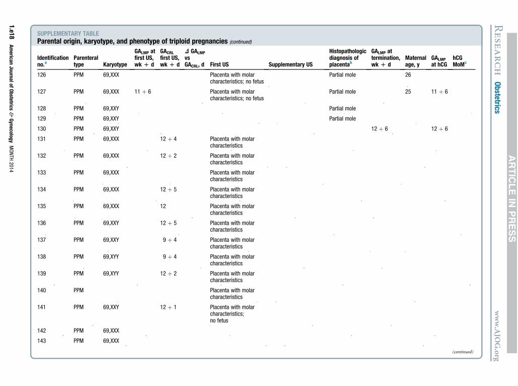
SUPPLEMENTARY TABLEParental origin, karyotype, and phenotype of triploid pregnancies
Identificationno.a
Parenteraltype Karyotype
GALMP atfirst US,wk D d
GACRLfirst US,wk D d
D GALMPvsGACRL, d First US Supplementary US
Histopathologicdiagnosis ofplacentab
GALMP attermination,wk D d
Maternalage, y
GALMPat hCG
hCGMoMc
1a MMP 69,XXY 12 þ 3 12 e3 Asymmetric IUGR,holoprosencephallicfetus with vaguefacial profile
Hydropicdegeneration
13 33
2a MMP 69,XXY 12 þ 4 11 þ 2 e9 Possible asymmetricalIUGR
15 39 9 þ 6 0.33c
3a MMP 69,XXY 21 þ 4 IUGR? Possibletransposition of thegreat arteries or doubleoutlet right ventricle,misalignment of wrists,and overlapping fingers
22 29
4a MMP 69,XXY NA NA NA NA 15 D after first US:no FHA, GA 10 þ 5
10 þ 5 41
5a MMP 69,XXX 12 þ 5 10 þ 1 e18 Early IUGR 13 þ 1 28
6a MMP 69,XXY 13 þ 1 12 þ 1 e7 No abnormalitiesdetected
13 þ 6 35
7a MMP 69,XXX 11 þ 5 11 þ 4 e1 No abnormalitiesdetected
12 D after first US:no abnormalitiesdetected; 2 d after:asymmetrical IUGR
14 þ 2 27 10 þ 1 0.3c
8a MMP 69,XXX 15 þ 2 13 þ 4 e12 Small placenta,oligohydramnios,asymmetrical IUGR
15 þ 4 27 12 þ 6 0.13c
9a MMP 69,XXX 14 þ 2 Possible IUGR 5 D after first US:IUGR; 1 wk after:no FHA
15 þ 5 37 14 þ 4 0.03c
10a MMP 69,XXX NA 10 þ 6 NA Thin small placenta 1 Mo after first US:asymmetric IUGR,hydrocephalic fetus withlemon sign, echogeniccardiac foci (“golf balls”)and overlapping fingers
16 þ 1 39
(continued)
OG.org
Obstetrics
Resea
rch

SUPPLEMENTARY TABLEParental origin, karyotype, and phenotype of triploid pregnancies (continued)
Identificationno.a
Parenteraltype Karyotype
GALMP atfirst US,wk D d
GACRLfirst US,wk D d
D GALMPvsGACRL, d First US Supplementary US
Histopathologicdiagnosis ofplacentab
GALMP attermination,wk D d
Maternalage, y
GALMPat hCG
hCGMoMc
11a MMP 69,XXY 12 þ 4 11 þ 1 e10 No abnormalitiesdetected
11 D after first US:asymmetric IUGR; 13 dafter: no FHA
16 þ 3 30 12 þ 5 0.17c
12a MMP 69,XXX 11 þ 6 10 þ 4 e9 Oligohydramnios 8 D after first US:oligohydramnios;asymmetric IUGR
16 þ 6 32 9 þ 6 0.48c
13a MMP 69,XXX 13 þ 4 12 þ 1 e10 Possible IUGR Sixteen days16 Dafter first US: noabnormalities detected;10 d after: no FHA
17 þ 3 30
14a MMP 69,XXX 10 NA 2 Mo after first US:large placenta, singleumbilical artery,negative bloodflow in diastole;oligohydramnios,asymmetrical IUGR
18 þ 4 23
15a MMP 69,XXY 16 þ 5 14 þ 2 e17 No abnormalitiesdetected
2 Mo after first US:IUGR; 10 d after: IUGRand slow heart rate
26 þ 3 29
16a MMP 69,XXY No abnormalitiesdetected
35
17a MMP NA
18a MMP 69,XXY 12 10 þ 4 e10 Possibleoligohydramnic
14 d after first US:oligohydramnios,asymmetric IUGR,possible hydrocephalic
Normal 15 þ 2 31
19a MMP 69,XXX 15 þ 2 14 e9 Asymmetrical IUGR Normal 15 þ 3 29
20a MMP 69,XXX 13 þ 1 11 þ 6 e9 No abnormalitiesdetected
1 mo after first US:oligohydramnios, IUGR,echogenic cardiac foci(“golf balls”)
Normal 15 þ 3 32 13 þ 1 0.17c
21a MMP 69,XXX 20 þ 1 17 þ 5 e17 Oligohydramnios,IUGR, hydrocephalicfetus
Normal 20 þ 5 27
(continued)
Resea
rch
Obstetrics
www.AJO
G.org
1.e10American
JournalofObstetrics
&Gynecology
MONTH
2014

SUPPLEMENTARY TABLEParental origin, karyotype, and phenotype of triploid pregnancies (continued)
Identificationno.a
Parenteraltype Karyotype
GALMP atfirst US,wk D d
GACRLfirst US,wk D d
D GALMPvsGACRL, d First US Supplementary US
Histopathologicdiagnosis ofplacentab
GALMP attermination,wk D d
Maternalage, y
GALMPat hCG
hCGMoMc
22a MMP 69,XXY 16 þ 6 14 e20 Oligohydramnios 2 wks after first US:oligohydramnios; 1 moafter second US:oligohydramnios,asymmetric IUGR
Normal 23 þ 6 26
23a MMP 69,XXX 11 þ 1 10 þ 6 e2 No abnormalitiesdetected
4 mos after first US:oligohydramnios,asymmetric IUGR
Normal 30 þ 3 30
24a MMP Doublepaternalcontribution
25 PPM 15 Fetus with no FHA Complete mole 15 40 15 4.02
26 PPM 17 þ 6 Placenta with molarcharacteristics; no fetus
Complete mole 18 22 18 11.35
27 PPM 12 þ 5 Spontaneous abortion Complete mole 12 þ 5 20 12 þ 5
28 PPM 69,XXX 14 Placenta with molarcharacteristics; no fetus
Complete mole 14 þ 1 33 14 þ 1 0.25
29 PPM 69,XXX 14 þ 4 Placenta with molarcharacteristics; no fetus
Complete mole 14 þ 5 26 14 þ 5 38.55
30 PPM NA 15 Placenta with molarcharacteristics; no fetus
Complete mole 15 þ 1 35 15 þ 1 18.27
31 PPM 69,XXY 15 Spontaneous abortion Complete mole 15 þ 1 25 15 þ 1 13.21
32 PPM 69,XYY 16 þ 4 Spontaneous abortion Complete mole 16 þ 5 24 16 þ 5
33 PPM 69,XXY 17 Placenta with molarcharacteristics; fetuswith no FHA;GA, 17 wks
Complete mole 17 þ 1 30 17 þ 1 15.54
34 PPM 69,XXX 18 Placenta with molarcharacteristics; no fetus
Complete mole 18 þ 1 24 18 þ 1
35 PPM 17 þ 6 Placenta with molarcharacteristics; no fetus
Complete mole 18 þ 3 26 18 þ 3 9.74
36 PPM 69,XXY 18 þ 4 Spontaneous abortion Complete mole 18 þ 4 26 18 þ 4 2.09
37 PPM 69,XXY 19 þ 1 Placenta with molarcharacteristics; no fetus
Complete mole 19 þ 2 38 19 þ 2 12.2
(continued)
www.AJO
G.org
Obstetrics
Resea
rch
MONTH
2014American
JournalofObstetrics
&Gynecology
1.e11

SUPPLEMENTARY TABLEParental origin, karyotype, and phenotype of triploid pregnancies (continued)
Identificationno.a
Parenteraltype Karyotype
GALMP atfirst US,wk D d
GACRLfirst US,wk D d
D GALMPvsGACRL, d First US Supplementary US
Histopathologicdiagnosis ofplacentab
GALMP attermination,wk D d
Maternalage, y
GALMPat hCG
hCGMoMc
38 PPM 69,XXX 17 þ 6 Placenta with molarcharacteristics; no fetus
Pregnancyproduct,not specified
18 28 18 15.45
39 PPM 69,XXY 12 Placenta with molarcharacteristics; no fetus
Pregnancyproduct,not specified
12 þ 1 23 12 þ 1 8.79
40 PPM 69,XXY 13 þ 5 Fetus with no FHA; GA,14 wks
Pregnancyproduct,not specified
13 þ 6 30 13 þ 6 54.2
41 PPM 69,XXY 13 þ 6 Placenta with molarcharacteristics; no fetus
Pregnancyproduct,not specified
13 þ 6 29 13 þ 6 2.94
42 PPM 69,XXX 15 þ 1 Placenta with molarcharacteristics; fetuswith no FHA; GA, 9 wks
Pregnancyproduct,not specified
15 þ 1 31 15 þ 1
43 PPM 69,XXY 15 þ 1 Placenta with molarcharacteristics; fetuswith no FHA;GA, 8 wks þ 5 d
Pregnancyproduct,not specified
15 þ 2 24 15 þ 2
44 PPM 69,XXX 15 þ 2 Placenta with molarcharacteristics; fetus(not specified)
Pregnancyproduct,not specified
15 þ 3 26 15 þ 3
45 PPM 16 þ 2 Spontaneous abortion Pregnancyproduct,not specified
16 þ 2 27 16 þ 2 1.4
46 PPM 69,XXY 16 þ 2 Placenta with molarcharacteristics; fetuswith no FHA
Pregnancyproduct,not specified
16 þ 3 31 16 þ 3 14.77
47 PPM 69,XXY 12 Placenta with molarcharacteristics; no fetus
Pregnancyproduct,not specified
16 þ 4 19 16 þ 4 104.28
48 PPM 69,XYY 15 Placenta with molarcharacteristics; no fetus
Hydropicdegeneration
15 29 15 15.81
49 PPM 69,XXY 16 þ 4 Placenta with molarcharacteristics; fetuswith no FHA
Hydropicdegeneration
17 24 17 12.8
(continued)
Resea
rch
Obstetrics
www.AJO
G.org
1.e12American
JournalofObstetrics
&Gynecology
MONTH
2014

SUPPLEMENTARY TABLEParental origin, karyotype, and phenotype of triploid pregnancies (continued)
Identificationno.a
Parenteraltype Karyotype
GALMP atfirst US,wk D d
GACRLfirst US,wk D d
D GALMPvsGACRL, d First US Supplementary US
Histopathologicdiagnosis ofplacentab
GALMP attermination,wk D d
Maternalage, y
GALMPat hCG
hCGMoMc
50 PPM 15 Hydropicdegeneration
15 þ 1 28 15 þ 1
51 PPM 69,XXX 15 þ 3 Spontaneous abortion Hydropicdegeneration
15 þ 3 30 15 þ 3
52 PPM 69,XXX 16 þ 2 Placenta with molarcharacteristics; fetuswith no FHA
Hydropicdegeneration
16 þ 3 25 16 þ 3
53 PPM 69,XYY 17 Placenta with molarcharacteristics; no fetus
Hydropicdegeneration
17 þ 1 19 17 þ 1 34.41
54 PPM 69,XXY 17 þ 2 Placenta with molarcharacteristics; fetuswith no FHA
Hydropicdegeneration
17 þ 2 19 17 þ 2 10.94
55 PPM 69,XXX 17 þ 3 Fetus with no FHA Hydropicdegeneration
17 þ 4 25 17 þ 4
56 PPM 69,XXY 18 Placenta with molarcharacteristics; no fetus
Hydropicdegeneration
18 þ 1 23 18 þ 1 20.07
57 PPM 69,XXX 18 þ 2 Placenta with molarcharacteristics; no fetus
Hydropicdegeneration
18 þ 3 23 18 þ 3 21.09
58 PPM 18 þ 3 Placenta with molarcharacteristics; fetuswith no FHA; GA, 11 wk
Hydropicdegeneration
18 þ 4 27 18 þ 4
59 PPM 69,XXY 18 þ 3 Placenta with molarcharacteristics, no FHA,asymmetric IUGR withsparring of head
Hydropicdegeneration
18 þ 4 25 18 þ 4 1.15
60 PPM 69,XXY 12 þ 4 Fetus with no FHA;GA, 12 wk
Molar notspecified
12 þ 4 31 12 þ 4 8.03
61 PPM 69,XXX 16 þ 5 Placenta with molarcharacteristics; no fetus
Molar notspecified
16 þ 6 31 16 þ 6 159.75
62 PPM 69,XXY 18 þ 3 Placenta with molarcharacteristics; no fetus
Molar notspecified
18 þ 5 37 18 þ 5 11.4
63a PPM 69,XXX 14 þ 5 12 þ 6 e13 No abnormalitiesdetected
18 d after first US: GA,15 þ 3 wk; IUGR;possible heartmalformation
17 þ 5 39
(continued)
www.AJO
G.org
Obstetrics
Resea
rch
MONTH
2014American
JournalofObstetrics
&Gynecology
1.e13

SUPPLEMENTARY TABLEParental origin, karyotype, and phenotype of triploid pregnancies (continued)
Identificationno.a
Parenteraltype Karyotype
GALMP atfirst US,wk D d
GACRLfirst US,wk D d
D GALMPvsGACRL, d First US Supplementary US
Histopathologicdiagnosis ofplacentab
GALMP attermination,wk D d
Maternalage, y
GALMPat hCG
hCGMoMc
64a PPM 69,XXY Cystic hygroma 30
65a PPM 69,XXX 11 þ 4 No abnormalitiesdetected
19 d after first US:placenta with molarcharacteristics; fetuswith possible heartdefects
28
66a PPM 69,XXY NT 3.7 mm 27
67a PPM 69,XXY NA Omphalocele 33
68a PPM NA
69a PPM 69,XXX 12 þ 2 12 e2 NT, 6 mm 35
70a PPM 69,XXX 9 9 0 No abnormalitiesdetected
1 wk after first US:no abnormalitiesdetected; 2 wk aftersecond US: placentawith molarcharacteristics; no FHA
Normal 12 þ 2 39
71 PPM 69,XXY 16 þ 5 Placenta with molarcharacteristics; fetuswithout abnormalities
7 D after first US: GA,15 þ 3 wk; placentawith molarcharacteristics
Normal 18 þ 3 32 18 þ 6 1339.5
72 PPM 69,XXY 12 Fetus with no FHA Partial mole 12 34 12 47.54
73 PPM 69,XXY 14 Placenta with molarcharacteristics; no fetus
Partial mole 12 26 12 5.45
74 PPM 69,XXY 12 þ 6 Fetus with no FHA Partial mole 13 31 13 8.8
75 PPM 69,XXX 16 þ 5 Fetus with no FHA Partial mole 17 38 17 10.43
76a PPM 69,XXY 16 þ 4 Fetus with no FHA Partial mole 19 36 16 þ 4
77 PPM 69,XXY 18 þ 6 Placenta with molarcharacteristics; fetus(not specified)
Partial mole 19 30 19
78 PPM 69,XXX 19 Placenta with molarcharacteristics; no fetus
Partial mole 19 24 19 7.09
79 PPM 69,XXY 19 þ 6 Placenta with molarcharacteristics; fetuswith no FHA
Partial mole 20 25 20 3.85
(continued)
Resea
rch
Obstetrics
www.AJO
G.org
1.e14American
JournalofObstetrics
&Gynecology
MONTH
2014

SUPPLEMENTARY TABLEParental origin, karyotype, and phenotype of triploid pregnancies (continued)
Identificationno.a
Parenteraltype Karyotype
GALMP atfirst US,wk D d
GACRLfirst US,wk D d
D GALMPvsGACRL, d First US Supplementary US
Histopathologicdiagnosis ofplacentab
GALMP attermination,wk D d
Maternalage, y
GALMPat hCG
hCGMoMc
80 PPM 69,XXY 20 þ 6 Placenta with molarcharacteristics; no fetus
Partial mole 21 26 21 0.54
81 PPM 69,XXX 21 þ 6 Spontaneous abortion Partial mole 22 24 22
82 PPM 69,XXY 11 þ 3 Placenta with molarcharacteristics; fetuswith no FHA
Partial mole 12 þ 5 30 12 þ 5 17.87
83 PPM 69,XXX 12 þ 5 Placenta with molarcharacteristics; no fetus
Partial mole 12 þ 6 32 12 þ 6
84 PPM 69,XXX 12 þ 6 Placenta with molarcharacteristics; no fetus
Partial mole 12 þ 6 30 12 þ 6 23.29
85 PPM 69,XXY 13 þ 3 Placenta with molarcharacteristics; no fetus
Partial mole 13 þ 4 19 13 þ 4 13.72
86a PPM 69,XXX 13 Placenta with molarcharacteristics; fetusNT, 6 mm
Partial mole 13 þ 6 26 12 þ 6 382.22
87 PPM 69,XXX 14 Placenta with molarcharacteristics; fetuswith no FHA
Partial mole 14 þ 1 29 14 þ 1
88 PPM 69,XXX 14 þ 2 Placenta with molarcharacteristics; fetuswith no FHA
Partial mole 14 þ 3 28 14 þ 3 53.4
89 PPM 69,XXY 14 þ 4 Fetus with no FHA;GA, 8 wk
Partial mole 14 þ 5 23 14 þ 5 54.65
90 PPM 69,XXY 15 þ 1 Placenta with molarcharacteristics; no fetus
Partial mole 15 þ 2 27 15 þ 2 5.06
91 PPM 69,XXY 15 þ 2 Fetus with no FHA;GA, 15 wk
Partial mole 15 þ 3 29 15 þ 3 8.37
92 PPM 69,XXY 15 þ 3 Placenta with molarcharacteristics; fetuswith no FHA
Partial mole 15 þ 3 35 15 þ 3 8.18
93 PPM 69,XXY 15 þ 5 Spontaneous abortion Partial mole 15 þ 5 31 15 þ 5 1.41
94 PPM 69,XXX 16 Fetus with no FHA;GA, 11 wk
Partial mole 16 þ 1 33 16 þ 1
(continued)
www.AJO
G.org
Obstetrics
Resea
rch
MONTH
2014American
JournalofObstetrics
&Gynecology
1.e15

SUPPLEMENTARY TABLEParental origin, karyotype, and phenotype of triploid pregnancies (continued)
Identificationno.a
Parenteraltype Karyotype
GALMP atfirst US,wk D d
GACRLfirst US,wk D d
D GALMPvsGACRL, d First US Supplementary US
Histopathologicdiagnosis ofplacentab
GALMP attermination,wk D d
Maternalage, y
GALMPat hCG
hCGMoMc
95 PPM 16 Placenta with molarcharacteristics; no fetus
Partial mole 16 þ 1 30 16 þ 1
96 PPM 69,XXY 15 þ 4 Placenta with molarcharacteristics; fetuswith FHA; GA, 13 wk
Partial mole 16 þ 2 25 16 þ 2 955.81
97 PPM 16 þ 2 Placenta with molarcharacteristics; no fetus
Partial mole 16 þ 2 19 16 þ 2
98 PPM 69,XXY 16 þ 2 Spontaneous abortion Partial mole 16 þ 3 34 16 þ 3 27.99
99 PPM 69,XXY 16 þ 4 Placenta with molarcharacteristics; fetuswith no FHA
Partial mole 16 þ 6 27 16 þ 6
100 PPM 69,XXY 16 þ 5 Placenta with molarcharacteristics; fetuswith no FHA
Partial mole 16 þ 6 30 16 þ 6 0.31
101 PPM 69,XXY 16 þ 5 Placenta with molarcharacteristics; fetuswith no FHA
Partial mole 16 þ 6 32 16 þ 6 16.57
102 PPM 69,XXY 17 Placenta with molarcharacteristics; no fetus
Partial mole 17 þ 1 29 17 þ 1 14.6
103 PPM 69,XXX 17 þ 1 Fetus with no FHA Partial mole 17 þ 2 29 17 þ 2
104 PPM 69,XXX 17 Placenta with molarcharacteristics; no fetus
Partial mole 17 þ 2 30 17 þ 2 26.78
105 PPM 69,XXY 17 þ 2 Placenta with molarcharacteristics; fetuswith no FHA; GA,12-13 wk
Partial mole 17 þ 3 26 17 þ 3 19.45
106 PPM 14 þ 6 Spontaneous abortion Partial mole 17 þ 4 26 17 þ 4 1.47
107 PPM 69,XXY 17 þ 4 NA Partial mole 17 þ 5 28 17 þ 5
108 PPM 69,XXX 17 þ 5 Placenta with molarcharacteristics; fetuswith no FHA
Partial mole 17 þ 5 23 17 þ 5 85.72
109 PPM 69,XXX 17 þ 3 Placenta with molarcharacteristics; no fetus
Partial mole 17 þ 5 23 17 þ 5 1556.9
(continued)
Resea
rch
Obstetrics
www.AJO
G.org
1.e16American
JournalofObstetrics
&Gynecology
MONTH
2014

SUPPLEMENTARY TABLEParental origin, karyotype, and phenotype of triploid pregnancies (continued)
Identificationno.a
Parenteraltype Karyotype
GALMP atfirst US,wk D d
GACRLfirst US,wk D d
D GALMPvsGACRL, d First US Supplementary US
Histopathologicdiagnosis ofplacentab
GALMP attermination,wk D d
Maternalage, y
GALMPat hCG
hCGMoMc
110 PPM 69,XXX 18 Placenta with molarcharacteristics; no fetus
Partial mole 18 þ 1 25 18 þ 1 197.85
111 PPM 69,XXX 11 þ 6 Fetus with no FHA Partial mole 18 þ 2 19 18 þ 2 4.76
112 PPM 69,XXX 18 þ 2 Fetus with no FHA;GA, 11 wk
Partial mole 18 þ 2 30 18 þ 2 35.34
113 PPM 18 þ 1 Placenta with molarcharacteristics; no fetus
Partial mole 18 þ 2 28 18 þ 2 22.46
114 PPM 69,XYY 18 þ 2 Placenta with molarcharacteristics; no fetus
Partial mole 18 þ 3 27 18 þ 3 22.72
115 PPM 69,XXY 18 þ 5 Placenta with molarcharacteristics; fetuswith no FHA
Partial mole 18 þ 5 28 18 þ 5 3.39
116 PPM 18 þ 5 Spontaneous abortion Partial mole 18 þ 6 26 18 þ 6 1.43
117 PPM 69,XXX 19 þ 1 Fetus with no FHA;GA, 9 wk
Partial mole 19 þ 2 25 19 þ 2 2.16
118 PPM 69,XXX 19 Placenta with molarcharacteristics; no fetus
Partial mole 19 þ 3 36 19 þ 3 80.92
119 PPM 19 þ 3 Spontaneous abortion Partial mole 19 þ 3 33 19 þ 3 12.2
120 PPM 69,XXX 20 Placenta with molarcharacteristics; fetus(not specified)
Partial mole 20 þ 1 24 20 þ 1 131.02
121 PPM 69,XXY 20 þ 1 Fetus with no FHA Partial mole 20 þ 2 25 20 þ 2 7.45
122 PPM 69,XXX 23 þ 5 Placenta with molarcharacteristics; fetuswith no FHA; GA, 14 wk
Partial mole 24 þ 5 29 24 þ 5 3.52
123a PPM 69,XXY 26 þ 2 Supposedly normal12-week scan (secondhand information, directconfirmation notpossible)
Partial mole 26 þ 2 31
124a PPM 69,XXY 13 þ 3 12 þ 2 e8 No abnormalitiesdetected
Partial mole 27
125 PPM 69,XXX 12 Placenta with molarcharacteristics; no fetus
Partial mole 31
(continued)
www.AJO
G.org
Obstetrics
Resea
rch
MONTH
2014American
JournalofObstetrics
&Gynecology
1.e17
SUPPLEMENTARY TABLEParental origin, karyotype, and phenotype of triploid pregnancies (continued)
Identificationno.a
Parenteraltype Karyotype
GALMP atfirst US,wk D d
GACRLfirst US,wk D d
D GALMPvsGACRL, d First US Supplementary US
Histopathologicdiagnosis ofplacentab
GALMP attermination,wk D d
Maternalage, y
GALMPat hCG
hCGMoMc
126 PPM 69,XXX Placenta with molarcharacteristics; no fetus
Partial mole 26
127 PPM 69,XXX 11 þ 6 Placenta with molarcharacteristics; no fetus
Partial mole 25 11 þ 6
128 PPM 69,XXY Partial mole
129 PPM 69,XXY Partial mole
130 PPM 69,XXY 12 þ 6 12 þ 6
131 PPM 69,XXX 12 þ 4 Placenta with molarcharacteristics
132 PPM 69,XXX 12 þ 2 Placenta with molarcharacteristics
133 PPM 69,XXX Placenta with molarcharacteristics
134 PPM 69,XXX 12 þ 5 Placenta with molarcharacteristics
135 PPM 69,XXX 12 Placenta with molarcharacteristics
136 PPM 69,XXY 12 þ 5 Placenta with molarcharacteristics
137 PPM 69,XXY 9 þ 4 Placenta with molarcharacteristics
138 PPM 69,XYY 9 þ 4 Placenta with molarcharacteristics
139 PPM 69,XYY 12 þ 2 Placenta with molarcharacteristics
140 PPM Placenta with molarcharacteristics
141 PPM 69,XXY 12 þ 1 Placenta with molarcharacteristics;no fetus
142 PPM 69,XXX
143 PPM 69,XXX
(continued)
Resea
rch
Obstetrics
www.AJO
G.org
1.e18American
JournalofObstetrics
&Gynecology
MONTH
2014

SUPPLEMENTARY TABLEParental origin, karyotype, and phenotype of triploid pregnancies (continued)
Identificationno.a
Parenteraltype Karyotype
GALMP atfirst US,wk D d
GACRLfirst US,wk D d
D GALMPvsGACRL, d First US Supplementary US
Histopathologicdiagnosis ofplacentab
GALMP attermination,wk D d
Maternalage, y
GALMPat hCG
hCGMoMc
144 PPM 69,XXX
145 PPM 69,XXX
146 PPM 69,XXX
147 PPM 69,XXX 12 þ 2
148 PPM 69,XXY
149 PPM 69,XXY
150 PPM 69,XXY
151 PPM 69,XXY
152 PPM 69,XXY
153 PPM 69,XXY
154 PPM 69,XXY
155 PPM 69,XYY
156 PPM
157 PPM
158 PPM
beta hCG, human chorionic gonadotropin; D, delta; FHA, fetal heart activity; GA, gestational age; GACRL, gestational age calculated from crown rump length; GALMP, gestational age calculated from last menstrual period; hCG, human chorionic gonadotropin; IUGR,intrauterine growth restriction; MMP, double maternal contribution; MoM, multiples of the median; NA, not available; PPM, double paternal contribution; US, ultrasound.
a From the Danish Cytogenetic Central Registry; b Reports on poor histopathologic interobserver and intraobserver reproducibility have been published.27-29 Because this part of the study was retrospective by many different pathologists over a period of 24 years, we haveno detailed information on sampling. Therefore, we have not commented on any potential difference between partial and complete moles because these are especially subject to sampling. Differentiation between the 2 molar diagnoses in this study should be made onlywith caution; c Beta hCG.
www.AJO
G.org
Obstetrics
Resea
rch
MONTH
2014American
JournalofObstetrics
&Gynecology
1.e19


![The [C II] 158 Micron Line Deficit in Ultraluminous Infrared Galaxies Revisited](https://static.fdokumen.com/doc/165x107/631ef5f5097e038f7c093ec0/the-c-ii-158-micron-line-deficit-in-ultraluminous-infrared-galaxies-revisited.jpg)