Glutamate receptor expression in rat striatum: Effect of deafferentation

Cystathionine Is a Novel Substrate of Cystine/GlutamateTransporterIMPLICATIONS FOR IMMUNE FUNCTION*
Received for publication, November 12, 2014, and in revised form, February 14, 2015 Published, JBC Papers in Press, February 20, 2015, DOI 10.1074/jbc.M114.625053
Sho Kobayashi‡§¶, Mami Sato‡, Takayuki Kasakoshi‡, Takumi Tsutsui‡, Masahiro Sugimoto�, Mitsuhiko Osaki**‡‡,Futoshi Okada**‡‡, Kiharu Igarashi‡, Jun Hiratake§§, Takujiro Homma¶¶, Marcus Conrad¶, Junichi Fujii¶¶,Tomoyoshi Soga�, Shiro Bannai‡, and Hideyo Sato‡§��1
From the ‡Department of Food and Applied Life Sciences, Faculty of Agriculture, Yamagata University, Tsuruoka, Yamagata 997-8555, Japan, the §Department of Functional Genomics and Biotechnology, United Graduate School of Agricultural Sciences, IwateUniversity, Morioka, Iwate 020-8550, Japan, the ¶Helmholtz Center Munich, German Research Center for Environmental Health,Institute of Developmental Genetics, Ingolstädter Landstrasse 1, 85764 Neuherberg, Germany, the �Institute of AdvancedBiosciences, Keio University, Tsuruoka, Yamagata 997-0052, Japan, the **Division of Pathological Biochemistry, Tottori UniversityFaculty of Medicine, Yonago 683-8503, Japan, the ‡‡Chromosome Engineering Research Center, Tottori University, Yonago, 683-8503, Japan, the §§Institute for Chemical Research, Kyoto University, Uji, Kyoto 611-0011, Japan, the ¶¶Department of Biochemistryand Molecular Biology, Graduate School of Medical Science, Yamagata University, Yamagata 990-9585, Japan, and the��Laboratory of Biochemistry and Molecular Biology, Department of Medical Technology, Faculty of Medicine, Niigata University,Chuo-ku, Niigata 951-8518, Japan
Background: System xc� is involved in various pathophysiological conditions, such as neurodegenerative disorders and
cancer.Results: Extracellular cystathionine competitively inhibited cystine uptake and could be exchanged with intracellular glutamatevia system xc
�.Conclusion: Cystathionine is exclusively transported into immune tissues as the third physiological substrate of system xc
�.Significance: Cystathionine can be exchanged with glutamate to reduce extracellular glutamate levels.
The cystine/glutamate transporter, designated as system xc�,
is important for maintaining intracellular glutathione levels andextracellular redox balance. The substrate-specific componentof system xc
�, xCT, is strongly induced by various stimuli,including oxidative stress, whereas it is constitutively expressedonly in specific brain regions and immune tissues, such as thethymus and spleen. Although cystine and glutamate are the wellestablished substrates of system xc
� and the knockout of xCTleads to alterations of extracellular redox balance, nothing isknown about other potential substrates. We thus performed acomparative metabolite analysis of tissues from xCT-deficientand wild-type mice using capillary electrophoresis time-of-flight mass spectrometry. Although most of the analyzed metab-olites did not show significant alterations between xCT-defi-cient and wild-type mice, cystathionine emerged as being absentspecifically in the thymus and spleen of xCT-deficient mice. Noexpression of either cystathionine �-synthase or cystathionine�-lyase was observed in the thymus and spleen of mice. Inembryonic fibroblasts derived from wild-type embryos, cystineuptake was significantly inhibited by cystathionine in a concen-tration-dependent manner. Wild-type cells showed an intracel-lular accumulation of cystathionine when incubated in cystathi-onine-containing buffer, which concomitantly stimulated an
increased release of glutamate into the extracellular space.By contrast, none of these effects could be observed in xCT-deficient cells. Remarkably, unlike knock-out cells, wild-typecells could be rescued from cystine deprivation-induced celldeath by cystathionine supplementation. We thus conclude thatcystathionine is a novel physiological substrate of system xc
�
and that the accumulation of cystathionine in immune tissues isexclusively mediated by system xc
�.
In mammalian cultured cells, the activity of the cystine/glu-tamate transporter, designated as system xc
� (1), has beenshown to be essential for maintaining intracellular glutathionelevels and the extracellular cystine/cysteine redox balance (2).The substrate-specific subunit of this transporter, xCT (3, 4), isstrongly induced by various stimuli, such as oxidative stress andelectrophile agents, whereupon intracellular glutathione levelsare increased. When cells are cultured in cystine-free mediumor the cystine uptake via xCT is inhibited, most of cultured cellsdie within 24 h due to glutathione depletion (5). Thus, systemxc
� is thought to be one of the adaptive cellular defense systemsagainst oxidative stress.
Constitutive expression of xCT mRNA is observed in specificregions of the brain, such as meninges and circumventricularorgans (6), and in immune tissues, such as thymus and spleen(7). We previously generated xCT-deficient mice and analyzedtheir phenotypes (8). Although mice were healthy in appear-ance and fertile, the plasma redox balance was shifted to a moreoxidative state. We further demonstrated that xCT-deficient
* This work was supported, in part, by the Strategic Young Researcher Over-seas Visits Program for Accelerating Brain Circulation from the Japan Soci-ety for the Promotion of Sciences; a research grant from the Naito Founda-tion; and Collaborative Research Program of the Institute for ChemicalResearch, Kyoto University, Grant 2013-43.
1 To whom correspondence should be addressed. Tel./Fax: 81-25-378-0959;E-mail: [email protected].
THE JOURNAL OF BIOLOGICAL CHEMISTRY VOL. 290, NO. 14, pp. 8778 –8788, April 3, 2015© 2015 by The American Society for Biochemistry and Molecular Biology, Inc. Published in the U.S.A.
8778 JOURNAL OF BIOLOGICAL CHEMISTRY VOLUME 290 • NUMBER 14 • APRIL 3, 2015
by guest on May 3, 2016
http://ww
w.jbc.org/
Dow
nloaded from

mice are more susceptible to the oxidative stress-inducingagent paraquat than wild-type mice and that glutathione levelsin lungs of xCT-deficient mice are lower than those of wild-typemice under these conditions (9). These results indicate thatxCT plays a protective role against oxidative stress in vivo andcontributes to maintaining glutathione levels of tissues exposedto oxidative stress. Hence, the function of xCT is to supply cellswith sufficient cysteine as a precursor of intracellular glutathi-one, which suggests a beneficial and protective role of xCT incell survival and function.
In recent years, it has been suggested that xCT is involved incancer development and other diseases (10). For instance, wehave demonstrated that increased glutathione via system xc
�
increases cisplatin resistance of human ovarian cancer (11). Loet al. (12) have reported that system xc
� plays a major role inpancreatic cancer growth and therapy resistance by enhancingglutathione synthesis. In this context, Ishimoto et al. (13), haverecently shown that a variant isoform of CD44, one of the cellsurface markers associated with cancer stem cells, stabilizesxCT and thereby causes a higher intracellular glutathione levelin tumor cells. As a result, the tumor stem cells may thusacquire an increased resistance to chemo- and radiotherapy.
On the other hand, because cystine is taken up into the cellvia xCT in exchange for intracellular glutamate with a molarratio of 1:1 (14), glutamate is inevitably secreted from xCT-expressing cells into the extracellular space. Glutamate releasedvia xCT has been suggested to cause glutamate excitotoxicityand/or oxidative glutamate toxicity in the brain, also known asoxytosis (15). In this context, xCT has been considered to belinked to the pathology of various neurodegenerative disorders,such as Alzheimer disease and Parkinson disease (16, 17). Theconcentration of extracellular glutamate in the hippocampusseems to be related to the activity of xCT (18). Sontheimer’sgroup (19, 20) provided evidence that inhibition of system xc
�
restrains tumor growth in the brain and that glutamate releasedvia xCT acts as an essential autocrine/paracrine signal for gli-oma cell invasion. Savaskan et al. (21) have shown that silencingof xCT by siRNA abrogates glioma-induced neurodegenerationand ameliorates brain edema. In addition to the effects in thebrain, glutamate released via xCT by dendritic cells may act as anovel modulator of T cell activation (22). These reports indicatethat xCT is also important as a supplier of glutamate into theextracellular space.
In the present study, we sought to investigate the metaboliteprofile in response to the targeted loss of xCT expression inmice beyond what had already been reported on cystine andglutathione levels in plasma of knock-out mice (8). To this end,we performed an extended, non-targeted analysis of metabo-lites in several tissues of wild-type and xCT-deficient miceusing capillary electrophoresis time-of-flight mass spectrome-try. Remarkably, we found that in xCT-deficient mice cysta-thionine was absent in the thymus and spleen of the xCT-defi-cient mice as compared with control mice, whereas all othermetabolites did not show any substantial differences betweenwild-type and knock-out animals. Cystathionine is known as anintermediary metabolite in cysteine synthesis from methioninein the transsulfuration pathway, although its function in theimmune system has remained enigmatic to date. We therefore
investigated the possibility that cystathionine is the third phys-iological substrate of system xc
�.
EXPERIMENTAL PROCEDURES
Materials—L-Cystathionine was obtained from Wako PureChemical Industries, Ltd. (Osaka, Japan). All other chemicalsand regents were purchased from Sigma unless otherwisestated.
Metabolome Analysis—C57BL/6J male mice (8 –9 weeks old)and xCT-deficient mice (8), which had been back-crossed withC57BL/6J mice for more than 10 generations, were housed at22 °C with a 12-h alternating light/dark cycle. All mice had freeaccess to standard rodent chow and water. Mice were anesthe-tized with pentobarbital, and heparinized blood was collectedfrom the inferior vena cava. Collected blood was immediatelycentrifuged, and 100 �l of plasma was moved into the othertube. In all cases, frozen tissues (�50 mg) were immediatelyplunged into methanol (500 �l) containing internal standards(20 �M methionine sulfone and D-camphor-10-sulfonic acidand homogenized for 3 min to inactivate the enzymes. Plasma(50 �l) was added to methanol (450 �l) containing the sameinternal standards and mixed without homogenizing. ThenMilli-Q water (200 �l) (Millipore, Billerica, MA) and chloro-form (500 �l) were added, and the mixture was thoroughlymixed. The solution was centrifuged at 4,600 � g for 15 min at4 °C, and the 450-�l upper aqueous layer was filtered by cen-trifugation through a Millipore 5 kDa cut-off filter to removelarge molecules. Plasma was centrifuged in the same mannerbut only for 5 min. The filtrate was lyophilized and dissolved inMilli-Q water (50 �l for tissue and 25 �l for plasma) containinga reference compound (200 �M each of 3-aminopyrrolidineand trimesate) prior to capillary electrophoresis-TOF/MS anal-ysis. Capillary electrophoresis-TOF/MS was carried out asdescribed previously (23).
Induction of Experimental Hypercystathionemia—To induceexperimental hypercystathionemia, male mice (8 –9 weeks)were injected with propargylglycine, a cystathionine �-lyase(CGL)2 inhibitor, diluted in saline (intraperitoneally; 50 mg/kg)daily for 3 days (24). Control male mice were injected with anequivalent volume of saline (intraperitoneally). After certaintreatment periods, blood was collected from inferior vena cava,and the liver, thymus, and spleen were removed. These experi-ments were approved by the Animal Research Committee ofYamagata University.
Measurement of Cystathionine Levels in Plasma, Liver, Thy-mus, and Spleen—Collected blood was immediately centri-fuged, and 100 �l of plasma was transferred into another tubecontaining 10 �l of 50% sulfosalicylic acid. After 30 min in an icebath, the mixture was stored at �20 °C until further processing.The samples were thawed and centrifuged at 10,000 � g for 15min, and the supernatant was removed. Isolated liver, thymus,and spleen were weighed and immediately homogenized in 5%trichloroacetic acid and then treated with ether to remove theacid. Then, samples were adjusted to pH 2.2 with 1 M LiOH.
2 The abbreviations used are: CGL, cystathionine �-lyase; CBS, cystathionine�-synthase; MEF, mouse embryonic fibroblast(s); ATF4, activating tran-scription factor 4.
Cystathionine Is a Novel Substrate of System xc�
APRIL 3, 2015 • VOLUME 290 • NUMBER 14 JOURNAL OF BIOLOGICAL CHEMISTRY 8779
by guest on May 3, 2016
http://ww
w.jbc.org/
Dow
nloaded from

Cystathionine levels were analyzed using an amino acid ana-lyzer (Hitachi model L-8800, Tokyo, Japan).
Reverse Transcription-PCR (RT-PCR)—Total RNA from tis-sues and cultured cells was isolated using Isogen (Nippon Gene,Toyama, Japan) by following the manufacturer’s instructions.First-strand cDNA synthesis and quantitative PCR were per-formed using the PrimeScript RT reagent kit (Takara Bio, Shiga,Japan) and SYBR� Premix Ex TaqTM (Takara Bio, Shiga, Japan)according to the manufacturer’s instructions. The primer sets formouse xCT, mouse Cgl, mouse cystathionine �-synthase (Cbs),and mouse glyceraldehyde-3-phosphate dehydrogenase (Gapdh)used for quantitative RT-PCR were 5�-CTCGTGACAGCTGTG-GGCAT-3� and 5�-GGCACTAGACTCAAGAACTGTG-3�, 5�-TGGATCGAGCTTTGAAGGCAGC-3� and 5�-CAGTTCTG-CGTATGCTCCGTAA-3�, 5�-GATTGGCTACGACTTCA-TCC-3� and 5�-AGTCCTTCCTGTGCGATGAG-3�, and 5�-GACCCCTTCATTGACCT-3� and 5�-CCACCACCCTGTTGC-TGT-3�, respectively.
Cell Culture—Mouse embryonic fibroblasts (MEF) isolatedfrom wild-type and xCT-deficient mice (8) and xCT-overex-pressing MEF (25) were cultured routinely in Dulbecco’s mod-ified Eagle’s medium supplemented with 10% fetal bovineserum, 2-mercaptoethanol (50 �M), insulin-transferrin-sele-nium A (Life Technologies, Inc.), penicillin (50 units/ml), andstreptomycin (50 �g/ml) at 37 °C in 5% CO2 and 95% air. Theaddition of 2-mercaptoethanol is necessary for xCT-deficientMEF to grow even under routine cell culture conditions (8, 26).For measurement of cell survival, cells were cultured in cystine-free medium (without 2-mercaptoethanol) with and without0.1 mM cystathionine for the time periods indicated. The num-ber of viable cells was measured by trypan blue exclusion.
Determination of Intracellular Total Glutathione (GSH andthe Oxidized Form of GSH (GSSG))—Cells were seeded on35-mm dishes (2 � 105 cells/dish) and cultured for 24 h in theroutine culture medium. Then cells were cultured in cystine-free medium with and without 0.1 mM cystathionine for thetime periods indicated. Cells were washed three times with ice-cold PBS, extracted with 5% trichloroacetic acid, and thentreated with ether to remove trichloroacetic acid. Total gluta-thione content in the aqueous layer was measured using anenzymatic method, which is based on the catalytic action ofglutathione in the reduction of 5,5�-dithiobis(2-nitrobenzoicacid) by the glutathione reductase system (27).
Cystine Uptake—Cells were seeded on 35-mm dishes (2 �105 cells/dish) and cultured for 24 h in the routine culturemedium. Then the cells were cultured with or without 50 �M
diethyl maleate for an additional 24 h. Cells were washed threetimes in prewarmed Na�-free PBS(�)G (10 mM phosphate-buffered saline (137 mM choline chloride, 3 mM KCl), pH 7.4,containing 0.01% CaCl2, 0.01% MgCl2�6H2O, and 0.1% glucose)and then incubated in 0.5 ml of prewarmed uptake medium at37 °C for 2 min. This uptake medium contained 20 �M cystineplus [14C]cystine (0.2 �Ci/ml) in Na�-free PBS(�)G. L-Cysta-thionine, L-glutamate, L-leucine, L-serine, or L-arginine each atfinal concentrations of 200 �M were added to the uptakemedium as inhibitors. Uptake was terminated by rapidly rinsingthe cells three times with ice-cold PBS, and the radioactivity inthe cells was counted.
Determination of Intracellular and Extracellular Cystathio-nine and Glutamate Levels—Cells were seeded on 35-mmdishes (2 � 105 cells/dish) and cultured for 24 h in the routineculture medium. Then the cells were cultured with or without50 �M diethyl maleate for an additional 24 h. Cells were washedthree times with prewarmed PBS(�)G (10 mM phosphate-buff-ered saline (137 mM NaCl, 3 mM KCl), pH 7.4, containing 0.01%CaCl2, 0.01% MgCl2�6H2O, and 0.1% glucose) and then incu-bated in 0.5 ml of prewarmed PBS(�)G containing 0.1 mM cys-tathionine or cystine at 37 °C for 15 min. PBS(�)G was col-lected, dried using a rotary evaporator, and dissolved in 100 �lof amino acid analysis buffer (pH 2.2) for measuring extracel-lular amino acids. Cells were washed three times with ice-coldPBS, extracted with 5% trichloroacetic acid, treated with etherto remove trichloroacetic acid, and used for measuring intra-cellular amino acids. Extracellular and intracellular amino acidswere analyzed using the amino acid analyzer.
Statistical Analyses—Statistical significances of the differ-ences were determined by Student’s t test (*, p � 0.05; **, p �0.01).
RESULTS
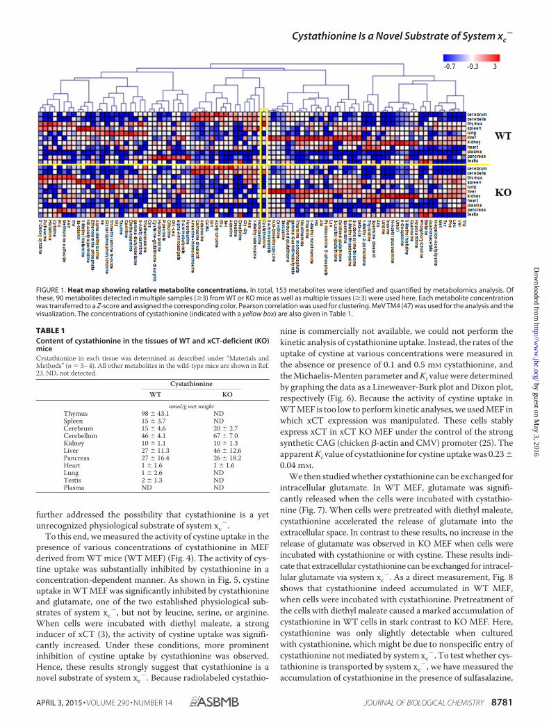
Previously, we showed that mice lacking xCT revealed per-turbations in extracellular cystine and GSH levels but otherwiseappeared healthy and fully viable (8). Here, we performed adetailed analysis of metabolites in cerebrum, cerebellum, thy-mus, spleen, lung, liver, kidney, heart, pancreas, testis, andplasma of wild-type (WT) and xCT-deficient (KO) mice usingcapillary electrophoresis time-of-flight mass spectrometry sothat all possible metabolite peaks were profiled. Although mostof the metabolite peaks showed no significant differencesbetween WT and KO mice, cystathionine was not detectable inthe thymus and spleen of KO mice (Fig. 1 and Table 1). How-ever, significant amounts of cystathionine were detected in thesame tissues of WT mice (Table 1), indicating that cystathio-nine might be a novel substrate for system xc
�.Therefore, we first examined expression of key enzymes
involved in cystathionine biosynthesis, CBS and CGL. Asshown in Fig. 2, the expression of Cbs and Cgl mRNA werehardly detectable in the thymus and spleen of both WT and KOmice, whereas they were markedly expressed in the livers ofthese mice. In WT mice, significant expression of xCT mRNAcould be detected in the thymus and spleen, whereas no xCTexpression could be observed in liver tissue. Although theamount of cystathionine in the thymus of WT mice was com-pletely different from that of KO mice, histological differencesin the thymus were not apparent between these mice (data notshown). To induce experimental hypercystathionemia, WTand KO mice were treated with propargylglycine, a CGL inhib-itor. Three days after treatment, cystathionine levels in liver,plasma, thymus, and spleen were measured by the amino acidanalyzer (Fig. 3). In plasma and liver of xCT-deficient and wild-type mice, cystathionine was markedly increased like in thethymus and spleen of wild-type mice. By stark contrast, it wasbarely detectable in the thymus and spleen of xCT-deficientmice. These results suggest that in the thymus and spleen, cys-tathionine is not synthesized but transported via system xc
�,whereupon it accumulates in these tissues. Therefore, we have
Cystathionine Is a Novel Substrate of System xc�
8780 JOURNAL OF BIOLOGICAL CHEMISTRY VOLUME 290 • NUMBER 14 • APRIL 3, 2015
by guest on May 3, 2016
http://ww
w.jbc.org/
Dow
nloaded from
further addressed the possibility that cystathionine is a yetunrecognized physiological substrate of system xc
�.To this end, we measured the activity of cystine uptake in the
presence of various concentrations of cystathionine in MEFderived from WT mice (WT MEF) (Fig. 4). The activity of cys-tine uptake was substantially inhibited by cystathionine in aconcentration-dependent manner. As shown in Fig. 5, cystineuptake in WT MEF was significantly inhibited by cystathionineand glutamate, one of the two established physiological sub-strates of system xc
�, but not by leucine, serine, or arginine.When cells were incubated with diethyl maleate, a stronginducer of xCT (3), the activity of cystine uptake was signifi-cantly increased. Under these conditions, more prominentinhibition of cystine uptake by cystathionine was observed.Hence, these results strongly suggest that cystathionine is anovel substrate of system xc
�. Because radiolabeled cystathio-
nine is commercially not available, we could not perform thekinetic analysis of cystathionine uptake. Instead, the rates of theuptake of cystine at various concentrations were measured inthe absence or presence of 0.1 and 0.5 mM cystathionine, andthe Michaelis-Menten parameter and Ki value were determinedby graphing the data as a Lineweaver-Burk plot and Dixon plot,respectively (Fig. 6). Because the activity of cystine uptake inWT MEF is too low to perform kinetic analyses, we used MEF inwhich xCT expression was manipulated. These cells stablyexpress xCT in xCT KO MEF under the control of the strongsynthetic CAG (chicken �-actin and CMV) promoter (25). Theapparent Ki value of cystathionine for cystine uptake was 0.23 �0.04 mM.
We then studied whether cystathionine can be exchanged forintracellular glutamate. In WT MEF, glutamate was signifi-cantly released when the cells were incubated with cystathio-nine (Fig. 7). When cells were pretreated with diethyl maleate,cystathionine accelerated the release of glutamate into theextracellular space. In contrast to these results, no increase in therelease of glutamate was observed in KO MEF when cells wereincubated with cystathionine or with cystine. These results indi-cate that extracellular cystathionine can be exchanged for intracel-lular glutamate via system xc
�. As a direct measurement, Fig. 8shows that cystathionine indeed accumulated in WT MEF,when cells were incubated with cystathionine. Pretreatment ofthe cells with diethyl maleate caused a marked accumulation ofcystathionine in WT cells in stark contrast to KO MEF. Here,cystathionine was only slightly detectable when culturedwith cystathionine, which might be due to nonspecific entry ofcystathionine not mediated by system xc
�. To test whether cys-tathionine is transported by system xc
�, we have measured theaccumulation of cystathionine in the presence of sulfasalazine,
FIGURE 1. Heat map showing relative metabolite concentrations. In total, 153 metabolites were identified and quantified by metabolomics analysis. Ofthese, 90 metabolites detected in multiple samples (�3) from WT or KO mice as well as multiple tissues (�3) were used here. Each metabolite concentrationwas transferred to a Z-score and assigned the corresponding color. Pearson correlation was used for clustering. MeV TM4 (47) was used for the analysis and thevisualization. The concentrations of cystathionine (indicated with a yellow box) are also given in Table 1.
TABLE 1Content of cystathionine in the tissues of WT and xCT-deficient (KO)miceCystathionine in each tissue was determined as described under “Materials andMethods” (n 3– 4). All other metabolites in the wild-type mice are shown in Ref.23. ND, not detected.
CystathionineWT KO
nmol/g wet weightThymus 98 � 43.1 NDSpleen 15 � 3.7 NDCerebrum 15 � 4.6 20 � 2.7Cerebellum 46 � 4.1 67 � 7.0Kidney 10 � 1.1 10 � 1.3Liver 27 � 11.3 46 � 12.6Pancreas 27 � 16.4 26 � 18.2Heart 1 � 1.6 1 � 1.6Lung 1 � 2.6 NDTestis 2 � 1.3 NDPlasma ND ND
Cystathionine Is a Novel Substrate of System xc�
APRIL 3, 2015 • VOLUME 290 • NUMBER 14 JOURNAL OF BIOLOGICAL CHEMISTRY 8781
by guest on May 3, 2016
http://ww
w.jbc.org/
Dow
nloaded from

a well established system xc� inhibitor (28). As expected from
our previous results, the accumulation of cystathionine in WTMEF was completely inhibited in the presence of sulfasalazine(Fig. 9).
When WT MEF were cultured in cystine-free medium, theyfailed to proliferate and died within 24 h as expected (Figs. 10and 11A). However, they survived and even proliferated by add-ing 0.1 mM cystathionine to cystine-free medium. On the con-
trary, KO MEF died within 24 h in cystine-free medium regard-less of cystathionine (0.1 mM) supplementation (Figs. 10 and11B). Although total glutathione in WT MEF was rapidlydecreased by culturing cells in cystine-free medium, it was sig-nificantly restored by culturing the cells in cystine-free mediumcontaining cystathionine (Fig. 11C). In contrast, the intracellu-lar glutathione levels were not increased in KO MEF by cultur-ing in the cystine-free medium with cystathionine (Fig. 11D).These data indicate that cystathionine is able to sustain intra-cellular glutathione levels even in the absence of exogenouscystine in WT cells but not in KO cells.
FIGURE 2. Expression of Cbs, Cgl, and xCT mRNA in the thymus, spleen, and liver of WT and xCT-deficient (KO) mice. Thymus, spleen, and liver werecollected from male xCT WT and KO mice. Total RNA was extracted, and Cbs, Cgl, and xCT mRNA expression was determined by quantitative RT-PCR as describedunder “Experimental Procedures.” GAPDH was used as the internal standard. Cbs and Cgl mRNA expression is represented as -fold of that of liver of wild type,and xCT mRNA expression is represented as -fold of that of thymus of wild-type (n 3). Error bars, S.D.
FIGURE 3. Cystathionine levels in liver, plasma, and thymus of WT andxCT-deficient (KO) mice treated with DL-propargylglycine (PPG). Male xCTWT and KO mice were injected with propargylglycine (intraperitoneally; 50mg/kg) daily for 3 days. Control male mice were injected with an equivalentvolume of saline. After treatment periods, blood, liver, thymus, and spleenwere collected. Plasma was collected from blood by centrifuge immediately.Cystathionine levels were determined as described under “Experimental Pro-cedures.” Values are means � S.D. (error bars) (n 3– 4).
FIGURE 4. Effect of cystathionine on cystine uptake in MEF derived fromWT mice. WT MEF were cultured for 24 h in the routine culture medium, andthen L-[14C]cystine (0.02 mM) uptake was measured in the absence or pres-ence of 0.1–2 mM cystathionine under Na�-free conditions. Each point repre-sents the mean � S.D. (error bars) (n 3– 4).
Cystathionine Is a Novel Substrate of System xc�
8782 JOURNAL OF BIOLOGICAL CHEMISTRY VOLUME 290 • NUMBER 14 • APRIL 3, 2015
by guest on May 3, 2016
http://ww
w.jbc.org/
Dow
nloaded from

When WT MEF were cultured for 8 h in cystine-free mediumwith or without cystathionine, the expression of xCT mRNAwas strongly induced, compared with cells cultured in the rou-
tine culture medium (Fig. 12A). Under these conditions, CglmRNA was also markedly induced (Fig. 12B). On the otherhand, Cbs mRNA was unchanged by culturing the cells in cys-tine-free medium with or without cystathionine (Fig. 12C). It isnoteworthy that Cgl mRNA in KO MEF was significantlyinduced even in the routine culture medium (Fig. 12B). Theaddition of 2-mercaptoethanol is necessary for KO MEF togrow even in the routine culture medium. In Fig. 12B, 2-mer-captoethanol was removed when culturing the cells in cystine-free medium started. Because KO MEF cannot take up extra-cellular cystine, it is likely that Cgl mRNA was induced inresponse to the decrease of intracellular cysteine by the removalof 2-mercaptoethanol in these cells.
DISCUSSION
In the present study, we have found that cystathionine isabsent in the thymus and spleen of xCT-deficient mice,although it was significantly detected in the same tissues ofwild-type mice. As reported previously (7), the expression ofxCT mRNA is constitutively expressed in the thymus andspleen. Cbs and Cgl mRNA are not expressed in these tissues ofxCT-deficient and wild-type mice (Fig. 2). Our present study
FIGURE 5. Effects of cystathionine and various amino acids on the cystineuptake in MEF derived from WT mice. WT MEF were cultured for 24 h in theroutine culture medium and then cultured further for 24 h with or without 50�M diethyl maleate (DEM). L-[14C]Cystine (0.02 mM) uptake was measured inthe absence or presence of 0.2 mM cystathionine, glutamate, leucine, serine,or arginine under Na�-free conditions. Each point represents the mean � S.D.(error bars) (n 6). **, p � 0.01 compared with control.
FIGURE 6. Lineweaver-Burk plot (A) and Dixon plot (B) for the inhibition of L-cystine uptake by cystathionine. xCT-overexpressing MEF were cultured for24 h in the routine culture medium and then cultured further for 24 h. L-[14C]cystine (0.0125, 0.025, and 0.05 mM) uptake was measured in the absence (Œ) orpresence of 0.1 mM (●) or 0.5 mM (�) cystathionine under Na�-containing conditions. The data points are the means of duplicate determinations from oneexperiment representative of three similar experiments.
FIGURE 7. Effects of cystathionine on glutamate release in MEF derived from WT and xCT-deficient (KO) mice. WT and KO MEF were cultured for 24 h inthe routine culture medium and then cultured further for 24 h with or without 50 �M diethyl maleate (DEM). Then the cells were incubated for 15 min in 1 mlof PBS(�)G (PBS containing 0.1% glucose, 0.01% Ca2�, and 0.01% Mg2�) with or without 0.1 mM cystathionine or 0.1 mM cystine. After a 15-min incubation, thesolution was collected, and the glutamate level in the PBS(�)G was determined as described under “Experimental Procedures.” Each point represents themean � S.D. (error bars) and is expressed as percentage of control (n 3– 8). **, p � 0.01 compared with control.
Cystathionine Is a Novel Substrate of System xc�
APRIL 3, 2015 • VOLUME 290 • NUMBER 14 JOURNAL OF BIOLOGICAL CHEMISTRY 8783
by guest on May 3, 2016
http://ww
w.jbc.org/
Dow
nloaded from

thus demonstrates that cystathionine is a novel substrate ofsystem xc
� and transported via system xc� in exchange for glu-
tamate in MEF (Fig. 13). From this, we conclude that cysta-thionine is exclusively transported via system xc
� from theextracellular space in the thymus and spleen. Recently, we haveobserved that xCT mRNA is constitutively expressed in Payer’s
patch, mesenteric and inguinal lymph nodes, and bone mar-row.3 It is thus likely that occurrence of cystathionine inthe immune tissues is dependent on the transport activity ofsystem xc
�.Patel et al. (29) have analyzed the differences of substrate and
non-substrate inhibitors of system xc� and revealed that potent
inhibitors such as L-quisqualate and (S)-4-carboxyphenylgly-cine were not always substrates for system xc
�. They alsoshowed that cystathionine significantly inhibits the activity ofglutamate uptake, although it has remained unclear whether itis a direct substrate for system xc
�. The present study nowclearly shows that cystathionine is a physiological substrate ofsystem xc
� and that it can be exchanged with intracellular glu-tamate. In general, some portion of cystine exists as a tripolar(monocationic and dianionic) form at pH 7.4, which occupies19.2% of total cystine molecule (pKa3 8.02). System xc
� onlyrecognizes the tripolar form of cystine, and this causes theexchange transport with intracellular glutamate (30). Calculat-ing from the pKa values (pKa3 8.54) of cystathionine (31),6.8% of cystathionine exists as a tripolar molecule at pH 7.4;thus, only a small part of cystathionine can be a substrate forsystem xc
�. As illustrated in Fig. 6, cystathionine was indeedshown to inhibit cystine uptake to a lesser extent than gluta-mate, which might be due to the fact that only the tripolar formof the cystathionine molecule can inhibit cystine uptake. Theapparent Ki value of cystathionine for cystine uptake was 0.23 �0.04 mM, which is similar to the calculated Km value of gluta-mate uptake (0.20 – 0.30 mM) via system xc
� in human fibro-blasts (1, 32). Therefore, under these conditions, less than 10%of cystathionine is able to inhibit cystine uptake. It is likely thatcystathionine with the tripolar form has even higher affinity toxCT than glutamate. In mammalian cells, cystine is transportedalso by a Na�-independent amino acid transporter called
3 S. Kobayashi, S. Azumi, M. Conrad, S. Bannai, and H. Sato, unpublished data.
FIGURE 8. Intracellular cystathionine levels in MEF derived from WT and xCT-deficient (KO) mice. WT and KO MEF were cultured for 24 h in the routineculture medium, and then cultured further for 24 h with or without 50 �M diethyl maleate (DEM). Then the cells were incubated for 15 min in 1 ml of PBS(�)G(PBS containing 0.1% glucose, 0.01% Ca2�, and 0.01% Mg2�) with or without 0.1 mM cystathionine or 0.1 mM cystine. After a 15-min incubation, intracellularcystathionine levels were determined as described under “Experimental Procedures.” Each point represents the mean � S.D. (error bars) (n 3– 8). **, p � 0.01compared with control.
FIGURE 9. Intracellular cystathionine levels in MEF derived from WTembryos exposed to sulfasalazine. WT MEF were cultured for 24 h in theroutine culture medium and then cultured further for 24 h with or without 50�M diethyl maleate (DEM). Then, the cells were incubated for 15 min in 1 ml ofPBS(�)G (PBS containing 0.1% glucose, 0.01% Ca2�, and 0.01% Mg2�) with orwithout 0.1 mM sulfasalazine (�SAS) in the presence of 0.1 mM cystathionine.After a 15-min incubation, intracellular cystathionine levels were determinedas described under “Experimental Procedures.” Each point represents themean � S.D. (error bars) (n 4; n.d., not detectable).
Cystathionine Is a Novel Substrate of System xc�
8784 JOURNAL OF BIOLOGICAL CHEMISTRY VOLUME 290 • NUMBER 14 • APRIL 3, 2015
by guest on May 3, 2016
http://ww
w.jbc.org/
Dow
nloaded from

b0,�AT, which was first described in blastocysts and is mainlyexpressed in kidney and intestine (33, 34). Therefore, the tet-rapolar form of cystathionine may also be transported by thistransporter, but because cystathionine was not detected in thethymus and spleen of xCT-deficient mice, b0,�AT is probablynot expressed in these tissues. Recently, a plasma membrane
cystine-specific transporter, CgCYN1, in Candida glabrata wasreported (35). Cystine uptake via this transporter is stronglyinhibited by cystathionine; however, this transporter shows nosimilarity to the hitherto known plasma membrane cystinetransporters, including xCT. Nonetheless, CgCYN1 may recog-nize the tetrapolar form of cystine and cystathionine. Recently,
FIGURE 10. Effect of cystathionine on cell viability in MEF derived from WT and xCT-deficient (KO) mice. WT and KO MEF were cultured for 24 h in theroutine culture medium and then cultured in cystine-free medium with or without 0.1 mM cystathionine for the indicated time periods in the absence of2-mercaptoethanol.
FIGURE 11. Effect of cystathionine on cell viability and intracellular glutathione levels in MEF derived from WT and xCT-deficient (KO) mice. WT and KOMEF were cultured for 24 h in the routine culture medium and then cultured in the cystine-free medium with (●) or without (E) 0.1 mM cystathionine for theindicated time periods in the absence of 2-mercaptoethanol. A and B, cell viability of WT MEF and KO MEF, respectively; C and D, intracellular glutathione levelsof WT and KO MEF, respectively. Each point represents the mean � S.D. (error bars) (n 4 – 8). *, p � 0.05; **, p � 0.01 compared with control (withoutcystathionine).
Cystathionine Is a Novel Substrate of System xc�
APRIL 3, 2015 • VOLUME 290 • NUMBER 14 JOURNAL OF BIOLOGICAL CHEMISTRY 8785
by guest on May 3, 2016
http://ww
w.jbc.org/
Dow
nloaded from

FIGURE 12. Expression of xCT, Cgl, and Cbs mRNA in MEF derived from xCT-deficient (KO) and WT mice. WT and KO MEF were cultured for 24 h in theroutine culture medium with 50 �M 2-mercaptoethanol and then in cystine-free medium with or without 0.1 mM cystine or cystathionine followed by another8 h without 2-mercaptoethanol (2-ME). Total RNA was extracted, and xCT (A), Cgl (B), and Cbs (C) mRNA expression was determined by quantitative RT-PCR asdescribed under “Experimental Procedures.” Gapdh was used as an internal standard (n 3). **, p � 0.01 compared with control (0 h). Error bars, S.D.
FIGURE 13. Physiological substrates for system xc�. Several exchange transport scenarios for (novel) system xc
� substrates are depicted. A, the wellestablished exchange of cystine (Cyss) and glutamate via system xc
� is shown. Cystine transported into cells is reduced to cysteine and used for GSH synthesisand protein synthesis. B, exchange of extracellular cystathionine (Cysta) for intracellular glutamate occurs in cells such as WT MEF, expressing CGL, and thusproviding cysteine for GSH synthesis. C, exchange of extracellular glutamate for intracellular cystathionine may occur in cells in the thymus, spleen, and brainto regulate extracellular glutamate concentrations. D, exchange of extracellular cystine for intracellular cystathionine may occur especially in the brain tosecure intracellular cysteine levels without increasing extracellular glutamate concentrations.
Cystathionine Is a Novel Substrate of System xc�
8786 JOURNAL OF BIOLOGICAL CHEMISTRY VOLUME 290 • NUMBER 14 • APRIL 3, 2015
by guest on May 3, 2016
http://ww
w.jbc.org/
Dow
nloaded from
a small molecule inhibitor specific for xCT, erastin, has beenreported (36, 37), a compound that might prove useful forstudying xCT function and other cystine transporters in thefuture.
The present study further suggests that the supply of cysteinefrom cystathionine is negligible in the thymus and spleen.There has been no known function for cystathionine other thanserving as an intermediate in the transsulfuration pathway.Maclean et al. (38) have recently found that cystathionine iscapable of blocking the induction of hepatic steatosis, kidneyinjury, and apoptotic cell death by mitigating endoplasmicreticulum stress. Thymus is known to undergo profound age-associated atrophy (i.e. thymus involution), which results in lessefficient T-cell development and decreased emigration of naiveT cells to the periphery (39). During the involution, the thymicepithelial space (cortex and medulla), in which T-cell develop-ment or thymopoiesis occurs, is gradually replaced with adi-pocytes. The present study shows that even if the transsulfura-tion pathway terminates, cystathionine can exist in the cell onlywhen xCT is expressed. We have observed that the averageweight of the thymus of xCT-deficient mice at the age of 8 –9weeks old is greater than that of wild-type mice.3 It seems anappealing hypothesis that cystathionine transported via systemxc
� plays an important role in preventing steatosis in the thy-mus. We have previously demonstrated that xCT is induced byendoplasmic reticulum stress caused by amino acid deprivationor the endoplasmic reticulum stress-inducing agent tunicamy-cin and that the induction is mediated by a genomic cis-elementtermed the amino acid response element and a transcriptionfactor, activating transcription factor 4 (ATF4) (40). Recently,Dickhout et al. (41) have demonstrated that Cgl is up-regulatedby the ATF4 pathway under endoplasmic reticulum stress con-ditions. In ATF4-deficient MEF, xCT is down-regulated, andintracellular GSH is significantly lower than in wild-type MEF(41). Because ATF4 seems to be one of the important regulatoryfactors for the expression of xCT (42, 43), the importance ofATF4 for the regulation of gene expression involved in thiolmetabolism deserves further investigation.
Glutamate is the major excitatory amino acid neurotransmit-ter in the mammalian central nervous system, whose function ismediated by several glutamate receptors. In addition, severalglutamate transporters play an important role in regulatingextracellular glutamate levels in the central nervous system.Recently, the importance of glutamate receptors in non-neuraltissues has been recognized (44). Especially, in the immune sys-tem, several glutamate receptors are expressed in T cells, andseveral glutamate transporters are expressed in antigen-pre-senting cells, such as dendritic cells and macrophages (45).Pacheco et al. (22) have demonstrated that glutamate releasedvia system xc
� by dendritic cells modulates T cell activation.Intracellular cystine transported via system xc
� is rapidly con-verted to cysteine, and thus it is hardly detectable in the cell. Ifcystathionine accumulates in dendritic cells, macrophages, orthymic stromal cells, it can be exchanged with extracellular glu-tamate, thereby possibly contributing to the regulation of extra-cellular glutamate levels also in immune tissues (Fig. 13C). It isnoteworthy that a substantial amount of cystathionine accu-mulates in human brain (46). These observations may result
from an imbalance between the relative activities of CBS andCGL. The accumulation of cystathionine in the brain has beenmainly considered to be a reservoir of cysteine for glutathionesynthesis until now; however, it is conceivable that cystathio-nine might play a role also in exchanging with glutamate toreduce and equilibrate extracellular glutamate levels in thebrain (Fig. 13C). In cases where CGL is not fully expressed insome parts of the brain, it is possible that accumulated cysta-thionine becomes exchanged for cystine, which is then reducedto cysteine and can be used for glutathione synthesis more effi-ciently than cystathionine (Fig. 13D). Under oxidative stressconditions in the brain, such as ischemia/reperfusion, it is likelythat xCT is induced, and cystine is actively transported intocells at the exchange for cystathionine to boost intracellularglutathione concentrations.
Conclusively, our data presented here identify cystathionineas a novel substrate of system xc
� and imply that cystathioninemay play an important role not only in the immune system (e.g.in thymopoiesis), but also in regulating extracellular glutamatehomeostasis in brain through system xc
� activity.
REFERENCES1. Bannai, S., and Kitamura, E. (1980) Transport interaction of L-cystine and
L-glutamate in human diploid fibroblasts in culture. J. Biol. Chem. 255,2372–2376
2. Bannai, S., and Tateishi, N. (1986) Role of membrane transport in metab-olism and function of glutathione in mammals. J. Membr. Biol. 89, 1– 8
3. Sato, H., Tamba, M., Ishii, T., and Bannai, S. (1999) Cloning and expres-sion of a plasma membrane cystine/glutamate exchange transporter com-posed of two distinct proteins. J. Biol. Chem. 274, 11455–11458
4. Sato, H., Tamba, M., Kuriyama-Matsumura, K., Okuno, S., and Bannai, S.(2000) Molecular cloning and expression of human xCT, the light chain ofamino acid transport system xc. Antioxid. Redox. Signal 2, 665– 671
5. Conrad, M., and Sato, H. (2012) The oxidative stress-inducible cystine/glutamate antiporter, system x(c)(�): cystine supplier and beyond. AminoAcids 42, 231–246
6. Sato, H., Tamba, M., Okuno, S., Sato, K., Keino-Masu, K., Masu, M., andBannai, S. (2002) Distribution of cystine/glutamate exchange transporter,system x(c)�, in the mouse brain. J. Neurosci. 22, 8028 – 8033
7. Taguchi, K., Tamba, M., Bannai, S., and Sato, H. (2007) Induction of cys-tine/glutamate transporter in bacterial lipopolysaccharide induced endo-toxemia in mice. J. Inflamm. (Lond.) 4, 20
8. Sato, H., Shiiya, A., Kimata, M., Maebara, K., Tamba, M., Sakakura, Y.,Makino, N., Sugiyama, F., Yagami, K., Moriguchi, T., Takahashi, S., andBannai, S. (2005) Redox imbalance in cystine/glutamate transporter-defi-cient mice. J. Biol. Chem. 280, 37423–37429
9. Kobayashi, S., Kuwata, K., Sugimoto, T., Igarashi, K., Osaki, M., Okada, F.,Fujii, J., Bannai, S., and Sato, H. (2012) Enhanced expression of cystine/glutamate transporter in the lung caused by the oxidative-stress-inducingagent paraquat. Free Radic. Biol. Med. 53, 2197–2203
10. Lo, M., Wang, Y. Z., and Gout, P. W. (2008) The x(c)� cystine/glutamateantiporter: a potential target for therapy of cancer and other diseases.J. Cell Physiol. 215, 593– 602
11. Okuno, S., Sato, H., Kuriyama-Matsumura, K., Tamba, M., Wang, H.,Sohda, S., Hamada, H., Yoshikawa, H., Kondo, T., and Bannai, S. (2003)Role of cystine transport in intracellular glutathione level and cisplatinresistance in human ovarian cancer cell lines. Br. J. Cancer 88, 951–956
12. Lo, M., Ling, V., Wang, Y. Z., and Gout, P. W. (2008) The xc- cystine/glutamate antiporter: a mediator of pancreatic cancer growth with a rolein drug resistance. Br. J. Cancer 99, 464 – 472
13. Ishimoto, T., Nagano, O., Yae, T., Tamada, M., Motohara, T., Oshima, H.,Oshima, M., Ikeda, T., Asaba, R., Yagi, H., Masuko, T., Shimizu, T.,Ishikawa, T., Kai, K., Takahashi, E., Imamura, Y., Baba, Y., Ohmura, M.,Suematsu, M., Baba, H., and Saya, H. (2011) CD44 variant regulates redox
Cystathionine Is a Novel Substrate of System xc�
APRIL 3, 2015 • VOLUME 290 • NUMBER 14 JOURNAL OF BIOLOGICAL CHEMISTRY 8787
by guest on May 3, 2016
http://ww
w.jbc.org/
Dow
nloaded from
status in cancer cells by stabilizing the xCT subunit of system xc(�) andthereby promotes tumor growth. Cancer Cell 19, 387– 400
14. Bannai, S. (1986) Exchange of cystine and glutamate across plasma mem-brane of human fibroblasts. J. Biol. Chem. 261, 2256 –2263
15. Lewerenz, J., Hewett, S. J., Huang, Y., Lambros, M., Gout, P. W., Kalivas,P. W., Massie, A., Smolders, I., Methner, A., Pergande, M., Smith, S. B.,Ganapathy, V., and Maher, P. (2013) The cystine/glutamate antiportersystem x(c)(�) in health and disease: from molecular mechanisms tonovel therapeutic opportunities. Antioxid. Redox. Signal. 18, 522–555
16. Qin, S., Colin, C., Hinners, I., Gervais, A., Cheret, C., and Mallat, M. (2006)System Xc- and apolipoprotein E expressed by microglia have oppositeeffects on the neurotoxicity of amyloid-� peptide 1– 40. J. Neurosci. 26,3345–3356
17. Massie, A., Schallier, A., Kim, S. W., Fernando, R., Kobayashi, S., Beck, H.,De Bundel, D., Vermoesen, K., Bannai, S., Smolders, I., Conrad, M., Ples-nila, N., Sato, H., and Michotte, Y. (2011) Dopaminergic neurons of sys-tem x(c)(�)-deficient mice are highly protected against 6-hydroxydop-amine-induced toxicity. FASEB J. 25, 1359 –1369
18. De Bundel, D., Schallier, A., Loyens, E., Fernando, R., Miyashita, H., VanLiefferinge, J., Vermoesen, K., Bannai, S., Sato, H., Michotte, Y., Smolders, I.,and Massie, A. (2011) Loss of system x(c)- does not induce oxidative stress butdecreases extracellular glutamate in hippocampus and influences spatialworking memory and limbic seizure susceptibility. J. Neurosci. 31, 5792–5803
19. Chung, W. J., Lyons, S. A., Nelson, G. M., Hamza, H., Gladson, C. L.,Gillespie, G. Y., and Sontheimer, H. (2005) Inhibition of cystine uptakedisrupts the growth of primary brain tumors. J. Neurosci. 25, 7101–7110
20. Lyons, S. A., Chung, W. J., Weaver, A. K., Ogunrinu, T., and Sontheimer,H. (2007) Autocrine glutamate signaling promotes glioma cell invasion.Cancer Res. 67, 9463–9471
21. Savaskan, N. E., Heckel, A., Hahnen, E., Engelhorn, T., Doerfler, A.,Ganslandt, O., Nimsky, C., Buchfelder, M., and Eyupoglu, I. Y. (2008)Small interfering RNA-mediated xCT silencing in gliomas inhibits neuro-degeneration and alleviates brain edema. Nat. Med. 14, 629 – 632
22. Pacheco, R., Oliva, H., Martinez-Navıo, J. M., Climent, N., Ciruela, F.,Gatell, J. M., Gallart, T., Mallol, J., Lluis, C., and Franco, R. (2006) Gluta-mate released by dendritic cells as a novel modulator of T cell activation.J. Immunol. 177, 6695– 6704
23. Sugimoto, M., Ikeda, S., Niigata, K., Tomita, M., Sato, H., and Soga, T.(2012) MMMDB: Mouse Multiple Tissue Metabolome Database. NucleicAcids Res. 40, D809 –D814
24. Barber, T., Triguero, A., Martınez-Lopez, I., Torres, L., Garcıa, C., Mi-ralles, V. J., and Vina, J. R. (1999) Elevated expression of liver �-cystathio-nase is required for the maintenance of lactation in rats. J. Nutr. 129,928 –933
25. Lewerenz, J., Baxter, P., Kassubek, R., Albrecht, P., Van Liefferinge, J.,Westhoff, M. A., Halatsch, M. E., Karpel-Massler, G., Meakin, P. J., Hayes,J. D., Aronica, E., Smolders, I., Ludolph, A. C., Methner, A., Conrad, M.,Massie, A., Hardingham, G. E., and Maher, P. (2014) Phosphoinositide3-kinases upregulate system xc(�) via eukaryotic initiation factor 2� andactivating transcription factor 4: a pathway active in glioblastomas andepilepsy. Antioxid. Redox. Signal. 20, 2907–2922
26. Ishii, T., Bannai, S., and Sugita, Y. (1981) Mechanism of growth stimula-tion of L1210 cells by 2-mercaptoethanol in vitro: role of the mixed disul-fide of 2-mercaptoethanol and cysteine. J. Biol. Chem. 256, 12387–12392
27. Tietze, F. (1969) Enzymic method for quantitative determination of nano-gram amounts of total and oxidized glutathione: applications to mamma-lian blood and other tissues. Anal. Biochem. 27, 502–522
28. Gout, P. W., Buckley, A. R., Simms, C. R., and Bruchovsky, N. (2001)Sulfasalazine, a potent suppressor of lymphoma growth by inhibition ofthe x(c)- cystine transporter: a new action for an old drug. Leukemia 15,1633–1640
29. Patel, S. A., Warren, B. A., Rhoderick, J. F., and Bridges, R. J. (2004) Differ-entiation of substrate and non-substrate inhibitors of transport systemxc(�): an obligate exchanger of L-glutamate and L-cystine. Neuropharma-cology 46, 273–284
30. Bannai, S., and Kitamura, E. (1981) Role of proton dissociation in the
transport of cystine and glutamate in human diploid fibroblasts in culture.J. Biol. Chem. 256, 5770 –5772
31. Aitken, S. M., and Kirsch, J. F. (2003) Kinetics of the yeast cystathionine�-synthase forward and reverse reactions: continuous assays and the equi-librium constant for the reaction. Biochemistry 42, 571–578
32. Bannai, S. (1984) Induction of cystine and glutamate transport activity inhuman fibroblasts by diethyl maleate and other electrophilic agents.J. Biol. Chem. 259, 2435–2440
33. Van Winkle, L. J., Campione, A. L., and Gorman, J. M. (1988) Na�-inde-pendent transport of basic and zwitterionic amino acids in mouse blasto-cysts by a shared system and by processes which distinguish between thesesubstrates. J. Biol. Chem. 263, 3150 –3163
34. Lee, W. S., Wells, R. G., Sabbag, R. V., Mohandas, T. K., and Hediger, M. A.(1993) Cloning and chromosomal localization of a human kidney cDNAinvolved in cystine, dibasic, and neutral amino acid transport. J. Clin.Invest. 91, 1959 –1963
35. Yadav, A. K., and Bachhawat, A. K. (2011) CgCYN1, a plasma membranecystine-specific transporter of Candida glabrata with orthologues preva-lent among pathogenic yeast and fungi. J. Biol. Chem. 286, 19714 –19723
36. Dixon, S. J., Lemberg, K. M., Lamprecht, M. R., Skouta, R., Zaitsev, E. M.,Gleason, C. E., Patel, D. N., Bauer, A. J., Cantley, A. M., Yang, W. S.,Morrison, B., 3rd, and Stockwell, B. R. (2012) Ferroptosis: an iron-depen-dent form of nonapoptotic cell death. Cell 149, 1060 –1072
37. Dixon, S. J., Patel, D. N., Welsch, M., Skouta, R., Lee, E. D., Hayano, M.,Thomas, A. G., Gleason, C. E., Tatonetti, N. P., Slusher, B. S., and Stock-well, B. R. (2014) Pharmacological inhibition of cystine-glutamate ex-change induces endoplasmic reticulum stress and ferroptosis. Elife 3,e02523
38. Maclean, K. N., Greiner, L. S., Evans, J. R., Sood, S. K., Lhotak, S.,Markham, N. E., Stabler, S. P., Allen, R. H., Austin, R. C., Balasubrama-niam, V., and Jiang, H. (2012) Cystathionine protects against endoplasmicreticulum stress-induced lipid accumulation, tissue injury, and apoptoticcell death. J. Biol. Chem. 287, 31994 –32005
39. Lynch, H. E., Goldberg, G. L., Chidgey, A., Van den Brink, M. R., Boyd, R.,and Sempowski, G. D. (2009) Thymic involution and immune reconstitu-tion. Trends Immunol. 30, 366 –373
40. Sato, H., Nomura, S., Maebara, K., Sato, K., Tamba, M., and Bannai, S.(2004) Transcriptional control of cystine/glutamate transporter gene byamino acid deprivation. Biochem. Biophys. Res. Commun. 325, 109 –116
41. Dickhout, J. G., Carlisle, R. E., Jerome, D. E., Mohammed-Ali, Z., Jiang, H.,Yang, G., Mani, S., Garg, S. K., Banerjee, R., Kaufman, R. J., Maclean, K. N.,Wang, R., and Austin, R. C. (2012) Integrated stress response modulatescellular redox state via induction of cystathionine �-lyase: cross-talk be-tween integrated stress response and thiol metabolism. J. Biol. Chem. 287,7603–7614
42. Lewerenz, J., and Maher, P. (2009) Basal levels of eIF2� phosphorylationdetermine cellular antioxidant status by regulating ATF4 and xCT expres-sion. J. Biol. Chem. 284, 1106 –1115
43. Lewerenz, J., Sato, H., Albrecht, P., Henke, N., Noack, R., Methner, A., andMaher, P. (2012) Mutation of ATF4 mediates resistance of neuronal celllines against oxidative stress by inducing xCT expression. Cell Death Dif-fer. 19, 847– 858
44. Julio-Pieper, M., Flor, P. J., Dinan, T. G., and Cryan, J. F. (2011) Excitingtimes beyond the brain: metabotropic glutamate receptors in peripheraland non-neural tissues. Pharmacol. Rev. 63, 35–58
45. Pacheco, R., Gallart, T., Lluis, C., and Franco, R. (2007) Role of glutamateon T-cell mediated immunity. J. Neuroimmunol. 185, 9 –19
46. Tallan, H. H., Moore, S., and Stein, W. H. (1958) L-Cystathionine in hu-man brain. J. Biol. Chem. 230, 707–716
47. Saeed, A. I., Sharov, V., White, J., Li, J., Liang, W., Bhagabati, N., Braisted,J., Klapa, M., Currier, T., Thiagarajan, M., Sturn, A., Snuffin, M., Rez-antsev, A., Popov, D., Ryltsov, A., Kostukovich, E., Borisovsky, I., Liu, Z.,Vinsavich, A., Trush, V., and Quackenbush, J. (2003) TM4: a free, open-source system for microarray data management and analysis. BioTech-niques 34, 374 –378
Cystathionine Is a Novel Substrate of System xc�
8788 JOURNAL OF BIOLOGICAL CHEMISTRY VOLUME 290 • NUMBER 14 • APRIL 3, 2015
by guest on May 3, 2016
http://ww
w.jbc.org/
Dow
nloaded from
Marcus Conrad, Junichi Fujii, Tomoyoshi Soga, Shiro Bannai and Hideyo SatoMitsuhiko Osaki, Futoshi Okada, Kiharu Igarashi, Jun Hiratake, Takujiro Homma,
Sho Kobayashi, Mami Sato, Takayuki Kasakoshi, Takumi Tsutsui, Masahiro Sugimoto,IMPLICATIONS FOR IMMUNE FUNCTION
Cystathionine Is a Novel Substrate of Cystine/Glutamate Transporter:
doi: 10.1074/jbc.M114.625053 originally published online February 20, 20152015, 290:8778-8788.J. Biol. Chem.
10.1074/jbc.M114.625053Access the most updated version of this article at doi:
Alerts:
When a correction for this article is posted•
When this article is cited•
to choose from all of JBC's e-mail alertsClick here
http://www.jbc.org/content/290/14/8778.full.html#ref-list-1
This article cites 47 references, 24 of which can be accessed free at
by guest on May 3, 2016
http://ww
w.jbc.org/
Dow
nloaded from
Copyright © 2022 FDOKUMEN