Pim3 negatively regulates glucose-stimulated insulin secretion

A Role for Glutamate Transporters in the Regulation ofInsulin SecretionRunhild Gammelsaeter1, Thierry Coppola2, Paıkan Marcaggi3, Jon Storm-Mathisen1, Farrukh A.
Chaudhry1,5, David Attwell3, Romano Regazzi4, Vidar Gundersen1,6*
1 Department of Anatomy and the CMBN, University of Oslo, Oslo, Norway, 2 Institut de Pharmacologie Moleculaire et Cellulaire, Universite de Nice, Nice, France,
3 Department of Physiology, University College London, London, United Kingdom, 4 Departement de Biologie Cellulaire et de Morphologie, Universite de Lausanne,
Lausanne, Switzerland, 5 The Biotechnology Centre of Oslo, University of Oslo, Oslo, Norway, 6 Department of Neurology, Rikshospitalet, Oslo, Norway
Abstract
In the brain, glutamate is an extracellular transmitter that mediates cell-to-cell communication. Prior to synaptic release it ispumped into vesicles by vesicular glutamate transporters (VGLUTs). To inactivate glutamate receptor responses afterrelease, glutamate is taken up into glial cells or neurons by excitatory amino acid transporters (EAATs). In the pancreaticislets of Langerhans, glutamate is proposed to act as an intracellular messenger, regulating insulin secretion from b-cells,but the mechanisms involved are unknown. By immunogold cytochemistry we show that insulin containing secretorygranules express VGLUT3. Despite the fact that they have a VGLUT, the levels of glutamate in these granules are low,indicating the presence of a protein that can transport glutamate out of the granules. Surprisingly, in b-cells the glutamatetransporter EAAT2 is located, not in the plasma membrane as it is in brain cells, but exclusively in insulin-containingsecretory granules, together with VGLUT3. In EAAT2 knock out mice, the content of glutamate in secretory granules ishigher than in wild type mice. These data imply a glutamate cycle in which glutamate is carried into the granules by VGLUT3and carried out by EAAT2. Perturbing this cycle by knocking down EAAT2 expression with a small interfering RNA, or byover-expressing EAAT2 or a VGLUT in insulin granules, significantly reduced the rate of granule exocytosis. Simulations ofgranule energetics suggest that VGLUT3 and EAAT2 may regulate the pH and membrane potential of the granules andthereby regulate insulin secretion. These data suggest that insulin secretion from b-cells is modulated by the flux ofglutamate through the secretory granules.
Citation: Gammelsaeter R, Coppola T, Marcaggi P, Storm-Mathisen J, Chaudhry FA, et al. (2011) A Role for Glutamate Transporters in the Regulation of InsulinSecretion. PLoS ONE 6(8): e22960. doi:10.1371/journal.pone.0022960
Editor: Kathrin Maedler, University of Bremen, Germany
Received June 23, 2011; Accepted July 2, 2011; Published August 11, 2011
Copyright: � 2011 Gammelsaeter et al. This is an open-access article distributed under the terms of the Creative Commons Attribution License, which permitsunrestricted use, distribution, and reproduction in any medium, provided the original author and source are credited.
Funding: The work was supported by grants from the Medical Faculty, University of Oslo, the Norwegian Research Council, the Norwegian Diabetes Association,the EU (DECG,QLG3-CT-2001-2004), and the Wellcome Trust (grants 017948 and 075232 to D. Attwell). The funders had no role in study design, data collectionand analysis, decision to publish, or preparation of the manuscript.
Competing Interests: The authors have declared that no competing interests exist.
* E-mail: [email protected]
Introduction
Glutamate is the transmitter used at most excitatory synapses in
the brain [1]. Glutamate is carried from the cytosol into synaptic
vesicles by vesicular glutamate transporters (VGLUTs) in a process
dependent on a vacuolar ATP-driven H+ pump. There are three
subtypes of VGLUT [2]. In the brain VGLUT1 and VGLUT2
are confined to known glutamatergic pathways, whereas
VGLUT3 is also present in non-glutamatergic neurons [3].
Transport of glutamate (and aspartate) from the extracellular
fluid into the cytosol of brain cells is accomplished by a family of
sodium dependent excitatory amino acid transporters (EAATs 1–
5), which are found in plasma membranes [4,5].
The role of glutamate transmission in non-neuronal organs has
been less investigated. In the pancreatic islets of Langerhans, which
consist mainly of insulin secreting b-cells and glucagon secreting a-
cells, several components of a glutamate signalling system have been
identified [6–8]. In a-cells VGLUT1 and VGLUT2 are reported to
package glutamate into secretory granules prior to exocytotic release
together with glucagon [8]. In b-cells, however, a novel role of
glutamate has been proposed. Glutamate acting through a VGLUT
is thought to regulate insulin secretion [9,10] without itself being
secreted [8]. The molecular identity and ultrastructural localization
of the vesicular transporter, as well as the concentration of
glutamate in b-cell secretory granules, are unknown. Expression
of mRNA for EAAT2 has also been reported [11], but the location
of these transporter proteins in islet cells is unresolved.
To study the role of glutamate transport processes in insulin
secretion, we used high resolution immunocytochemistry and
EAAT2 knock out (KO) mice, in combination with functional
secretion studies exploiting small interfering RNA (siRNA)
techniques and simulations of granule energetics. Our data suggest
a glutamate cycle in which glutamate is carried into the granules
by VGLUT3 and carried out by EAAT2. The concerted action of
these glutamate transporters may regulate the pH and membrane
potential of the granules and thereby modulate insulin secretion.
Results and Discussion
VGLUT3 is present in membranes of insulin containingsecretory granules
Immunoblots revealed that VGLUT3 is present in islet tissue
(Figure 1G), and immunofluorescent microscopy demonstrated
that VGLUT3 is present in both b-cells and a-cells (Figure 1A),
PLoS ONE | www.plosone.org 1 August 2011 | Volume 6 | Issue 8 | e22960

whereas VGLUT2 is present in a-cells only (Figure 1B). We could
not detect VGLUT1 in any islet cell type (not shown). Thus, the
only VGLUT detected in the b -cells is VGLUT3. VGLUT3 was
co-localized with insulin containing granules (Figure 1A, C), and
there was also a significant co-localization with the synaptic-like
microvesicle (SLMV) marker synaptophysin (Figure 1D). Electron
microscopic immunogold labelling confirmed that VGLUT3 is
located in both secretory granules and SLMVs (Figure 1E).
Quantitative immunogold analysis showed that the VGLUT3
labelling densities in the membranes of secretory granules and
SLMVs were much higher than in plasma membranes or in
cytosol (where the densities were not significantly different from
background labelling over mitochondria: Figure 1F). Consistent
with these immunocytochemical data, subcellular fractionation of
cells from the b -cell insulinoma line INS-1E showed that
VGLUT3 is present in secretory granules as well as in SLMVs
(Figure S1). These data suggest that VGLUT3 is the previously
hypothesized [9,10] protein that mediates glutamate uptake into
insulin containing secretory vesicles.
The glutamate levels in insulin granules are lowIf VGLUT3 transports glutamate into secretory granules, we
would expect a high concentation of glutamate in the granules.
Surprisingly, although electron immunogold microscopy using
antibodies to glutamate showed that glutamate is indeed located in
secretory granules in b-cells, it is present at lower levels than in the
cytosol and much lower than in SLMVs (Figure 2A–B, E). In a-
cells the glutamate concentration is also high in SLMVs
(Figure 2C–D), but in contrast to the situation in b-cells, the
glutamate levels in the granules are higher than in the cytosol
(Figure 2E). In B cells the granule:cytosol ratio for glutamate is
about 0.70, compared to about 2.4 in a-cells (Figure 2E,
significantly different, p,0.05). Both ratios are significantly
different from 1 (Mann Whitney U test, p,0.05), suggesting that
the glutamate levels in the a- and b-granules are higher and lower,
respectively, than in the cytosol.
EAAT2 is present in insulin granulesThe observation that the glutamate concentration is low in b-
cell secretory granules, despite the presence of VGLUT3,
motivated a search for proteins that could actively transport
glutamate out of the secretory granules. The plasma membrane
glutamate transporters could be such proteins. We performed
Figure 1. The vesicular glutamate transporter VGLUT3 islocalized in secretory granules and synaptic-like microvesicles(SLMVs) in pancreatic b-cells. (A) VGLUT3 (green) co-localizes withinsulin (red), but is also found in peripheral non-B islet cells. (B) VGLUT2(green) does not co-localize with insulin (red). (C) A single b-cell fromthe islet presented in panel A: VGLUT3 (red) co-localizes partly withinsulin (green). The circles highlight some of the overlapping VGLUT3and insulin dots. (D) A single b-cell: VGLUT3 (green) co-localizes partlywith synaptophysin (red) in small dots. (E) Electron micrograph showingimmunogold particles for VGLUT3 in two b-cells. Secretory granules areindicated by transparent yellow. m, membranes of secretory granules. c,core of the secretory granule (of which some disappeared in thepreparation procedure). SLMVs are indicated by arrowheads and redcircles. (F) Quantification of VGLUT3 in b-cells (n = 8 cells). Immunogoldparticle densities (mean number of gold particles/mm26SD) in themembrane of the granules (SGm) and in SLMVs are significantly higherthan in the core of the b-cell granules (SGc), cytosol and plasmamembranes (PM) (background labeling, quantified over mitochondria, issubtracted, see Methods) (p,0.001, Mann-Whitney-U test, two tails). (G)Western blots of rat brain tissue (B) and isolated rat islets (I) probing forVGLUT3. (C), islet blot without the VGLUT3 primary antibodies.doi:10.1371/journal.pone.0022960.g001
Glutamate Transporters and Insulin Secretion
PLoS ONE | www.plosone.org 2 August 2011 | Volume 6 | Issue 8 | e22960

immunocytochemical experiments with antibodies against the
plasma membrane glutamate transporters known to be present in
the brain or in non-neuronal tissues (EAAT1, EAAT2, EAAT3
and EAAT4); as EAAT5 is restricted to the retina [12], this
transporter was not included in the study. Of these transporters
only EAAT2 was detected in the endocrine pancreas. EAAT2 is
present only in insulin positive b-cells (Figure 3A) and not in
glucagon positive a-cells (Figure 3B). In contrast to what is
observed in the brain [4], EAAT2 was not present at the cell
membrane, but co-localized with insulin in secretory granule-like
dots in the cytoplasm (Fig. 3A inset). This staining pattern was
found with all available EAAT2 antibodies (the anti-B12, anti-
B483, anti-B518, anti-B568, and anti-73kDa antibodies, as well as
the monoclonal 9C4 antibody, which are described in the
Methods), directed to different parts of the protein and in several
species (rat (Fig. 3A, B, C), mouse (Fig. 4E) and human (not
shown)).
In order to directly visualize and quantify EAAT2 in insulin
containing granules in b-cells we performed electron microscopic
immunogold cytochemistry. A high density of EAAT2 gold
particles was detected in b-cell secretory granules (Figure 3D, E),
while a-cell secretory granules showed a labelling density at
background level (Figure 3E). The EAAT2 level was high both in
the granule limiting membranes and in the cores of the secretory
granules (perhaps suggesting that there is some sort of protein
reservoir within the granules), but very low both in a- and b-cell
plasma membranes and in the cytosol of both cell types (Figure 3D,
E). Furthermore, EAAT2 was found to be co-localized with
VGLUT3 (Figure 3C), suggesting that both types of glutamate
transporter are situated in the same secretory granule.
The presence of EAAT2 protein in the islet tissue was detected
with antibodies against different epitopes of EAAT2 on Western
blots from acutely isolated islets, giving bands with an appropriate
molecular mass [13] (Figure 3F, 3G), and at the mRNA level using
RT-PCR (Fig. 3H). In contrast, EAAT1 (Figure 3F) and EAAT3
(not shown) immunoblots were negative. An additional demon-
stration of the presence of EAAT2 in b-cell secretory granules was
obtained from Western blots of subcellular fractions from INS-1E
cells, which showed that the EAAT2 antibodies only labelled the
insulin-containing fractions and not the synaptophysin-containing
ones (Figure S1).
EAAT2 keeps the glutamate levels in the insulin granuleslow
Based on the data above, we propose that there is a flux of
glutamate through the secretory granules provided by VGLUT3,
which mediates uptake of glutamate into the granules, and
EAAT2, which carries glutamate out of the granules back into the
cytosol. To further test this hypothesis we labelled ultrathin
sections from EAAT2 knock out (KO) and wild type (wt) [14] mice
with antibodies against EAAT2 and glutamate using the
immunogold method (Figure 4A–D). In the KOs the EAAT2
signal in b-cell secretory granules was at background level (Fig. 4B,
D), while the glutamate levels were twice as high as in the wt mice
(Figure 4A–C), consistent with an abolition of glutamate export
from the granules. The level of glutamate in b-cells was
significantly lower in the granules than in the cytosol in the wt
mice (9.060.9 vs. 12.262.1 gold particles/mm26SEM p,0.05,
n = 4 mice), confirming the glutamate data from the rats
(Figure 2E), whereas the opposite was observed for the KO mice
(17.762.0 vs. 10.563.9 gold particles/mm26SEM, p,0.05, n = 4
mice). The wt mice showed an EAAT2 labelling pattern similar to
that in the rats (compare Figures 4E and 3A).
To further confirm the EAAT2 results, we immunofluorescently
labelled islets and brains from KO and wt mice. Islets from wt
mice show EAAT2 labelling in b-cells, whereas there was no
significant EAAT2 labelling in islets from KO mice (Figure 4E).
Brain sections, processed with the EAAT2 antibodies along with
the islet sections, showed labelling of the wt brain and no labelling
Figure 2. The glutamate concentration in SLMVs is muchhigher than in secretory granules, and lower in b-cell secretorygranules than in a-cell secretory granules. (A) Immunogoldparticles representing glutamate in b-cell cytoplasm are scarce oversecretory granules (transparent yellow). (B) Close up showing glutamategold particles in b-cell SLMVs (arrowheads, red circle). (C) Immunogoldparticles representing glutamate in a-cell cytoplasm. (D) Close upshowing glutamate gold particles in a-cell SLMVs (arrowheads, redcircle). Scale bars A–D, 100 nm. (E) Immunogold quantification showsthat the secretory granule (SG)/cytosol (cyto) ratio (mean6SD) of netglutamate labelling (background subtracted, see Methods) is signifi-cantly lower in b-cells than in a-cells (p,0.05, n = 5 cells of each kind)and that the SLMV/cytosol ratio is much higher than the SG/cytosolratio in both a- and b-cells (p,0.01, n = 5 cells of each kind) (Mann-Whitney-U test, two tails). The mean glutamate labelling density(average number of gold particles/mm26SD) in a-cell granules was33.6614.9, whereas the value in a-cell cytosol was 21.164.6 (p,0.05,Mann-Whitney-U test, two tails). In b-cells the glutamate densities were19.7612.4 in the granules and 30.267.6 in the cytosol (p,0.05, Mann-Whitney-U test, two tails). From ultrathin test sections with conjugatescontaining known concentrations of glutamate, which were processedwith the glutamate antibodies in parallel with the glutamate labelling ofislet tissue, a relationship between the concentration of fixed glutamateand the gold particle density in islet tissue can be approximatelyestimated. In b-cells the approximate concentration of glutamate wasestimated to be in the lower mM range (2–3 mM in secretory granulesand cytosolic matrix, respectively).doi:10.1371/journal.pone.0022960.g002
Glutamate Transporters and Insulin Secretion
PLoS ONE | www.plosone.org 3 August 2011 | Volume 6 | Issue 8 | e22960

of the KO brain (Figure 4F) [15], further substantiating that the
EAAT2 antibodies give a specific labelling signal in the tissue.
EAAT2 carries glutamate out of the granulesAs we could not detect any immunocytochemical signal for
EAAT2 (or any of the other known glutamate transporters) in the
b-cell plasma membrane, we tested for glutamate transport activity
in the plasma membrane using exogenous D-aspartate as a tracer
[16], since this is transported by all the EAATs. We could not find
any evidence for transport across the b-cell plasma membrane
(Figure 5A–C), supporting the idea that EAAT2 has a function
other than plasma membrane transport in b-cells. The lower
glutamate content in the b-cell secretory granules than in the
cytosol (Figure 2E), and the increase in granule glutamate level
when EAAT2 is knocked out (Figure 4C), suggest that EAAT2 is
not carrying glutamate from the cytosol into the secretory
granules, but in the opposite direction. To test for transport from
the cytosol into the granule, we employed cells of an insulin
secreting cell line (INS-1E cells) that, like intact b-cells, express
EAAT2 (not shown). These cells were permeabilized with
streptolysin-O to allow entry into the cytosol of exogenous D-
aspartate (which is a substrate for EAAT2 [4,5,16], but not for the
VGLUTs, including VGLUT3 [3,17,18]). Immunofluorescence
and immunogold staining showed that exogenous D-aspartate was
Figure 3. The glutamate transporter EAAT2 is selectively localized in b-cell secretory granules. (A–B) EAAT2 (green) co-localizes withinsulin (red in A) in b-cells but not with glucagon in a-cells (red B). (C) EAAT2 (red) co-localizes partly with the vesicular glutamate transporter VGLUT3(green). At higher magnification (right panels) it is evident that there are granules containing both EAAT2 and VGLUT3, some of which are indicatedby circles. (D1–D3) Electron micrographs showing that EAAT2 immunogold particles are localized in secretory granules (transparent yellow) in threedifferent b-cells. m, granule membrane. c, granule core. Arrowheads (/\), plasma membrane. E, Quantification of EAAT2 in different cellularcompartments in a- and b-cells. The values are mean number of EAAT2 gold particles/mm26SD in the various tissue compartments in 7 a- and 7 b-cells. The EAAT2 density is significantly higher in the membrane (B SG m) and core (B SG c) of the granules in b-cells, than in the cytosol of a- and b-cells (A cyto and B cyto), the plasma membrane of a- and b-cells (A PM and B PM) and the core (A SG c) and limiting membrane (A SG m) of a-cellgranules (p,0.001, Mann-Whitney-U test, two tails). (Background labelling was subtracted, see Methods. Except for B SG m and B SG c, all othercompartments observed were at background levels.) The quantitative data presented are from one animal and similar results were obtained in twoother animals. F, Western blots of isolated rat pancreatic islets (I) and rat brain tissue (B) using EAAT2 antibodies raised against both the C-terminal(B12) and the N-terminal (monoclonal (M)) parts. EAAT1 showed no band in islet tissue. Controls (C) without primary antibody showed no band. Notethat the Westerns were run with several different homogenate concentrations, so that the lanes to be directly compared could mostly not be on thesame membrane, however all membranes were run in the same set of experiments. (G) Western blot of isolated rat pancreatic islets (I), and rat braintissue (2 separate brains, B1 and B2) all on the same membrane using EAAT2 antibodies raised against the EAAT2 C-terminal (B12). (H) RT-PCR of ratbrain tissue (B) and isolated islets (I) in which there was a PCR signal for EAAT2 at about 100 bp (expected value), but not for human GAPDH.doi:10.1371/journal.pone.0022960.g003
Glutamate Transporters and Insulin Secretion
PLoS ONE | www.plosone.org 4 August 2011 | Volume 6 | Issue 8 | e22960

Figure 4. Lack of EAAT2 and increased concentration of glutamate in secretory granules in EAAT2 KO mice. (A–B) Immunogoldelectron micrographs showing EAAT2 (large gold particles, long arrows) and glutamate (small gold particles, short arrows) in b-cell secretory granules(transparent yellow) in wt (A) and KO (B) islets (two examples of secretory granules shown for each phenotype). m, granule membrane. c, granulecore. (C–D) Quantitative representation of the glutamate (C) and EAAT2 (D) gold particle densities in b-cell secretory granules in wild type (wt) andknock out (KO) mice. The values are mean numbers of gold particles/mm26SEM in 4 KO and 4 wt animals (background subtracted, see Methods). 119secretory granules in wt mice and 179 secretory granules in KO mice were included in the quantifications. *, the value in KO secretory granules issignificantly higher than the value in wt secretory granules (p,0.01, Mann-Whitney-U test, two tails) and **, the values in the limiting membrane (m)
Glutamate Transporters and Insulin Secretion
PLoS ONE | www.plosone.org 5 August 2011 | Volume 6 | Issue 8 | e22960
not co-localised with insulin, but rather represents D-aspartate
fixed to extragranular compartments (Figure 5E and 5F).
Thus, we conclude that EAAT2 is not transporting glutamate
into the secretory granules, but instead mediates extrusion of
glutamate from the vesicular lumen to cytosol. Glutamate
transport by EAAT2 is coupled [19] to the co-transport of 3
Na+ and 1 H+ and the counter-transport of 1 K+. Secretory
granules contain high concentrations of H+ compared with the
cytosol which, along with a positive granule membrane potential
relative to the cytoplasm generated by the vacuolar H+-ATPase,
will favour transport of glutamate out of the granule by EAAT2.
Little is known about the intragranular concentration of sodium in
insulin containing secretory granules, however secretory granules
in neurohypophysial cells have been shown to contain sodium
[20]. Thus, the membrane potential and ion concentration
gradients across the membranes of insulin-containing secretory
granules may be in favour of an outward (lumen-to-cytosol)
transport of glutamate.
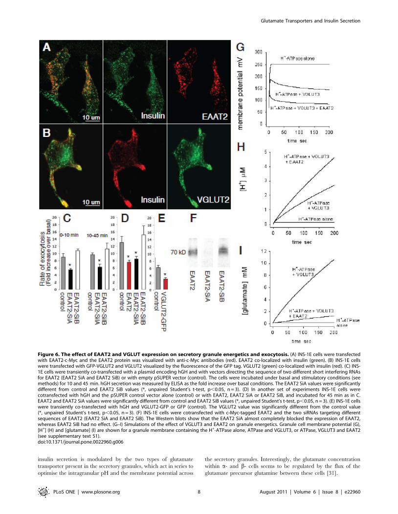
EAAT2 and VGLUT regulate insulin secretionTo assess the function of EAAT2 in the regulation of insulin
secretion from b-cells, we diminished its expression level in INS-
1E cells using RNA interference [21,22]. We generated two
plasmids that allow the synthesis of short double-stranded RNA
molecules (siRNAs) directed against different sequences of rat
EAAT2. As shown in Figure 6F, transient transfection of EAAT2-
SilA led to a very strong reduction of rat EAAT2 expression. In
contrast, EAAT2-SilB had no effect. To evaluate the impact of a
reduction in EAAT2 expression on exocytosis the two silencers
were then transiently co-transfected in INS-1E cells with a plasmid
encoding the human growth hormone (hGH). hGH release was
used to selectively monitor exocytosis in the cells receiving the
silencers, since hGH is targeted to b-cell secretory granules and co-
released with insulin [22]. In cells transfected with the EAAT2-
SilA we found that hormone secretion, evoked by high K+
(40 mM) and glucose (20 mM) concentrations (in the presence of
the cAMP raising agents forskolin and IBMX: see Methods), was
significantly reduced compared to that in mock (pSUPER)
transfected cells or in cells receiving the inactive silencer
(EAAT2-SilB). This effect was evident both during the first
10 min after raising the [K+] and [glucose] (first secretory phase)
and during the following 35 min (sustained secretory phase)
(Figure 6C). There was no effect of the EAAT2-SilA on basal
insulin secretion (i.e. without the glucose/K+ stimulation regime,
data not shown).
When the cells were stimulated with only a high glucose
concentration (20 mM) the increase in secretion over basal
secretion was significantly higher in the mock and EAAT2-SilB
transfected cells (2.7- and 3.1-fold) than in EAAT2-SilA
transfected cells (1.7-fold) (p,0.05, two-sample t-test). This was
true also when only a high K+ concentration was used, for which
the increase in secretion was significantly higher in the mock and
EAAT2-SilB transfected cells (2.4-fold) than in cells treated with
EAAT2-SilA (1.4-fold) (p,0.05). Furthermore, in INS-1E cells, in
which exogenously transfected EAAT2 was expressed in secretory
granules (Figure 6A), over-expression of EAAT2 caused a
significant reduction in secretion (Figure 6D, an explanation of
this effect is suggested below). Thus, EAAT2 activity contributes to
regulation of secretion in b-cells, and either an increase or a
decrease in its expression reduces secretion.
The low glutamate levels in insulin containing secretory
granules, together with the observation that EAAT2 is not
carrying glutamate into these granules, suggest that EAAT2 may
optimize insulin secretion by generating an outward transport of
glutamate, keeping the glutamate concentration low inside the
secretory granules. To test this we expressed a glutamate
transporter that carries glutamate into vesicles [2] in the INS-1E
cells. All the cloned VGLUTs transport glutamate with similar
substrate specificities and kinetics [2]. To ensure insertion of an
excess of VGLUTs in the membranes of secretory granules we
chose to use VGLUT2, which gave a high transfection yield, and is
not expressed endogenously in b-cells. After transfection of INS-
1E cells with c-myc tagged VGLUT2, the transporter was
translocated to secretory granules as judged by its co-localization
with insulin (Figure 6B). In such cells over-expressing a VGLUT,
insulin secretion evoked by the high K+ and glucose concentra-
tions was significantly reduced (Figure 6E). An explanation for this
is suggested below.
EAAT2 and VGLUT3 regulate the pH and membranepotential of the granules
To clarify the mechanism by which the VGLUT3 and EAAT2
glutamate transporters regulate the glutamate concentration in the
granule and modulate exocytosis, we simulated the effect of
including a VGLUT and EAAT2 in the secretory granule
membrane on the pH and the membrane potential of the
secretory granule (see Supplementary Text S1). With just the H+-
ATPase in the granule membrane, the membrane becomes
polarized to over +200 mV as soon as the ATPase is active,
because there is no route for charge to leave the granule, which
inhibits further ATPase activity (Figure 6G). Consequently,
essentially no H+ or glutamate are accumulated (Figure 6H, I).
Including a VGLUT in the membrane of the secretory granule
provides a counter-charge movement (glutamate2 entering the
granule; this may also occur via Cl2 channels: see Supplementary
Text S1) which reduces the polarization of the granule membrane
(Figure 6G) and thus allows maintained operation of the ATPase,
leading to H+ accumulation via the ATPase (Figure 6H) and
glutamate accumulation via the VGLUT (Figure 6I). Finally,
inserting EAAT2 in addition to a VGLUT provides a mechanism
for the release of accumulated glutamate (which would otherwise
start to inhibit the VGLUT), and provides extra counter-charge
movement out of the granule (because glutamate2 leaves the
granule with 3Na+ and an H+, while 1 K+ enters the granule)
leading to a less positive granule membrane potential (Figure 6G).
Despite the efflux of H+ on EAAT2, the overall result is to increase
the H+ concentration further (Figure 6H), while the glutamate
level inside the granule is very much reduced (Figure 6I). Thus,
including both glutamate transporters reduces the vesicle mem-
brane potential and increases H+ accumulation. Consequently,
glutamate may regulate exocytosis via two mechanisms. First,
fluxing glutamate through the secretory granules via the two types
of glutamate transporter promotes the acidification of the granules,
which is known to be important both for the maturation of insulin
[23] and for the exocytotic process [24]. Secondly, the concerted
action of the two transporters limits the membrane potential
reached across the secretory granule, which may be important for
and the core (c) of the secretory granules are significantly lower in KO than in wt mice (p,0.01, Mann-Whitney-U test, two tails). (E–F) Islet (E) andbrain (F) tissue from wild type (EAAT2+/+) and EAAT2 KO (EAAT22/2) mice labelled with the EAAT2 antibodies.doi:10.1371/journal.pone.0022960.g004
Glutamate Transporters and Insulin Secretion
PLoS ONE | www.plosone.org 6 August 2011 | Volume 6 | Issue 8 | e22960

optimising exocytosis. An intact membrane potential across the
granule, acting through trans-SNARE pairing mechanisms, is
needed for exocytosis, possibly independent of the H+ accumula-
tion produced by this potential [25]. Thus, the decrease of
membrane potential that occurs when putting more EAAT2 and
VGLUT into the secretory granules (Figure 6G) might explain
why exocytosis is decreased by overexpression of these transporters
(Figure 6D, E).
The role of glutamate in modulating insulin releaseThe discussion above assumes that glutamate itself is not the
initial trigger for the exocytosis of insulin-containing granules (we
assume that the trigger is a glucose-evoked rise of ATP level
closing ATP-gated K+ channels), but that the flux of glutamate
through the secretory granules leads to the granules being more
acidic and less positive in potential, and thus modulates the
amount of insulin release occurring. Initial claims that glutamate
potentiated secretion [9,10,26] were based on the observation
that a rise in cytoplasmic glutamate concentration correlated with
increased insulin release, but were disputed [27,28] when it was
found that insulin release did not always correlate with the total
islet glutamate concentration. These discrepancies may be partly
resolved by our data, which suggest that it is not so much the total
islet, nor even cytoplasmic glutamate concentration that is the
important regulatory factor, but the flux of glutamate through the
secretory granules, which regulates the granule pH and
membrane potential. Thus, we think that alteration of the
glutamate flux through secretory granules is the mechanism by
which altered glutamate levels modulate insulin release. Even
though a simple relationship between cytoplasmic glutamate
concentration and secretion is not to be expected, because the
simulations depict that secretion is largely dependent on both the
granule [H+] and its membrane potential, granule energetics
could be affected by cytoplasmic glutamate levels (cytoplasmic
glutamate must present to be transported through the granule).
Consistent with this is recent data showing that knocking down a
mitochondrial glutamate transporter (which will presumably alter
the cytoplasmic glutamate level) also leads to a decrease of insulin
secretion [30].
Another consequence of having EAAT2 in the granule
membrane to maintain a low glutamate concentration in the
granule could be that glutamate taken up into the granule is not
lost into the extracellular space during granule exocytosis, but is
recycled back to the cytoplasm where it may fuel the mitochon-
drial TCA cycle. Indeed, there is experimental evidence that b-cell
secretory granules do not release a significant amount of glutamate
[8]. In line with the present and previous in vitro results [9,10,26]
we did not detect any changes in the basal (unstimulated) serum
concentrations of insulin and glucose in EAAT2 KO mice (Figure
S2). In summary, our data suggest that there is a glutamate cycle
through insulin-containing secretory granules in b-cells, and that
Figure 5. The glutamate analogue D-aspartate is neither takenup through the plasma membrane in intact b-cells nor into SGsin permeabilized b-cells. A–C, Acutely prepared slices of islet tissuewere incubated with exogenous D-aspartate (100 mM) before aldehydefixation and labelling with antibodies that selectively recognize D-aspartate. (A–B) Immunoperoxidase labelling shows that the tissue notexposed to D-aspartate (Control) is unlabelled, while in islets exposedto D-aspartate (D-Asp) labelling is observed only in the peripheral a-cellarea, not in the central b-cell area of the islet. (C) Immunofluorescenceshows that the central insulin positive b-cells are negative forexogenous D-aspartate and that the peripheral non-insulin a-cells arelabelled. (D–F) Streptolysin-O permeabilized INS-1E cells were exposedto different concentrations of exogenous D-aspartate (0–3 mM) before
fixation and labelling with the D-aspartate antibodies. (D) In cells notexposed to D-aspartate (Control) there was no labelling for D-aspartate,only for insulin (red). (E) In cells exposed to 1 mM D-aspartate stainingwith the D-aspartate antibodies (green) is observed. There was someweak co-localization (yellow) with insulin (red) that is attributable toextra granular fixation of D-aspartate (see F). (F) Electron micrograph ofpermeabilized INS cells exposed to 1 mM D-aspartate shows nosignificant D-aspartate labelling inside the secretory garnules (indicatedin transparent yellow). Note some labelling along the limitingmembrane of secretory granules and in the cytosol, reflecting fixationof exogenous D-aspartate to extragranular proteins.doi:10.1371/journal.pone.0022960.g005
Glutamate Transporters and Insulin Secretion
PLoS ONE | www.plosone.org 7 August 2011 | Volume 6 | Issue 8 | e22960
insulin secretion is modulated by the two types of glutamate
transporter present in the secretory granules, which act in series to
optimise the intragranular pH and the membrane potential across
the secretory granules. Interestingly, the glutamate concentration
within a- and b- cells seems to be regulated by the flux of the
glutamate precursor glutamine between these cells [31].
Figure 6. The effect of EAAT2 and VGLUT expression on secretory granule energetics and exocytosis. (A) INS-1E cells were transfectedwith EAAT2-c-Myc and the EAAT2 protein was visualized with anti-c-Myc antibodies (red). EAAT2 co-localized with insulin (green). (B) INS-1E cellswere transfected with GFP-VGLUT2 and VGLUT2 visualized by the fluorescence of the GFP tag. VGLUT2 (green) co-localized with insulin (red). (C) INS-1E cells were transiently co-transfected with a plasmid encoding hGH and with vectors directing the sequence of two different short interfering RNAsfor EAAT2 (EAAT2 SiA and EAAT2 SiB) or with empty pSUPER vector (control). The cells were incubated under basal and stimulatory conditions (seemethods) for 10 and 45 min. hGH secretion was measured by ELISA as the fold increase over basal conditions. The EAAT2 SiA values were significantlydifferent from control and EAAT2 SiB values (*, unpaired Student’s t-test, p,0.05, n = 3). (D) In another set of experiments INS-1E cells werecotransfected with hGH and the pSUPER control vector alone (control) or with EAAT2, EAAT2 SiA or EAAT2 SiB, and incubated for 45 min as in C.EAAT2 and EAAT2 SiA values were significantly different from control and EAAT2 SiB values (*, unpaired Student’s t-test, p,0.05, n = 3). (E) INS-1E cellswere transiently co-transfected with hGH and VGLUT2-GFP or GFP (control). The VGLUT2 value was significantly different from the control value(*, unpaired Student’s t-test, p,0.05, n = 3). (F) INS-1E cells were cotransfected with c-Myc-tagged EAAT2 and the two siRNAs targeting differentsequences of EAAT2 (EAAT2 SiA and EAAT2 SiB). The Western blots show that the EAAT2 SiA almost completely blocked the expression of EAAT2,whereas EAAT2 SiB had no effect. (G–I) Simulations of the effect of VGLUT3 and EAAT2 on granule energetics. Granule cell membrane potential (G),[H+] (H) and [glutamate] (I) are shown for a granule membrane containing the H+-ATPase alone, ATPase and VGLUT3, or ATPase, VGLUT3 and EAAT2(see supplementary text S1).doi:10.1371/journal.pone.0022960.g006
Glutamate Transporters and Insulin Secretion
PLoS ONE | www.plosone.org 8 August 2011 | Volume 6 | Issue 8 | e22960

A somewhat analogous role for VGLUT3 has recently been
proposed for cholinergic synaptic vesicles in the striatum [29],
where VGLUT3 mediated uptake of cytoplasmic glutamate
amplifies cholinergic transmission by providing a compensatory
anion influx to the vesicles, similar to what we propose for the
insulin granules. Our data and simulations may therefore be
important for understanding, not only the modulation of insulin
secretion in the pancreas, but also the control of exocytosis of other
membrane vacuole compartments more generally.
Methods
Ethics StatementAnimal experiments were carried out according to international
legislations (European Communities Council Directive of 24
November 1986 (86/609/EEC)) and Norwegian (1996-01-15
no 23) and British (UK Animals (Scientific Procedures) Act
1986) regulations. Formal approval to conduct the experiments
was obtained from the animal subjects review boards of our
institutions. Every effort was made to minimize suffering and the
number of animals used. The animals were maintained under
standard colony conditions in a temperature- (2361uC) and
humidity- (40%) controlled animal room under a 12 h light/dark
cycle (7:00–19:00), with ad libitum access to food and water.
Primary antibodiesPolyclonal antibodies against L-glutamate and D-aspartate were
raised in rabbits as originally described [32]. The no. 607
glutamate and the no. 482 D-aspartate antisera have been
extensively characterized [16,33]. The rabbit anti-73kDa antibod-
ies to EAAT2 were raised against the EAAT2 protein isolated
from rat brain [34]. A monoclonal EAAT2-antibody (9C4)
binding to residues 518–525 (rat numbering) was produced in
mouse [35], and a mouse monoclonal EAAT2 antibody was
purchased from Novo Castra, UK. Anti-peptide antibodies against
glutamate transporters were prepared as described [36]. Different
EAAT2 antibodies were produced in rabbits against residues 12–
26 (anti-B12) [36], residues 518–536 (anti-B518) [37], residues
493–517 (anti-B493) [37], or residues 563–573 (anti-B563). These,
as well as the EAAT1 [34], EAAT3 [38] and EAAT4 [39]
antibodies have been extensively charcterized. In addition, goat
anti-EAAT2 antibodies (Santa Cruz Biotechnology, Santa Cruz,
USA) were used in double labelling immunogold experiments.
Rabbit VGLUT1, VGLUT2 and VGLUT3 antibodies were
gifts from R. H. Edwards (University of California, San Francisco,
USA) [3,17,18]. Guinea pig VGLUT3 antibody was from
Chemicon (Temecula, CA, USA).
A monoclonal mouse anti-glucagon antibody and polyclonal
guinea pig anti-insulin antibodies were from Sigma (St. Louis,
MO, USA), a monoclonal synaptophysin anitbody (mouse) from
Boehringer Mannheim Biochemica (Mannheim, Germany), and a
monoclonal mouse anti-insulin was from Zymed Laboratories, Inc.
(San Francisco, USA). The anti-Myc mouse monoclonal antibody
was purified from the culture medium of myeloma cells (clone
9E10) using an IgG affinity column.
Tissue and cellsAdult Wistar rats were fed ad libitum and perfused [40] through
the left cardiac ventricle with one of the following mixtures of
fixatives in 0.1 M sodium phosphate buffer (pH 7.4) (NaPi): 2.5%
glutaraldehyde and 1% formaldehyde (amino acid immunoper-
oxidase and immunogold cytochemistry), 4% formaldehyde and
0.1% glutaraldehyde (protein immunogold cytochemistry and
immunofluorescence) or 4% formaldehyde (immunofluorescence).
Wild type and KO mice [14] were fed ad libitum. After cervical
dislocation (at P14), pancreatic and brain tissue from EAAT2 KO
mice and wt littermates were immediately immersion fixed in a
mixture of 4% formaldehyde and 0.1% glutaraldehyde. INS-1E
and COS7 cells were cultured as previously described [41].
Cellular uptake sites for D-aspartatePancreatic slices (0.5 mm thick) from adult Wistar rats were
prepared by hand cutting, before they were transferred to vials
with Krebs solution (pH 7.4 with 140 mM NaCl, 15 mM NaPi,
5 mM KCl, 5 mM glucose, 1.2 mM CaCl2, 1.2 mM MgSO4) that
were continuously gassed with O2 at 30uC. The slices were
preincubated for 30 minutes before incubation in the presence and
absence of 100 mM D-aspartate for 20 minutes.
D-asparate uptake in secretory granuleAs D-aspartate is a substrate for EAAT2 [4,5,16], but not for
the VGLUTs [3,17,18], we used D-aspartate as a tracer to explore
whether secretory granules in b-cells show EAAT2 uptake activity.
INS-1E cells were permeabilized with 1.5 IU/ml streptolysin-O
and incubated with 0, 0.5, 0.3 and 1.0 mM D-aspartate in an
intracellular medium containing 20 mM HEPES, pH 7.0,
140 mM KCl, 5 mM NaCl, 7 mM MgSO4, 5 mM Na2ATP,
10.2 mM EGTA and 0.1 mM CaCl2 for 7 min at 37uC.
Both the tissue slices and the INS-1E cells were fixed by either a
mixture of 1% formaldehyde and 2.5% glutaraldehyde (for
immunperoxidase and immunogold cytochemistry) or 4% form-
aldehyde and 0.5% glutaraldehyde (for immunfluorescence).
Immunofluorescence and immunoperoxidaseAfter fixation, pancreas and brain specimens were cryostat sectioned
(5–15 mm) after cryoprotection in 30% sucrose at 4uC overnight. The
sections were processed with the antibodies according to an
immunofluorescence or a streptavidin-biotin-peroxidase method as
previously described [16,31,40]. The following antibody dilutions were
used: rabbit anti-D-aspartate 482 (1:1000), anti-glutamate 607
(1:1000–1:3000), and the anti-EAAT2 immunoglobulins anti-B12
(0.3–3 mg/ml), anti-B518 (1 mg/ml), anti-B493 (3 mg/ml), anti-B563
(0.3–1 mg/ml), mouse monoclonal EAAT2 antibody (1:30), rabbit anti-
EAAT1 (1–6 mg/ml), anti-EAAT3 (1–6 mg/ml), anti-EAAT4 (1–
6 mg/ml), anti-VGLUT1 (1:1000), anti-VGLUT2 (1:2000), guinea-pig
anti-VGLUT2 (1:3000), anti-VGLUT3 (1:300), guinea-pig anti-
VGLUT3 (1:1500), anti-insulin (1:50–1:100), anti-glucagon (1:2000),
mouse anti-c-Myc (1:10). Primary antibodies were visualized with
FITC-, Cy3- or Alexa (488 or 555)- fluorescent secondary antibodies.
In control immunofluorescence experiments in which the
primary antibodies were omitted or substituted with preimmune-
sera, there was no significant staining of the tissue sections, but
some experiments produced a diffuse fluorescence background
that was subtracted from the labelling produced by the primary
antibodies. In immunoperoxidase experiments, the glutamate and
D-aspartate antisera were used with the addition of 0.2 mM
complexes of glutaraldehyde/formaldehyde treated L-aspartate
plus glutamine and L-aspartate, respectively. Addition of 0.3 mM
complexes of L-glutamate and D-aspartate to the primary
antibodies abolished the glutamate and D-aspartate labeling.
Sections were observed with a Zeiss Pascal laser scanning
microscope or a Zeiss fluorescence microscope at wavelengths of
488 nm and 568 nm.
Immunogold staining and quantitationTissue for electron microscopy was embedded in Lowicryl
HM20 according to a freeze substitution protocol, and immuno-
Glutamate Transporters and Insulin Secretion
PLoS ONE | www.plosone.org 9 August 2011 | Volume 6 | Issue 8 | e22960

gold labelling was performed as described [40,42]. Ultrathin
sections were processed with the primary antibodies at the
following dilutions: anti-D-aspartate (1:300), anti-glutamate
(1:3000), anti-EAAT2 B12 (6–40 mg/ml) anti-EAAT2 73 kDa
(5 mg/ml) and anti-VGLUT3 from guinea pig (1:100). In single
labelling experiments the primary antibodies were visualized with
anti-rabbit or anti-guniea pig Ig specific secondary antibodies
coupled to 15 nm gold particles (Europrobe, Lyon, France). In the
double labelling experiments of KO and wt tissue, rabbit anti-
glutamate (1:3000) and goat anti-EAAT2 antibodies (2 mg/ml)
were visualized with goat anti-rabbit (10 nm gold particles (BBI,
UK)) and donkey anti-goat (15 nm gold particles (Aurion, The
Netherlands)) secondary antibodies. In each amino acid experi-
ment ultrathin test sections [43] were processed along with the islet
sections, ascertaining the specificity of the labelling produced.
When tested on ultrathin test sections containing known
concentrations of glutamate and D-aspartate, both the glutamate
and the D-aspartate antisera have been shown to produce a close
to linear relation between the immunogold labelling densities and
the concentrations of amino acids in the test sections [44,45].
The sections were viewed in a Philips CM10 electron
microscope and a- and b-cells were identified on morphological
grounds [40,46]. Immunogold particles signalling glutamate,
EAAT2 and VGLUT3, and grid points for area determination
[33,40,42] (see below) were recorded over secretory granules,
SLMVs, plasma membranes, cytosol and mitochondria (back-
ground labelling). Particles and grid points were included in the
SLMV when the particle centres or grid points were within a
30 nm distance from the outer border of the SLMV. This is a
distance within which most of the immunogold particles are
expected to occur [42,47]. Similarily, EAAT2 and VGLUT3 gold
particles with centres within 30 nm on either side of plasma
membranes and membranes limiting secretory granules were
recorded as belonging to these membranes (for details, see [40]).
The EAAT2 and VGLUT3 values over tissue membranes were
corrected for background labelling (measured over mitochondrial
outer membranes: average 3.7 and 1.3 gold particles/mm2), while
EAAT2 and VGLUT3 values over non-membrane compartments
(cytosol, secretory granule cores) were corrected for background
labelling measured over the mitochondrial matrix (average 4.9 and
2.7 gold particles/mm2). Similar quantifications were done for gold
particles representing glutamate. The glutamate values were
corrected for background labelling over empty resin (average 1.7
gold particles/mm2). The results were statistically evaluated by a
non-parametric test (Mann-Whitney-U, two tails) (Statistica) and a
paired-sample t-test (Excel).
Western blottingPancreatic islets were acutely isolated as described [48].
Homogenates of isolated islets and COS-7 cells were separated
by SDS-PAGE, blotted onto nitrocellulose, and proteins were
immunodetected by primary antibodies, HRP-conjugated second-
ary antibodies and chemiluminescence reagents. COS-7 cell
homogenates were also processed by secondary antibodies coupled
to alkaline phosphatase (AP) and detected with AP-substrates.
Cloning of rat EAAT2 and generation of EAAT2 silencersThe open reading frame of rat EAAT2 was cloned by PCR from
an INS-1E cDNA library in pBK vector (Stratagene). The PCR
reaction was performed using the forward primer 59_CGCGGA-
TCCATGGCATCAACCGAGGGT_39 and the reverse primer
59_GCTCTAGATTATTTTTCACGTTTCCA _39. Both prim-
ers were designed according to the sequence of rat EAAT2
(NM_017215) [49]. For sequencing and expression experiments the
PCR products were inserted in the BamHI and Xba I cloning sites
of myc-pcDNA3. Sequence analysis of the inserts was performed by
MWG Biotech Company (Germany). Mammalian expression
vectors directing the synthesis of small interfering RNAs (siRNAs)
targeted against EAAT2 were prepared according to the method of
Brummelkamp [49]. Two cDNA fragments encoding a 19-
nucleotide sequence derived from the target transcript and
separated from its reverse 19-nucleotide complement by a short
spacer were synthesized by MWG Biotech Company (Ebersberg,
Germany), annealed and cloned in front of the H1-RNA promoter
in the pSUPER vector16 nucleotides: 59_GATCCCCCCGAGGG-
TGCCAACAATATTTCAAGAGAATATTGTTGGCACCC C-
GGTTTTTGGAAA_39 and 59_AGCTTTTCCAAAAACCGA-
GGGTGCCAACAATATTCTCTTGAAATATTGTT GCACC-
CTCGGGGG_39 (SilA) and 59_GATCCCCGATGCTCATC-
CTCCCTCTCTTCAAGAGAGAGAGGGAGGATGAGCAT-
CTTTTTGGAAA _39 and 59_AGCTTTTCCAAAAAGAT-
GCTCATCCTCCCTCTCTCTCTTGAAGAGAGGGAGGAT-
GAGCATCGGG_39 (SilB). To test for the silencing activity of SilA
and SilB, each plasmid was transiently co-transfected with the
plasmid encoding rat myc-tagged EAAT2 in COS 7 cells. The
expression of EAAT2 was assessed two days later by Western
blotting.
Secretion assayINS-1E cells were transiently cotransfected with a plasmid
encoding human growth hormone (hGH) and with plasmids
encoding the siRNAs or EAAT2-c-Myc or VGLUT2-green
fluorescent protein (GFP) constructs. Three days later, the cells
were preincubated for 30 min in 20 mM HEPES, pH 7.4,
128 mM NaCl, 5 mM KCl, 1 mM MgCl2, 2.7 mM CaCl2 and
2 mM glucose. The cells were then incubated for 45 min at 37uCeither in the same buffer or stimulated with the 20 mM HEPES
buffer containing 40 mM KCl, 20 mM glucose, 1 mM forskolin,
and 1 mM 3-isobutyl-L-methylxantine (IBMX). In some experi-
ments the cells were stimulated with either 20 mM glucose or
40 mM K+ alone. Exocytosis from transfected cells was deter-
mined by measuring by ELISA the amount of hGH (Roche,
Rotkreuz, Switzerland) released into the medium during the
incubation period, and expressed as an increase over basal
(exocytosis = (stimulated2basal)/basal). The results were statisti-
cally evaluated by a two-sample t-test.
At the time of stimulation some cells were fixed with 4%
formaldehyde and processed for confocal immunofluorescent
microscopy with c-Myc and insulin antibodies.
Blood samples from EAAT2 KO mice and littermates (n = 8 of
each +/+ and 2/2) were taken from the aorta after cervical
dislocation and serum was prepared for ELISA measurements.
Insulin was analysed using the Mouse Ultrasensitive Insulin ELISA
kit (Mercodia, Uppsala, Sweden). Serum samples for each animal
were analysed in triplicate and absorbance read at 450 nm in a
Photometer for microtitration plate. Blood glucose concentrations
from each sample were analysed by Accu-Chek compact (Roche
Diagnostics GmbH, Mannheim, Germany).
RT-PCRPCR was conducted on RNA from isolated islets of Langerhans
and brain tissue using Qiagen OneStep RT-PCR kit (35 3-step
cycles). Primer control and -RT controls were included. Primers
used were EAAT2 P1F (GAAAAAACCCATTCTCCTTTTT)
and EAAT2 P1R (CCGACTGGGAGGACGAATC). Primers
were from Oligold Eurogentec. 20 ng RNA template of each tissue
was used per reaction. Electrophoresis was conducted on 2%
agarose gels, samples diluted with Fermentas 66Orange buffer.
Glutamate Transporters and Insulin Secretion
PLoS ONE | www.plosone.org 10 August 2011 | Volume 6 | Issue 8 | e22960

Simulation of granule energeticsDetails of these simulations are given in the Supplementary
Material.
Supporting Information
Figure S1 The figure shows the subcellular distribution of
VGLUT3 and EAAT2 in INS1-E cells. After elimination of nuclei
and cell debris the postnuclear supernatants from INS1-E cells
were separated on a sucrose density gradient (0.45–2 M). (A) The
insulin content was measured by ELISA in each fraction of the
gradient. Dense-core insulin-containing secretory granules are
recovered in the fractions corresponding to 1.3–1.6 M sucrose. (B)
The fractions were analysed by Western blotting using antibodies
against VGLUT3, EAAT2 and synaptophysin. In contrast to
insulin, synaptophysin, a marker of synaptic-like microvesicles, is
recovered at 0.9–1.3 M sucrose. EAAT2 was located in the insulin
containing fraction, suggesting that it is present in insulin granules.
VGLUT3 was located both in the insulin and the synaptophysin
containing fractions, suggesting that it is present in secretory
granules and SLMVs. Method: Subcellular fractionation of
approximately 108 INS-1E cells was performed as described (Ref
1 Supplementary Text S1). Briefly, a postnuclear supernatant
obtained after disruption of the cells by sonication, was loaded on
a continuous sucrose density gradient (8 ml; 0.45–2 M sucrose).
After centrifugation for 18 h at 110,0006 g, 16 fractions of 0.5 ml
were collected from the top of the tube. The concentration of
sucrose in the fractions was determined by measuring the
refractive index of the solution. The amount of insulin present
in the fractions was measured by ELISA (Mercodia). The
distribution throughout the gradient of EAAT2, VGLUT3 and
synaptophysin was measured by Western blotting. The bound
antibody was visualized using an HRP-conjugated goat anti mouse
(synaptophysin) and rabbit anti-goat (EEAT2, VGLUT3) followed
by chemiluminescence reagents.
(TIF)
Figure S2 To examine whether EAAT2 affects insulin secretion
in intact animals, we analysed insulin serum concentrations in the
EAAT2 KO mice (KO), in which the transporter is absent from
the b-cell SGs (see Fig. 4), and compared these values to results
obtained in wild type mice (wt). The values are the mean serum
concentration of glucose and insulin 6 SD in 8 wt and 8 KO
animals. We could not detect any significant difference in insulin
or glucose concentrations between mice lacking and expressing
EAAT2. As the serum glucose concentrations in the EAAT2 KO
mice is low (4.960.7 mM) the hormone values reflect basal
secretion, which has been shown not to be significantly affected by
glutamate (see text). This is also in accordance with our in vitro
data showing that silencing of EAAT2 is not important for
regulating basal insulin secretion. We are not in a position to keep
the EAAT2 knock out animals for doing in vivo animal research.
The EAAT2 KO mice get epileptic after 2 weeks of age and,
according to the animal welfare regulations that we presently have
to abide by, they must be killed before the age of 2 weeks. This
precludes further detailed analysis, such as insulin measurements
after glucose tolerance tests, which would be extremely demanding
to do on mice younger than 14 days of age.
(TIF)
Text S1
(DOC)
Acknowledgements
We thank Professor Susan Bonner-Weir (Joslin Diabetes Center, Boston
USA) for help with confocal microscopy of VGLUTs. Antibodies to
EAAT1-4 were from N.C. Danbolt (University of Oslo, Norway). The
rabbit VGLUT1-3 antibodies were a gift from R.H. Edwards (UCSF
School of Medicine, San Francisco, USA). The pBK vector (Stratagene)
was kindly provided by C. Bonny (University of Lausanne, Switzerland).
We also thank Kohichi Tanaka for generously supplying GLT-1 KO mice
to D. Attwell.
Author Contributions
Conceived and designed the experiments: RG DA RR VG. Performed the
experiments: RG TC PM DA VG. Analyzed the data: RG TC JS-M FAC
DA RR VG. Contributed reagents/materials/analysis tools: DA FAC VG.
Wrote the paper: RG DA RR VG.
References
1. Ottersen OP, Storm-Mathisen J (1984) Glutamate- and GABA-containing
neurons in the mouse and rat brain, as demonstrated with a new
immunocytochemical technique. J Comp Neurol 229: 374–392.
2. Fremeau RT, Jr., Voglmaier S, Seal RP, Edwards RH (2004) VGLUTs define
subsets of excitatory neurons and suggest novel roles for glutamate. Trends
Neurosci 27: 98–103.
3. Fremeau RT, Jr., Burman J, Qureshi T, Tran CH, Proctor J, et al. (2002) The
identification of vesicular glutamate transporter 3 suggests novel modes of
signaling by glutamate. Proc Natl Acad Sci U S A 99: 14488–93.
4. Danbolt NC (2001) Glutamate uptake. Prog Neurobiol 65: 1–105.
5. Balcar V, Johnston GA (1972) The structural specificity of the high affinity uptake
of L-glutamate and L-aspartate by rat brain slices. J Neurochem 19: 2657–66.
6. Inagaki N, Kuromi H, Gonoi T, Okamoto Y, Ishida H, et al. (1995) Expression
and role of ionotropic glutamate receptors in pancreatic islet cells. FASEB J 9:
686–691.
7. Weaver CD, Yao TL, Powers AC, Verdoorn TA (1996) Differential expression
of glutamate receptor subtypes in rat pancreatic islets. J Biol Chem 271:
12977–12984.
8. Hayashi M, Yamada H, Uehara S, Morimoto R, Muroyama A, et al. (2003)
Secretory granule-mediated co-secretion of L-glutamate and glucagon triggers
glutamatergic signal transmission in islets of Langerhans. J Biol Chem 278:
1966–1974.
9. Maechler P, Wollheim CB (1999) Mitochondrial glutamate acts as a messenger
in glucose-induced insulin exocytosis. Nature 402: 685–689.
10. Hoy M, Maechler P, Efanov AM, Wollheim CB, Berggren PO, et al. (2002)
Increase in cellular glutamate levels stimulates exocytosis in pancreatic beta-cells.
FEBS Lett 531: 199–203.
11. Manfras BJ, Rudert WA, Trucco M, Boehm BO (1994) Cloning and
characterization of a glutamate transporter cDNA from human brain and
pancreas. Biochim Biophys Acta 1195: 185–188.
12. Arriza JL, Eliasof S, Kavanaugh MP, Amara SG (1997) Excitatory amino acid
transporter 5, a retinal glutamate transporter coupled to a chloride conductance.
Proc Natl Acad Sci U S A 94: 4155–4160.
13. Lehre KP, Levy LM, Ottersen OP, Storm-Mathisen J, Danbolt NC (1995)
Differential expression of two glial glutamate transporters in the rat brain:
quantitative and immunocytochemical observations. J Neurosci 15: 1835–53.
14. Tanaka K, Watase K, Manabe T, Yamada K, Watanabe M, et al. (1997)
Epilepsy and exacerbation of brain injury in mice lacking the glutamate
transporter GLT-1. Science 276: 1699–1702.
15. Hamann M, Rossi DJ, Marie H, Attwell D (2002) Knocking out the glial
glutamate transporter GLT-1 reduces glutamate uptake but does not affect
hippocampal glutamate dynamics in early simulated ischaemia. Eur J Neurosci
15: 308–314.
16. Gundersen V, Danbolt NC, Ottersen OP, Storm-Mathisen J (1993) Demon-
stration of glutamate/aspartate uptake activity in nerve endings by use of
antibodies recognizing exogenous D-aspartate. Neuroscience 57: 97–111.
17. Bellocchio EE, Hu H, Pohorille A, Chan J, Pickel VM, et al. (1998) The
localization of the brain-specific inorganic phosphate transporter suggests a
specific presynaptic role in glutamatergic transmission. J Neurosci 18:
8648–8659.
18. Fremeau RT, Jr., Troyer MD, Pahner I, Nygaard GO, Tran CH, et al. (2001)
The expression of vesicular glutamate transporters defines two classes of
excitatory synapse. Neuron 31: 247–260.
19. Levy L, Warr O, Attwell D (1998) Stoichiometry of the glial glutamate
transporter GLT-1 expressed inducibly in a Chinese hamster ovary cell line
selected for low endogenous Na+-dependent glutamate uptake. J Neurosci 18:
9620–9628.
20. Thirion S, Troadec JD, Pivovarova NB, Pagnotta S, Andrews SB, et al. (1999)
Stimulus-secretion coupling in neurohypophysial nerve endings: a role for
intravesicular sodium. Proc Natl Acad Sci USA 96: 3206–3210.
Glutamate Transporters and Insulin Secretion
PLoS ONE | www.plosone.org 11 August 2011 | Volume 6 | Issue 8 | e22960

21. Waselle L, Coppola T, Fukuda M, Iezzi M, El-Amraoui A, et al. (2003)
Involvement of the Rab27 binding protein Slac2c/MyRIP in insulin exocytosis.Mol Biol Cell 14: 4103–4113.
22. Coppola T, Perret-Menoud V, Luthi S, Farnsworth CC, Glomset JA, et al.
(1999) Disruption of Rab3-calmodulin interaction, but not other effectorinteractions, prevents Rab3 inhibition of exocytosis. EMBO J 18: 5885–5891.
23. Orci L, Ravazzola M, Amherdt M, Madsen O, Perrelet A, et al. (1986)Conversion of proinsulin to insulin occurs coordinately with acidification of
maturing secretory vesicles. J Cell Biol 103: 2273–2281.
24. Barg S, Huang P, Eliasson L, Nelson DJ, Obermuller S, et al. (2001) Priming ofinsulin granules for exocytosis by granular Cl- uptake and acidification. J Cell Sci
114: 2145–2154.25. Ungermann C, Wickner W, Xu Z (1999) Vacuole acidification is required for
trans-SNARE pairing, LMA1 release, and homotypic fusion. Proc Natl Acad SciUSA 96: 11194–11199.
26. Rubi B, Ishihara H, Hegardt FG, Wollheim CB, Maechler P (2001) GAD65-
mediated glutamate decarboxylation reduces glucose-stimulated insulin secretionin pancreatic beta cells. J Biol Chem 276: 36391–36396.
27. MacDonald MJ, Fahien LA (2000) Glutamate is not a messenger in insulinsecretion. J Biol Chem 275: 34025–34027.
28. Bertrand G, Ishiyama N, Nenquin M, Ravier MA, Henquin JC (2002) The
elevation of glutamate content and the amplification of insulin secretion inglucose-stimulated pancreatic islets are not causally related. J Biol Chem 277:
32883–32891.29. Gras C, Amilhon B, Lepicard EM, Poirel O, Vinatier J, et al. (2008) The
vesicular glutamate transporter VGLUT3 synergizes striatalacetylcholine tone.Nat Neurosci 11: 292–300.
30. Casimir M, Rubi B, Frigerio F, Chaffard G, Maechler P. Silencing of the
mitochondrial NADH shuttle component aspartate-glutamate carrier AGC1/Aralar1 in INS-1E cells and rat islets. Biochem J 2009 Dec 10;424(3): 459–66.
31. Gammelsaeter R, Jenstad M, Bredahl MK, Gundersen V, Chaudhry FA (2009)Complementary expression of SN1 and SAT2 in the islets of Langerhans
suggests concerted action of glutamine transport in the regulation of insulin
secretion. Biochem Biophys Res Commun 381: 378–382.32. Storm-Mathisen J, Leknes AK, Bore AT, Vaaland JL, Edminson P, et al. (1983)
First visualization of glutamate and GABA in neurones by immunocytochem-istry. Nature 301: 517–520.
33. Gundersen V, Chaudhry FA, Bjaalie JG, Fonnum F, Ottersen OP, et al. (1998)Synaptic vesicular localization and exocytosis of L-aspartate in excitatory nerve
terminals: a quantitative immunogold analysis in rat hippocampus. J Neurosci
18: 6059–6070.34. Danbolt NC, Storm-Mathisen J, Kanner BI (1992) An [Na++K+]coupled L-
glutamate transporter purified from rat brain is located in glial cell processes.Neuroscience 51: 295–310.
35. Levy LM, Lehre KP, Walaas SI, Storm-Mathisen J, Danbolt NC (1995) Down-
regulation of glial glutamate transporters after glutamatergic denervation in therat brain. Eur J Neurosci 7: 2036–2041.
36. Lehre KP, Levy LM, Ottersen OP, Storm-Mathisen J, Danbolt NC (1995)
Differential expression of two glial glutamate transporters in the rat brain:
quantitative and immunocytochemical observations. J Neurosci 15: 1835–53.
37. Beckstrom H, Julsrud L, Haugeto O, Dewar D, Graham DI, et al. (1999)
Interindividual differences in the levels of the glutamate transporters GLAST
and GLT, but no clear correlation with Alzheimer’s disease. J Neurosci Res 55:
218–229.
38. Haugeto O, Ullensvang K, Levy LM, Chaudhry FA, Honore T, et al. (1996)
Brain glutamate transporter proteins form homomultimers. J Biol Chem 271:
27715–27722.
39. Dehnes Y, Chaudhry FA, Ullensvang K, Lehre KP, Storm-Mathisen J, et al.
(1998) The glutamate transporter EAAT4 in rat cerebellar Purkinje cells: a
glutamate-gated chloride channel concentrated near the synapse in parts of the
dendritic membrane facing astroglia. J Neurosci 18: 3606–3619.
40. Gammelsaeter R, Frøyland M, Aragon C, Danbolt NC, Fortin D, et al. (2004)
Glycine, GABA and their transporters in pancreatic islets of Langerhans:
evidence for a paracrine transmitter interplay. J Cell Sci 117: 3749–3758.
41. Asfari M, Janjic D, Meda P, Li G, Halban PA, et al. (1992) Establishment of 2-
mercaptoethanol-dependent differentiated insulin-secreting cell lines. Endocri-
nology 130: 167–178.
42. Bergersen LH, Storm-Mathisen J, Gundersen V (2008) Immunogold quantifi-
cation of amino acids and proteins in complex subcellular compartments. Nat
Protoc 3: 144–152.
43. Ottersen OP (1989) Postembedding light- and electron microscopic immuno-
cytochemistry of amino acids: description of a new model system allowing
identical conditions for specificity testing and tissue processing. Exp Brain Res
69: 167–174.
44. Ottersen OP (1989) Postembedding immunogold labelling of fixed glutamate: an
electron microscopic analysis of the relationship between gold particle density
and antigen concentration. J Chem Neuroanat 2: 57–66.
45. Gundersen V, Shupliakov O, Brodin L, Ottersen OP, Storm-Mathisen J (1995)
Quantification of excitatory amino acid uptake at intact glutamatergic synapses
by immunocytochemistry of exogenous D-aspartate. J Neurosci 15: 4417–4428.
46. Cell and Tissue Biology: A Textbook of Histology (ed. Weiss, L.) (Urban and
Schwartzenberg, Baltimore, 1988).
47. Chaudhry FA, Lehre KP, van Lookeren Campagne M, Ottersen OP,
Danbolt NC, et al. (1995) Glutamate transporters in glial plasma membranes:
highly differentiated localizations revealed by quantitative ultrastructural
immunocytochemistry. Neuron 15: 711–720.
48. Thorens B, Weir GC, Leahy JL, Lodish HF, Bonner-Weir S (1990) Reduced
expression of the liver/beta-cell glucose transporter isoform in glucose-insensitive
pancreatic beta cells of diabetic rats. Proc Natl Acad Sci U S A 87: 6492–6496.
49. Brummelkamp TR, Bernards R, Agami R (2002) A system for stable expression
of short interfering RNAs in mammalian cells. Science 296: 550–553.
Glutamate Transporters and Insulin Secretion
PLoS ONE | www.plosone.org 12 August 2011 | Volume 6 | Issue 8 | e22960
Copyright © 2022 FDOKUMEN