Domain Organization, Catalysis and Regulation of Eukaryotic Cystathionine Beta-Synthases
14
Domain Organization, Catalysis and Regulation of Eukaryotic Cystathionine Beta-Synthases Tomas Majtan 1 , Angel L. Pey 2 , Roberto Ferna ´ ndez 3 , Jose ´ A. Ferna ´ ndez 3 , Luis A. Martı´nez-Cruz 4 , Jan P. Kraus 1 * 1 Department of Pediatrics, University of Colorado, School of Medicine, Aurora, Colorado, United States of America, 2 Department of Physical Chemistry, Faculty of Sciences, University of Granada, Granada, Spain, 3 Department of Physical Chemistry, Faculty of Science and Technology, University of the Basque Country (UPV/EHU), Leioa, Spain, 4 Structural Biology Unit, CIC bioGUNE, Derio, Bizkaia, Spain Abstract Cystathionine beta-synthase (CBS) is a key regulator of sulfur amino acid metabolism diverting homocysteine, a toxic intermediate of the methionine cycle, via the transsulfuration pathway to the biosynthesis of cysteine. Although the pathway itself is well conserved among eukaryotes, properties of eukaryotic CBS enzymes vary greatly. Here we present a side-by-side biochemical and biophysical comparison of human (hCBS), fruit fly (dCBS) and yeast (yCBS) enzymes. Preparation and characterization of the full-length and truncated enzymes, lacking the regulatory domains, suggested that eukaryotic CBS exists in one of at least two significantly different conformations impacting the enzyme’s catalytic activity, oligomeric status and regulation. Truncation of hCBS and yCBS, but not dCBS, resulted in enzyme activation and formation of dimers compared to native tetramers. The dCBS and yCBS are not regulated by the allosteric activator of hCBS, S- adenosylmethionine (AdoMet); however, they have significantly higher specific activities in the canonical as well as alternative reactions compared to hCBS. Unlike yCBS, the heme-containing dCBS and hCBS showed increased thermal stability and retention of the enzyme’s catalytic activity. The mass-spectrometry analysis and isothermal titration calorimetry showed clear presence and binding of AdoMet to yCBS and hCBS, but not dCBS. However, the role of AdoMet binding to yCBS remains unclear, unlike its role in hCBS. This study provides valuable information for understanding the complexity of the domain organization, catalytic specificity and regulation among eukaryotic CBS enzymes. Citation: Majtan T, Pey AL, Ferna ´ndez R, Ferna ´ndez JA, Martı ´nez-Cruz LA, et al. (2014) Domain Organization, Catalysis and Regulation of Eukaryotic Cystathionine Beta-Synthases. PLoS ONE 9(8): e105290. doi:10.1371/journal.pone.0105290 Editor: Vladimir N. Uversky, University of South Florida College of Medicine, United States of America Received May 30, 2014; Accepted July 23, 2014; Published August 14, 2014 Copyright: ß 2014 Majtan et al. This is an open-access article distributed under the terms of the Creative Commons Attribution License, which permits unrestricted use, distribution, and reproduction in any medium, provided the original author and source are credited. Data Availability: The authors confirm that all data underlying the findings are fully available without restriction. All relevant data are within the paper and its Supporting Information files. Funding: This work was supported by Postdoctoral Fellowship 0920079G from the American Heart Association (to TM), by National Institutes of Health Grant HL065217, by American Heart Association Grant In-Aid 09GRNT2110159, by a grant from the Jerome Lejeune Foundation (all to JPK) and by a research contract RYC2009-04147 (to ALP). In addition, grant support (P11-CTS-07187, CSD2009-00088 and BIO2012-34937) to Dr. Jose M. Sanchez-Ruiz (University of Granada) and SGIker technical and human support (UPV/EHU, MICINN, GV/EJ, ESF) are gratefully acknowledged. The funders had no role in study design, data collection and analysis, decision to publish, or preparation of the manuscript. Competing Interests: The authors have declared that no competing interests exist. * Email: [email protected] Introduction Methionine (Met) is an essential sulfur amino acid for mammals and its metabolism comprises two intersecting metabolic pathways: the methionine cycle, found in all tissues, and the transsulfuration pathway, which occurs in a limited number of tissues, but mainly in liver and kidney [1]. Both pathways compete for homocysteine (Hcy), a central intermediate that has been formed from Met. While in the methionine cycle Hcy is converted back to Met by either methionine synthase or betaine-homocysteine methyltrans- ferase, in the transsulfuration pathway Hcy is irreversibly converted to cysteine (Cys). The transsulfuration pathway is believed to be the sole route for Cys synthesis in vertebrates [2]. Thus, Hcy formation and its distribution between these two pathways represents an occasion for regulatory intervention. Cystathionine beta-synthase (CBS) is the enzyme, which regulates the flux of Hcy through the transsulfuration pathway and thus commits Hcy to the synthesis of Cys [3]. Deficiency in CBS results in a serious metabolic disorder, homocystinuria, clinically mani- fested chiefly by connective tissue defects, mental retardation and thromboembolism [4]. Considering the importance of CBS in sulfur amino acid metabolism, it is interesting that domain organization, quaternary structure and regulatory mechanism of CBS enzymes are not conserved across phyla (Fig. 1A–B). The extensively studied human CBS (hCBS) is a homotetrameric enzyme of 63 kDa polypeptides, each consisting of three distinct domains (reviewed in [3,5]). The N-terminal domain of hCBS binds heme, which binds via a Cys/His ligation [6]. The origin and role of the heme in CBS is still an enigma and it is believed to function as a redox sensor [5] and/or to play a structural role facilitating a correct folding [7,8]. The highly conserved central region forms a catalytic domain containing the PLP cofactor. The C-terminal domain houses a tandem of CBS domains, a structural motif known to bind adenosine nucleotides and to regulate protein function [9]. Indeed, the catalytic activity as well as kinetic stability of hCBS is increased upon interaction of S-adenosyl-L-methionine (AdoMet) with the CBS domains [10,11]. In comparison to hCBS, the variability in PLOS ONE | www.plosone.org 1 August 2014 | Volume 9 | Issue 8 | e105290
-
Upload
independent -
Category
Documents
-
view
1 -
download
0
Transcript of Domain Organization, Catalysis and Regulation of Eukaryotic Cystathionine Beta-Synthases
Domain Organization, Catalysis and Regulation ofEukaryotic Cystathionine Beta-SynthasesTomas Majtan1, Angel L. Pey2, Roberto Fernandez3, Jose A. Fernandez3, Luis A. Martınez-Cruz4,
Jan P. Kraus1*
1 Department of Pediatrics, University of Colorado, School of Medicine, Aurora, Colorado, United States of America, 2 Department of Physical Chemistry, Faculty of
Sciences, University of Granada, Granada, Spain, 3 Department of Physical Chemistry, Faculty of Science and Technology, University of the Basque Country (UPV/EHU),
Leioa, Spain, 4 Structural Biology Unit, CIC bioGUNE, Derio, Bizkaia, Spain
Abstract
Cystathionine beta-synthase (CBS) is a key regulator of sulfur amino acid metabolism diverting homocysteine, a toxicintermediate of the methionine cycle, via the transsulfuration pathway to the biosynthesis of cysteine. Although thepathway itself is well conserved among eukaryotes, properties of eukaryotic CBS enzymes vary greatly. Here we present aside-by-side biochemical and biophysical comparison of human (hCBS), fruit fly (dCBS) and yeast (yCBS) enzymes.Preparation and characterization of the full-length and truncated enzymes, lacking the regulatory domains, suggested thateukaryotic CBS exists in one of at least two significantly different conformations impacting the enzyme’s catalytic activity,oligomeric status and regulation. Truncation of hCBS and yCBS, but not dCBS, resulted in enzyme activation and formationof dimers compared to native tetramers. The dCBS and yCBS are not regulated by the allosteric activator of hCBS, S-adenosylmethionine (AdoMet); however, they have significantly higher specific activities in the canonical as well asalternative reactions compared to hCBS. Unlike yCBS, the heme-containing dCBS and hCBS showed increased thermalstability and retention of the enzyme’s catalytic activity. The mass-spectrometry analysis and isothermal titration calorimetryshowed clear presence and binding of AdoMet to yCBS and hCBS, but not dCBS. However, the role of AdoMet binding toyCBS remains unclear, unlike its role in hCBS. This study provides valuable information for understanding the complexity ofthe domain organization, catalytic specificity and regulation among eukaryotic CBS enzymes.
Citation: Majtan T, Pey AL, Fernandez R, Fernandez JA, Martınez-Cruz LA, et al. (2014) Domain Organization, Catalysis and Regulation of Eukaryotic CystathionineBeta-Synthases. PLoS ONE 9(8): e105290. doi:10.1371/journal.pone.0105290
Editor: Vladimir N. Uversky, University of South Florida College of Medicine, United States of America
Received May 30, 2014; Accepted July 23, 2014; Published August 14, 2014
Copyright: � 2014 Majtan et al. This is an open-access article distributed under the terms of the Creative Commons Attribution License, which permitsunrestricted use, distribution, and reproduction in any medium, provided the original author and source are credited.
Data Availability: The authors confirm that all data underlying the findings are fully available without restriction. All relevant data are within the paper and itsSupporting Information files.
Funding: This work was supported by Postdoctoral Fellowship 0920079G from the American Heart Association (to TM), by National Institutes of Health GrantHL065217, by American Heart Association Grant In-Aid 09GRNT2110159, by a grant from the Jerome Lejeune Foundation (all to JPK) and by a research contractRYC2009-04147 (to ALP). In addition, grant support (P11-CTS-07187, CSD2009-00088 and BIO2012-34937) to Dr. Jose M. Sanchez-Ruiz (University of Granada) andSGIker technical and human support (UPV/EHU, MICINN, GV/EJ, ESF) are gratefully acknowledged. The funders had no role in study design, data collection andanalysis, decision to publish, or preparation of the manuscript.
Competing Interests: The authors have declared that no competing interests exist.
* Email: [email protected]
Introduction
Methionine (Met) is an essential sulfur amino acid for mammals
and its metabolism comprises two intersecting metabolic pathways:
the methionine cycle, found in all tissues, and the transsulfuration
pathway, which occurs in a limited number of tissues, but mainly
in liver and kidney [1]. Both pathways compete for homocysteine
(Hcy), a central intermediate that has been formed from Met.
While in the methionine cycle Hcy is converted back to Met by
either methionine synthase or betaine-homocysteine methyltrans-
ferase, in the transsulfuration pathway Hcy is irreversibly
converted to cysteine (Cys). The transsulfuration pathway is
believed to be the sole route for Cys synthesis in vertebrates [2].
Thus, Hcy formation and its distribution between these two
pathways represents an occasion for regulatory intervention.
Cystathionine beta-synthase (CBS) is the enzyme, which regulates
the flux of Hcy through the transsulfuration pathway and thus
commits Hcy to the synthesis of Cys [3]. Deficiency in CBS results
in a serious metabolic disorder, homocystinuria, clinically mani-
fested chiefly by connective tissue defects, mental retardation and
thromboembolism [4].
Considering the importance of CBS in sulfur amino acid
metabolism, it is interesting that domain organization, quaternary
structure and regulatory mechanism of CBS enzymes are not
conserved across phyla (Fig. 1A–B). The extensively studied
human CBS (hCBS) is a homotetrameric enzyme of 63 kDa
polypeptides, each consisting of three distinct domains (reviewed in
[3,5]). The N-terminal domain of hCBS binds heme, which binds
via a Cys/His ligation [6]. The origin and role of the heme in CBS
is still an enigma and it is believed to function as a redox sensor [5]
and/or to play a structural role facilitating a correct folding [7,8].
The highly conserved central region forms a catalytic domain
containing the PLP cofactor. The C-terminal domain houses a
tandem of CBS domains, a structural motif known to bind
adenosine nucleotides and to regulate protein function [9]. Indeed,
the catalytic activity as well as kinetic stability of hCBS is increased
upon interaction of S-adenosyl-L-methionine (AdoMet) with the
CBS domains [10,11]. In comparison to hCBS, the variability in
PLOS ONE | www.plosone.org 1 August 2014 | Volume 9 | Issue 8 | e105290

domain organization can be illustrated on CBS species that have
been experimentally documented so far [12–15]. The presence of
heme is unique among the PLP-dependent enzymes and,
moreover, not all eukaryotic CBS enzymes contain this cofactor.
As an example, Saccharomyces cerevisiae CBS (yCBS) does not
contain heme [13], while Drosophila melanogaster CBS (dCBS)
does [14,16]. Similarly, while majority of CBS enzymes contains
the regulatory domain, only the mammalian CBS, such as hCBS,
appears to be regulated by AdoMet; dCBS and yCBS are not
[13,14]. The regulatory domain seems to be essential for
oligomerization as its removal from a full-length yCBS and hCBS
native tetramers yields dimeric truncated enzymes [17,18]. On the
contrary, the full-length dCBS forms native homodimers [14].
In addition to the canonical condensation of Hcy with L-serine
(Ser) into cystathionine (Cth) and water (Fig. 1C, reaction (1)),
several alternative reactions have been described for the previously
characterized CBS enzymes [3,19–21]. A recent recognition of
H2S as a physiologically important gaseous signaling molecule and
the implication of the two transsulfuration enzymes, CBS and
cystathionine gamma-lyase (CGL), in H2S biosynthesis [22] led to
exploration of alternative H2S-generating reactions (Fig. 1C,
reactions (2–4)). These reactions utilize Cys instead of Ser
following either b-replacement or b-elimination mechanism to
yield H2S. In addition, as the conserved CBS catalytic core is
shared by both CBS and O-acetylserine sulfhydrase (OASS; also
known as cysteine synthase (CS)) enzymes [6,23], it was not
surprising that CBS can catalyze a formation of Cys by utilizing
H2S (Fig. 1C, reactions (5–7)) [19,24]. However, physiological
relevance of these alternative reactions remains to be answered.
In this study, we addressed several structural, catalytic and
regulatory features of this crucial enzyme of sulfur amino acid
metabolism. Side-by-side comparison of three eukaryotic CBS
enzymes from yeast, fruit fly and human revealed their unique
characteristics and properties. We discuss how the acquisition or
loss of specific features shaped the enzyme properties and provide
hypothesis explaining the complexity of domain organization,
regulation and catalytic specificity among eukaryotic CBS
enzymes.
Materials and Methods
ChemicalsUnless stated otherwise, all chemicals were purchased from
Sigma or Fisher Scientific. L-[14C(U)]-serine was obtained from
PerkinElmer Life Sciences.
Preparation of CBS constructsWe used our established constructs for hCBS as models for
cloning of yCBS and dCBS into pGEX-6P1 (GE Healthcare) and/
or pET-28a (Novagen) vectors and for preparation of their C-
terminally truncated forms [8,25]. Table S1 lists all the
oligonucleotides used for subcloning and mutagenesis.
Briefly, the coding sequence of yCBS was PCR amplified from a
previously prepared pGEX-561-yCBS plasmid [13] using the 656
and 657 oligonucleotides. The ApaI- and NotI-digested, gel-
extracted PCR product was ligated into a similarly prepared
pGEX-6P1 vector using T4 DNA ligase (NEB Biolabs). The dCBS
coding sequence was PCR amplified from a recently reported
pGEX-6P1-DMCBS plasmid [24] using the 824 and 825
oligonucleotides. The NcoI- and HindIII-digested, gel-extracted
PCR product was ligated into a similarly prepared pET-28a vector
using T4 DNA ligase (NEB Biolabs). Both yCBS and dCBS
constructs, designated as pGEX-6P1-yCBS and pET28-C-dCBS,
respectively, were transformed into E. coli XL1-Blue cells (Agilent)
and their authenticity was confirmed by DNA sequencing. The
pGEX-6P1-yCBS construct was later used as a template for PCR
for recloning of full-length yCBS WT and truncated yCBS L345*
into a pET-28a vector using the 794 and 795 and 794 and 796
oligonucleotides, respectively. The NcoI- and XhoI-digested, gel-
extracted PCR product was ligated into a similarly prepared pET-
28a vector using T4 DNA ligase (NEB Biolabs). Both pET28-C-
yCBS WT and pET28-C-yCBS L345* constructs were trans-
formed into E. coli XL1-Blue cells (Agilent) and their authenticity
and the presence of a C-terminal 6xHis tag was confirmed by
DNA sequencing.
In order to prepare the various truncated forms of yCBS and
dCBS lacking the regulatory domain (Fig. 2), the translational
STOP codons were introduced using a QuikChange II XL site
directed mutagenesis kit (Agilent). For yCBS, we introduced and
tested three STOP codons in positions S323, L345 and K370
using the 788 and 789, 790 and 791 and 792 and 793
oligonucleotide pairs, respectively. For dCBS, we mutated four
residues into STOP codons in the previously reported pGEX-6P1-
DMCBS vector in positions S356, K366, L379 and P387 using the
816 and 817, 818 and 819, 820 and 821 and 822 and 823
oligonucleotide pairs, respectively. Later, the coding sequences of
the truncated dCBS forms were PCR amplified using the 824 and
826, 824 and 827, 824 and 828 or 824 and 829 oligonucleotide
pairs and subcloned into pET-28a vector following an analogous
strategy as for the full-length form. The presence of the desired
STOP codons was confirmed by DNA sequencing. The verified
plasmids for full-length and truncated forms of yCBS and dCBS
were finally transformed into E. coli Rosetta2 (DE3) expression
host cells (Novagen).
Expression and purificationBacterial growth and protein expression were carried out at
30uC and together with preparation of soluble and insoluble crude
extracts followed the previously published procedure [8,25].
The purification of yCBS, yCBS L345* and dCBS carrying a
permanent 6xHis affinity tag at the C-terminus followed the
procedure recently described for human CBS variants with a few
modifications [25]. After the first immobilized metal affinity
chromatography step (TALON column; Clontech) and subsequent
desalting on Sephadex G-25 (GE Healthcare) column, the sample
was loaded onto a DEAE Sepharose (GE Healthcare) column
equilibrated in the DEAE loading buffer (15 mM potassium
phosphate, pH 7.2, 1 mM EDTA, 1 mM DTT, 10% ethylene
glycol). The bound yCBS or yCBS L345* was washed with 2
column volumes of the DEAE loading buffer followed by 5 column
volumes of the DEAE wash buffer (50 mM potassium phosphate
pH 7.2, 1 mM EDTA, 1 mM DTT, 10% ethylene glycol). The
enzymes were then eluted with 150 mM potassium phosphate in
the DEAE loading/wash buffer. The bound dCBS was washed
with 5 column volumes of DEAE loading/wash buffer (15 mM
potassium phosphate, pH 7.2, 1 mM EDTA, 1 mM DTT, 10%
ethylene glycol) and eluted with 50 mM potassium phosphate in
the DEAE loading/wash buffer. The enzymes were buffer
exchanged into the final storage buffer (20 mM HEPES pH 7.4,
1 mM TCEP) on a Sephadex G-25 column and subsequently
concentrated using an ultrafiltration device (Amicon) equipped
with an YM-30 (Millipore) membrane. Finally, the enzymes were
aliquoted, flash-frozen in liquid nitrogen and stored at –80uC.
Protein gel electrophoresis and Western blot analysisProtein concentrations were determined by the Bradford
method (Thermo Pierce) using bovine serum albumin (BSA) as a
standard according to the manufacturer’s recommendations.
Domain Organization, Catalysis and Regulation of Eukaryotic CBSs
PLOS ONE | www.plosone.org 2 August 2014 | Volume 9 | Issue 8 | e105290

Figure 1. Domain architecture and structure of CBS enzymes and reactions catalyzed by CBS. (A) Domain architecture of CBS enzymesfrom H. sapiens (hCBS), D. melanogaster (dCBS) and S. cerevisae (yCBS). Regions corresponding to the central catalytic domain (green) and CBS domain(blue) as well as presence of the cofactors (heme in red, PLP in yellow) are indicated. (B) Crystal structures of dCBS (PDB #3PC2) and hCBSD516–525(PDB #4L0D). Subunits within the dimers are distinguished by red or blue color, while the linker connecting the catalytic domain with the regulatorydomain (dashed box) is highlighted in green. Yellow color highlights the residues in the connecting linker, which were targeted by mutagenesis (formore details see Fig. 2). Cofactors, heme and PLP, are shown as sticks. (C) Reactions catalyzed by CBS leading to Cth, H2S and Cys generation thatwere characterized in this study.doi:10.1371/journal.pone.0105290.g001
Domain Organization, Catalysis and Regulation of Eukaryotic CBSs
PLOS ONE | www.plosone.org 3 August 2014 | Volume 9 | Issue 8 | e105290
Denatured proteins were separated by SDS-PAGE using a 9%
separating gel with a 4% stacking gel. Native samples were
separated in 4–15% polyacrylamide gradient precast gels (Mini-
PROTEAN TGX, Bio-Rad). For visualization, the denatured gels
were stained with Simple Blue (Invitrogen). Western blot analysis
of crude cell lysates under denaturing or native conditions was
performed as described previously [8].
CBS activity measurementsThe CBS activity in the classical reaction was determined by a
previously described radioisotope assay using [14C(U)] L-serine as
the labeled substrate [26]. Briefly, a purified enzyme (420 ng) was
assayed in a 100 mL reaction for 30 min at 37uC. The reaction
mixture contained 100 mM Tris–HCl pH 8.6, 10 mM L-serine,
0.2 mM PLP, 0.3 mCi L-[14C(U)]-serine and 0.5 mg/ml BSA.
The reaction was performed in the presence or absence of AdoMet
in a final concentration of 0.3 mM. The reaction mixture with
enzyme was incubated at 37uC for 5 min and the reaction was
initiated by addition of L-homocysteine to a final concentration of
10 mM. The reaction was terminated by an immediate cooling of
the mixture in ice water and the labeled product was separated
from the substrates by paper chromatography. Spots correspond-
ing to Cth were cut-out and radioactivity was determined by using
a scintillation counter.
The thermal pre-treatment of the enzyme prior the CBS activity
assay was performed as described before [25]. Briefly, the purified
enzyme was diluted to a final concentration of 0.1 mg/ml in Tris-
buffered saline pH 8.6, 100 mM PLP. For isothermal incubation,
the enzyme solutions (4650 ml) were incubated at 37uC in 200-ml
thin-walled PCR tubes in a Mastercycler gradient PCR thermal
cycler (Eppendorf) for up to 96 hours. For gradual thermal
denaturation, the enzyme solutions (4650 ml) were heated in 200-
ml tubes in a PCR thermal cycler from 37uC to 60uC in 0.5uC-
increments with a 1 min incubation at each temperature. Aliquots
(20 ml) were collected into separate tubes at designated times or
temperatures and assayed for the CBS activity as described above.
The activities in the H2S-generating alternative reactions were
measured using a colorimetric determination of H2S described
earlier (methylene blue method [27]) with the following modifi-
cations. The reaction mixture (200 ml) contained 200 mM Tris–
HCl, pH 8.6, 40 mM L-cysteine, 20 mM L-homocysteine
(omitted in the cysteine b-elimination/b-replacement reaction),
0.5 mM PLP and 0.5 mg/ml BSA. The reaction was performed in
the absence or presence of AdoMet in a final concentration of
0.3 mM. The mixture was incubated at 37uC for 2 min and the
reaction was initiated by addition of CBS enzyme (2.5 mg) and
carried at 37uC for 6 min. The reaction was terminated by a 40-
fold diluting a 25 ml assay aliquot in water and mixing it with a N,N-dimethyl-p-phenylenediamine reagent and ferric chloride solu-
tion. The samples were stored in the dark at room temperature for
20 min for color development. The concentration of sulfide was
determined from the absorbance at 650 nm using a standard
curve prepared from sodium sulfide solutions of known concen-
tration.
The Cys-producing activities were determined by using a
colorimetric detection of generated cysteine as described earlier
(ninhydrin method [28]) with the following modifications. The
reaction mixture (200 ml) contained 200 mM Tris–HCl, pH 8.6,
15 mM sodium sulfide, 10 mM L-serine (or O-acetylserine or O-
phosphoserine), 0.5 mM PLP and 0.5 mg/ml BSA. The reaction
was performed in the absence or presence of AdoMet in a final
concentration of 0.3 mM. The mixture was incubated at 37uC for
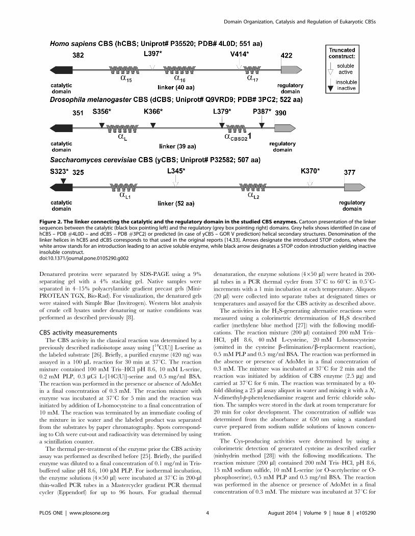
Figure 2. The linker connecting the catalytic and the regulatory domain in the studied CBS enzymes. Cartoon presentation of the linkersequences between the catalytic (black box pointing left) and the regulatory (grey box pointing right) domains. Grey helix shows identified (in case ofhCBS – PDB #4L0D – and dCBS – PDB #3PC2) or predicted (in case of yCBS – GOR V prediction) helical secondary structures. Denomination of thelinker helices in hCBS and dCBS corresponds to that used in the original reports [14,33]. Arrows designate the introduced STOP codons, where thewhite arrow stands for an introduction leading to an active soluble enzyme, while black arrow designates a STOP codon introduction yielding inactiveinsoluble construct.doi:10.1371/journal.pone.0105290.g002
Domain Organization, Catalysis and Regulation of Eukaryotic CBSs
PLOS ONE | www.plosone.org 4 August 2014 | Volume 9 | Issue 8 | e105290
2 min and the reaction was initiated by addition of the enzyme
(10 mg) and carried out at 37uC for 6 min. Reaction was
terminated by removing a 50 mml assay aliquot and mixing it
with equal amounts of glacial acetic acid and acidic ninhydrin
reagent. After 10 min boiling and immediate cooling, the color
was stabilized by addition of denatured ethanol. The concentra-
tion of cysteine was determined from the absorbance at 560 nm
using a standard curve prepared from cysteine solutions of known
concentration.
One unit of activity is defined as the amount of CBS that
catalyzes the formation of 1 mmol of the product in 1 hour at 37uCunder above described assay conditions.
Differential scanning calorimetry (DSC)DSC measurements were performed in a capillary VP-DSC
microcalorimeter (GE Healthcare) as described previously [11].
Briefly, samples containing 5 mM of CBS enzymes (in protein
subunit) were prepared in 20 mM HEPES pH 7.4 in the presence
of 50 mM PLP, and in some cases up to 400 mM AdoMet. Protein
concentration was measured spectrophotometrically using the
following extinction coefficients determined from acid hydrolysis:
e280 = 103,800 M21cm21 (hCBS), 79,000 M21cm21 (yCBS) and
119,700 M21cm21 (dCBS). Scans were performed in a 4–100uCrange at 2–4uC/min scan rates.
Analysis of DSC transitions was performed in all cases using a
two-state irreversible denaturation model [11,29]. In this model,
denaturation of the protein (or domain) is assumed to follow
equation 1:
N ?k
F ð1Þ
Where N and F stand for the native and irreversible denatured
states and k is the first-order rate constant. The expression used to
fit the experimental DSC traces (apparent molar heat capacities,
Cp(app), vs. Temperature) explicitly considers the experimental
chemical baseline [30] as follows:
Cp(app)~Cp(pre)z(Cp(post){Cp(pre)):(1{XN )
{DH: dXN
dT
� � ð2Þ
XN~ exp { expEaD T
RTm2
� �� �(3)
dXN
dT~{
Ea
RTm2: exp
EaDT
RTm2
� �: exp { exp
EaDT
RTm2
� �� �ð4Þ
Where XN is the mole fraction of native state, DH is the
denaturation enthalpy and Cp(pre) and Cp(post) are the pre- and
post-transition baselines, which are considered to be a linear
function of temperature, Ea stands for the activation energy and
Tm for the temperature of the maximum of the transition. The two
first terms in the right-hand-side of Equation 1 represent the
chemical baseline and the last term describes the denaturation
transition (‘‘the peak’’).
Theoretical DH values were evaluated using the correlation
between unfolding enthalpies (at 60u) and heat capacities on the
protein size (in number of residues) described by Robertson &
Murphy [31]. Experimental DH values were determined at the
experimental Tm of each domain/protein using the Kirchoffs
equation, to compare them with the theoretical DH values.
Mass spectrometrySamples containing purified hCBS (18.4 mg/ml), yCBS
(43.9 mg/ml) and dCBS (12.6 mg/ml) were examined using
MALDI (matrix assisted laser desorption/ionization) mass spec-
trometry as described previously with a few modifications [32]. A
saturated solution of CHCA (a-cyano-4-hydroxycinnamic acid) in
acetonitrile/water (4:1 v:v) was used as matrix. Each sample was
mixed with the matrix solution and 1 ml aliquots were deposited in
the MALDI target for analysis. The AdoMet solution (10 mM
AdoMet in 0.001N H2SO4, pH 3.0) and proteins were mixed with
the matrix in sample:matrix (1:10 v:v) ratio and analyzed using
either Synapt G2 (Waters) with a nominal resolution of 40,000 or
LTQ Orbitrap XL (Thermo) with a nominal resolution of
100,000. In both cases, a minimum of 100 shots were averaged
to build the final spectrum. The G2 spectrometer was calibrated
before each experiment, using the peaks from polyethyleneglycol
to ensure that the mass accuracy was better than 5 mDa.
Isothermal titration calorimetry (ITC)ITC measurements were carried out in an ITC200 microcal-
orimeter (GE Healthcare) essentially as described previously [11].
Briefly, protein solutions (,20 mM in protein subunit) were
prepared in 20 mM HEPES pH 7.4 and titrated at 25uC by
adding 40–50 injections of 300 mM AdoMet (0.8–1 ml each).
Binding thermodynamic parameters were obtained from fittings to
a one- or two-independent type of sites using the software provided
by the manufacturer.
Results
Structure of the linker between the catalytic and theregulatory domains
We analyzed and compared the primary sequence and the
secondary structure elements of the region connecting the two
functional domains in the studied enzymes (Fig. 2). The length of
the linker in both heme-containing CBS enzymes is almost
identical: 40 and 39 amino acid residues long in hCBS and dCBS,
respectively. However, the linker in yCBS, containing only the
PLP cofactor, is significantly longer containing 52 residues. The
crystal structure of hCBS (PDB #4L0D) showed three helices
within the linker (a15, a16 and a17), while the dCBS structure (PDB
#3PC2) revealed only two helices (aL and aCBSD21) [14,33]. By
using the CDM protein secondary structure prediction server
(http://gor.bb.iastate.edu/cdm/), we identified two helices in
yCBS designated as aL1 and aL2. Presence of an additional helix in
hCBS (a16) significantly differs from the unorganized central
regions of the linkers in dCBS and yCBS, which may have
implications for the flexibility of the linker and thus conformation
of the regulatory domain towards the catalytic core as illustrated in
Fig. 1B.
The C-terminal truncations of dCBS and yCBSTo experimentally define the importance of the linker for a
relationship between the catalytic and the regulatory domains, we
introduced four and three STOPs into the sequence of dCBS and
yCBS, respectively: S356*, K366*, L379* and P387* in dCBS and
S323*, L345* and K370* in yCBS (Fig. 2). To minimize the effect
of the affinity tag on the linker analysis, two types of truncated
enzymes carrying either the N-terminal GST tag or the C-terminal
6xHis tag were tested.
As illustrated in Fig. 3 the removal of the regulatory domain
had totally different effect on dCBS compared to yCBS. None of
the four tested truncated dCBS constructs yielded a soluble
enzyme, which is supported by lack of CBS activity in the soluble
Domain Organization, Catalysis and Regulation of Eukaryotic CBSs
PLOS ONE | www.plosone.org 5 August 2014 | Volume 9 | Issue 8 | e105290

fraction (Fig. 3A). As this result might likewise suggest possible
problems with protein expression, we also analyzed the insoluble
fractions. Strong signals from Western blot analysis after SDS-
PAGE corresponding to the truncated dCBS species in insoluble
fractions ruled out any problems with protein expression and
similar results were obtained for the pGEX-6P1-derived constructs
(data not shown). On the contrary, two out of the three tested
truncated yCBS constructs (L345* and K370*) yielded active,
soluble enzymes with similar results for both types of constructs:
pGEX-6P1-derived GST-yCBS fusion proteins (Fig. 3B) as well
as pET28-derived 6xHis tagged proteins (data not shown). Failure
to obtain soluble active truncated enzyme for the yCBS S323*
construct served as a control experiment showing that any
interference within the central catalytic region (e.g. removal of
the very last two residues of the catalytic domain along with the
linker and regulatory domain of yCBS) yielded insoluble, inactive
truncated protein.
Initial characterization of the purified full-length andtruncated CBS enzymes
In order to compare all three eukaryotic CBS enzymes side-by-
side, we successfully purified three full-length (hCBS, dCBS and
yCBS) and two truncated (45 kDa hCBS i.e. hCBS V414* and
yCBS L345*) CBS enzymes to homogeneity (.95%) carrying the
permanent 6xHis tag at the C-terminus (Fig. 4A) [25]. UV-visible
spectroscopic analysis showed the presence of heme in hCBS,
dCBS and 45 kDa hCBS (Soret peak at 430 nm) and its absence in
yCBS and yCBS L345* (only the PLP peak at 412 nm was
present) (Fig. 4B). Interestingly, unlike its truncated variant, the
full-length yCBS showed a shoulder at around 320 nm, which may
correspond to different protonation states of the PLP cofactor [34].
In case of hCBS and yCBS, removal of the regulatory domain lead
to a change of the oligomeric status from tetramers to dimers as
determined by native gel electrophoresis (Fig. 4C) and was
accompanied with an increase of the catalytic activity of the
truncated enzymes (Fig. 4D). The hCBS was the only enzyme
regulated by AdoMet showing ,3.2-fold increase in CBS specific
activity upon addition of 300 mM AdoMet. Removal of the
regulatory region from the AdoMet-responsive tetrameric hCBS
resulted in formation of a highly active (,3.5-fold higher activity
compared to basal hCBS activity), AdoMet-unresponsive dimeric
45 kDa hCBS. Even though the basal activity of yCBS is even
higher than that of 45 kDa hCBS, the truncation of yCBS yielded
an almost 2-fold more active catalytic core compared to the full-
Figure 3. Removal of the regulatory C-terminal domain in dCBS and yCBS. Four and three STOP codons were introduced to the linkerregion of dCBS (A) and yCBS (B), respectively, in order to prepare the truncated enzymes. 25 ug of soluble clarified bacterial crude extract (left panels)or solubilized denatured insoluble fraction (right panels) were loaded per lane and separated on 9% SDS-PAGE gels, transferred to a PVDF membraneand probed with either monoclonal anti-6xHis antibody (ABM) (for dCBS) or monoclonal anti-GST antibody (ABM) (for yCBS). CBS specific activities areshown below the respective lanes of the soluble fractions for each construct.doi:10.1371/journal.pone.0105290.g003
Domain Organization, Catalysis and Regulation of Eukaryotic CBSs
PLOS ONE | www.plosone.org 6 August 2014 | Volume 9 | Issue 8 | e105290

Domain Organization, Catalysis and Regulation of Eukaryotic CBSs
PLOS ONE | www.plosone.org 7 August 2014 | Volume 9 | Issue 8 | e105290

length enzyme, which was also accompanied with an oligomeric
status change from a native tetramer (yCBS) to a truncated dimer
(yCBS L345*). Unlike hCBS and yCBS, the full-length dCBS
forms native dimers, whose specific activity in the canonical CBS
reaction is similar to that of yCBS.
Catalytic variability of CBS enzymesWe assayed the purified full-length CBS enzymes and deter-
mined their specific catalytic activities in the canonical as well as
alternative reactions depicted in Fig. 1C. Both dCBS and yCBS
showed over a 5-fold higher specific activity compared to hCBS in
the canonical condensation of Ser and Hcy (Fig. 5A). As
expected, hCBS was the only studied enzyme regulated by
AdoMet showing ,3-fold increase in the presence of 300 mM
AdoMet. In the alternative H2S-producing condensation of Cys
and Hcy, all three enzymes expressed higher specific activities
compared to the canonical reaction (Fig. 5B). While dCBS
appeared to have similar activity as the AdoMet-stimulated hCBS,
yCBS showed a dramatic ,3.5-fold increase compared to the
heme-containing CBS enzymes. By measuring the production of
H2S from Cys alone, we could not differentiate between Cys
desulfurase activity (b-elimination) and Cys+Cys condensation (b-
replacement) (Fig. 5C). Again, both dCBS and yCBS showed
,3.7x and ,2.6x higher specific activities, respectively, compared
to hCBS. Interestingly, we observed significantly lower response of
hCBS to AdoMet stimulation in this reaction compared to all
other tested combinations of substrates. The capability of CBS
enzymes to utilize H2S and to form Cys was demonstrated by
using either Ser as a substrate (Ser sulfhydrylase – SS – activity) or
its activated forms, such as OAS and OPS (CS/OASS activity). As
Fig. 5D shows, Ser appeared to be the only relevant substrate for
CBS enzymes to form Cys. Even though CBSs are evolutionary
the closest relatives to CS/OASSs, they could barely utilize the
activated forms of Ser as substrates for Cys production. The yCBS
showed the highest activity (,10-fold compared to hCBS),
followed by dCBS displaying ,3.2x higher specific activity than
hCBS. Taken together, both yCBS and dCBS were not regulated
by AdoMet and showed higher specific activity in all tested
reactions compared to hCBS.
Thermal stability of CBS enzymesWe have already demonstrated in our previous work, that a
gradual thermal denaturation of hCBS resulted in enzyme
activation due to irreversible denaturation of the regulatory
domains [11], thus mimicking the stimulation by AdoMet
[8,25]. In order to assess the thermal stability of the purified
CBS enzymes, we subjected dCBS and yCBS to a similar thermal
pre-treatment assay (Fig. 6A) supplemented with an isothermal
incubation at 37uC for 96 hours (Fig. 6B) and compared them
with the hCBS. From the former analysis, it is obvious that the
heme-containing CBSs (dCBS, 45 kDa hCBS) are significantly
more resistant towards heat-induced denaturation and unfolding
than the hemeless yCBS or its truncated form yCBS L345*
(Fig. 6A). Clearly, such dramatic decrease in yCBS activity could
not be explained by the loss of PLP as the catalytically required
cofactor, present in the incubation buffer, should have efficiently
compensated for its eventual loss. On the other hand, the
isothermal incubation of CBS enzymes at 37uC for up to 96
hours did not show any dramatic changes (Fig. 6B). Altogether,
our data demonstrate that, unlike yCBS, catalytic activities of the
heme-containing dCBS and 45 kDa hCBS are unaffected by
heating up to 60uC thus further supporting the proposed structural
role of heme cofactor in CBS.
Thermal denaturation of CBS enzymes by DSCTo study the rapid activity loss of hemeless yCBS and the
resistance toward thermal denaturation of the heme-containing
dCBS in more detail, we employed DSC (Fig. 7). As previously
found for hCBS [11], denaturation of yCBS and dCBS is
irreversible, strongly scan-rate dependent and described well (for
the main transitions) by a simple 2-state irreversible denaturation
model.
In the case of yCBS, one unfolding transition was detected with
a Tm of 59.9uC and a DH of 15867 kcal/mol (Fig. 7A).
Theoretical DH values corresponding to denaturation of the
catalytic domain alone and both the catalytic and the regulatory
domains together are 227 kcal/mol and 354 kcal/mol, respec-
tively. The unfolding of the regulatory domain (377–508) gives a
theoretical value of 91 kcal/mol, and including the linker (325–
508), 128 kcal/mol. Since the denaturation transition by DSC
agrees well with the half-inactivation temperature (Fig. 6A), it is
likely that this thermal transition corresponds to the irreversible
denaturation of at least the catalytic domain. Therefore, based on
the unfolding enthalpy we cannot say whether both domains
partially unfolded or only the catalytic domain was unfolding. A
kinetic analysis of yCBS denaturation provides a half-life for
denaturation at 37uC of about 226 h, which is 2.2-fold lower than
for the catalytic domain of hCBS [11]. The presence of 100 mM
AdoMet only slightly increased the Tm about 0.2uC (Fig. 7A)
indicating low affinity binding, low binding stoichiometry or that
the ligand is not released prior to the rate limiting step of
unfolding. Taken together, the data presented here suggest that
yCBS is not stable when gradually heated, unfolds rapidly and
AdoMet does not affect this process.
Unfolding of dCBS displays one main transition with a Tm of
70.8uC and DH of 38863 kcal/mol and a second small transition
with a Tm of about 55uC (Fig. 7B). The small transition probably
reflects the unfolding of the N-terminal heme-binding domain, but
it is difficult to model due to its very small signal [11]. The main
transition seems to reflect the unfolding of both the regulatory and
the catalytic domain, since the theoretical DH value of 398 kcal/
mol is in excellent agreement with the experimental value. The
regulatory domain of dCBS is largely stabilized reminiscent of the
previously published effect of AdoMet on the regulatory domain of
hCBS [11] (Fig. 7C). A kinetic analysis of dCBS denaturation
provides a half-life for denaturation extrapolated to 37uC of about
7.46107 h, which is five orders of magnitude higher than for the
catalytic domain of hCBS [11].
Figure 4. Comparison of the purified full-length and truncated CBS enzymes. (A) Homogenous purified full-length (FL; hCBS, dCBS andyCBS) and truncated (T; 45 kDa hCBS and yCBS L345*) CBS enzymes were separated on 9% polyacrylamide gel under reducing denaturing conditions(100 ng CBS per lane), transferred to a PVDF membrane and probed with monoclonal anti-6xHis antibody (ABM). (B) UV-visible spectra of purifiedCBSs. The enzymes were diluted in 1x Tris-buffered saline, pH 8.6 and spectra were recorded on an Agilent 8453 UV-visible spectrophotometer. (C)Purified enzymes (100 ng CBS per lane) were separated in a 4–15% polyacrylamide gel under native conditions, transferred to a PVDF membrane andprobed with monoclonal anti-6xHis antibody (ABM). The 2x and 4x corresponds to native dimers and tetramers, respectively. (D) The enzymes weretested for their catalytic activity and response to the allosteric activator AdoMet in the canonical reaction. Activities are expressed as CBS specificactivities (for clarity without standard deviations), where ‘‘2’’ designates the absence of AdoMet and ‘‘+’’ denotes the presence of 300 mM AdoMet.doi:10.1371/journal.pone.0105290.g004
Domain Organization, Catalysis and Regulation of Eukaryotic CBSs
PLOS ONE | www.plosone.org 8 August 2014 | Volume 9 | Issue 8 | e105290

Figure 5. Specific activities of studied full-length CBS enzymes in various reactions. The reaction, for which the specific activities aredisplayed in each panel, and the detected compound (in italic and underlined) are shown above the corresponding graphs: (A) canonical reaction, (B,C) H2S-generating reactions and (D) Cys-producing reactions. Error bars represent standard deviations from a minimum of three independentmeasurements.doi:10.1371/journal.pone.0105290.g005
Figure 6. Thermal stability of the purified CBS enzymes. (A) The effect of thermal pre-treatment of full-length dCBS (squares), yCBS (circles),truncated 45 kDa hCBS (triangles) and yCBS L345* (diamonds) on their specific activities in the canonical reaction. (B) The effect of isothermal incubation(at 37uC) of the truncated 45 kDa hCBS (triangles), full-length dCBS (squares) and yCBS (circles) on their specific activities in the canonical reaction.doi:10.1371/journal.pone.0105290.g006
Domain Organization, Catalysis and Regulation of Eukaryotic CBSs
PLOS ONE | www.plosone.org 9 August 2014 | Volume 9 | Issue 8 | e105290

MALDI-MS analysis of CBS enzymesThe presence of only one main transition in DSC profiles of
yCBS and dCBS suggested the absence of an independent
transition for the regulatory domain. When compared to hCBS
(Fig. 7C), the presence of AdoMet bound within the dCBS
regulatory domain might have explained such observation. For
this reason, we carried out a MALDI-MS analysis of CBS
enzymes. Despite the large abundance of peaks due to the buffer,
the MS spectra of hCBS and yCBS, unlike dCBS, unequivocally
showed the presence of AdoMet. Peaks at positions of 399.140 Da,
298.093 Da and 250.091 Da (Fig. 8) correspond to the proton-
ated parent ion and two protonated fragments. Even though the
assay was not carried out in a quantitative manner, one can
assume from the size of AdoMet-corresponding peaks that yCBS
preparation likely contains much less bound AdoMet than hCBS,
especially taking into account the relatively higher concentration
of yCBS sample (see Methods section). Moreover, these amounts
of AdoMet must be present in hCBS in very limited quantities,
since every preparation of hCBS tested by the MS exhibited a 3-5-
fold increase in the presence of AdoMet (data not shown).
Taken together, the MS analysis suggests that a significant
stabilization of dCBS regulatory domain is not due to the presence
of bound AdoMet within the CBS domains. Furthermore, high
catalytic activity and unresponsiveness to AdoMet stimulation of
yCBS as well as dCBS cannot be explained by the presence of
bound AdoMet in the protein preparations.
AdoMet binding to purified CBS enzymes by ITCThe MALDI-MS analysis suggested that yCBS may bind
AdoMet. Therefore, we determined binding of AdoMet to yCBS
and dCBS by using ITC (Fig. 9). Representative raw calorimetric
titrations of CBS proteins with AdoMet clearly show that yCBS
indeed binds AdoMet, while dCBS does not (Fig. 9A). However,
translation of raw data into binding isotherms (Fig. 9B) and
subsequent calculation of binding stoichiometry and thermody-
namic parameters for AdoMet binding (Fig. 9C) showed signif-
icant differences between the values for yCBS and those previously
published on hCBS [11]. Unlike hCBS, AdoMet binding to yCBS
is consistent with the presence of just one type of sites with binding
stoichiometry of ,2 molecules of AdoMet per yCBS tetramer with
significantly lower binding affinity (Kd = 5.060.5 mM). The lack of
yCBS activation, the similar stoichiometries and binding enthal-
pies between the AdoMet site in yCBS and the high-affinity site in
hCBS suggest those sites are alike, even though the reduction in
the rate of denaturation (kinetic stabilization) exerted in yCBS by
AdoMet is negligible. Taken together, yCBS binds AdoMet, but
the significance of AdoMet binding in yCBS remains unclear.
Discussion
In this study, we characterized side-by-side three CBS enzymes,
from Saccharomyces cerevisiae, Drosophila melanogaster and Homosapiens. Despite the fact that all three enzymes are from
eukaryotes and have similar protein organization containing a
catalytic domain and a tandem of CBS domains in the C-terminal
regulatory region (Fig. 1A), we have shown that they significantly
differ in the domain cooperativity, oligomeric status, catalysis,
thermal stability, binding of AdoMet and allosteric regulation.
However, there are several exemptions to this canonical
structural organization. While CBSs from parasitic protozoans
T. cruzi and L. major entirely lack the regulatory domains [12,19],
C. elegans CBS contains a unique tandem repeat of two catalytic
regions in a single polypeptide chain [15]. These examples suggest
that CBS catalytic cores represent self-sustained, fully catalytically-
competent independent modules. In this study, removal of the
regulatory domain in hCBS and yCBS yielded highly active
dimeric enzymes, while similar constructs of dCBS yielded
insoluble inactive proteins. Koutmos et al. [14] suggested that
the arrangement of the regulatory domain in dCBS activates the
enzyme and thus is different from that of found in hCBS, where it
imposes an intrasteric block [33]. Unexpectedly, removal of the
CBS domains from dCBS yielded insoluble protein in spite of the
fact that the crystal structure of dCBS (PDB #3PC2) does not
show any significant interface or communication between the
catalytic and the regulatory domains. Moreover, yCBS, which is as
active as dCBS, can be further activated by a removal of the
regulatory CBS domains. These contrasting results suggest that
there is a communication between the catalytic core and the
regulatory domain in CBS enzymes regardless of size of the shared
interface or extent of physical interactions between the domains.
Our results support the notion of CBS enzymes with the canonical
organization adopting one of at least two different conformations
e.g. as depicted in Fig. 1B. One conformation is similar to that of
dCBS [14], where the regulatory CBS domains form a compact
disk-like CBS module clearly separated from the catalytic core, as
Figure 7. Differential scanning calorimetry (DSC) profiles of the studied CBS enzymes. DSC thermograms of yCBS (A) and dCBS (B) in theabsence and the presence of 100 mM AdoMet (solid lines) overlaid with the best fit curves using a 2-state unfolding model (dashed lines). (C)Illustration of the stabilization effect of AdoMet on hCBS showing the up-shift of the first thermal transition (corresponding to the regulatory domain)by comparing this species to a largely stabilized, well-defined, sharp DSC peak of dCBS. The scan rate was 3uC/min and the protein concentration was5 mM in protein subunit.doi:10.1371/journal.pone.0105290.g007
Domain Organization, Catalysis and Regulation of Eukaryotic CBSs
PLOS ONE | www.plosone.org 10 August 2014 | Volume 9 | Issue 8 | e105290

previously found in many CBS domain proteins [9,32]. In the
other conformation, represented by hCBS [33], the CBS domains
physically interact with the catalytic cores and thus have a
significant impact on catalytic properties of the enzyme. Indeed,
we showed that removal of the regulatory domain activates both
hCBS and yCBS. Moreover, similar activation of hCBS can be
achieved by other means as well [17,18]. Denaturation of the
regulatory domain (e.g. by heating) [8,35], missense mutation in
the CBS domains, such as pathogenic S466L or I435T in hCBS
[8,36] or binding of the allosteric activator, such as AdoMet for the
mammalian CBS enzymes [11,37] all leads to activation of hCBS,
which is most likely accompanied with a conformational change.
The relaxed substrate specificity represents the most complex
property of the CBS enzymes. Comparison of kinetic parameters
for the canonical Ser and Hcy condensation as well as alternative
H2S-producing condensation of Cys and Hcy of the previously
studied hCBS and yCBS suggests that, while apparent affinities of
the enzymes for the substrates (Km) are similar, their maximal
reaction velocities (Vmax) differ substantially [20,23,38]. The
specific activities determined at saturating levels of substrates, as
presented in Fig. 5, thus directly relate to the enzyme catalytic
activity and efficiency. The dCBS and yCBS specific activities in
the canonical reaction correlate nicely with the ones we reported
previously [24] as well as with the Vmax value for yCBS reported
earlier [20]. As far as the alternative reactions are concerned, the
insect CBS is at least as capable producing H2S in the studied
reactions as is the AdoMet-stimulated hCBS. High catalytic
activities of the investigated CBSs, mainly the yCBS, could
translate in vivo into a higher flux of Hcy through the
transsulfuration pathway and thus production of Cys and
subsequently glutathione. Increased flux rate through the
transsulfuration pathway is important for maintaining the cellular
redox balance [39] and has been implicated in enhanced defense
against reactive oxygen species and subsequently in increased life
Figure 8. MALDI-MS analysis of full-length CBS enzymes for the presence of AdoMet. In the spectrum of authentic AdoMet, three peakswere clearly identified: 399.140 Da, 298.093 Da and 250.091 Da, which corresponds to the AdoMet protonated species and to its two protonatedfragments, as indicated in the inset with dashed arrows. These exact peaks were also identified in the preparations of hCBS and yCBS, but not in thedCBS sample. To further confirm the identification, the fragmentation of the peak at 399.140 Da was also recorded (data not shown), obtaining thesame fragments as for the pure substance.doi:10.1371/journal.pone.0105290.g008
Domain Organization, Catalysis and Regulation of Eukaryotic CBSs
PLOS ONE | www.plosone.org 11 August 2014 | Volume 9 | Issue 8 | e105290

span of mice, fruit fly or yeast [40–42]. However, concurrent
production of H2S in these alternative reactions must be tightly
controlled due to its strong reducing nature, regulatory and signal
transduction function at low concentrations and cellular cytotox-
icity at elevated levels [22,43,44].
The OASS enzymes represent the closest relatives to CBS
enzymes [45]; thus, it is not surprising that CBS can produce Cys
Figure 9. Binding of AdoMet to the examined CBS enzymes as determined by isothermal titration calorimetry (ITC). (A)Representative raw calorimetric titrations of CBS proteins (,20 mM in protein subunit) with 300 mM AdoMet. In each experiment, 40–50 injections of0.8–1 ml were performed. (B) Binding isotherms corresponding to full-length hCBS (squares), dCBS (circles) and yCBS (diamonds). (C) A table withthermodynamic parameters for AdoMet binding as determined from ITC measurements for yCBS and compared to the previously published data onhCBS [11]. Values of AdoMet binding sites (N) are expressed per CBS tetramer.doi:10.1371/journal.pone.0105290.g009
Figure 10. Sequence alignment of residues constituting the two potential AdoMet binding sites (S1 and S2) in hCBS, dCBS andyCBS. The residues comprising each region were extracted from a careful analysis of both the amino acid sequences and the crystal structures ofdCBS and hCBS [14,33] or 3D model of yCBS. The numbering of secondary elements shown above the sequences was adopted from the crystalstructure of hCBS. The location of the conserved aspartate that most typically stabilizes the ribose ring of bound nucleotides in CBS domains ismarked with an asterisk.doi:10.1371/journal.pone.0105290.g010
Domain Organization, Catalysis and Regulation of Eukaryotic CBSs
PLOS ONE | www.plosone.org 12 August 2014 | Volume 9 | Issue 8 | e105290

by utilizing H2S and either Ser (SS reaction) or its activated form
required by the related OASS: OAS or OPS (CS reactions). Our
results suggest that Cys production from Ser could be the only
relevant Cys-producing activity of CBS enzymes with yCBS being
more than 5-fold more active than dCBS or hCBS (Fig. 5D).
Clear distinction between yCBS and heme-containing dCBS and
hCBS in SS reaction suggests significant differences in the active
site pocket [19,23,24]. In support of this notion, a side-by-side
comparison of CBS and CS from the protozoan parasite
Leishmania major or Trypanosoma cruzi showed that their CBSs
can efficiently process both Ser as well as OAS [12,19]. In fact,
catalytic efficiency of L. major CBS was comparable to that of CS,
when using OAS and H2S as substrates. The significant difference
in Km values for both substrates suggested that both enzymes were
adapted to different physiological conditions, most likely in
response to various developmental stages of the parasite [19]. It
is important to note, that, in addition to the N-terminal heme-
binding domain, both CBS enzymes from the above mentioned
parasites also lack the C-terminal regulatory domain, whose
presence in the compared CBS enzymes can also affect the active
site pocket geometry.
Catalytic cores of hCBS and dCBS contain the heme cofactor,
which is axially coordinated by Cys/His residues in their N-
terminal parts. It is unknown when and why the heme cofactor
was acquired by the PLP-dependent CBS enzymes. While its
function is still disputed [5,8,46,47], our data presented here
further support the structural role of heme in CBS. Unlike yCBS,
the heme-containing dCBS and hCBS showed significantly higher
resistance towards heating and thermal denaturation. The
presence or absence of the regulatory domain did not play any
role in thermal denaturation/stability assays as truncated yCBS or
hCBS performed similarly to their full-length counterparts. We
showed previously that such treatment of the full-length hCBS
leads to its activation [25], which is in line with the notion of
removing the intrasteric block imposed by the regulatory domain
(Fig. 1B) [33] and supported by our recent calorimetric study
[11].
We showed in our previous work that the regulatory domain of
hCBS has two types of AdoMet binding sites with a different
stoichiometry and function (Fig. 9C) [11]. Here we showed that,
unlike dCBS [24], yCBS and hCBS contain traces of AdoMet. An
amino acid sequence alignment of the regulatory CBS domains
from the three CBS enzymes (Fig. 10) demonstrates that
mammalian and yeast enzymes share significant sequence
similarity in the proposed site S2, which may be the primary
binding site for AdoMet in hCBS [33]. In particular, the b16–a22
region of CBS2 domain contains a recognition motif (GhxS/
TxhD/E, where h is a hydrophobic and x is any amino acid) that
has been proved to favor binding of adenosine analogs in various
CBS domain proteins [32]. The presence of conserved threonine
and aspartate residues, the aspartate preceded by a hydrophobic
amino acid, most likely favors the accommodation of the ribose
and methionyl moiety of AdoMet within the site S2 of both hCBS
and yCBS (Fig. 10). As found in other CBS domain proteins
[8,32], residues F443 (in hCBS) and F398 (in yCBS) located at the
opposite side of the crevice may contribute to stabilize the adenine
ring of AdoMet within the S2 cavity. The presence of a V411
residue at this position in dCBS most probably precludes binding
of AdoMet at site S2 in the insect enzyme, despite the presence of
conserved threonine and aspartate residues in the b16–a22 region
(Fig. 10). Similarly, the lack of threonine/aspartate and/or a
hydrophobic residue in the preceding position discards the site S1
as a suitable AdoMet binding site in all three enzymes.
Nevertheless, significance of AdoMet binding in yCBS remains
unclear. The absence of activation/regulation of yCBS by AdoMet
(Fig. 5), rapid thermal denaturation (Fig. 6 and 7), relatively low
binding affinity and stoichiometry (Fig. 9) contributed to the lack
of kinetic stabilization as observed previously for hCBS [11].
Increased transsulfuration pathway flux mediated by the consti-
tutively activated dCBS was found critical for adaptation of the
fruit fly to its amino acid-poor diet and for the increased lifespan
during dietary restriction [14,42]. Therefore, we argue that an
additional regulation of dCBS by AdoMet would be counterintu-
itive. Furthermore, regulation of yCBS by AdoMet would be
redundant due to a concurrent presence of the transsulfuration
pathway operating in the reverse direction in yeast [48,49]. Thus
the flux of sulfur through the competitive pathways can be
maintained e.g. by transcriptional/translational regulation of the
corresponding enzymes.
As our previous structural work suggested [33], here we have
further confirmed by using series of biochemical and biophysical
techniques that eukaryotic CBS with a tandem of CBS domains
can adopt strikingly different conformations. While the presence of
heme within the N-terminal extensions of dCBS and hCBS
correlates with their increased thermal stability, the binding of
AdoMet to the C-terminal regulatory domain of yCBS and hCBS
does not universally activate the enzyme or kinetically stabilize its
regulatory domain [11]. All three CBS enzymes showed relaxed
substrate specificity catalyzing various b-replacement or b-
elimination reactions. Such alternative reactivity of hCBS is
integral to its proposed role as the main H2S generator in human
body [50]. Taken together, data presented here helps our
understanding of the complexity of domain organization, regula-
tion and catalytic specificity among eukaryotic CBS enzymes.
Supporting Information
Table S1 Oligonucleotides used in this study.
(DOCX)
Author Contributions
Conceived and designed the experiments: TM ALP JPK. Performed the
experiments: TM ALP RF. Analyzed the data: TM ALP JAF LAMC JPK.
Contributed reagents/materials/analysis tools: TM ALP JAF LAMC JPK.
Contributed to the writing of the manuscript: TM ALP LAMC JPK.
References
1. Finkelstein JD (2006) Inborn errors of sulfur-containing amino acid metabolism.
J Nutr 136: 1750S–1754S.
2. Cooper AJL (1983) Biochemistry of sulfur-containing amino acids. Annual
Review of Biochemistry 52: 187–222.
3. Miles EW, Kraus JP (2004) Cystathionine beta-synthase: structure, function,
regulation, and location of homocystinuria-causing mutations. J Biol Chem 279:
29871–29874.
4. Mudd SH, Levy HL, Kraus JP (2001) Disorders of transsulfuration. In: Scriver
CR, Beaudet AL, Sly WS, Valle D, Childs B et al., editors. The Metabolic and
Molecular Bases of Inherited Disease. 8 ed. New York: McGraw-Hill. 2007–
2056.
5. Banerjee R, Zou CG (2005) Redox regulation and reaction mechanism of
human cystathionine-beta-synthase: a PLP-dependent hemesensor protein. Arch
Biochem Biophys 433: 144–156.
6. Meier M, Janosik M, Kery V, Kraus JP, Burkhard P (2001) Structure of human
cystathionine beta-synthase: a unique pyridoxal 59-phosphate-dependent heme
protein. Embo J 20: 3910–3916.
7. Majtan T, Singh LR, Wang L, Kruger WD, Kraus JP (2008) Active
cystathionine beta-synthase can be expressed in heme-free systems in the
presence of metal-substituted porphyrins or a chemical chaperone. The Journal
of biological chemistry 283: 34588–34595.
Domain Organization, Catalysis and Regulation of Eukaryotic CBSs
PLOS ONE | www.plosone.org 13 August 2014 | Volume 9 | Issue 8 | e105290

8. Majtan T, Liu L, Carpenter JF, Kraus JP (2010) Rescue of cystathionine beta-
synthase (CBS) mutants with chemical chaperones: purification and character-
ization of eight CBS mutant enzymes. The Journal of biological chemistry 285:15866–15873.
9. Baykov AA, Tuominen HK, Lahti R (2011) The CBS domain: a protein module
with an emerging prominent role in regulation. ACS Chem Biol 6: 1156–1163.
10. Prudova A, Bauman Z, Braun A, Vitvitsky V, Lu SC, et al. (2006) S-
adenosylmethionine stabilizes cystathionine beta-synthase and modulates redox
capacity. Proc Natl Acad Sci U S A 103: 6489–6494.
11. Pey AL, Majtan T, Sanchez-Ruiz JM, Kraus JP (2013) Human cystathioninebeta-synthase (CBS) contains two classes of binding sites for S-adenosylmethi-
onine (SAM): complex regulation of CBS activity and stability by SAM.
Biochem J 449: 109–121.
12. Nozaki T, Shigeta Y, Saito-Nakano Y, Imada M, Kruger WD (2001)
Characterization of transsulfuration and cysteine biosynthetic pathways in the
protozoan hemoflagellate, Trypanosoma cruzi. Isolation and molecular
characterization of cystathionine beta-synthase and serine acetyltransferasefrom Trypanosoma. J Biol Chem 276: 6516–6523.
13. Maclean KN, Janosik M, Oliveriusova J, Kery V, Kraus JP (2000)
Transsulfuration in Saccharomyces cerevisiae is not dependent on heme:purification and characterization of recombinant yeast cystathionine beta-
synthase. J Inorg Biochem 81: 161–171.
14. Koutmos M, Kabil O, Smith JL, Banerjee R (2010) Structural basis for substrate
activation and regulation by cystathionine beta-synthase (CBS) domains incystathionine {beta}-synthase. Proceedings of the National Academy of Sciences
of the United States of America 107: 20958–20963.
15. Vozdek R, Hnizda A, Krijt J, Kostrouchova M, Kozich V (2012) Novelstructural arrangement of nematode cystathionine beta-synthases: characteriza-
tion of Caenorhabditis elegans CBS-1. Biochem J 443: 535–547.
16. Kery V, Bukovska G, Kraus JP (1994) Transsulfuration depends on heme inaddition to pyridoxal 59-phosphate. Cystathionine beta-synthase is a heme
protein. J Biol Chem 269: 25283–25288.
17. Kery V, Poneleit L, Kraus JP (1998) Trypsin cleavage of human cystathionine
beta-synthase into an evolutionarily conserved active core: structural andfunctional consequences. Arch Biochem Biophys 355: 222–232.
18. Jhee KH, McPhie P, Miles EW (2000) Domain architecture of the heme-
independent yeast cystathionine beta-synthase provides insights into mechanismsof catalysis and regulation. Biochemistry 39: 10548–10556.
19. Williams RA, Westrop GD, Coombs GH (2009) Two pathways for cysteine
biosynthesis in Leishmania major. Biochem J 420: 451–462.
20. Singh S, Padovani D, Leslie RA, Chiku T, Banerjee R (2009) Relative
contributions of cystathionine beta-synthase and gamma-cystathionase to H2S
biogenesis via alternative trans-sulfuration reactions. J Biol Chem 284: 22457–
22466.
21. Marciano D, Santana M, Nowicki C (2012) Functional characterization of
enzymes involved in cysteine biosynthesis and H(2)S production in Trypano-
soma cruzi. Mol Biochem Parasitol 185: 114–120.
22. Szabo C (2007) Hydrogen sulphide and its therapeutic potential. Nat Rev Drug
Discov 6: 917–935.
23. Frank N, Kent JO, Meier M, Kraus JP (2008) Purification and characterizationof the wild type and truncated human cystathionine beta-synthase enzymes
expressed in E. coli. Arch Biochem Biophys 470: 64–72.
24. Su Y, Majtan T, Freeman KM, Linck R, Ponter S, et al. (2013) ComparativeStudy of Enzyme Activity and Heme Reactivity in Drosophila melanogaster and
Homo sapiens Cystathionine beta-Synthases. Biochemistry 52: 741–751.
25. Majtan T, Kraus JP (2012) Folding and activity of mutant cystathionine beta-
synthase depends on the position and nature of the purification tag:Characterization of the R266K CBS mutant. Protein Expr Purif 82: 317–324.
26. Kraus JP (1987) Cystathionine beta-synthase (human). Methods Enzymol 143:
388–394.
27. Kayastha AM, Miles EW (1991) A colorimetric assay for a pyridoxal phosphate-
dependent beta-replacement reaction with L-cysteine: application to studies of
wild-type and mutant tryptophan synthase alpha 2 beta 2 complexes. AnalBiochem 193: 200–203.
28. Gaitonde MK (1967) A spectrophotometric method for the direct determination
of cysteine in the presence of other naturally occurring amino acids. Biochem J104: 627–633.
29. Sanchez-Ruiz JM, Martinez-Carrion M (1988) A Fourier-transform infrared
spectroscopic study of the phosphoserine residues in hen egg phosvitin andovalbumin. Biochemistry 27: 3338–3342.
30. Rodriguez-Larrea D, Minning S, Borchert TV, Sanchez-Ruiz JM (2006) Role ofsolvation barriers in protein kinetic stability. J Mol Biol 360: 715–724.
31. Robertson AD, Murphy KP (1997) Protein Structure and the Energetics of
Protein Stability. Chem Rev 97: 1251–1268.32. Lucas M, Encinar JA, Arribas EA, Oyenarte I, Garcia IG, et al. (2010) Binding
of S-methyl-59-thioadenosine and S-adenosyl-L-methionine to protein MJ0100triggers an open-to-closed conformational change in its CBS motif pair. J Mol
Biol 396: 800–820.33. Ereno-Orbea J, Majtan T, Oyenarte I, Kraus JP, Martinez-Cruz LA (2013)
Structural basis of regulation and oligomerization of human cystathionine beta-
synthase, the central enzyme of transsulfuration. Proc Natl Acad Sci U S A 110:E3790–3799.
34. Taoka S, West M, Banerjee R (1999) Characterization of the heme andpyridoxal phosphate cofactors of human cystathionine beta-synthase reveals
nonequivalent active sites. Biochemistry 38: 7406.
35. Janosik M, Kery V, Gaustadnes M, Maclean KN, Kraus JP (2001) Regulation ofhuman cystathionine beta-synthase by S-adenosyl-L-methionine: evidence for
two catalytically active conformations involving an autoinhibitory domain in theC-terminal region. Biochemistry 40: 10625–10633.
36. Maclean KN, Gaustadnes M, Oliveriusova J, Janosik M, Kraus E, et al. (2002)High homocysteine and thrombosis without connective tissue disorders are
associated with a novel class of cystathionine beta-synthase (CBS) mutations.
Hum Mutat 19: 641–655.37. Finkelstein JD, Kyle WE, Martin JJ, Pick A-M (1975) Activation of cystathionine
synthase by adenosylmethionine and adenosylethionine. Biochemical andBiophysical Research Communications 66: 81–87.
38. Taoka S, Banerjee R (2002) Stopped-flow kinetic analysis of the reaction
catalyzed by yeast cystathionine beta - synthase. J Biol Chem 10: 10.39. Banerjee R (2012) Redox outside the box: linking extracellular redox remodeling
with intracellular redox metabolism. J Biol Chem 287: 4397–4402.40. Uthus EO, Brown-Borg HM (2006) Methionine flux to transsulfuration is
enhanced in the long living Ames dwarf mouse. Mech Ageing Dev 127: 444–450.
41. Zuin A, Gabrielli N, Calvo IA, Garcia-Santamarina S, Hoe KL, et al. (2008)
Mitochondrial dysfunction increases oxidative stress and decreases chronologicallife span in fission yeast. PLoS One 3: e2842.
42. Kabil H, Kabil O, Banerjee R, Harshman LG, Pletcher SD (2011) Increasedtranssulfuration mediates longevity and dietary restriction in Drosophila. Proc
Natl Acad Sci U S A 108: 16831–16836.
43. Lloyd D (2006) Hydrogen sulfide: clandestine microbial messenger? TrendsMicrobiol 14: 456–462.
44. Li L, Rose P, Moore PK (2011) Hydrogen sulfide and cell signaling. Annu RevPharmacol Toxicol 51: 169–187.
45. Christen P, Mehta PK (2001) From cofactor to enzymes. The molecularevolution of pyridoxal-59-phosphate-dependent enzymes. Chem Rec 1: 436–
447.
46. Taoka S, Ojha S, Shan X, Kruger WD, Banerjee R (1998) Evidence for heme-mediated redox regulation of human cystathionine b-synthase activity. Journal of
Biological Chemistry 273: 25179–25184.47. Janosik M, Oliveriusova J, Janosikova B, Sokolova J, Kraus E, et al. (2001)
Impaired heme binding and aggregation of mutant cystathionine beta-synthase
subunits in homocystinuria. Am J Hum Genet 68: 1506–1513.48. Cherest H, Thomas D, Surdin-Kerjan Y (1993) Cysteine biosynthesis in
Saccharomyces cerevisiae occurs through the transsulfuration pathway which hasbeen built up by enzyme recruitment. Journal of Bacteriology 175: 5366–5374.
49. Hebert A, Casaregola S, Beckerich JM (2011) Biodiversity in sulfur metabolism
in hemiascomycetous yeasts. FEMS Yeast Res 11: 366–378.50. Singh S, Banerjee R (2011) PLP-dependent H(2)S biogenesis. Biochim Biophys
Acta 1814: 1518–1527.
Domain Organization, Catalysis and Regulation of Eukaryotic CBSs
PLOS ONE | www.plosone.org 14 August 2014 | Volume 9 | Issue 8 | e105290