Graphene-modified nanostructured vanadium pentoxide ... - Nature
Analyst
PAPER
Publ
ishe
d on
21
Oct
ober
201
4. D
ownl
oade
d on
13/
01/2
015
14:5
0:58
.
View Article OnlineView Journal | View Issue
An electrochemi
aSelcuk University, Institute of Science, De
E-mail: [email protected]; Fax: +90 332bVilnius University, Faculty of Chemistry, C
Science, Vilnius, Lithuania. E-mail: almyra@cNecmettin Erbakan University, A.K. Educ
Department, Konya, TurkeydNecmettin Erbakan University, A.K. Edu
Konya, Turkey. E-mail: [email protected] University, Advanced Technology R
Turkey. E-mail: [email protected]
Cite this: Analyst, 2015, 140, 313
Received 26th September 2014Accepted 21st October 2014
DOI: 10.1039/c4an01751j
www.rsc.org/analyst
This journal is © The Royal Society of C
cal and computational study fordiscrimination of D- and L-cystine by reducedgraphene oxide/b-cyclodextrin
Erhan Zor,*abc Haluk Bingol,d Almira Ramanaviciene,b Arunas Ramanaviciusb
and Mustafa Ersoze
Here, we report a novel enantioselective electrochemical biosensor for the discrimination of cystine
enantiomers (D- and L-cystine) using a chiral interface for the specific recognition of D- and L-cystine.
The biosensor is based on reduced graphene oxide modified by b-cyclodextrin (rGO/b-CD) at the GCE
surface. During the preparation of rGO/b-CD/GCE, the modified electrode surfaces were characterized
by cyclic voltammetry (CV), electrochemical impedance spectroscopy (EIS) and scanning electron
microscopy (SEM). The electrochemical behaviours of the D- and L-cystine were investigated using the
rGO/b-CD/GCE by CV and compared to bare GCE. A clear separation between the oxidation peak
potentials of D- and L-cystine was observed at 1.32 and 1.42 V, respectively. The electrochemical
discrimination performance of the fabricated chiral sensor was also examined by differential pulse
voltammetry (DPV) in a mixed solution of D- and L-cystine. In addition, the DPV technique was used for
the determination of D- and L-cystine at low concentration values in the range of 1.0–10.0 mM. To
investigate the amperometric response of rGO/b-CD/GCE towards D- and L-cystine, the
chronoamperometry technique was used in the concentration range of 10.0–100.0 mM. The interactions
of the enantiomers with rGO/b-CD were modelled by molecular docking using AutoDock Vina, and the
interaction energies were predicted to be �4.8 and �5.3 kcal mol�1 for D- and L-cystine, respectively.
The corresponding values of binding constants were calculated to be 3.32 � 103 and 7.71 � 103 M�1,
respectively. The experimental and molecular docking results indicate that the rGO/b-CD/GCE has a
different affinity for each enantiomer.
1. Introduction
The investigation of chiral compounds has a fundamentalsignicance for understanding the intrinsic properties of thebiomolecular building blocks of life and broad classes ofchemical processes occurring in living organisms.1–3 Because ofthis, the distinction of chiral interactions is crucial in drugdiscovery, pharmaceuticals and biochemical processes.4–6
Numerous compounds used in such areas are known to exist asa mixture of enantiomers. One enantiomer may be ineffective orfrequently possess a different physiological role, which maycause serious side-effects, while the other may exhibit some
partment of Chemistry, Konya, Turkey.
3238225; Tel: +90 332 3238220 - 5566
entre of Nanotechnology and Materials
imi.lt; [email protected]
ation Faculty, Science and Technology
cation Faculty, Chemistry Department,
com
esearch and Application Center, Konya,
hemistry 2015
desirable properties.7 In addition, the development of simple,rapid, sensitive, highly selective, and time-saving methods forthe detection and quantication of one individual enantiomerin very complex samples containing a mixture of enantiomersstill remains a challenge.8,9 Although various techniques, suchas UV-vis spectroscopy,10 uorescence spectroscopy,11 high-performance liquid chromatography,12 capillary electropho-resis13 and electrochemical investigations14,15 have been per-formed for the detection of the enantiomers of differentcompounds, a few studies have been successfully carried out forthe electrochemical discrimination of enantiomers. Forinstance, Kataky and Lopes have achieved the discrimination offacilitated chiral transfer of the ephedrinium ion at liquid–liquid interface,4 Nie et al. have prepared cysteic acid modiedglassy carbon electrodes to discriminate tyrosine enantiomers,16
and Zor et al. have developed an electrochemical biosensorbased on the human serum albumin/graphene oxide/3-amino-propyltriethoxysilane modied indium tin oxide electrode forthe discrimination of tryptophan enantiomers.7
In recent years, the experimental studies combined withtheoretical calculations have received increasing attention17
because theoretical approaches can provide an explanation of
Analyst, 2015, 140, 313–321 | 313
Analyst Paper
Publ
ishe
d on
21
Oct
ober
201
4. D
ownl
oade
d on
13/
01/2
015
14:5
0:58
. View Article Online
experimental observations.18,19 In this respect, molecular dock-ing is one of the widely used molecular modeling methods toexplain the molecular recognition of chiral molecules,17 and itcan provide an insight into the preferred binding location.20
The key step for producing a chiral biosensor is to build achiral surface having recognition sites with different affinitiestowards enantiomers.5,7 This surface serves to be a host forbiologically active target compounds.21 Cyclodextrins (CDs) area class of cyclic oligosaccharides, which are toroidal in shapewith a hydrophobic inner cavity and a hydrophilic exterior.22–24
These interesting characteristics can enable them to selec-tively bind targeted molecules into their cavities throughvarious types of intermolecular interactions, such as hydro-phobic interaction, van der Waals force, electrostatic affinity,and hydrogen bonding.25 CDs can be attached on the surface ofreduced graphene oxide (rGO) sheets not only tomake graphenemore hydrophilic but also to enable a high surface area andconductivity. Therefore, the integration of rGO and CD isexpected to expand potential applications in various elds, suchas sensors, electrocatalysis and biological probes.26 Recently,rGO-CDs modied electrodes have attracted signicant interestbecause of their potential application as selective and sensitiveelectrochemical biosensors.22,23,25,27,28
Here, we present the discrimination of two enantiomericforms of cystine (D- and L-cystine), which is a dimeric amino acidderivative and is a constituent of hair and nail keratin.29 Thechemical oxidation of disuldes, such as cystine by bromineand other oxidizing agents, is a well-known commercialprocess.30 However, to the best of our knowledge, there is noprior report on the electrochemical enantiomeric discrimina-tion of D- and L-cystine by CV and DPV techniques. To achievethis discrimination, we prepared and characterized a glassycarbon electrode surface modied with rGO/b-CD compositeand electrochemically investigated the electrode performancein detail. The interactions between cystine enantiomers andrGO/b-CD were investigated by molecular docking studiesbecause of the fact that it is a useful tool to conrm the bindingmode, and the interaction energies were evaluated. The resultsshow that the rGO/b-CD/GCE can be used as an effective elec-trochemical biosensor to discriminate D- and L-cystine.
2. Experimental2.1. Chemicals and apparatus
All the chemicals were of analytical grade and were used withoutfurther purication. Concentrated H3PO4 and H2SO4, H2O2
(30%), hydrazine hydrate solution (50 wt%), ammonia solution(28 wt%), graphite powder (99.99%), K2S2O8, KMnO4 (99%),P2O5, D-and L-cystine (cystine) and b-cyclodextrin werepurchased from Sigma-Aldrich. All the aqueous solutions werefreshly prepared using ultrapure water with a resistivity of 18.2MU cm.
Electrode morphologies were investigated by scanning elec-tron microscopy (SEM), performed on a ZEISS EVO LS 10 SEM atan accelerating voltage of 20 kV and 5.00 k�magnication. UV-vis absorption spectra were obtained on a Shimadzu UV-1800double beam spectrophotometer. Fourier transform infrared
314 | Analyst, 2015, 140, 313–321
(FT-IR) spectra of the samples were recorded between 550 and4000 cm�1 using an ATR FT-IR spectrometer (Perkin Elmer 100FT-IR). Electrochemical impedance spectroscopy (EIS)measurements were conducted in A-PBS solution of pH 7.01containing 1.0 mM [Fe(CN)6]
�3/�4 redox couples. The imped-ance measurements were performed in the frequency rangefrom 10 Hz to 100 kHz with 5 mV signal amplitude. Electro-chemical measurements were performed in an electrochemicalcell comprised of a three-electrode conguration system usingan Autolab PGSTAT 30 Potentiostat/Galvanostat operated by theGPES soware Eco Chemie (Utrecht, Netherlands). Glassycarbon electrode (GCE), Ag/AgCl in saturated KCl (Ag/AgCl/KClsat) and platinum wire were used as the working electrode,reference electrode and counter electrode, respectively. All themeasurements were carried out in a mixed solution of 50 mMsodium acetate and 50 mM phosphate buffer (A-PBS) of pH 7.4with 100 mM KCl at ambient temperature.
2.2. Preparation of b-cyclodextrin functionalized graphene
Graphene oxide (GO) was synthesized from graphite powder bythe improved method,31 which has signicant advantages overHummers' method,32 with an additional preoxidation process.Firstly, 3.0 g graphite was pre-oxidized in a mixture containing15 mL concentrated H2SO4, 1.5 g K2S2O8 and 1.5 g P2O5. Themixture was then diluted with ultrapure water, ltered anddried in a vacuum oven at 50 �C. Secondly, the preoxidizedgraphite was reoxidized by the improved method involvingKMnO4 (18.0 g) in a 9 : 1 mixture of concentrated H2SO4/H3PO4
(360 : 40 mL) for the production of GO. b-Cyclodextrin func-tionalized graphene was synthesized as indicated in literatureas follows:22 Briey, a 10.0 mL graphene oxide solution (0.5 mgmL�1) was sonicated for 1 h to obtain a homogeneous disper-sion. Then, it was mixed with 10.0 mL of 40 mg mL�1 aqueoussolution of b-cyclodextrin and 150.0 mL of ammonia solution,followed by the addition of 10 mL of hydrazine solution. Aershaking for few minutes, the solution was immersed in a waterbath (60 �C) for 3.5 h to obtain a stable black dispersion. Thedispersion was ltered to obtain reduced graphene oxide/b-cyclodextrin nanocomposite (rGO/b-CD), which can be readilyredispersed in water by ultrasonication.
2.3. Preparation of modied electrodes
Prior to modication, GCE surfaces were pre-cleaned withacetone, ethanol and ultra-pure water, respectively. Then, GCEsurfaces were polished with 1.0, 0.3 and 0.05 mm alumina slurry(PACE Technologies, USA) on a felt pad, and washed with acopious amount of ultra-pure water. The GCE was thenimmersed in water and methanol for 15 minutes, respectively,in an ultrasonic bath (Sonorex Super RK 106, Germany) toremove residual alumina particles by sonication. The electrodewas dried at room temperature before the modication step.Aer drying, the rGO/b-CD/GCE was prepared by casting 5.0 mLof rGO/b-CD suspension (0.2 mg mL�1). Finally, the obtainedelectrode was dried at room temperature overnight. The rGO/b-CD/GCE was then washed with ultra-pure water and driedbefore the use. A brief schematic diagram of producing an
This journal is © The Royal Society of Chemistry 2015
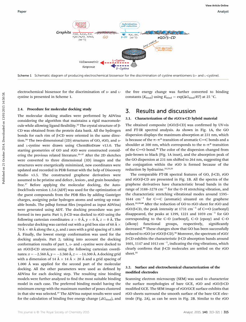
Scheme 1 Schematic diagram of producing electrochemical biosensor for the discrimination of cystine enantiomers (D- and L-cystine).
Paper Analyst
Publ
ishe
d on
21
Oct
ober
201
4. D
ownl
oade
d on
13/
01/2
015
14:5
0:58
. View Article Online
electrochemical biosensor for the discrimination of D- and L-cystine is presented in Scheme 1.
2.4. Procedure for molecular docking study
The molecular docking studies were performed by ADVinaconsidering the algorithm that maintains a rigid macromole-cule while allowing ligand exibility.33 The crystal structure of b-CD was obtained from the protein data bank. All the hydrogenbonds for each rim of b-CD were oriented in the same direc-tion.34 The two-dimensional (2D) structures of GO, rGO, and D-and L-cystine were drawn using ChemBioDraw v13.0. Thestarting geometries of GO and rGO were constructed consid-ering the previous related literature.35–37 Aer the 2D sketcheswere converted to three dimensional (3D) images and thestructures were energetically minimized, new coordinates wereupdated and recorded in PDB format with the help of DiscoveryStudio v3.5. The constructed graphene derivatives wereassumed to be pristine and defect-, lesion-, and grain boundary-free.17 Before applying the molecular docking, the Auto-DockTools version 1.5.6 (ADT) was used for the optimization ofthe guest compounds from the PDB les by adding Gasteigercharges, assigning polar hydrogen atoms and setting up rotat-able bonds. The pdbqt format les (required as input ADVina)were generated using ADT. The docking procedure was per-formed in two parts: Part 1; b-CD was docked to rGO using thefollowing cartesian coordinates: x ¼ 0 A, y ¼ 0 A, z ¼ 0 A. Themolecular docking was carried out with a grid box size of 60 A �70 A � 40 A along the x, y, and z axes with a grid spacing of 1.000A. Finally, the lowest energy conformation was used for thedocking analysis. Part 2; taking into account the dockingconformation results of part 1, D- and L-cystine were docked toan rGO/b-CD structure using the following cartesian coordi-nates: x¼�2.560 A, y¼�3.560 A, z¼�14.500 A. A docking gridwith a dimension of 14 A � 14 A � 20 A and a grid spacing of1.000 A was applied for the second part of the moleculardocking. All the other parameters were used as dened byADVina for each docking step. The resulting nine bindingmodels were further analyzed to nd the most suitable bindingmodel in each case. The preferred binding model having theminimum energy with the maximum number of poses clusteredin that site was selected.37 The ADVina output results were usedfor the calculation of binding free energy change (DGbind), and
This journal is © The Royal Society of Chemistry 2015
the free energy change was further converted to bindingconstants (Kbind) using Kbind ¼ exp(DGbind/RT) at 25 �C.
3. Results and discussion3.1. Characterization of the rGO/a-CD hybrid material
The obtained composite (rGO/b-CD) was conrmed by UV-visand FT-IR spectral analysis. As shown in Fig. 1A, the GOdispersion displays the maximum absorption at 231 nm, whichis because of the p–p* transition of aromatic C]C bonds and ashoulder at 300 nm, which corresponds to the n–p* transitionof the C]O bond.38 The color of the dispersion changed frompale-brown to black (Fig. 1A inset), and the absorption peak ofthe GO dispersion at 231 nm shied to 264 nm, suggesting thatthe conjugation within the rGO is formed because of thereduction by hydrazine.22,23,25
The comparable FT-IR spectral features of GO, b-CD, rGOand rGO/b-CD are presented in Fig. 1B. All the spectra of thegraphene derivatives have characteristic broad bands in therange of 3188–3278 cm�1 for the O–H stretching vibration, andthe characteristic stretching vibrational modes around 1595–1644 cm�1 for C]C (aromatic) situated on the graphenesheet.22,39,40 Aer the reduction of GO to rGO sheet for rGO andrGO/b-CD, the peak intensity at 1731 cm�1 of C]O (carbonyl)disappeared, the peaks at 1399, 1221 and 1059 cm�1 for GOcorresponding to the C–O (carboxyl), C–O (epoxy) and C–O(alkoxy) functional groups, respectively, signicantlydecreased.40 These changes show that GO has been successfullyreduced to rGO (or rGO/b-CD).41 Moreover, the spectrum of rGO/b-CD exhibits the characteristic b-CD absorption bands around1601, 1147 and 1013 cm�1, indicating the ring vibrations, whichclearly conrms that b-CD molecules are settled on the rGOsheet.22
3.2. Surface and electrochemical characterization of themodied electrodes
Scanning electron microscopy (SEM) was used to characterizethe surface morphologies of bare GCE, rGO and rGO/b-CDmodied GCE. The SEM image of rGO/GCE surface exhibits thatrGO sheets surround the smooth surface of the bare GCE elec-trode (Fig. 2A), as can be seen in Fig. 2B. Similar to the rGO
Analyst, 2015, 140, 313–321 | 315
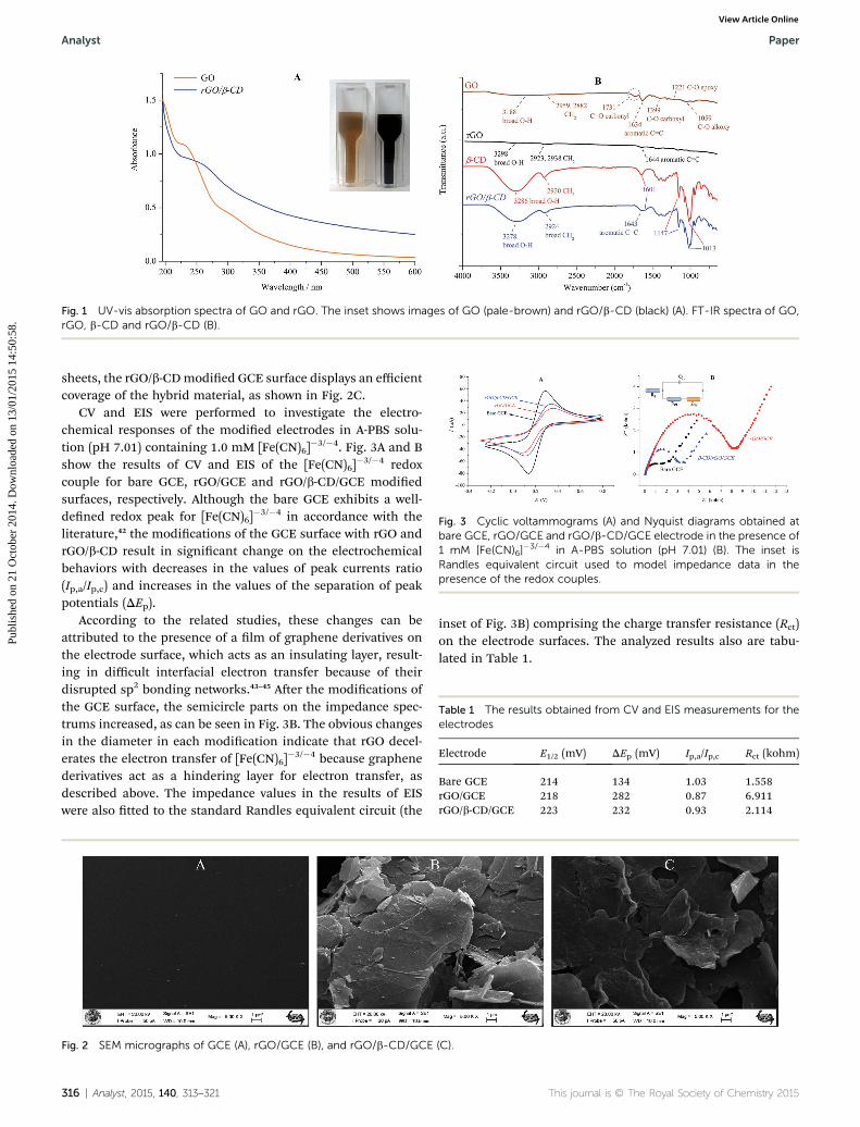
Fig. 1 UV-vis absorption spectra of GO and rGO. The inset shows images of GO (pale-brown) and rGO/b-CD (black) (A). FT-IR spectra of GO,rGO, b-CD and rGO/b-CD (B).
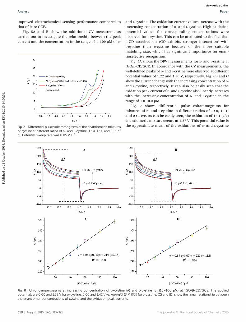
Fig. 3 Cyclic voltammograms (A) and Nyquist diagrams obtained atbare GCE, rGO/GCE and rGO/b-CD/GCE electrode in the presence of1 mM [Fe(CN)6]
�3/�4 in A-PBS solution (pH 7.01) (B). The inset isRandles equivalent circuit used to model impedance data in thepresence of the redox couples.
Table 1 The results obtained from CV and EIS measurements for theelectrodes
Electrode E1/2 (mV) DEp (mV) Ip,a/Ip,c Rct (kohm)
Bare GCE 214 134 1.03 1.558rGO/GCE 218 282 0.87 6.911rGO/b-CD/GCE 223 232 0.93 2.114
Analyst Paper
Publ
ishe
d on
21
Oct
ober
201
4. D
ownl
oade
d on
13/
01/2
015
14:5
0:58
. View Article Online
sheets, the rGO/b-CDmodied GCE surface displays an efficientcoverage of the hybrid material, as shown in Fig. 2C.
CV and EIS were performed to investigate the electro-chemical responses of the modied electrodes in A-PBS solu-tion (pH 7.01) containing 1.0 mM [Fe(CN)6]
�3/�4. Fig. 3A and Bshow the results of CV and EIS of the [Fe(CN)6]
�3/�4 redoxcouple for bare GCE, rGO/GCE and rGO/b-CD/GCE modiedsurfaces, respectively. Although the bare GCE exhibits a well-dened redox peak for [Fe(CN)6]
�3/�4 in accordance with theliterature,42 the modications of the GCE surface with rGO andrGO/b-CD result in signicant change on the electrochemicalbehaviors with decreases in the values of peak currents ratio(Ip,a/Ip,c) and increases in the values of the separation of peakpotentials (DEp).
According to the related studies, these changes can beattributed to the presence of a lm of graphene derivatives onthe electrode surface, which acts as an insulating layer, result-ing in difficult interfacial electron transfer because of theirdisrupted sp2 bonding networks.43–45 Aer the modications ofthe GCE surface, the semicircle parts on the impedance spec-trums increased, as can be seen in Fig. 3B. The obvious changesin the diameter in each modication indicate that rGO decel-erates the electron transfer of [Fe(CN)6]
�3/�4 because graphenederivatives act as a hindering layer for electron transfer, asdescribed above. The impedance values in the results of EISwere also tted to the standard Randles equivalent circuit (the
Fig. 2 SEM micrographs of GCE (A), rGO/GCE (B), and rGO/b-CD/GCE
316 | Analyst, 2015, 140, 313–321
inset of Fig. 3B) comprising the charge transfer resistance (Rct)on the electrode surfaces. The analyzed results also are tabu-lated in Table 1.
(C).
This journal is © The Royal Society of Chemistry 2015

Fig. 4 Cyclic voltammograms in the absence and presence of D- andL-cystine (50 mM) in A-PBS solution at rGO/b-CD/GCE. Potentialsweep rate was 0.1 V s�1.
Scheme 2 Possible oxidation mechanism of cystine to cysteic acid atrGO/b-CD/GCE.
Fig. 5 Cyclic voltammograms at increasing concentration of D-cystinepotential sweep rate was 0.1 V s�1.
Fig. 6 Differential pulse voltammograms in the absence and presence ograms at increasing concentration of D-cystine (B) and L-cystine (C) in lorate was 0.05 V s�1.
This journal is © The Royal Society of Chemistry 2015
Paper Analyst
Publ
ishe
d on
21
Oct
ober
201
4. D
ownl
oade
d on
13/
01/2
015
14:5
0:58
. View Article Online
3.3. Enantioselective discrimination of D- and L-cystine atrGO/b-CD/GCE by voltammetry
Fig. 4 shows the CV measurements performed with rGO/b-CD/GCE. No well-dened electrochemical response for the D- and L-cystine were obtained at bare GCE (the inset), which is inaccordance with the previous report.16 Aer the modication ofthe surface with rGO/b-CD, strong irreversible oxidation peaksof D- and L-cystine were observed at different potential values of1.32 and 1.42 V, respectively. The possible oxidation mecha-nism is provided in Scheme 2 as previously reported.30
The voltammetric response of cystine oxidation to cysteicacid is changed according to enantiomeric structure. The highsensitivity and selective recognition capability of rGO/b-CD/GCEcan be attributed to three factors: (i) the rGO/b-CD/GCE has alarger surface area; (ii) the attached b-CD prevents the rGO fromagglomeration; thus, rGO/b-CD/GCE has more accessible activesites than bare GCE and considerably higher catalytic activity;46
(iii) these active sites of b-CD have a different binding affinitytowards target cystine enantiomers that might lead to thedifferences in the free Gibbs energy; this effect is observed as apotential shi in electrochemical measurements.16 The CVmeasurements illustrate that by the combination of the uniqueelectronic properties of rGO with the high selective b-CDcompound, the rGO/b-CD nanocomposite shows signicantly
(A) and L-cystine (B) (1–100 mM) in A-PBS solution at rGO/b-CD/GCE;
f D- and L-cystine in A-PBS solution (A). Differential pulse voltammo-w concentration range (1–10 mM) at rGO/b-CD/GCE; potential sweep
Analyst, 2015, 140, 313–321 | 317
Analyst Paper
Publ
ishe
d on
21
Oct
ober
201
4. D
ownl
oade
d on
13/
01/2
015
14:5
0:58
. View Article Online
improved electrochemical sensing performance compared tothat of bare GCE.
Fig. 5A and B show the additional CV measurementscarried out to investigate the relationship between the peakcurrent and the concentration in the range of 1–100 mM of D-
Fig. 7 Differential pulse voltammograms of the enantiomericmixturesof cystine at different ratios of D- and L-cystine (1 : 0, 1 : 1, and 0 : 1 c/c). Potential sweep rate was 0.05 V s�1.
Fig. 8 Chronoamperograms at increasing concentration of D-cystinepotentials are 0.00 and 1.32 V for D-cystine, 0.00 and 1.42 V vs. Ag/AgCl (the enantiomer concentrations of cystine and the oxidation peak curren
318 | Analyst, 2015, 140, 313–321
and L-cystine. The oxidation current values increase with theincreasing concentration of D- and L-cystine. High oxidationpotential values for corresponding concentrations wereobserved for L-cystine. This can be attributed to the fact thatb-CD attached on rGO exhibits stronger interaction7 withL-cystine than D-cystine because of the more suitablematching size, which has signicant importance for enan-tioselective recognition.
Fig. 6A shows the DPV measurements for D- and L-cystine atrGO/b-CD/GCE. In accordance with the CV measurements, thewell-dened peaks of D- and L-cystine were observed at differentpotential values of 1.22 and 1.36 V, respectively. Fig. 6B and Cshow the current change with the increasing concentration of D-and L-cystine, respectively. It can also be easily seen that theoxidation peak current of D- and L-cystine also linearly increaseswith the increasing concentration of D- and L-cystine in therange of 1.0-10.0 mM.
Fig. 7 shows differential pulse voltammograms formixtures of D- and L-cystine in different ratios of 1 : 0, 1 : 1,and 0 : 1 c/c. As can be easily seen, the oxidation of 1 : 1 (c/c)enantiomeric mixture occurs at 1.27 V. This potential value isthe approximate mean of the oxidations of D- and L-cystine
(A) and L-cystine (B) (10–100 mM) at rGO/b-CD/GCE. The applied3 M KCl) for L-cystine. (C) and (D) show the linear relationship betweents.
This journal is © The Royal Society of Chemistry 2015
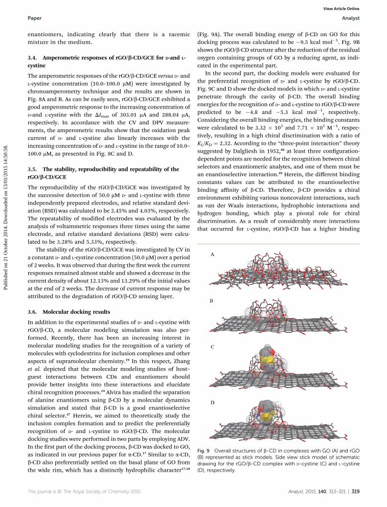
Fig. 9 Overall structures of b-CD in complexes with GO (A) and rGO(B) represented as stick models. Side view stick model of schematicdrawing for the rGO/b-CD complex with D-cystine (C) and L-cystine(D), respectively.
Paper Analyst
Publ
ishe
d on
21
Oct
ober
201
4. D
ownl
oade
d on
13/
01/2
015
14:5
0:58
. View Article Online
enantiomers, indicating clearly that there is a racemicmixture in the medium.
3.4. Amperometric responses of rGO/b-CD/GCE for D-and L-cystine
The amperometric responses of the rGO/b-CD/GCE versus D- andL-cystine concentration (10.0–100.0 mM) were investigated bychronoamperometry technique and the results are shown inFig. 8A and B. As can be easily seen, rGO/b-CD/GCE exhibited agood amperometric response to the increasing concentration ofD-and L-cystine with the DImax of 303.01 mA and 288.04 mA,respectively. In accordance with the CV and DPV measure-ments, the amperometric results show that the oxidation peakcurrent of D- and L-cystine also linearly increases with theincreasing concentration of D- and L-cystine in the range of 10.0–100.0 mM, as presented in Fig. 8C and D.
3.5. The stability, reproducibility and repeatability of therGO/b-CD/GCE
The reproducibility of the rGO/b-CD/GCE was investigated bythe successive detection of 50.0 mM D- and L-cystine with threeindependently prepared electrodes, and relative standard devi-ation (RSD) was calculated to be 2.45% and 4.03%, respectively.The repeatability of modied electrodes was evaluated by theanalysis of voltammetric responses three times using the sameelectrode, and relative standard deviations (RSD) were calcu-lated to be 3.28% and 5.33%, respectively.
The stability of the rGO/b-CD/GCE was investigated by CV ina constant D- and L-cystine concentration (50.0 mM) over a periodof 2 weeks. It was observed that during the rst week the currentresponses remained almost stable and showed a decrease in thecurrent density of about 12.13% and 13.29% of the initial valuesat the end of 2 weeks. The decrease of current response may beattributed to the degradation of rGO/b-CD sensing layer.
3.6. Molecular docking results
In addition to the experimental studies of D- and L-cystine withrGO/b-CD, a molecular modeling simulation was also per-formed. Recently, there has been an increasing interest inmolecular modeling studies for the recognition of a variety ofmolecules with cyclodextrins for inclusion complexes and otheraspects of supramolecular chemistry.19 In this respect, Zhanget al. depicted that the molecular modeling studies of host–guest interactions between CDs and enantiomers shouldprovide better insights into these interactions and elucidatechiral recognition processes.19 Alvira has studied the separationof alanine enantiomers using b-CD by a molecular dynamicssimulation and stated that b-CD is a good enantioselectivechiral selector.47 Herein, we aimed to theoretically study theinclusion complex formation and to predict the preferentiallyrecognition of D- and L-cystine to rGO/b-CD. The moleculardocking studies were performed in two parts by employing ADV.In the rst part of the docking process, b-CD was docked to GO,as indicated in our previous paper for a-CD.17 Similar to a-CD,b-CD also preferentially settled on the basal plane of GO fromthe wide rim, which has a distinctly hydrophilic character17,18
This journal is © The Royal Society of Chemistry 2015
(Fig. 9A). The overall binding energy of b-CD on GO for thisdocking process was calculated to be �9.5 kcal mol�1. Fig. 9Bshows the rGO/b-CD structure aer the reduction of the residualoxygen containing groups of GO by a reducing agent, as indi-cated in the experimental part.
In the second part, the docking models were evaluated forthe preferential recognition of D- and L-cystine by rGO/b-CD.Fig. 9C and D show the docked models in which D- and L-cystinepenetrate through the cavity of b-CD. The overall bindingenergies for the recognition of D- and L-cystine to rGO/b-CD werepredicted to be �4.8 and �5.3 kcal mol�1, respectively.Considering the overall binding energies, the binding constantswere calculated to be 3.32 � 103 and 7.71 � 103 M�1, respec-tively, resulting in a high chiral discrimination with a ratio ofKL/KD ¼ 2.32. According to the “three-point interaction” theorysuggested by Dalgliesh in 1952,48 at least three conguration-dependent points are needed for the recognition between chiralselectors and enantiomeric analytes, and one of them must bean enantioselective interaction.49 Herein, the different bindingconstants values can be attributed to the enantioselectivebinding affinity of b-CD. Therefore, b-CD provides a chiralenvironment exhibiting various noncovalent interactions, suchas van der Waals interactions, hydrophobic interactions andhydrogen bonding, which play a pivotal role for chiraldiscrimination. As a result of considerably more interactionsthat occurred for L-cystine, rGO/b-CD has a higher binding
Analyst, 2015, 140, 313–321 | 319
Analyst Paper
Publ
ishe
d on
21
Oct
ober
201
4. D
ownl
oade
d on
13/
01/2
015
14:5
0:58
. View Article Online
energy than D-cystine. The higher binding energy and bindingconstant for the interactions occurring between L-cystine andrGO/b-CD are consistent with the electrochemical results,indicating the higher binding affinity of rGO/b-CD for L-cystine,which also provide a better insight into the interactionsbetween cystine enantiomers and rGO/b-CD on a molecularlevel.
4. Conclusions
In the present work, we report a simple and fast method for thediscrimination of D- and L-cystine by rGO/b-CD/GCE electrode.In the experimental part of the study, CV, DPV and chro-noamperometry techniques were employed to investigate theelectrochemical response of the modied electrode for thediscrimination of D- and L-cystine. The enantioselective prop-erties of b-CD provide an opportunity for the discrimination ofD- and L-cystine. The rGO layer provides a large surface area forthe deposition of b-CD. The described method is very simple,cheap and time saving. To the best of our knowledge, it is therst report on an electrochemical chiral sensor for thediscrimination of D- and L-cystine. In the second part of thestudy, molecular interactions were investigated by moleculardocking to compare the experimental results with the compu-tational results. The molecular docking results supported theelectrochemical results, indicating the different enantiose-lective affinity of rGO/b-CD towards D- and L-cystine. As aconsequence, this study is expected to be a useful and prom-ising platform in the sensor area for the electrochemicaldiscrimination of some chiral analytes.
Acknowledgements
This work was supported by TUBITAK (113Z664) and wasproduced from a part of E. Zor's PhD thesis. We also express ourdeep thanks to the Turkish Academy of Sciences (TUBA).
Notes and references
1 M. Trojanowicz and M. Kaniewska, Electroanalysis, 2009, 21,229–238.
2 L. Wu and F. G. Vogt, J. Pharm. Biomed. Anal., 2012, 69, 133–147.
3 L. Song, S. Wang, N. A. Kotov and Y. Xia, Anal. Chem., 2012,84, 7330–7335.
4 R. Kataky and P. Lopes, Chem. Commun., 2009, 12, 1490–1492.
5 Q. Zhang, L. Guo, Y. Huang, Y. Wang, Q. Han and Y. Fu, Anal.Methods, 2013, 5, 4397–4401.
6 Y. Tao, N. R. Quebbemann and R. R. Julian, Anal. Chem.,2012, 84, 6814–6820.
7 E. Zor, I. Hatay Patir, H. Bingol and M. Ersoz, Biosens.Bioelectron., 2013, 42, 321–325.
8 D. Patterson, M. Schnell and J. M. Doyle, Nature, 2013, 497,475–477.
320 | Analyst, 2015, 140, 313–321
9 L. Challier, F. Mavre, J. Moreau, C. Fave, B. Schollhorn,D. Marchal, E. Peyrin, V. Noel and B. Limoges, Anal. Chem.,2012, 84, 5415–5420.
10 J. Athilakshmi, M. Mohan and D. K. Chand, TetrahedronLett., 2013, 54, 427–430.
11 Z. Huang, S. Yu, K. Wen, X. Yu and L. Pu, Chem. Sci., 2014, 5,3457–3462.
12 L. Qin, X.-W. Hea, W.-Y. Li and Y.-K. Zhang, J. Chromatogr. A,2008, 1187, 94–102.
13 L. Sanchez-Hernandez, M. L. Marina and A. L. Crego, J.Chromatogr. A, 2011, 1218, 4944–4951.
14 Y. Wang, L. Luo, Y. Ding, X. Zhang, Y. Xu and X. Liu, J.Electroanal. Chem., 2012, 667, 54–58.
15 E. Khaled, M. S. Kamel, H. N. A. Hassan and H. Y. Abou-Enein, J. Electroanal. Chem., 2011, 661, 239–244.
16 R. Nie, X. Bo, H. Wang, L. Zeng and L. Guo, Electrochem.Commun., 2013, 27, 112–115.
17 E. Zor, M. E. Saglam, S. Alpaydin and H. Bingol, Anal.Methods, 2014, 6, 6522–6530.
18 K. B. Lipkowitz, Chem. Rev., 1998, 98, 1829–1873.19 X.-H. Zhang, H.-L. Wu, X.-L. Yin, L.-H. Li, J.-Y. Wang,
Y. Chen, C. Kang and Ru-Q. Yu, Anal. Methods, 2013, 5,710–717.
20 X. Li, G. Wang, D. Chen and Y. Lu, RSC Adv., 2014, 4, 7301–7312.
21 L. Szente and J. Szeman, Anal. Chem., 2013, 85, 8024–8030.22 Y. Guo, S. Guo, J. Ren, Y. Zhai, S. Dong and E. Wang, ACS
Nano, 2010, 4, 4001–4010.23 Y. Guo, S. Guo, J. Li, E. Wang and S. Dong, Talanta, 2011, 84,
60–64.24 Z. Wang, S. Xiao and Y. Chen, J. Electroanal. Chem., 2006,
589, 237–242.25 D. Lu, S. Lin, L. Wang, X. Shi, C. Wang and Y. Zhang,
Electrochim. Acta, 2012, 85, 131–138.26 M. Chen, Y. Meng, W. Zhang, J. Zhou, J. Xie and G. Diao,
Electrochim. Acta, 2013, 108, 1–9.27 C. Xu, X. Wang, J. Wang, H. Hu and L. Wan, Chem. Phys.
Lett., 2010, 498, 162–167.28 Q. Han, Y. Wang, Y. Huang, L. Guo and Y. Fu, Analyst, 2013,
138, 2051–2056.29 A. Nosal-Wiercinska, Electrochim. Acta, 2013, 92, 397–403.30 K. Firoz Babu, R. Sivasubramanian, M. Noel and M. Anbu
Kulandainathan, Electrochim. Acta, 2011, 56, 9797–9801.31 D. C. Marcano, D. V. Kosynkin, J. M. Berlin, A. Sinitskii,
Z. Sun, A. Slesarev, L. B. Alemany, W. Lu and J. M. Tour,ACS Nano, 2010, 4, 4806–4814.
32 W. S. Hummers and R. E. Offeman, J. Am. Chem. Soc., 1958,80, 1339.
33 O. Trott and A. J. Olson, J. Comput. Chem., 2010, 31, 455–461.34 J. Kohler and N. Grczelschak-Mick, Beilstein J. Org. Chem.,
2013, 9, 118–134.35 S. Mao, H. Pu and J. Chen, RSC Adv., 2012, 2, 2643–2662.36 Y. Zhang, C. Wu, S. Guo and J. Zhang, Nanotechnology
Reviews, 2013, 2, 27–45.37 G. P. Kotchey, B. L. Allen, H. Vedala, N. Yanamala,
A. A. Kapralov, Y. Y. Tyurina, J. Klein-Seetharaman,V. E. Kagan and A. Star, ACS Nano, 2011, 5, 2098–2108.
This journal is © The Royal Society of Chemistry 2015
Paper Analyst
Publ
ishe
d on
21
Oct
ober
201
4. D
ownl
oade
d on
13/
01/2
015
14:5
0:58
. View Article Online
38 J.-L. Chen, X.-P. Yan, K. Meng and S.-F. Wang, Anal. Chem.,2011, 83, 8787–8793.
39 E. Zor, M. E. Saglam, I. Akin, A. O. Saf, H. Bingol andM. Ersoz, RSC Adv., 2014, 4, 12457–12466.
40 V. Chandra and K. S. Kim, Chem. Commun., 2011, 47, 3942–3944.
41 X. Gao, J. Jang and S. Nagase, J. Phys. Chem. C, 2010, 114,832–842.
42 F. C. Moraesa, I. Cesarinoa, V. Cesarinoa, L. H. Mascaroband S. A. S. Machado, Electrochim. Acta, 2012, 85, 560–565.
43 Y. Hu, K. Wang, Q. Zhang, F. Li, T. Wu and L. Niu,Biomaterials, 2012, 33, 1097–1106.
This journal is © The Royal Society of Chemistry 2015
44 Y. Hu, F. Li, X. Bai, D. Li, S. Hua, K. Wang and L. Niu, Chem.Commun., 2011, 47, 1743–1745.
45 E. Zor, A. O. Saf, H. Bingol and M. Ersoz, Anal. Biochem.,2014, 449, 83–89.
46 L. Tan, K.-G. Zhou, Y.-H. Zhang, H.-X. Wang, X.-D. Wang,Y.-F. Guo and H.-L. Zhang, Electrochem. Commun., 2010,12, 557–560.
47 E. Alvira, Tetrahedron: Asymmetry, 2013, 24, 1198–1206.48 C. E. Dalgliesh, J. Chem. Soc., 1952, 3940–3942.49 C. Chang, X. Wang, Y. Bai and H. Liu, TrAC, Trends Anal.
Chem., 2012, 39, 195–206.
Analyst, 2015, 140, 313–321 | 321
Copyright © 2022 FDOKUMEN