Mice deficient for p53 are developmentally normal but susceptible to spontaneous tumours
Upload
independentCategory
view
2download
0
The
Journ
al o
f Exp
erim
enta
l M
edic
ine
ARTICLE
JEM © The Rockefeller University Press $30.00
Vol. 204, No. 11, October 29, 2007 2579-2590 www.jem.org/cgi/doi/
2579
10.1084/jem.20071351
DCs are professional APCs that activate na-ive T cells, and thereby control the initiation of adaptive immune responses ( 1, 2 ). Several types of DC, varying in hematopoietic origin, surface phenotype, and function, have been described ( 3, 4 ). The two major groups are the CD11c int CD45R � plasmacytoid DCs (pDCs), which are characterized by their high capacity to secrete type I IFN ( 5 ), and the CD11c high CD45R � conventional DCs (cDCs), which play major roles in T cell priming ( 6, 7 ). One recently described DC type is the IFN-producing killer DC (IKDC), which has been proposed to provide a link between innate and adap-tive immune responses ( 8, 9 ). IKDCs resemble cells of the innate immune system by lysing NK cell targets and producing large amounts of IFN- � ( 8 – 10 ), although in contrast with previous
reports we recently found they do not pro-duce IFN- � ( 10 ). Upon activation, IKDCs have been claimed to up-regulate MHC class II and acquire the ability to present antigen-like cDCs, thereby directing adaptive T cell responses ( 9 ). Related hybrid cells have been previously de-scribed in mice ( 11, 12 ), rats ( 13 – 15 ), and hu-mans ( 16 – 18 ), and although the phenotypic defi nition varies between reports, dual func-tion remains the central theme. In humans, the converse situation of NK cells that behave like DCs has been demonstrated; human NK cells have been shown on activation to express MHC class II, and then to process and present antigen to T cells ( 19 – 21 ). Such conversion of NK cells into APCs has not been demonstrated in mice ( 22 ).
In the studies proposing a dual function ( 8, 9 ), the IKDCs were initially found to express in-termediate levels of the DC marker CD11c,
CORRESPONDENCE
Irina Caminschi:
Abbreviations used: cDC, con-
ventional DC; CHO, chinese
hamster ovary; IKDC, IFN-
producing killer DC; pDC,
plasmacytoid DC; PI, propid-
ium iodide; SA, streptavidin.
The online version of this article contains supplemental material.
Putative IKDCs are functionally and developmentally similar to natural killer cells, but not to dendritic cells
Irina Caminschi, 1 Fatma Ahmet, 1 Klaus Heger, 1 Jason Brady, 1 Stephen L. Nutt, 1 David Vremec, 1 Suzanne Pietersz, 1 Mireille H. Lahoud, 1 Louis Schofi eld, 1 Diana S. Hansen, 1 Meredith O ’ Keeff e, 2 Mark J. Smyth, 3 Sammy Bedoui, 1 Gayle M. Davey, 1 Jose A. Villadangos, 1 William R. Heath, 1 and Ken Shortman 1
1 The Walter and Eliza Hall Institute of Medical Research, Melbourne, Victoria, 3050, Australia
2 Bavarian Nordic GmbH, Munich D-82152, Germany
3 Cancer Immunology Program, Peter MacCallum Cancer Centre, East Melbourne, 3002, Victoria, Australia
Interferon-producing killer dendritic cells (IKDCs) have been described as possessing the
lytic potential of NK cells and the antigen-presenting capacity of dendritic cells (DCs). In
this study, we examine the lytic function and antigen-presenting capacity of mouse spleen
IKDCs, including those found in DC preparations. IKDCs effi ciently killed NK cell targets,
without requiring additional activation stimuli. However, in our hands, when exposed to
protein antigen or to MHC class II peptide, IKDCs induced little or no T cell proliferation
relative to conventional DCs or plasmacytoid DCs, either before or after activation with
CpG, or in several disease models. Certain developmental features indicated that IKDCs
resembled NK cells more than DCs. IKDCs, like NK cells, did not express the transcription
factor PU.1 and were absent from recombinase activating gene-2 – null, common � -chain –
null (Rag2 � / � Il2rg � / � ) mice. When cultured with IL-15 and -18, IKDCs proliferated exten-
sively, like NK cells. Under these conditions, a proportion of expanded IKDCs and NK cells
expressed high levels of surface MHC class II. However, even such MHC class II � IKDCs
and NK cells induced poor T cell proliferative responses compared with DCs. Thus, IKDCs
resemble NK cells functionally, and neither cell type could be induced to be effective
antigen-presenting cells.

2580 IKDCS ARE POOR ANTIGEN-PRESENTING CELLS | Caminschi et al.
CD11c, but unlike pDCs, they also express CD49b (DX5) and NK1.1 ( 8, 9 ). Using our DC isolation procedure that enriches for splenic pDCs and cDCs ( Fig. 1 A ), we noted that a proportion of cells expressed CD49b ( Fig. 1 B ). These CD49b � cells expressed variable levels of CD45R (and CD45RA; not depicted), ranging from undetectable to high ( Fig. 1 C ). Furthermore, the majority of CD49b � cells ex-pressed low to intermediate levels of CD11c, although a small proportion were CD11c hi ( Fig. 1 C ). The subpopulation of the CD49b � cells that were CD11c int CD45R � therefore matched the description of IKDCs ( Fig. 1 C ). Additionally, there appeared to be another minor population of CD49b � cells that expressed high levels of CD11c and MHC class II, like cDCs; we termed these CD49b � DCs. Further analysis showed that these IKDCs and NK cells, but not CD49b � cDCs, pDCs, or cDCs, expressed NKG2D and NK1.1 ( Fig. 1 D ). The freshly isolated IKDCs expressed only low levels of MHC class II ( Fig. 1 D ), as previously reported ( 9 ). However, the level appeared a little lower than in the earlier study, which may refl ect diff erences in reagents and staining strategies, or diff erences in the IKDC isolation procedures.
to express CD45R like pDCs and NK cells, and to express CD49b like NK cells, but to have low surface MHC class II expression. We have identifi ed cells with this surface pheno-type and isolated them from our DC preparations, as well as from the total spleen population. The IKDCs that we isolated were immediately capable of killing NK cell targets, but in response to CpG and a range of other stimuli, failed to up-regulate MHC class II and failed to effi ciently activate naive T cells. In terms of developmental requirements and functional capacity, the IKDCs that we isolated resembled NK cells rather than DCs. Importantly, we show that mouse IKDCs and NK cells proliferated, and a proportion up-regulated MHC class II molecules upon exposure to IL-15 and -18 in cell culture, in this respect aligning mouse and human NK cell biology; however, both cell types in our hands remained poor activators of naive T cells.
RESULTS
Surface phenotype analysis
IKDCs have been defi ned as cells that are similar to pDCs in that they express CD45R (B220) and intermediate levels of
Figure 1. Surface phenotype of IKDCs. Splenic DCs were isolated and enriched as described in the Materials and methods. The enriched DC prepara-
tion was stained with anti-CD11c-Cy7-PE, anti-CD45R (B220-FITC), and anti-CD49b (DX5-biotin), followed by the secondary reagent SA-Alexa Fluor 594,
and then analyzed by fl ow cytometry. Dead cells were stained with PI. (A) Gating for cDCs (CD11c hi CD45R � ) and pDCs (CD11c int CD45R � ) without exclud-
ing CD49b � cells. (B) Gating for CD49b � within the DC-enriched preparation. (C) Expression of CD11c and CD45R on the CD49b � cells, showing the gates
used for IKDC and CD49b � DC isolation. (D) The expression of NK1.1, NKG2D, and MHC class II on splenic DC subsets. For NK1.1 expression analysis,
splenic DC preparations were stained with anti-NK1.1 (PK136-APC), anti-CD45R (B220-PE), anti-MHC class II (M5/114-FITC), anti-CD11c (N418-Alexa
Fluor 594), and anti-CD49b (DX5-biotin; followed by secondary reagent SA-Cy7PE). For NKG2D expression analysis, splenic DC preparations were stained
with anti-NKG2D (CX5-biotin), anti-CD49b (DX5-FITC), anti-CD11c (N418-Alexa Fluor 594), and anti-CD45R (B220-PE), followed by SA-Cy7-PE secondary
reagent. For MHC class II expression analysis, splenic DC preparations were stained with anti-MHC class II (M5/114-FITC), anti-NK1.1 (P136-APC),
anti-CD11c (N418-Alexa-594), and anti-CD45R (B220-PE). Based on the expression of CD11c, CD45R, and CD49b, subsets were defi ned as IKDC
(CD11c int CD45R � CD49b � ), CD49b � DC (CD11c hi CD49b � ), pDC (CD11c int CD45R � CD49b � ), and cDC (CD11c hi CD45R � CD49b � ). Spleen suspensions,
which were depleted of red blood cells using 1.091 g/cm 3 Nycodenz density centrifugation, were the source of NK cells. Splenocytes were stained with
anti-NK1.1 (PK136-APC), anti-CD49b (DX5-Biotin; followed by secondary reagent SA-Cy7-PE), and anti-CD3 (KT3-Alexa-594), then NK cells sorted as
NK1.1 � CD49b � CD3 � . The continuous line represents staining on gated cells and the dotted line represents background.

JEM VOL. 204, October 29, 2007
ARTICLE
2581
However, culturing our purifi ed IKDCs with CpG and GM-CSF for 48 h resulted in < 10% survival ( Fig. 4 A ), despite the presence of GM-CSF, which enhances the in vitro survival of cDC. This questioned the origin of the few remaining cells because the starting cultures of IKDCs were 95% pure, with 5% of cells likely to be DC contaminants. To circumvent problems associated with poor IKDC survival in culture, we isolated IKDCs and pDCs from transgenic mice expressing
Lytic function of IKDCs
The initial study describing IKDCs suggested that preincuba-tion with CpG was required for induction of lytic capacity ( 9 ). Al though we confi rmed that IKDCs (CD11c int CD45R � CD49b � ) killed YAC-1 target cells after incubation with CpG and GM-CSF (not depicted), we observed, in contrast to the previous study, that even freshly isolated IKDCs (but not pDCs or cDCs) also effi ciently killed YAC-1 ( Fig. 2 A ) or chinese hamster ovary (CHO)-K1-OVA ( Fig. 2, B and C ) cells. The lytic activity of IKDCs was comparable to, or even slightly better than, that of NK cells ( Fig. 2 ). Those CD11 int CD49b � cells, which expressed lower levels of CD45R, also killed these NK cell – sensitive target cells (unpublished data).
T cell stimulation capacity
To assess our isolated IKDCs for their ability to present anti-gen and activate naive T cells, which is a hallmark of DCs, we compared them to cDCs, pDCs, and NK cells by coculturing each population with MHC class II – restricted, OVA-specifi c CD4 T cells (OT-II) in the presence of whole OVA protein or doses of OVA peptide up to and beyond the optimum for cDCs. The proliferative response of CFSE-labeled OT-II cells was enumerated 3 or 5 d later. We also compared the eff ects of adding CpG (1668) to the cultures, as this had been identifi ed as a factor that matures IKDCs in vitro to become APCs ( 9 ). However, our B6 IKDCs failed to induce OT-II proliferation to either the whole protein or to the peptide, whereas cDCs induced potent proliferative responses and pDCs induced moderate responses ( Fig. 3, A and C ). Coculture in the pres-ence of CpG and GM-CSF did not enhance the stimulatory capacity of IKDCs ( Fig. 3, B and D ). Similar results were ob-tained when we isolated IKDCs from BALB/c mice and used these as APCs (Fig. S1, available at http://www.jem.org/cgi/content/full/jem.20071351/DC1). The lack of APC function was also observed when B6 DCs were fi rst preincu-bated overnight with CpG and GM-CSF, and then sorted for IKDCs (Fig. S2).
Because CpG failed to enhance antigen presentation by IKDCs in our studies, we checked the reported capacity of CpG to up-regulate MHC class II on these cells ( 9 ).
Figure 2. IKDCs kill YAC-1 and CHO-K1 target cells. Splenic DCs were enriched and sorted based on their expression of CD11c, CD45R, and CD49b,
as described in the Materials and methods. CD11c hi CD45R � (cDC), CD11c int CD45R � CD49b � (pDC), and CD11 int CD45R � CD49b � (IKDC) were purifi ed and
incubated at various ratios with 51 Cr-labeled YAC-1 (A) and CHO-OVA (B) target cells in a 4-h 51 Cr release assay, or in a 5-h 51 Cr release assay at an E/T
ratio of 2:1 (C). Lysis at each effector to target (E/T) cell ratio was determined as described in the Materials and methods. Each assay point was done in
triplicate or duplicate, and the result is representative of a minimum of two experiments. Error bars indicate the SEM.
Figure 3. IKDCs cultured in the presence of OVA protein or peptide
do not induce naive OVA-specifi c transgenic T cells to proliferate.
Splenic pDCs (CD11c int CD45R � CD49b � ), cDCs (CD11c hi CD45R � CD49b � ),
IKDCs (CD11c int CD45R � CD49b � ), and NK cells (CD49b � NK1.1 � CD3 � )
were isolated and purifi ed by fl ow cytometry. The purifi ed cells (10 4 ) were
cocultured for 3 d (A and B) with graded doses of class II – restricted
OVA 323-339 peptide and 5 � 10 4 CFSE-labeled OT-II cells in the absence
(A) or presence (B) of CpG and GM-CSF. APCs and CFSE-labeled OT-II cells
were also incubated for 3 and 5 d with 0.5 mg/ml whole OVA protein in
the absence (C) or presence (D) of CpG and GM-CSF. The proliferative
response of OT-II cells was enumerated by fl ow cytometry, as described in
the Materials and methods. Sample points were prepared in duplicates,
and results are representative of several experiments.
2582 IKDCS ARE POOR ANTIGEN-PRESENTING CELLS | Caminschi et al.
compared with cDCs ( Fig. 6, A and B ). Thus, in vitro infection with virus did not enhance the ability of IKDCs to stimulate naive T cells to proliferate.
In the third model, we examined IKDCs in mice infected with malaria ( Plasmodium berghei ). We observed that such mice had increased numbers of IKDCs in the spleen (Fig. S3, available at http://www.jem.org/cgi/content/full/jem.20071351/DC1). Furthermore, infection with P. berghei results in systemic DC activation ( 24 ). We reasoned that the factors that governed the
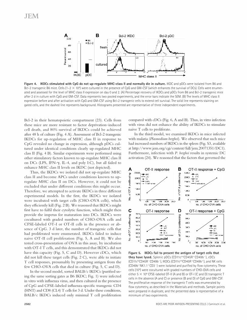
Bcl-2 in their hematopoietic compartment ( 23 ). Cells from these mice are more resistant to factor deprivation – induced cell death, and 80% survival of IKDCs could be achieved after 48 h of culture ( Fig. 4 A ). Assessment of Bcl-2 transgenic IKDCs for up-regulation of MHC class II in response to CpG revealed no change in expression, although pDCs cul-tured under identical conditions clearly up-regulated MHC class II ( Fig. 4 B ). Similar experiments were performed using other stimulatory factors known to up-regulate MHC class II on DCs (LPS, IFN- � , IL-4, and poly I:C), but all failed to enhance MHC class II levels on IKDC (not depicted).
Thus, the IKDCs we isolated did not up-regulate MHC class II and become APCs under conditions known to up-regulate MHC class II on DCs. However, it could not be excluded that under diff erent conditions this might occur. Therefore, we attempted to activate IKDCs in three diff erent experimental models. In the fi rst, the IKDCs we isolated were incubated with target cells (CHO-OVA cells), which they effi ciently kill ( Fig. 2 B ). We reasoned that IKDCs might fi rst have to fulfi l their cytolytic function, which might then provide the impetus for maturation into DCs. IKDCs were cocultured with graded numbers of CHO-OVA cells and CFSE-labeled OT-I or OT-II cells in the presence or ab-sence of CpG. 3 d later, the number of transgenic cells that had proliferated were enumerated. IKDCs failed to induce naive OT-II cell proliferation ( Fig. 5, A and B ). We also tested cross-presentation of OVA in this assay, by incubation with OT-I T cells, and this demonstrated that IKDCs did not have this capacity ( Fig. 5, C and D ). However cDCs, which did not kill these target cells ( Fig. 2 C ), were able to initiate T cell responses, presumably by presenting antigen from the few CHO-OVA cells that died in culture ( Fig. 5, C and D ).
In the second model, sorted BALB/c IKDCs (purifi ed us-ing the same sorting gates as B6 IKDC; Fig. 1 ) were infected in vitro with infl uenza virus, and then cultured in the presence of CpG and CFSE-labeled infl uenza-specifi c transgenic CD4 (HNT) and CD8 (CL4) T cells for 3 d. Under these conditions, BALB/c IKDCs induced only minimal T cell proliferation
Figure 4. IKDCs stimulated with CpG do not up-regulate MHC-class II and normally die in culture. IKDC and pDCs were isolated from B6 and
Bcl-2 transgenic B6 mice. Cells (1 – 2 � 10 4 ) were cultured in the presence of CpG and GM-CSF (which enhances the survival of DCs). Cells were enumer-
ated and assessed for the level of MHC class II expression on day 0 and 2. (A) Percentage recovery of IKDCs and pDCs from B6 and Bcl-2 transgenic mice
after 2 d in culture with CpG and GM-CSF. Data represents two pooled experiments, and the error bars indicate the SEM. (B) The levels of MHC class II
expression before and after activation with CpG and GM-CSF using Bcl-2 transgenic cells to extend cell survival. The solid line represents staining on
gated cells, and the dashed line represents background. Histograms presented are representative of three independent experiments.
Figure 5. IKDCs fail to present the antigen of target cells that
they have lysed. Splenic pDCs (CD11c int CD45R � CD49b � ), cDCs
(CD11c hi CD45R � CD49b � ), IKDCs (CD11c int CD45R � CD49b � ), and NK cells
(CD49b � NK1.1 � CD3 � ) were isolated and purifi ed by fl ow cytometry. These
cells (10 4 ) were cocultured with graded numbers of CHO-OVA cells and
either 5 � 10 4 CFSE-labeled OT-II (A and B) or OT-I (C and D) transgenic T
cells in the absence (A and C) or presence (B and D) of CpG and GM-CSF.
The proliferative response of the transgenic T cells was enumerated by
fl ow cytometry, as described in the Materials and methods. Sample points
were prepared in duplicate, and the presented data is representative of a
minimum of two experiments.

JEM VOL. 204, October 29, 2007
ARTICLE
2583
http://www.jem.org/cgi/content/full/jem.20071351/DC1). We also followed the Chan et al. ( 9 ) procedure in using only anti-CD3 and -CD19 as depletion mAbs, but the IKDCs again failed to become eff ective APCs ( Fig. S4 B ). We also noted that some cells with the IKDC surface phenotype were denser than the 1.077 g/cm 3 used in this study and by Chan et al. ( 9 ) to enrich DCs, although all IKDCs appeared less dense than 1.082 g/cm 3 . To ensure we recovered all spleen cells with the surface phenotype of IKDCs, we isolated IKDCs using a much denser cut, which recovered all spleen-viable cells (eliminating only erythrocytes and dead cells), as well as omitting anti-Thy1.1 mAb from the depletion mix. However, this “ total ” IKDC fraction likewise failed to acquire APC function on culture with CpG (Fig. S4 C). We concluded that the behavior of the IKDCs in our DC-enriched preparations refl ected that of the total IKDC population.
Absence of IKDC in Rag2 � / � Il2rg � / � mice
The effi cient lytic function of IKDC, but poor capacity to present antigen to naive T cells, suggested that IKDCs were a type of NK cell rather than a subtype of DCs. One important argument against this conclusion was the reported presence of IKDCs in recombinase-activating gene-2 null, common � -chain null (Rag2 � / � Il2rg � / � ) mice, which are defi cient in NK cells ( 8, 25 ). Therefore, we reexamined this issue. We found that a major complication in the analysis of Rag2 � / � Il2rg � / � mouse spleen is the high frequency of
accumulation of IKDCs and systemic activation of DCs in the spleens of mice infected with malaria might also activate the antigen-presenting capacity of IKDCs. Thus, we isolated IKDCs from malaria-infected mice and assessed their ability to induce antigen-specifi c CD4 � T cell proliferation by cocul-turing them with graded doses of MHC class II – restricted OVA peptide for 3 or 5 d. These IKDCs also failed to induce sig-nifi cant OT-II proliferation ( Fig. 6, C and D ). In comparison, and consistent with the results shown in Fig. 3 , cDCs cultured with OVA peptide induced potent OT-II proliferation, whereas pDCs induced more moderate proliferation, regardless of whether they were isolated from malaria-infected mice or healthy control mice ( Fig. 6, C and D ).
T cell stimulatory capacity of total spleen IKDCs
It was possible our failure to activate IKDCs to become APCs was because we had isolated an IKDC population that was dif-ferent from that of Chan et al. ( 9 ), despite the similarities in the isolation procedure. One diff erence was that, when depleting our DC preparations of lymphoid cells, we normally include several mAbs for maximum purity, including anti-CD90 (Thy1.1; T24/31.7), which Chan et al. ( 9 ) did not. Indeed, we found that � 50% of IKDCs expressed CD90, and that some of these were likely removed from our IKDC sample. How-ever, when CD90 depletion was omitted, the resulting IKDCs in our DC preparations still failed to become eff ective APCs on activation with CpG (Fig. S2 B and Fig. S4 A, available at
Figure 6. IKDCs exposed to two different pathogens do not acquire the ability to stimulate naive T cells into proliferation. Splenic pDCs
(CD11c int CD45R � CD49b � ), cDCs (CD11c hi CD45R � CD49b � ), IKDCs (CD11c int CD45R � CD49b � ), and NK cells (CD49b � NK1.1 � CD3 � ) were isolated and
purifi ed by fl ow cytometry. (A and B) These cells were infected in vitro with infl uenza (PR8 infl uenza virus), washed, and used at the indicated numbers
to stimulate fl u-specifi c transgenic CD4 (HNT; A) and CD8 (CL4) T cells (B). Proliferating T cells were identifi ed and enumerated by fl ow cytometry.
(C and D) Mice were infected with malaria ( P. berghei ) and killed 3 d later. Spleens from healthy or malaria-infected mice were used to isolate and purify pDCs
(CD11c int CD45R � CD49b � ), cDCs (CD11c hi CD45R � CD49b � ), IKDCs (CD11c int CD45R � CD49b + ), and NK cells (CD49b � NK1.1 � CD3 � ), and 10 4 APCs were cocul-
tured for 3 (C) and 5 d (D) with graded doses of class II-restricted OVA 323-339 peptide and 5 × 10 4 CFSE-labeled OT-II. Proliferating OT-II were enumerated
as indicated in the Materials and methods. The presented data is representative of three independent experiments.

2584 IKDCS ARE POOR ANTIGEN-PRESENTING CELLS | Caminschi et al.
very few cells that had this phenotype ( Fig. 7 C ). Because even these may have been residual autofl uorescent cells or other noise, we then used the more stringent defi nition that specifi es IKDC to be NK1.1 � , as well as CD49b � CD11c int CD45R � ( Fig. 7 D ), and found that these cells were at ex-tremely low levels in Rag2 � / � Il2rg � / � mice ( Fig. 7 E ). Further-more, the NK cell – sensitive targets RMA-S and RMA-S Rae- � , which are killed in a perforin- and NKG2D-depen-dent manner ( 27 ), were growth suppressed (RMA-S) and re-jected (RMA-S Rae- � ) in Rag � / � mice, but not eliminated in Rag2 � / � Il2rg � / � mice, or in Rag � / � mice depleted of NK1.1 � cells (Fig. S5, available at http://www.jem.org/cgi/content/full/jem.20071351/DC1). This argues that the residual CD49b � cells present in Rag2 � / � Il2rg � / � mice are neither NK cells nor IKDCs, which have been shown to kill targets
autofl uorescent cells, which can generate false positives in the analysis of rare DC populations ( 10 ). Thus, particular care was taken to exclude autofl uorescent cells and dead cells, and to avoid and gate out doublets and aggregates ( Fig. 7 A ). Us-ing this approach, Rag2 � / � Il2rg � / � mice were shown to have pDCs and cDCs ( Fig. 7 B ), albeit at reduced total num-bers compared with wild-type B6 mice ( Fig. 7 E ). This re-duction likely refl ects the absence of B cells ( 26 ) and the smaller spleen size. Rag2 � / � Il2rg � / � mouse spleen appeared to contain some CD49b � cells ( Fig. 7 C ), although this pop-ulation diff ered from the CD49b � cells seen in normal B6 mice ( Fig. 7 C ). As expected ( 25 ), there were virtually no CD49b � NK1.1 � NK cells in Rag2 � / � Il2rg � / � mice ( Fig. 7 D ). To enumerate IKDC, we used two separate defi nitions. First, we gated for CD49b � CD11c int CD45R � cells and found
Figure 7. IKDCs are severely reduced in Rag2 � / � Il2rg � / � mice. Spleens of age- and gender-matched B6 and Rag2 � / � Il2rg � / � mice were chopped
and digested, and lighter density cells were separated using a 1.082-g/cm 3 Nycodenz density cut. Irrelevant cell types were removed, as indicated in the
Materials and methods. Remaining cells were stained with the following cocktail of fl uorescently labeled mAbs: anti-CD11c (N418-Cy7-PE), anti-CD45R-
(B220-FITC), anti-NK1.1 (PK136-APC), and anti-CD49b (DX5-biotin, followed by SA-PE secondary; A). Excluded from the analysis were cells that incorpo-
rated PI (indicative of dead cells), had large forward scatter width (indicative of cell doublets), and autofl uorescent cells (cells that fl uoresce in the FL4
channel in the absence of fl uorochromes that are detected in FL4). (B) DCs in Rag2 � / � Il2rg � / � and B6 mice. CD49b � cells (C) or CD49b � NK1.1 � cells (D)
were gated on and analyzed for CD11c and CD45R expression. (E) The total number of DCs, IKDCs, and NK cells were determined and expressed as the
ratio of Rag2 � / � Il2rg � / � to B6. Three independent experiments were conducted using 3 – 4 Rag2 � / � Il2rg � / � and B6 mice. Representative data is presented.
The ratios of recovered of Rag2 � / � Il2rg � / � to B6 cells (E) is the mean of the three experiments, and error bars represent the SEM.

JEM VOL. 204, October 29, 2007
ARTICLE
2585
at http://www.jem.org/cgi/content/full/jem.20071351/DC1). The emerging MHC class II � cells are unlikely to represent a contaminating DC population because: (a) DCs do not prolif-erate under these conditions ( Fig. 9 A ), and (b) the number of cells with up-regulated MHC class II was 10-fold higher than the total number of cells initially seeded into culture.
Cultured MHC class II � IKDCs do not induce
T cell proliferation
Because some cultured, growth factor – expanded IKDCs ex-pressed MHC class II, in contrast to our CpG activated IKDC, we tested their ability to present antigen and activate T cells. Day 4 bulk cultures of IKDC containing 10% of MHC class II � IKDCs were incubated with OVA peptide and OT-II cells. The cultures of IKDC failed to induce considerable OT-II cell proliferation compared with DCs at levels equiva-lent to the number of MHC class II � cells present in the IKDC cultures ( Fig. 9 C ). In addition, cultures of IKDCs and NK cells were sorted based on their MHC class II expression, and then incubated with whole OVA protein or peptide and OT-II cells. Peptide-coated MHC class II � IKDC and NK cells induced OT-II proliferation responses that were 100 – 1,000-fold less effi cient than that induced by cDCs ( Fig. 9 E ), or by DCs labeled with equivalent levels of anti – MHC class II mAb. In addition, no response was obtained to whole OVA protein ( Fig. 9 F ). Consistent with the observation that MHC class II � IKDCs and NK cells were poor activators of naive T cells, these cultured cells did not express signifi cant levels of the costimulatory molecules CD80 and CD86 ( Fig. 9 D ).
DISCUSSION
In our DC-enriched preparations, we found cells with the reported surface phenotype of IKDCs, namely CD11 int CD45R � CD49b � . These cells also expressed the NK cell markers NK1.1 and NKG2D. We found that these IKDCs had a potent ability to kill NK cell targets, even without preactivation. However, in our experiments, the IKDCs had little or no anti-gen presentation capacity, even after activation.
Despite the use of a range of DC activation stimuli (CpG, LPS, poly I:C, IL-4, and IFN- � ), and the use of bcl-2 trans-genic IKDCs to overcome cell death in culture, in our hands, the IKDCs failed to up-regulate MHC class II under normal conditions of DC activation and failed to activate naive antigen-specifi c T cells, even after incubation with synthetic peptide antigen, which is in contrast to an earlier work ( 9 ). Although much higher levels of peptide were used in the ear-lier study, we titrated and tested levels of peptide beyond that which saturated DC responses, demonstrating that IKDCs were 100 – 1,000-fold less effi cient APCs than DCs. Several disease models that might have provided alternate maturation signals also failed to induce IKDCs to become APCs.
What then is the basis of the diff erence between our data and that of Chan et al. ( 9 )? Although the experimental approach was similar, there remain some diff erences in procedures. Selec-tion of diff erent types of IKDCs seems unlikely to be the expla-nation because when we took care to isolate all spleen cells with
via NKG2D ( 8, 9 ). Our overall conclusion was that IKDCs, like NK cells, are at a very low level or absent from Rag2 � / � Il2rg � / � mice.
Lack of PU.1 expression by IKDCs
To further determine whether the gene expression program of IKDCs is more closely related to DCs or to NK cells, we ex-amined the expression of PU.1, which is a transcription factor involved in hematopoiesis. PU.1 is not expressed in mature NK cells, but is expressed at moderate and high levels in pDCs and cDCs, respectively ( 28, 29 ). Using the PU.1 gfp reporter mice that express the GFP protein under the control of the endogenous PU.1 promoter, we assessed the levels of PU.1 expression in the IKDCs isolated from our DC preparations and compared these to NK cells and DCs. Like mature NK cells, these IKDCs failed to express PU.1, although, as ex-pected, expression was detected in DCs ( Fig. 8 ).
Response of IKDCs to NK cell growth factors
In our experiments, IKDCs failed to respond to DC growth or maturation factors such as CpG and GM-CSF. However, both cDCs and pDCs can effi ciently be generated by culturing bone marrow with Flt-3 ligand ( 30 ). Thus, we analyzed Flt-3 ligand BM cultures for the presence of IKDCs (Fig. S6, available at http://www.jem.org/cgi/content/full/jem.20071351/DC1). Although both cDCs and pDCs were effi ciently generated, IKDCs were not produced, further delineating the growth requirements of these cells from that of DCs.
Because IKDCs resembled NK cells by function and many surface markers, we tested their response to NK cell growth factors. When our IKDCs were cultured in the presence of IL-15 and -18, they proliferated extensively, like NK cells, whereas DCs did not ( Fig. 9 A ). Interestingly, a proportion of these expanded IKDCs then expressed surface MHC class II ( Fig. 9 B ). Importantly, a small proportion of expanded NK cells also expressed MHC class II ( Fig. 9 B ); this proportion was up to 25% under some culture conditions (Fig. S7, available
Figure 8. IKDCs fail to express the transcription factor PU.1. DCs
were isolated from PU.1 GFP and B6 mice, as described in the Materials and
methods. Enriched DC preparations or a splenic lymphocyte cell suspen-
sion containing NK cells were stained with mAb against CD11c (N418-
Alexa-594), CD45R (B220-Cy7-PE), CD49b (DX5-Biotin, followed by
SA-PE), and NK1.1 (PK136-APC). The levels of PU.1 GFP expression on gated
pDCs (CD11c int CD45R � CD49b � ), cDCs (CD11c hi CD45R � CD49b � ), IKDCs
(CD11c int CD45R � CD49b � ), and NK cells (CD49b � NK1.1 � ) are represented
by the solid lines, and B6 backgrounds are represented by the dashed
lines. Data presented is representative of two experiments.

2586 IKDCS ARE POOR ANTIGEN-PRESENTING CELLS | Caminschi et al.
should have been included in our total spleen IKDC prepara-tion, which included all spleen CD11c � cells. It may be that the beads introduced some additional signals or generated cell dou-blets between IKDCs and pDCs. However, overlap of their IKDC fraction with true APCs is a more likely explanation for
the IKDC surface phenotype, they showed the same lack of T cell stimulation capacity as the IKDC in our DC preparations. Another possible diff erence is the additional use of a positive bead selection of CD11c � cells by Chan et al. ( 9 ). This may have excluded some types of IKDCs, but the selected IKDCs
Figure 9. IKDCs and NK cells proliferate, and a proportion express high levels of class II MHC in response to IL-15 and -18, but do not
become APCs. Splenic IKDCs (CD11c int CD45R � CD49b � ), NK cells (NK1.1 � CD49b � CD3 � ), and DCs (CD11c hi CD49b � ; 5 � 10 3 cells) were cultured in
100 � l of media containing 50 ng/ml IL-18 and 50 ng/ml IL-15. At the indicated time points, cells were counted (A), and level of MHC class II was
determined by staining with M5/114-FITC and analysis by fl ow cytometry (B). (C) Bulk-cultured IKDCs do not activate OT-II cells. IKDCs were cultured
as above, harvested on day 4, and determined by fl ow cytometry to contain 10% MHC class II � cells. 10 4 -cultured IKDCs (containing the equivalent of
10 3 MHC class II � cells) or 10 3 cells from an enriched DC preparation (DC), were incubated with CFSE-labeled OT-II cells (5 � 10 4 ) and 1,000 ng/ml of
class II – restricted OVA 323-339 peptide. (D) The levels of the costimulatory markers expressed on cultured MHC class II � and class II � IKDC and NK cells
compared with fresh cDCs. Expression profi les were confi rmed in two independent experiments. (E and F) Purifi ed MHC class II � IKDCs and NK cells
were ineffective at inducing OT-II cell proliferation. MHC class II + and class II � cultured IKDCs and NK cells, and cDCs (10 4 cells) were incubated with
CFSE-labeled OT-II cells (5 � 10 4 ) and graded doses of class II – restricted OVA 323-339 peptide (E) or 0.5 mg/ml of whole OVA protein (F) for 4 d. Prolifer-
ating OT-II cells were enumerated as indicated in the Materials and methods. The presented data is representative of three independent experiments.
Error bars indicate the SEM.

JEM VOL. 204, October 29, 2007
ARTICLE
2587
but unlike DCs, proliferated vigorously in culture to IL-15 and -18.
Culture of IKDCs in IL-15 and -18 not only induced cell proliferation, but produced some cells expressing high levels of MHC class II. Importantly, a proportion of cul-tured NK cells showed a similarly high surface expression of MHC class II. To our knowledge, this is the fi rst study of growth factors directly inducing MHC class II expression in mouse NK cells and provides some alignment with hu-man NK cells, which express MHC class II upon activation ( 19 – 21 ). In addition, mouse NK cells and NKDCs can be induced to express MHC class II in vivo ( 32 ). These fi nd-ings are in contrast to our inability to induce MHC class II by activation of IKDC with CpG and other DC stimuli in vitro. However, despite the induction of MHC class II, our culture-expanded IKDCs did not signifi cantly up-regulate the costimulator molecules CD80 or CD86, and they still failed to induce naive T cell proliferation. They could not be classed as eff ective APCs.
In conclusion, in our studies, IKDCs resemble NK cells in their lytic function, and do not acquire the antigen-presenting functions of DCs even after activation. As we pre-viously reported ( 10 ), they produce IFN � like NK cells, but do not produce IFN � like pDCs. Their transcription factor profi le and response to cytokines resembles that of NK cells, not that of DCs. Their developmental relationship to NK cells is not entirely clear. They have the appearance of an ac-tivated form of NK cell, but one study ( 31 ) points to diff er-ences in early stages of development. In either case, IKDCs should be considered as a subtype of NK cells, rather than a subtype of DCs.
MATERIALS AND METHODS Mice. C57BL/6J wehi (B6), BALB/c, Rag2 � / � Il2rg � / � (backcrossed onto B6),
and various transgenic mice (vav-bcl-2, PU.1 gfp , OT-I, OT-I bm-1 , OT-II,
DO11.10, CL4, and HNT) were bred under specifi c pathogen-free con-
ditions at the Walter and Eliza Hall Institute. Female mice were used at
6 � 12 wk of age and handled according to the guidelines of the National
Health and Medical Research Council of Australia. Experimental proce-
dures were approved by the Animal Ethics Committee, Melbourne Health
Research Directorate.
Cell lines and media. YAC-1 and CHO-K1 cell lines were maintained in
DME or RPMI-1640 containing 10% FCS, and then harvested during ex-
ponential growth phase for use in assays. Freshly isolated cells were cultured
in DC culture medium (modifi ed RPMI-1640 medium containing 10%
FCS, 100 U/ml penicillin, 100 � g/ml streptomycin, and 10 � 4 M 2-mercap-
toethanol). Where indicated, 50 ng/ml IL-18 (R & D Systems) and 50 ng/ml
IL-15 (rhIL-15; R & D Systems) were added to DC culture medium.
Antibodies. The following fl uorochrome-conjugated mAbs were used: anti-
CD11c (N418)-APC, -Cy5, or -FITC; anti-CD45RA (14.8) or anti-CD45R
(CD45R; RA-6B2) -PE or -APC; anti-CD49b (DX5)-biotin or -FITC; anti-
NK1.1 (PK136)-biotin or APC; anti-NKG2D (CX5-biotin); anti-MHC class
II (M5/114)-FITC, PE, or Alexa Fluor 594. For biotin conjugates, streptavi-
din (SA)-PE, SA-Alexa Fluor 594, or SA-Cy7-Pe was used in the second-
stage reaction (BD PharMingen). To assure comparative MHC class II staining,
our M5/114 conjugates contained suffi cient unconjugated mAb to saturate
class II binding, but maintain bright staining on scale. In all staining procedures,
the diff erences. One candidate contaminant is CD49b � DC. These cells express high levels of MHC class II, CD80, and CD86, and thus would be predicted to be potent APCs. It is notable that, in the B6 mice we mainly used, these CD49b � DCs were at a low level in the DC preparations, but their frequency varied in BALB/c DC preparations, on occa-sions reaching a 1:1 ratio with IKDC. Accordingly, they would be harder to exclude from the IKDC fraction. In addition, BALB/c mice lack the NK1.1 marker useful for distinguishing these two subpopulations. The developmental relationship between CD49b � DC and IKDC is not clear, but we obtained no transformation of IKDC to CD49b � DC on incubation with maturation factors such as CpG. However, it is possible that in culture, selective death of IKDCs could masquerade as transformation of one into the other, so giving the impres-sion that IKDCs became eff ective APCs. A further possible contaminant which might lead to induced APC function is some residual pDC. We note that the IKDC fraction of Chan et al. ( 9 ) produced IFN- � on stimulation with CpG. Our recent study ( 10 ) suggests that pDCs, but not IKDCs, produce IFN- � under these conditions.
An alignment of IKDC with NK cells, rather than with DCs, was reinforced by our analysis of the expression of the hematopoietic transcription factor PU1. This factor is ex-pressed at high levels in cDCs and at moderate levels in pDCs, but is not expressed on mature NK cells ( 28 ). Our IKDCs did not express PU.1, aligning their transcription factor expression pattern more closely with NK cells. Importantly, IKDC, like NK cells, are also dependent on the Id-2 transcription inhibi-tor, but unlike DCs, they do not express the transcription factor Spi-B ( 31 ). Therefore, both the cytokine signaling re-quirements and transcription factor expression profi les argue against a close association of IKDCs and DCs.
A compelling argument delineating IKDCs from NK cells had been the observation that IKDCs, like DCs, ap-peared to be present in Rag2 � / � Il2rg � / � mice ( 8 ), whereas NK cells are absent ( 25 ). However, our analysis now suggests that IKDC cells were markedly reduced, if present at all, in such mice. One possible explanation for the discrepancy is the disproportionate number of autofl uorescent cells in DC prepa-rations from these mice, which may give the false-positive signals in the IKDC gates. Multiple markers are required to clearly delineate an IKDC population.
Responses to growth factors can clarify the lineage rela-tionships between diff erent cell types. In our hands IKDCs did not respond to the DC growth factor GM-CSF. Admin-istration of Flt-3 ligand in vivo enhances DC numbers, and has been reported to increase a population of “ NKDCs, ” a subset of which expresses CD45R, and so might be classed as an IKDC. However, our kinetic study of cultures of bone marrow with Flt-3 ligand, which effi ciently generates both cDCs and pDCs, indicated that IKDCs were not produced. Secondary events of Flt-3 ligand administration may cause the elevation of IKDC numbers in vivo. Our GM-CSF and Flt-3 ligand culture results suggest IKDC are not respon-sive to DC growth factors. In contrast, IKDCs, like NK cells,

2588 IKDCS ARE POOR ANTIGEN-PRESENTING CELLS | Caminschi et al.
in DC culture medium with 10 ng/ml GM-CSF and 0.5 � M CpG-1668
(GeneWorks). The level of MHC class II expression by the various DC
populations was assessed before and 1 – 2 d after activation.
51 Chromium release lysis assay. Purifi ed eff ector cells were washed in
DC culture medium to remove residual EDTA, and then plated in serial
dilution in U-bottomed 96-well plates in medium with or without GM-CSF
and CpG. Target cells (YAC-1 or CHO-K1-OVA) were harvested in expo-
nential growth phase, washed, and resuspended in DC culture medium at
10 7 /ml. 1 million target cells were incubated at 37 ° C for 1.5 h with 100 � l
of sodium chromate ( 51 Cr; 100 � Ci). Labeled targets were washed 4 times
and plated at 10 4 cells/well onto prediluted eff ector cells. Cells were centri-
fuged for 1 min at 335 g to facilitate cell – cell contact, and incubated for 4 h
at 37 ° C. The plates were centrifuged (for 5 min at 400 g ), and 100 � l of su-
pernatant was removed and counted for 1 min for � emission. Spontaneous
(S) chromium release was determined by incubating target cells in the ab-
sence of eff ectors, and maximum (M) cell lysis was obtained by exposing tar-
get cells to 10% Triton X-100. Specifi c lysis was calculated according to the
following formula: percentage of lysis = 100 × (E � S)/(M � S), where E
is the experimental chromium release. All eff ector/target cell ratios were
conducted in duplicate or triplicate. The spontaneous chromium release was
routinely 10%.
Infl uenza infection of APCs. For in vitro infections, purifi ed IKDCs,
CD49b � DCs, pDCs, cDCs, and NK cells were incubated with 5 PFU/cell
of PR8 infl uenza virus for 60 min at 37 ° C. Cells were washed three times,
and then resuspended in culture media at 1.5 × 10 5 cells/ml. Graded doses
of APC were incubated with 50,000 purifi ed CFSE-labeled HNT or CL4 T
cells, and after 60 h of coculture, proliferating T cells were enumerated.
Malaria infection of mice. Mice were injected intraperitoneally with 10 6
P. berghei ANKA-infected erythrocytes. Parasitemia was assessed from
Giemsa-stained smears of tail blood prepared after 2 – 3 d. 3 d after infection,
spleens were extracted, and DCs, IKDCs, and NK cells were isolated and
purifi ed by fl ow cytometry.
Generation of OVA-FLAG expressing CHO cells. Flag-tagged, mem-
brane-bound OVA (OVA-FLAG) protein was expressed on the surface of
CHO cells as a C-terminal (extracellular) FLAG-tagged protein. To gener-
ate the FLAG – tagged proteins, OVA cDNA was amplifi ed using Advan-
tage high fi delity 2 polymerase (CLONTECH Laboratories, Inc.) and the
primers 5 -TAGTAGATGGCGCGCCATGATCAAGCTAGATCAG-
CATTC-3 and 5 -TAGTAGACGCGTAGGGGAAACACATCTGC-
CAAA-3 , using the previously described membrane-bound OVA (fi rst 118
amino acids of transferrin receptor linked to amino acids 138 – 385 of OVA)
( 35 ) as a template. The amplifi ed cDNA was restriction digested with AscI
and Mlu-1 and subcloned into the AscI site of a pEF-Bos vector modifi ed
to contain the FLAG epitope (donated by T. Willson, Walter and Eliza Hall
Institute, Melbourne, Australia). CHO cells were cotransfected with the
pEF-Bos-OVA-FLAG and a pGK-neo plasmid containing the neomycin
phosphotransferase gene by electroporation (Gene Pulsar; Bio-Rad Lab-
oratories) and transfectants selected with 1 mg/ml G418 (Geneticin; Life
Technologies). OVA-FLAG – positive cells were stained with biotinylated
anti-FLAG mAb 9H10 ( 36 ), followed by SA-PE, and then isolated by fl ow
cytometric sorting.
Online supplemental material. Fig. S1 shows that BALB/c IKDCs do
not activate naive CD4 T cells. Fig. S2 shows that IKDCs preactivated with
CpG did not acquire APC function. Fig. S3 shows that IKDCs accumulate
in the spleens of mice infected with malaria. Fig. S4 shows that total spleen
IKDCs do not activate naive T cells. Fig. S5 shows that parental and Rae-
1 � – expressing RMA-S tumor cells are rejected by NK cells in Rag � / � mice,
but not by cells in Rag2 � / � Il2rg � / � mice. Fig. S6 shows that Flt3 ligand
bone marrow cultures produce pDCs and cDCs, but not IKDCs. Fig. S7
blocking with rat Ig anti-Fc receptor mAb (2.4G2) was used to reduce non-
specifi c staining.
Isolation of DC, IKDC, and NK cells. The isolation of DC subpopulations
has been previously described ( 10, 33, 34 ). In brief, tissues were chopped,
digested with collagenase and DNase at room temperature, and treated
with EDTA. Low-density cells were enriched by density centrifugation
(1.077 g/cm 3 Nycodenz, mouse osmolarity). In some experiments, which
were designed to select IKDCs from all viable nucleated spleen cells, spleen
cells were centrifuged instead in 1.091 g/cm 3 Nycodenz medium, eliminat-
ing only the pellet of dead cells and erythrocytes. Non – DC lineage cells were
coated with mAbs (KT3-1.1, anti-CD3; T24/31.7, anti-Thy1; TER119,
anti-erythrocytes; ID3, anti-CD19; and 1A8, anti-Ly6G or RB6-8C5, anti-
Ly6C/G), and then removed using anti – rat Ig magnetic beads (BioMag
beads; QIAGEN). Coating with RB6-8C5 mAb (anti-Gr-1) did not result
in the depletion of pDCs ( 34 ). In some experiments, anti-Thy1.1 mAb was
omitted from the depletion mix, and in others only anti-CD3 and -CD19
were used, without changing the IKDC functional results. Dead cells were
stained with propidium iodide (PI) and gated out. Freshly isolated splenic DCs
were fl uorescent cell sorted based on the expression of CD11c, CD45RA
(14.8), or CD45R (B220) and CD49b, into CD11c hi CD45RA � (cDC),
CD11c int CD45RA � CD49b � (pDC), and CD11 int CD45RA � CD49b � (IKDC)
subsets. Splenic NK cells were isolated by chopping and enzymic digestion,
as mentioned earlier in this section, and removing red and dead cells by
density centrifugation (1.091 g/cm 3 Nycodenz, mouse osmolarity). Other
irrelevant cells were then coated with mAbs against MHC class II (M5/114),
CD24 (M1/69), CD4 (GK1.5), and CD8 (53 – 6.7) and removed using
anti – rat Ig magnetic beads. Remaining cells were stained with mAb against
CD3 � -PE (BD PharMingen), CD49b-FITC, and NK1.1-APC, and NK cells
(CD49b � NK1.1 � CD3 � ) were isolated to � 99% purity by sorting. Depending
on the fl uorochromes used, sorting was performed on a DiVa instrument
(Becton Dickinson), FACStar Plus (Becton Dickinson), or FACSAria (Becton
Dickinson), but most commonly on a MoFlo instrument (DakoCytomation);
the data generated with the sorted populations was consistent between
instruments. Analysis was performed on LSR1 (Becton Dickinson), FACScan
(Becton Dickinson), or FACStar Plus instruments.
Purifi cation of transgenic T cells. CL4 and HNT, OT-I, and OT-II
transgenic T cells were isolated from lymph node cell suspensions by staining
irrelevant cells with mAbs (anti-erythrocytes, TER-119; anti-Gr1, RB6-6C5;
anti-Mac-1, M1/70; anti-class II MHC, M5/114; anti-F4/80, and anti-
CD8, 53 – 6.7; or anti-CD4, GK1.5), and then removing these using anti – rat
Ig – coupled magnetic beads at a 1:10 cell/bead ratio; purity was verifi ed to
be � 95%.
CFSE-labeled T cell proliferation assays. Purifi ed T cells were washed
once in 0.1% BSA PBS and resuspended at 10 7 cell/ml. 5 mM CFSE was
added (1 � l/10 7 cells), and incubated at 37 ° C for 10 min. RPMI-1640 me-
dium containing 2.5% FCS was added, and cells were washed twice. T cells
(5 × 10 4 cells/well) were incubated with APCs (10 4 cells, or as otherwise
stated) in U-bottomed 96-well plates in 200 � l DC culture medium. Where
indicated, 10 ng/ml GM-CSF and 0.5 � M CpG 1668 were added. Graded
doses (2 – 2,000 ng/ml) of MHC class II – specifi c OVA peptide (323 – 339)
or whole OVA (0.5 mg/ml) were added to the cells. To enumerate T cells
after culture, 2.5 × 10 4 calibration beads (BD Bioscience) were added
per well, and T cells were visualized by staining with appropriate markers
(OT-I or OT-II cells were stained with anti-TCR-V � 2 mAb [B20.1-PE; BD
PharMingen] and HNT or CL4 transgenic T cells were stained with anti-
CD4 [GK1.5-APC] or anti-CD8 [53 – 6.7-APC], respectively). Dead cells were
excluded using propidium iodide. Analysis was performed on a FACScan or
FACSCalibur (Becton Dickinson). Proliferating T cells were identifi ed by
loss of CFSE fl uorescence and enumerated relative to the beads, thus allow-
ing a count of total proliferating T cells per well.
IKDC and DC activation/maturation. Isolated DCs and IKDCs (up
to 10 6 cells/ml) were cultured in U-bottomed 96-well plates for 18 – 44 h

JEM VOL. 204, October 29, 2007
ARTICLE
2589
and nontumor cells and phagocytose their victims. J. Immunol. 175 : 2408 – 2417 .
15 . Trinite , B. , C. Voisine , H. Yagita , and R. Josien . 2000 . A subset of cytolytic dendritic cells in rat. J. Immunol. 165 : 4202 – 4208 .
16 . Schmitz , M. , S. Zhao , Y. Deuse , K. Schakel , R. Wehner , H. Wohner , K. Holig , F. Wienforth , A. Kiessling , M. Bornhauser , et al . 2005 . Tumoricidal potential of native blood dendritic cells: direct tumor cell killing and activation of NK cell-mediated cytotoxicity. J. Immunol. 174 : 4127 – 4134 .
17 . Fanger , N.A. , C.R. Maliszewski , K. Schooley , and T.S. Griffi th . 1999 . Human dendritic cells mediate cellular apoptosis via tumor ne-crosis factor-related apoptosis-inducing ligand (TRAIL). J. Exp. Med. 190 : 1155 – 1164 .
18 . Stary , G. , C. Bangert , M. Tauber , R. Strohal , T. Kopp , and G. Stingl . 2007 . Tumoricidal activity of TLR7/8-activated infl ammatory den-dritic cells. J. Exp. Med. 204 : 1441 – 1451 .
19 . Hanna , J. , T. Gonen-Gross , J. Fitchett , T. Rowe , M. Daniels , T.I. Arnon , R. Gazit , A. Joseph , K.W. Schjetne , A. Steinle , et al . 2004 . Novel APC-like properties of human NK cells directly regulate T cell activation. J. Clin. Invest. 114 : 1612 – 1623 .
20 . Zingoni , A. , T. Sornasse , B.G. Cocks , Y. Tanaka , A. Santoni , and L.L. Lanier . 2004 . Cross-talk between activated human NK cells and CD4� T cells via OX40-OX40 ligand interactions. J. Immunol. 173 : 3716 – 3724 .
21 . Roncarolo , M.G. , M. Bigler , J.B. Haanen , H. Yssel , R. Bacchetta , J.E. de Vries , and H. Spits . 1991 . Natural killer cell clones can effi ciently process and present protein antigens. J. Immunol. 147 : 781 – 787 .
22 . Spits , H. , and L.L. Lanier . 2007 . Natural killer or dendritic: what ’ s in a name? Immunity . 26 : 11 – 16 .
23 . Ogilvy , S. , D. Metcalf , C.G. Print , M.L. Bath , A.W. Harris , and J.M. Adams . 1999 . Constitutive Bcl-2 expression throughout the hemato-poietic compartment aff ects multiple lineages and enhances progenitor cell survival. Proc. Natl. Acad. Sci. USA . 96 : 14943 – 14948 .
24 . Wilson , N.S. , G.M. Behrens , R.J. Lundie , C.M. Smith , J. Waithman , L. Young , S.P. Forehan , A. Mount , R.J. Steptoe , K.D. Shortman , et al . 2006 . Systemic activation of dendritic cells by Toll-like receptor ligands or malaria infection impairs cross-presentation and antiviral immunity. Nat. Immunol. 7 : 165 – 172 .
25 . Colucci , F. , C. Soudais , E. Rosmaraki , L. Vanes , V.L. Tybulewicz , and J.P. Di Santo . 1999 . Dissecting NK cell development using a novel alymphoid mouse model: investigating the role of the c-abl proto-oncogene in murine NK cell diff erentiation. J. Immunol. 162 : 2761 – 2765 .
26 . Kabashima , K. , T.A. Banks , K.M. Ansel , T.T. Lu , C.F. Ware , and J.G. Cyster . 2005 . Intrinsic lymphotoxin-beta receptor requirement for homeostasis of lymphoid tissue dendritic cells. Immunity . 22 : 439 – 450 .
27 . Hayakawa , Y. , J.M. Kelly , J.A. Westwood , P.K. Darcy , A. Diefenbach , D. Raulet , and M.J. Smyth . 2002 . Cutting edge: tumor rejection medi-ated by NKG2D receptor-ligand interaction is dependent upon perfo-rin. J. Immunol. 169 : 5377 – 5381 .
28 . Nutt , S.L. , D. Metcalf , A. D ’ Amico , M. Polli , and L. Wu . 2005 . Dynamic regulation of PU.1 expression in multipotent hematopoietic progenitors. J. Exp. Med. 201 : 221 – 231 .
29 . Dakic , A. , L. Wu , and S.L. Nutt . 2007 . Is PU.1 a dosage-sensitive regu-lator of haemopoietic lineage commitment and leukaemogenesis? Trends Immunol. 28 : 108 – 114 .
30 . Naik , S.H. , A.I. Proietto , N.S. Wilson , A. Dakic , P. Schnorrer , M. Fuchsberger , M.H. Lahoud , M. O ’ Keeff e , Q.X. Shao , W.F. Chen , et al . 2005 . Cutting edge: generation of splenic CD8� and CD8- dendritic cell equivalents in Fms-like tyrosine kinase 3 ligand bone marrow cultures. J. Immunol. 174 : 6592 – 6597 .
31 . Welner , R.S. , R. Pelayo , K.P. Garrett , X. Chen , S.S. Perry , X.H. Sun , B.L. Kee , and P.W. Kincade . 2007 . Interferon-producing killer dendritic cells (IKDCs) arise via a unique diff erentiation pathway from primitive c-kitHiCD62L� lymphoid progenitors. Blood . 109 : 4825 – 4931 .
32 . Chaudhry , U.I. , T.P. Kingham , G. Plitas , S.C. Katz , J.R. Raab , and R.P. DeMatteo . 2006 . Combined stimulation with interleukin-18 and CpG induces murine natural killer dendritic cells to produce IFN-gamma and inhibit tumor growth. Cancer Res. 66 : 10497 – 10504 .
shows that a proportion of purifi ed NK cells cultured in IL-15 and -18 up-
regulate MHC class II expression. There is also a Supplemental materials and
methods section. The online version of this article is available at http://
www.jem.org/cgi/content/full/jem.20071351/DC1.
M.J. Smyth thanks Ms. Shannon Griffi ths for technical support. We would like to
thank Dr. Li Wu for her helpful discussions.
This work was supported by the National Health and Medical Research Council,
Australia. I. Caminschi was supported by the Melbourne University Career
Interruption Fellowship. S. Bedoui was supported by the Deutsche
Forschungsgemeinschaft BE 3285/1-1 and 1-2. K. Heger was supported by the Dr.
Karl Wamsler Foundation and the German Academic Exchange Service. J.A.
Villadangos is a Leukemia and Lymphoma Society Scholar. W.R. Heath is a Howard
Hughes Medical Institute International Scholar.
The authors have no confl icting fi nancial interests.
Submitted: 2 July 2007
Accepted: 24 September 2007
REFERENCES 1 . Steinman , R.M. 1991 . The dendritic cell system and its role in immuno-
genicity. Annu. Rev. Immunol. 9 : 271 – 296 . 2 . Reis e Sousa , C. 2006 . Dendritic cells in a mature age. Nat. Rev.
Immunol. 6 : 476 – 483 . 3 . Villadangos , J.A. , and W.R. Heath . 2005 . Life cycle, migration and an-
tigen presenting functions of spleen and lymph node dendritic cells: lim-itations of the Langerhans cells paradigm. Semin. Immunol. 17 : 262 – 272 .
4 . Shortman , K. , and S.H. Naik . 2007 . Steady-state and infl ammatory dendritic-cell development. Nat. Rev. Immunol. 7 : 19 – 30 .
5 . O ’ Keeff e , M. , H. Hochrein , D. Vremec , B. Scott , P. Hertzog , L. Tatarczuch , and K. Shortman . 2003 . Dendritic cell precursor popula-tions of mouse blood: identifi cation of the murine homologues of hu-man blood plasma cytoid pre-DC2 and CD11c� DC1 precursors. Blood . 101 : 1453 – 1459 .
6 . Probst , H.C. , and M. van den Broek . 2005 . Priming of CTLs by lym-phocytic choriomeningitis virus depends on dendritic cells. J. Immunol. 174 : 3920 – 3924 .
7 . Jung , S. , D. Unutmaz , P. Wong , G. Sano , K. De los Santos , T. Sparwasser , S. Wu , S. Vuthoori , K. Ko , F. Zavala , et al . 2002 . In vivo depletion of CD11c(�) dendritic cells abrogates priming of CD8(�) T cells by exogenous cell-associated antigens. Immunity . 17 : 211 – 220 .
8 . Taieb , J. , N. Chaput , C. Menard , L. Apetoh , E. Ullrich , M. Bonmort , M. Pequignot , N. Casares , M. Terme , C. Flament , et al . 2006 . A novel dendritic cell subset involved in tumor immunosurveillance. Nat. Med. 12 : 214 – 219 .
9 . Chan , C.W. , E. Crafton , H.N. Fan , J. Flook , K. Yoshimura , M. Skarica , D. Brockstedt , T.W. Dubensky , M.F. Stins , L.L. Lanier , et al . 2006 . Interferon-producing killer dendritic cells provide a link between innate and adaptive immunity. Nat. Med. 12 : 207 – 213 .
10 . Vremec , D. , M. O ’ Keeff e , H. Hochrein , M. Fuchsberger , I. Caminschi , M. Lahoud , and K. Shortman . 2007 . Production of interferons by den-dritic cells, plasmacytoid cells, natural killer cells, and interferon-pro-ducing killer dendritic cells. Blood . 109 : 1165 – 1173 .
11 . Homann , D. , A. Jahreis , T. Wolfe , A. Hughes , B. Coon , M.J. van Stipdonk , K.R. Prilliman , S.P. Schoenberger , and M.G. von Herrath . 2002 . CD40L blockade prevents autoimmune diabetes by induction of bitypic NK/DC regulatory cells. Immunity . 16 : 403 – 415 .
12 . Pillarisetty , V.G. , S.C. Katz , J.I. Bleier , A.B. Shah , and R.P. Dematteo . 2005 . Natural killer dendritic cells have both antigen presenting and lytic function and in response to CpG produce IFN-gamma via auto-crine IL-12. J. Immunol. 174 : 2612 – 2618 .
13 . Josien , R. , M. Heslan , J.P. Soulillou , and M.C. Cuturi . 1997 . Rat spleen dendritic cells express natural killer cell receptor protein 1 (NKR-P1) and have cytotoxic activity to select targets via a Ca2�-dependent mechanism. J. Exp. Med. 186 : 467 – 472 .
14 . Trinite , B. , C. Chauvin , H. Peche , C. Voisine , M. Heslan , and R. Josien . 2005 . Immature CD4- CD103� rat dendritic cells induce rapid caspase-independent apoptosis-like cell death in various tumor

2590 IKDCS ARE POOR ANTIGEN-PRESENTING CELLS | Caminschi et al.
33 . Vremec , D. , J. Pooley , H. Hochrein , L. Wu , and K. Shortman . 2000 . CD4 and CD8 expression by dendritic cell subtypes in mouse thymus and spleen. J. Immunol. 164 : 2978 – 2986 .
34 . O ’ Keeff e , M. , H. Hochrein , D. Vremec , I. Caminschi , J.L. Miller , E.M. Anders , L. Wu , M.H. Lahoud , S. Henri , B. Scott , et al . 2002 . Mouse plasmacytoid cells: long-lived cells, heterogeneous in surface phenotype and function, that diff erentiate into CD8 � dendritic cells only after micro-bial stimulus. J. Exp. Med. 196 : 1307 – 1319 .
35 . Boyle , J.S. , C. Koniaras , and A.M. Lew . 1997 . Infl uence of cellular location of expressed antigen on the effi cacy of DNA vaccination: cytotoxic T lym-phocyte and antibody responses are suboptimal when antigen is cytoplasmic after intramuscular DNA immunization. Int. Immunol. 9 : 1897 – 1906 .
36 . Caminschi , I. , A.J. Corbett , C. Zahra , M. Lahoud , K.M. Lucas , M. Sofi , D. Vremec , T. Gramberg , S. Pohlmann , J. Curtis , et al . 2006 . Functional comparison of mouse CIRE/mouse DC-SIGN and human DC-SIGN. Int. Immunol. 18 : 741 – 753 .
Copyright © 2022 FDOKUMEN