Spectral and temporal modulation tradeoff in the inferior colliculus
Research paper
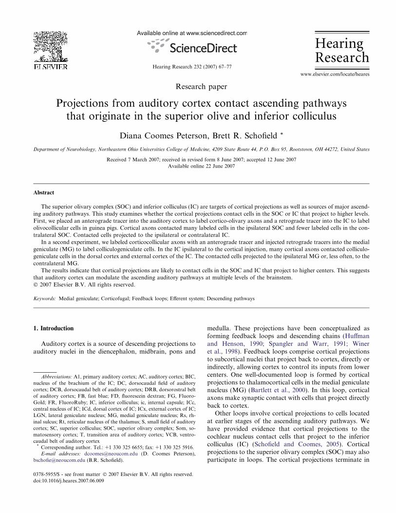
Projections from auditory cortex contact ascending pathwaysthat originate in the superior olive and inferior colliculus
Diana Coomes Peterson, Brett R. Schofield *
Department of Neurobiology, Northeastern Ohio Universities College of Medicine, 4209 State Route 44, P.O. Box 95, Rootstown, OH 44272, United States
Received 7 March 2007; received in revised form 8 June 2007; accepted 12 June 2007Available online 22 June 2007
Abstract
The superior olivary complex (SOC) and inferior colliculus (IC) are targets of cortical projections as well as sources of major ascend-ing auditory pathways. This study examines whether the cortical projections contact cells in the SOC or IC that project to higher levels.First, we placed an anterograde tracer into the auditory cortex to label cortico-olivary axons and a retrograde tracer into the IC to labelolivocollicular cells in guinea pigs. Cortical axons contacted many labeled cells in the ipsilateral SOC and fewer labeled cells in the con-tralateral SOC. Contacted cells projected to the ipsilateral or contralateral IC.
In a second experiment, we labeled corticocollicular axons with an anterograde tracer and injected retrograde tracers into the medialgeniculate (MG) to label colliculogeniculate cells. In the IC ipsilateral to the cortical injection, many cortical axons contacted colliculo-geniculate cells in the dorsal cortex and external cortex of the IC. The contacted cells projected to the ipsilateral MG or, less often, to thecontralateral MG.
The results indicate that cortical projections are likely to contact cells in the SOC and IC that project to higher centers. This suggeststhat auditory cortex can modulate the ascending auditory pathways at multiple levels of the brainstem.� 2007 Elsevier B.V. All rights reserved.
Keywords: Medial geniculate; Corticofugal; Feedback loops; Efferent system; Descending pathways
1. Introduction
Auditory cortex is a source of descending projections toauditory nuclei in the diencephalon, midbrain, pons and
medulla. These projections have been conceptualized asforming feedback loops and descending chains (Huffmanand Henson, 1990; Spangler and Warr, 1991; Wineret al., 1998). Feedback loops comprise cortical projectionsto subcortical nuclei that project back to cortex, directly orindirectly, allowing cortex to control its inputs from lowercenters. One well-documented loop is formed by corticalprojections to thalamocortical cells in the medial geniculatenucleus (MG) (Bartlett et al., 2000). In this loop, corticalaxons make synaptic contact with cells that project directlyback to cortex.
Other loops involve cortical projections to cells locatedat earlier stages of the ascending auditory pathways. Wehave provided evidence that cortical projections to thecochlear nucleus contact cells that project to the inferiorcolliculus (IC) (Schofield and Coomes, 2005). Corticalprojections to the superior olivary complex (SOC) may alsoparticipate in loops. The cortical projections terminate in
0378-5955/$ - see front matter � 2007 Elsevier B.V. All rights reserved.
doi:10.1016/j.heares.2007.06.009
Abbreviations: A1, primary auditory cortex; AC, auditory cortex; BIC,nucleus of the brachium of the IC; DC, dorsocaudal field of auditorycortex; DCB, dorsocaudal belt of auditory cortex; DRB, dorsorostral beltof auditory cortex; FB, fast blue; FD, fluorescein dextran; FG, Fluoro-Gold; FR, FluoroRuby; IC, inferior colliculus; ic, internal capsule; ICc,central nucleus of IC; ICd, dorsal cortex of IC; ICx, external cortex of IC;LGN, lateral geniculate nucleus; MG, medial geniculate nucleus; Rs, rh-inal sulcus; Rt, reticular nucleus of the thalamus; S, small field of auditorycortex; SC, superior colliculus; SOC, superior olivary complex; Som, so-matosensory cortex; T, transition area of auditory cortex; VCB, ventro-caudal belt of auditory cortex* Corresponding author. Tel.: +1 330 325 6655; fax: +1 330 325 5916.E-mail addresses: [email protected] (D. Coomes Peterson),
[email protected] (B.R. Schofield).
www.elsevier.com/locate/heares
Hearing Research 232 (2007) 67–77
HearingResearch

the periolivary nuclei as well as the lateral superior olivarynucleus (Coomes and Schofield, 2004), both of which aremajor sources of projections to the IC (Schofield and Cant,1992). However, whether cortical axons contact olivarycells that project to the IC is unknown.
Cortical projections to the IC may also participate inloops. The IC is a major target of cortical projectionsand is the primary source of ascending auditory projectionsto the MG. Mitani et al. (1983) recorded intracellularlyfrom colliculogeniculate cells identified by antidromic stim-ulation of the MG in cats. These cells showed short-latencyexcitatory postsynaptic potentials (as well as longer latencyexcitatory and inhibitory postsynaptic potentials) follow-ing electrical stimulation of the ipsilateral auditory cortex.The authors suggested that the short-latency excitationresulted from direct cortical inputs to the colliculogenicu-late cells. These inputs have yet to be demonstrated ana-tomically, and questions remain as to where in the ICsuch contacts might occur.
For the present study, we combined anterograde label-ing of corticofugal axons with retrograde labeling of brain-stem cells that give rise to ascending projections. In the firstexperiment, we injected one fluorescent tracer into auditorycortex to label corticofugal axons and different fluorescenttracers into one or both inferior colliculi to label olivocol-licular cells. We then examined the SOC for contactsbetween the labeled axons and the labeled cells. In the sec-ond experiment, we labeled cortical axons as before andinjected fluorescent tracers into one or both medial genicu-late nuclei to label colliculogeniculate cells. We then exam-ined the IC for evidence of contacts between the corticalaxons and the colliculogeniculate cells.
2. Materials and methods
Experiments were performed on adult albino guinea pigs(270–690 g; either sex) obtained from Charles River Labo-
ratories, Inc. (Wilmington, MA). All procedures wereapproved by the Institutional Animal Care and Use Com-mittee and administered following the National Institutesof Health guidelines for the care and use of laboratoryanimals.
2.1. Surgery and perfusion
Prior to surgery, each guinea pig was anesthetized withhalothane (3.5% for induction, 2.5–2.75% for maintenance)in a mixture of oxygen and nitrous oxide. The guinea pigwas premedicated with atropine sulfate (0.08 mg/kg, i.m.)to reduce bronchial secretions, and an antibiotic ointment(Neosporin Ophthalmic) was placed in each eye to preventdrying of the cornea during anesthesia. The animal wasthen placed in a stereotaxic holder on a feedback-con-trolled heating pad to maintain body temperature.
During surgery, an incision was made in the scalp andthe margins of the incision were injected with a long-lastinglocal anesthetic (0.25% bupivacaine; Sensorcaine; AstraUSA, Inc., Westborough, MA). Stereotaxic coordinateswere used to guide all injections. A dental drill was usedto open the skull at appropriate locations.
2.1.1. Injections into cortex
The skull was opened over the temporal lobe. Fluores-cein dextran (FD, 10% solution in saline; molecularweight = 10,000: Molecular Probes, Inc., Eugene, OR) orFluoroRuby (FR, 10% solution in saline; molecularweight = 10,000: Molecular Probes) was injected at 6–56sites with a 10 ll Hamilton syringe (Table 1). A volumeof 0.1–0.2 ll was injected at each site, yielding a total injec-tion volume of 0.9–6.4 ll.
2.1.2. Injections into the MG
Fast Blue (FB, 5% aqueous solution; Sigma) wasinjected into one MG in 7 cases and FR was injected into
Table 1Locations of tracer injections
Case # Left AC Right AC Left MG Right MG Left IC Right IC
GP330 x FD x FB x xGP331 x FD x FB x xGP332 x FD x FB x xGP345 FD x FR FB x xGP346 FD x FB FR x xGP347 FD x FR FB x xGP348 FD x FR FB x xGP365 FR x x x FD xGP367 FR x x x FD xGP368 FR x x x FD xGP369 FD x x x FG FRGP372 FD x x x x FRGP376 FR x x x FD FBGP377 FR x x x FB FDGP380 FR x x x FD FB
Abbreviations: AC – auditory cortex; FB – Fast Blue; FD – fluorescein dextran, FG – FluoroGold; FR – FluoroRuby; IC – inferior colliculus; MG –medial geniculate; x – indicates no tracer injection.
68 D. Coomes Peterson, B.R. Schofield / Hearing Research 232 (2007) 67–77

the opposite MG in 4 cases (Table 1). In the first 3 cases,FB was injected with a micropipette (25–28 lm insidediameter) attached to a Nanoliter Injector (World Preci-sion Instruments, Sarasota, FL). A total of 0.23 ll (10pulses, 23 nl/pulse; 1 min between pulses) was injected ata single site in the MG. In the other four cases, the tracerswere injected into each MG using 10 ll Hamilton syringes.A different syringe was used for each tracer. On each side ofthe brain, a volume of 0.15–0.2 ll was injected at each offour sites in the thalamus.
2.1.3. Injections into the IC
Eight animals received injections into one or both IC(Table 1). Tracers included FD, FR and FB as describedabove, as well as FluoroGold (FG; 4% aqueous solution;FluoroChrome, Inc., Englewood, CO). Each tracer wasinjected with a different 10 ll Hamilton syringe. For eachIC injection, tracer was deposited at four sites (0.2 ll/site).
Following the injections, the exposed brain was coveredwith Gelfoam and the scalp was sutured. To provide 48 hof post-operative analgesia, Ketoprofen (3 mg/kg, i.p.)was injected immediately after surgery and again a daylater. If the animal was to survive more than ten days, itwas re-anesthetized and the sutures were removed on thetenth post-operative day.
After 5–15 days, the animal was sacrificed with CO2 gas(inhalation, 10 min) or overdose of sodium pentobarbital(440 mg/kg, i.p.), and the animal was perfused throughthe aorta with approximately 100 ml of Tyrode’s solution(pH 7.4), 350 ml of 4% paraformaldehyde in 0.1 M phos-phate buffer, pH 7.4 (PB), and 350 ml of 4% paraformalde-hyde/10% sucrose in PB. The brain was then removed andstored at 4 �C in 4% paraformaldehyde/30% sucrose in PB.On the following day the injected cortex was photographedfor reconstruction of the cortical injection site (describedbelow). The cortex was then separated from the brainstemand thalamus. The cortex was frozen and cut in the trans-verse plane on a sliding microtome. The brainstem (withthalamus) was split at the midline and each half was frozenand cut in the parasagittal plane. All pieces of tissue werecut on a sliding microtome into 40, 50, or 80 lm thick sec-tions. Sections were collected in six series, at least four ofwhich were mounted on gelatin-coated slides and allowedto dry. One series was counterstained with thionin for theidentification of cytoarchitectonic borders. All sectionswere coverslipped with DPX (Aldrich Chemical Company,Inc., Milwaukee, WI).
2.2. Data analysis
Surface photographs of cortical injections were takenwith both natural light and fluorescence filtering using adigital camera (Magnafire, Optronics, Inc.; Goleta, CA).The images were then related to physiological maps ofauditory cortex in the guinea pig (Wallace et al., 2000,2002) using reconstruction techniques described previously(Coomes and Schofield, 2004). Transverse sections through
cortex were also evaluated to assess injections relative tocortical layers. Cortical layers were identified in adjacentthionin-stained sections according to criteria described pre-viously (Schofield et al., 2006).
Injections into the MG or IC were drawn with a Neu-rolucida system (MicroBrightField, Williston, VT)attached to a Zeiss Axioplan 2 fluorescence microscope.Adjacent sections stained with thionin were used to drawnuclear borders. The MG was identified as described byRedies et al. (1989). The subdivisions of the IC – centralnucleus, dorsal cortex (ICd), and external cortex (ICx) –were identified using criteria described in guinea pigs andrats (Faye-Lund and Osen, 1985; Malmierca et al., 1995).Nuclei within the SOC were distinguished according to cri-teria described previously (Schofield and Cant, 1991; Coo-mes and Schofield, 2004).
At least every 6th section through the IC and SOC wasexamined with a 40· lens or 63· oil immersion lens on theZeiss Axioplan 2 microscope. Cortical boutons were iden-tified as swellings along the course or at the end of an axon.Boutons that appeared to contact retrogradely-labeled cellswere photographed with an Optronics Magnafire cameraattached to the microscope. We will refer to these apparentcontacts simply as ‘‘contacts’’; of course, conclusionsregarding synapses will have to be verified with subsequentelectron microscopic studies. Adobe Photoshop was usedto adjust brightness, contrast, and color balance, toarrange and label photomicrographs, and to overlayimages collected with different fluorescence filters.
3. Results
We did two series of experiments. The first focused oncortical inputs to olivocollicular cells and the second on cor-tical inputs to colliculogeniculate cells. We first describe thecortical injections, which were similar in both experiments.
3.1. Cortical injections
Fig. 1a shows a representative injection site in temporalcortex. The injections generally spread through most of thecortical layers. Some of the injections spread into whitematter (not shown). Cases with these injections showedpatterns of terminal labeling similar to cases in which theinjections were restricted to layers I–VI. Examination ofthe thalamus, where retrograde labeling was concentratedin the MG, confirmed that the cortical injections were cen-tered in auditory cortex. The injections typically spreadacross temporal cortex to include multiple auditory areas(Fig. 1b). According to the maps described by Wallaceet al. (2000,2002), the injections generally included bothcore and belt areas of auditory cortex. The injection inthe case illustrated appeared to spread into some surround-ing areas that are not well-defined. In another case(GP369), the injection appeared to encroach on somatosen-sory cortex. The results in this case were similar to those inother cases. Several animals had smaller injections that
D. Coomes Peterson, B.R. Schofield / Hearing Research 232 (2007) 67–77 69

appeared to be confined to a few (2–3) cortical areas. Thesesmaller injections always encompassed a large portion ofprimary auditory cortex (A1) or the dorsocaudal field(DC). The smaller injections labeled fewer corticofugalaxons, but the results were similar qualitatively to thosefrom other cases.
3.2. Experiments to identify cortical contacts on
olivocollicular cells
Fig. 2 shows two representative injection sites in the IC.All injections centered on the central nucleus and includedparts of the dorsal and external cortices. None of the injec-tions spread into the contralateral IC or into adjacentstructures. The IC injections labeled many cells in numer-ous auditory areas, including the cochlear nuclei, SOC,nuclei of the lateral lemniscus and auditory cortex. In mostareas, the retrograde label was limited to the cell body andproximal dendrites. Within the SOC, cells were observed ina pattern described previously (Schofield and Cant, 1992).
Briefly, cells were located in the medial and lateral superiorolivary nuclei and in all periolivary nuclei except the pos-teroventral periolivary nucleus. Many more cells werelabeled on the ipsilateral side than on the contralateral side.
Cortico-olivary axons were distributed in a patterndescribed previously (Coomes and Schofield, 2004). Axonsand boutons were more numerous on the ipsilateral sidethan on the contralateral side. On both sides, the majorityof boutons were located in the ventral nucleus of the trap-ezoid body and the superior paraolivary nucleus. Addi-tional boutons were present in the lateral superior olivarynucleus and in the remaining periolivary nuclei. We exam-ined each area that contained labeled axons for contactswith labeled cells.
The majority of labeled boutons were located in the neu-ropil, among labeled cells but not in obvious contact. Theretrograde tracers rarely labeled the dendrites beyond thefirst branch point, so it was impossible to determinewhether contacts occur on distal dendrites. However,numerous boutons appeared to contact labeled cell bodiesor proximal dendrites. The majority of contacts wereobserved in the SOC ipsilateral to the cortical injection(Fig. 3). Of these contacts, most were located within thesuperior paraolivary nucleus and the ventral nucleus ofthe trapezoid body. A few contacts were also observed inthe dorsal periolivary nuclei (dorsal and dorsolateral peri-olivary nuclei), the lateral periolivary nuclei (anterolateralperiolivary nucleus and lateral nucleus of the trapezoidbody), and the lateral superior olivary nucleus. Amongthe cells that were contacted, the majority projected tothe ipsilateral IC (Figs. 3a–c); fewer projected to the con-tralateral IC (Figs. 3d and e). No contacts were observedin the medial superior olivary nucleus, the medial nucleusof the trapezoid body or the posteroventral periolivarynucleus.
Cortical projections to the contralateral SOC are muchless dense than those to the ipsilateral SOC. Contacts
Fig. 1. Representative injection site in temporal cortex. (a) Photomicro-graph of a transverse section showing an injection of fluorescein dextran(dark area) in the left temporal cortex of case GP345. The image wascaptured in color, converted to grayscale, and the contrast inverted (blackto white) to improve visibility. Cortical surface is at the left. Layers areindicated by Roman numerals. wm – white matter. Scale bar = 0.5 mm.(b) Surface reconstruction showing the extent of temporal cortex injectedwith FluoroRuby (black area) in case GP365. Overlaid on the recon-struction are outlines of cortical areas as mapped by Wallace et al.(2000,2002); see text for details). A1 – primary auditory cortex; DC –dorsocaudal auditory cortex; DCB – dorsocaudal belt; DRB – dorsoros-tral belt; rs – rhinal sulcus; S – small field; Som – somatosensory cortex; T– transition area; VCB – ventrocaudal belt; Vis – visual cortex; VRB –ventrorostral belt. Scale bar = 1 mm.
Fig. 2. Fluorescence photomicrographs showing representative injectionsin parasagittal sections through the inferior colliculus (IC). (a) Center of afluorescein dextran injection site. (b) Center of a Fast Blue injection site.Both panels: Case GP376. Rostral is right; dorsal is up. ICc – centralnucleus of the IC; ICd – dorsal cortex of the IC; ICx – external cortex ofthe IC; SC – superior colliculus. Scale bar = 0.5 mm.
70 D. Coomes Peterson, B.R. Schofield / Hearing Research 232 (2007) 67–77

Fig. 3. Photomicrographs showing cortical axons in contact with cell bodies (arrows) or dendrites (arrowheads) of olivocollicular cells. Cortical axonswere labeled by an injection of FluoroRuby into left auditory cortex. Olivocollicular cells were labeled by injections of fluorescein dextran (green) into theleft inferior colliculus (IC) and Fast Blue into the right IC. (a–c). Cortical contacts on cells in the left superior olivary complex (SOC; ipsilateral to theinjected cortex) that projected to the left IC. The cells were located in the ventral nucleus of the trapezoid body (a) or the superior paraolivary nucleus (b,c). (d, e). Cortical contacts on cells in the left SOC that projected to the right IC. The cells were located in the lateral superior olivary nucleus (d) orsuperior paraolivary nucleus (e). (f). Cortical contact on a cell in the right lateral superior olivary nucleus, contralateral to injected cortex, that projected tothe left IC. (g, h). Cortical contacts on cells in the right SOC that projected to the right IC. The cells were located in the ventral nucleus of the trapezoidbody (g) or the dorsolateral periolivary nucleus (h). Case GP376. Scale bar = 10 lm.
D. Coomes Peterson, B.R. Schofield / Hearing Research 232 (2007) 67–77 71

between labeled axons and labeled cells were observed(Figs. 3f–h), but they were far fewer than on the ipsilateralside. Contacts were observed most often in the superiorparaolivary nucleus and ventral nucleus of the trapezoidbody. Additional contacts were observed in most of theperiolivary nuclei and the lateral superior olivary nucleus.Contacts occurred on somas or proximal dendrites of cellsthat projected to the IC ipsilateral (Figs. 3g and h) or, lessoften, contralateral (Fig. 3f) to the SOC in which the cellwas located.
In summary, cortical axons contacted olivocollicularcells in the ipsilateral SOC much more often than in thecontralateral SOC. On each side, more contacts wereobserved on cells that projected to the ipsilateral IC thanonto cells that projected to the contralateral IC.
3.3. Experiments to identify cortical contacts on
colliculogeniculate cells
We placed injections into temporal cortex and the MGin seven animals to search for corticocollicular contactson colliculogeniculate cells. Injections into the temporalcortex were similar to those described above (Fig. 1). Cor-tico-collicular axons were observed bilaterally in the ICwith an ipsilateral predominance. On both sides, axonswith en passant and terminal boutons were located primar-ily in the ICx and ICd. On the ipsilateral side, labeled axonsand boutons were also scattered sparsely in the ICc.
A representative injection into the thalamus is shown inFig. 4. The injection involved parts of the MG andencroached on some of the surrounding nuclei. In 4 of the11 thalamic injections, the injection site included parts ofthe nucleus of the brachium of the IC or posterior thalamicnucleus (not shown). The IC sends axons to these areas(Andersen et al., 1980), so it is possible that the some ofthe labeled IC cells project to these regions instead of theMG. The results in these cases were similar to those in caseswith injections confined to the MG. Labeled cells were pres-ent bilaterally in each subdivision of the IC. More cells werepresent in the ipsilateral IC than the contralateral IC.
We examined the IC ipsilateral to the cortical injectionsfor contacts between labeled axons and labeled cells.Numerous contacts were observed in every case, with indi-vidual sections often containing multiple examples. Thecontacts were observed in ICd and ICx; labeled axonsand cells were intermingled in the ICc, but no contacts wereobserved.
Most of the contacts occurred on cells that projected tothe ipsilateral thalamus (Figs. 5a–l). Contacts occurred onboth cell bodies and proximal dendrites, and were presentin both the external cortex (Figs. 5a–h) and the dorsal cor-tex (Figs. 5i–l) of the IC. Although contacts were numer-ous, the majority of labeled boutons were located in theneuropil, among the labeled cells but not in obvious con-tact. Whether contacts occur on distal dendrites couldnot be determined. Fewer contacts were observed on ICcells that project to the contralateral MG (Figs. 5m–o).
The contacts were observed in the external cortex and dor-sal cortex of the IC.
4. Discussion
The present paper provides evidence for cortical inputsto cells in the SOC and IC that project to higher centers.This is the first evidence that ascending pathways fromthe SOC may be under direct cortical influence. The resultsare consistent with physiological studies that suggest directcortical inputs to IC cells that project to the ipsilateral MG(Mitani et al., 1983). We found additional contacts on ICcells that project to the contralateral MG. In the followingdiscussion, we first consider some technical issues impor-tant for interpreting the data. We then discuss functionalimplications of cortical inputs to ascending brainstem audi-tory pathways.
4.1. Technical considerations
The most important technical issue is the use of lightmicroscopy to assess the likelihood of synaptic contacts.
Fig. 4. Photomicrographs of parasagittal sections showing a representa-tive injection (black) of Fast Blue into the medial geniculate nucleus (MG).Thionin-stained sections through the thalamus were photographed.Adjacent sections were photographed with fluorescence to reveal theinjection site. The images were overlaid and the injection sites (black) weredrawn on the thionin images. Sections are arranged from lateral to medial(top to bottom), and are spaced 600 lm apart. Scale bar = 1 mm. BIC –nucleus of the brachium of the inferior colliculus; ic – internal capsule;LGN – lateral geniculate nucleus; Rt – reticular nucleus of the thalamus.
72 D. Coomes Peterson, B.R. Schofield / Hearing Research 232 (2007) 67–77

Fig. 5. Photomicrographs showing cortical axons in contact with cell bodies (arrows) or dendrites (arrowheads) of colliculogeniculate cells. Cortical axonswere labeled by an injection of fluorescein dextran into left auditory cortex. Colliculogeniculate cells were labeled by injections of FluoroRuby into the leftmedial geniculate nucleus (MG) or Fast Blue into the right MG. (a–h). Photomicrographs of cortical contacts on cells in the external cortex of the leftinferior colliculus that project to the ipsilateral (left) MG. (i–l). Photomicrographs of cortical contacts on cells in the dorsal cortex of the left inferiorcolliculus that project to the ipsilateral (left) MG. (m–o). Photomicrographs of cortical contacts on cells in the dorsal cortex of the left inferior colliculusthat project to the contralateral (right) MG. Cases GP345 (a, b, d–m); GP348 (c, n, o). Scale bar = 10 lm.
D. Coomes Peterson, B.R. Schofield / Hearing Research 232 (2007) 67–77 73

Our results do not prove that cortical axons contact thelabeled olivary or collicular cells, but they do provide evi-dence strongly suggesting that such contacts exist. Further,they provide important guidance for future physiologicalor electron microscopic studies by indicating the likelylocations of contacts and the projections of the targetedcells. It is noteworthy in this context that some areas con-tained labeled cortical axons and retrogradely labeled cells,but did not have evidence for contacts. This occurred in theSOC (e.g., in the medial nucleus of the trapezoid body) andin the central nucleus of the IC. Our data do not prove thatthere are no contacts in these areas, but suggest that furtherstudies should be focused on other regions.
Two issues must be addressed with regard to the size ofthe injection sites. The first issue regards the spread of theinjections beyond the intended target. In both experimentscortex was injected with an anterograde tracer. Corticalprojections to the IC (Druga et al., 1988; Winer et al.,1998; Bajo and Moore, 2005) and to the SOC (Coomesand Schofield, 2004) arise from multiple cortical areas.We made large injections into the temporal cortex to max-imize the number of labeled axons and thus the likelihoodof discovering any cortical targets that might exist. It isprobable that all of our cortical injections spread into mul-tiple auditory areas (see discussion in Coomes and Scho-field, 2004). Future experiments, with injections confinedto single, physiologically-identified cortical areas, will beneeded to assess the contributions of specific cortical areasto the pathways seen in the present study.
In the first experiment, we made large injections of a ret-rograde tracer into the IC. These injections did not spreadbeyond the IC borders. The importance of large injectionswas to maximize the number of labeled cells and thus thechance of observing cortical inputs. Consequently, ourinjections routinely involved multiple IC subdivisions,including the central nucleus, dorsal cortex and externalcortex. Most olivary projections terminate in the centralnucleus, but some (e.g., those from the superior paraoli-vary nucleus) may also terminate in dorsal cortex (Gonza-lez-Hernandez et al., 1996). Consequently, we cannotalways specify the subdivision targeted by the olivocollicu-lar cells that receive cortical inputs.
In the second experiment, we made large injections of aretrograde tracer into the MG to maximize the number oflabeled colliculogeniculate cells. Some injections spreadbeyond the MG into parts of the nucleus of the brachiumof the IC or posterior thalamic nucleus. Because the ICsends axons to these areas (Andersen et al., 1980), IC cellsthat project to these regions could have been labeled. How-ever, we also observed contacts following smaller injec-tions. Thus even if some of the labeled cells from thelarger injections were not colliculo-geniculate cells, thesmaller injections confirm our primary conclusion (i.e., cor-tical axons contact colliculogeniculate cells).
The second issue regarding injection site size concernsincomplete filling of the target. None of our injectionsare likely to have labeled every cell or axon in the relevant
pathways. Of additional importance is the fact that the ret-rograde tracers labeled only the soma and proximal den-drites of the cells; we would have missed any contacts ondistal dendrites or dendritic spines. The distribution of cor-tical axon terminals on olivary cells is not known, but theirdistribution on IC cells (in cats, at least) appears to favordendrites and spines, with relatively few contacts on somas(Jones and Rockel, 1972; Granstrem, 1984). If the same istrue in guinea pigs, the limited labeling of spines and distaldendrites means that we may have missed a significantnumber of contacts. This issue is relevant for both the ICand the SOC, where the majority of labeled boutons werelocated in neuropil and not clearly associated with alabeled cell. These limitations suggest that cortical inputsto the pathways under study may be far more numerousthan we observed.
4.2. Cortical effects on SOC cells
To our knowledge, there have been no physiologicalstudies of cortico-olivary projections. Anatomical studiesshow that these projections, like those to the IC, originatefrom cortical layer V cells and therefore likely use an excit-atory neurotransmitter such as glutamate (Mulders andRobertson, 2000; Doucet et al., 2002).
The majority of contacts in the SOC occurred on cellsin the periolivary nuclei. As a group, the periolivary nucleiare a significant source of projections to the IC (Schofieldand Cant, 1992). Unfortunately, very little is known aboutthe physiological properties of olivocollicular cells. In gen-eral, olivocollicular cells are interspersed with cells thatproject to other targets, so physiological data from periol-ivary nuclei cannot definitely be attributed to olivocollicu-lar cells (Schwartz et al., 1992). The superior paraolivarynucleus in rats provides an important exception becausevirtually all the cells in this nucleus project to the IC(Saldana and Berrebi, 2000). Kulesza et al. (2003) charac-terized the acoustically-driven responses of single units inthe rat superior paraolivary nucleus. They concluded thatthe cells in this nucleus are important for encoding tempo-ral features of sounds. Presumably, the cortical projectionsto the superior paraolivary nucleus are involved in regulat-ing temporal coding at this level, but this remains to betested. The effects of cortical inputs to olivocollicular cellsin other olivary nuclei must await further physiologicalstudies.
Numerous studies have demonstrated an important roleof inhibition in shaping collicular responses to sound(Kelly and Caspary, 2005). In rats, apparently all the cellsin the superior paraolivary nucleus use GABA as a neuro-transmitter (Kulesza and Berrebi, 2000). Many additionalolivocollicular cells use GABA or glycine as neurotransmit-ters and are likely to inhibit their targets in the IC (SaintMarie and Baker, 1990; Gonzalez-Hernandez et al.,1996). The present data suggest that some of theseinhibitory inputs to the IC are under the influence of thecortex.
74 D. Coomes Peterson, B.R. Schofield / Hearing Research 232 (2007) 67–77

4.3. Cortical effects on IC cells
Stimulation of auditory cortex has been shown to haveexcitatory, inhibitory, or mixed effects on cells in the IC(Mitani et al., 1983; Syka and Popelar, 1984; Goldstein-Daruech et al., 2002; Bledsoe et al., 2003; Torteroloet al., 1998; reviewed by Casseday et al., 2002; Suga andMa, 2003). For cells in the ICc, cortical stimulation canaffect spontaneous or sound-evoked activity and can alterthe cell’s selectivity for various parameters of an acousticstimulus (Mitani et al., 1983; Sun et al., 1996; Yan andSuga, 1996a,b, 1999; Jen et al., 2001; Ma and Suga, 2001;Jen and Zhou, 2003; Zhou and Jen, 2005; Yan et al.,2005). Fewer studies have examined corticofugal effectson cells in the ICd or ICx (Jen et al., 2002; Syka et al.,1988; Nwabueze-Ogbo et al., 2002; Bledsoe et al., 2003;Popelar et al., 2003). These studies have shown that corticalstimulation produces facilitation and/or suppression ofspontaneous or acoustically-evoked activity in ICd andICx; however, less is known about effects on selectivityfor specific stimulus parameters. Additional experiments,similar to those conducted in ICc, are needed to betterunderstand corticofugal effects in these regions.
The study by Mitani et al. (1983) is the only one to useintracellular recording of IC cells to assess the influence ofcortical inputs. The results show that cortical stimulationinduces both excitatory and inhibitory post-synaptic poten-tials in ICc cells, consistent with expectations from extra-cellular recording. The shortest latency responses tocortical stimulation (1.0–1.4 ms) were always excitatory,and the authors concluded they were probably monosynap-tic. This is consistent with work suggesting that the cortico-collicular axons use glutamate as a neurotransmitter(Feliciano and Potashner, 1995). Longer latency responses(1.8–4.2 ms) included excitatory and inhibitory effects, andare likely to include di- or multisynaptic circuits. Some ofthese effects are almost certainly due to cortical activationof cells in ICd or ICx that project in turn to the ICc (Jenet al., 2002).
One question raised by comparison of the present workwith that of Mitani and colleagues concerns the locationswithin the IC of contacts between cortical axons and colli-culogeniculate cells. Mitani et al. (1983) identified pre-sumptive monosynaptic inputs onto cells in the ICc incats. We observed many contacts in ICx and ICd, but nonein the ICc. There are several possible explanations for thisdiscrepancy. First, contacts in the ICc may have goneunobserved in the present study because of technical limita-tions (as discussed above). Second, there may be a speciesdifference, with contacts occurring in ICc in cats but notin guinea pigs. It is also quite possible that the ICc has beenidentified differently in the two species. On the one hand,Mitani et al. described monosynaptic inputs to cells thathad narrow frequency tuning and short-latency responsesto acoustic stimuli (features usually associated with ICc).On the other hand, the authors stated, by comparison withanatomical studies, that these units occupy a region of the
IC that receives dense cortical inputs (a feature generallyassociated with ICd, not ICc [reviewed by Winer et al.(1998)]). Further studies will be needed to resolve this issue.
4.4. Organization of corticofugal projections: loops and
chains
Cortical projections have been implicated in descendingpathways through both the IC and the SOC. The corticalprojections to the SOC contact cells that project to thecochlea (Mulders and Robertson, 2000) or cochlearnucleus (Schofield and Coomes, 2006). Cortical projectionsto the IC contact cells that project to the ipsilateral or con-tralateral cochlear nucleus (Schofield and Coomes, 2006).These projections form descending chains that parallelthe direct projections to the cochlear nucleus from cortex.Therefore, neural processing in the cochlear nucleus couldpresumably be affected by direct cortical projections as wellas by cortical influences on projections from the IC and theSOC to the cochlear nucleus. The multitude of descendingpathways presumably allows for a variety of effects onlower centers (Schofield and Coomes, 2006).
Potential loops from cortex to the IC to the MG andback to cortex have been described previously (e.g., Huff-man and Henson, 1990; Spangler and Warr, 1991; Wineret al., 1998). The present study supports the hypothesisof loops through the ICd and ICx to the thalamus. The tha-lamic projections from these subdivisions terminate pri-marily in the dorsal and medial subdivisions of the MG,which together project widely to auditory cortex (de Rib-aupierre, 1997). These loops could thus affect activityacross most or all of auditory cortex and contribute tonumerous auditory functions.
Cortical projections to the SOC provide additionalopportunities for interaction with ascending pathways.Some of these pathways could form extended loops thatproject back to cortex on the same side. Axons that origi-nate, for example, from the left auditory cortex terminateon cells in both the left and right SOC that project to theleft IC. The left IC provides the major input to the leftMG which in turn projects to the left auditory cortex, com-pleting the feedback loop.
On the other hand, axons from the left cortex, for exam-ple, can also contact olivary cells that project to the rightIC. These projections would presumably have their pri-mary influence on the right MG and right cortex. A simi-larly ‘‘crossed’’ circuit exists for projections from leftcortex to left IC with contacts onto cells that project tothe right MG. These pathways could allow corticofugalprojections from one side of cortex to affect the ascendinginputs to the opposite cortex. Whether or not one callsthese pathways ‘‘loops’’, it is clear that the corticofugalprojections are positioned to influence a large number ofascending auditory pathways.
In conclusion, the auditory cortex is in a position toaffect auditory processing at virtually every level of theauditory system. The auditory cortex projects directly
D. Coomes Peterson, B.R. Schofield / Hearing Research 232 (2007) 67–77 75

and indirectly, via the SOC and IC, to the cochlear nucleus(Schofield and Coomes, 2006). The cortical projections tothe SOC and the IC appear to make direct contact with anumber of ascending and descending projections fromthese regions (present study; Mulders and Robertson,2000; Schofield and Coomes, 2006). At the same time, theSOC and the IC give rise to other projections. For example,many periolivary cells project to other olivary nuclei, suchas the medial and lateral superior olivary nuclei (reviewedby Thompson and Schofield, 2000). These projections playimportant roles in binaural processing; are they also con-tacted by cortical axons? Cortical projections to the ICappear to contact cells that project to the thalamus (presentstudy) or to the cochlear nucleus (Schofield and Coomes,2006). Do cortical axons also contact IC cells that projectto other targets such as the pons, the superior colliculus,or the contralateral IC? A full understanding of the func-tions of the corticofugal system awaits answers to theseand similar questions.
Acknowledgments
Special thanks to Mr. Arkadiusz Slusarczyk for experttechnical assistance. We thank Dr. Yong Lu for valuablecomments on an earlier draft. This work was supportedby the National Institutes of Health DC04391 andDC05277.
References
Andersen, R.A., Roth, G.L., Aitkin, L.M., Merzenich, M.M., 1980. The
efferent projections of the central nucleus and the pericentral nucleus of
the inferior colliculus in the cat. J. Comp. Neurol. 194, 649–662.
Bajo, V.M., Moore, D.R., 2005. Descending projections from the auditory
cortex to the inferior colliculus in the Gerbil, Meriones unguiculatus. J.
Comp. Neurol. 486, 101–116.
Bartlett, E.L., Stark, J.M., Guillery, R.W., Smith, P.H., 2000. Comparison
of the fine structure of cortical and collicular terminals in the rat
medial geniculate. Neuroscience 100, 811–828.
Bledsoe, S.C., Shore, S.E., Guitton, M.J., 2003. Spatial representation of
corticofugal input in the inferior colliculus: a multicontact silicon
probe approach. Exp. Brain Res. 153, 530–540.
Casseday, J.H., Fremouw, T., Covey, E., 2002. The inferior colliculus: a
hub for the central auditory system. In: Oertel, D., Fay, R., Popper, A.
(Eds.), Integrative Functions in the Mammalian Auditory Pathway,
Vol. 15. Springer-Verlag, New York, NY, pp. 238–318.
Coomes, D.L., Schofield, B.R., 2004. Projections from the auditory cortex
to the superior olivary complex in guinea pigs. Eur. J. Neurosci. 19,
2188–2200.
de Ribaupierre, F., 1997. Acoustical information processing in the
auditory thalamus and cerebral cortex. In: Ehret, G., Romand, R.
(Eds.), The Central Auditory System. Oxford University Press, New
York, pp. 317–397.
Doucet, J.R., Rose, L., Ryugo, D.K., 2002. The cellular origin of
corticofugal projections to the superior olivary complex in the rat.
Brain Res. 925, 28–41.
Druga, R., Syka, J., Rajkowska-Markow, G., 1988. Localization of
cortical neurons projecting to the inferior colliculus in the rat and
guinea pig. In: Syka, J., Masterton, B.R. (Eds.), Auditory Pathway
Structure and Function. Plenum Press, New York, pp. 293–298.
Faye-Lund, H., Osen, K.K., 1985. Anatomy of the inferior colliculus in
rat. Anat. Embryol. 171, 1–20.
Feliciano, M., Potashner, S.J., 1995. Evidence for a glutamatergic
pathway from the guinea pig auditory cortex to the inferior colliculus.
J. Neurochem. 65, 1348–1357.
Goldstein-Daruech, N., Pedemonte, M., Inderkum, A., Velluti, R.A.,
2002. Effects of excitatory amino acid antagonists on the activity of
inferior colliculus neurons during sleep and wakefulness. Hearing Res.
3896, 1–7.
Gonzalez-Hernandez, T., Mantolan-Sarmiento, B., Gonzalez-Gonzalez,
B., Perez-Gonzalez, H., 1996. Sources of GABAergic input to the
inferior colliculus of the rat. J. Comp. Neurol. 372, 309–326.
Granstrem, E.E., 1984. Ultrastructural organization of AI and AIV
projection zones of the cat auditory cortex in the inferior colliculi of
the lamina tecti. Neurosci. Behav. Physiol. 14, 296–302.
Huffman, R.F., Henson, O.W., 1990. The descending auditory pathway
and acousticomotor systems: connections with the inferior colliculus.
Brain Res. Rev. 15, 295–323.
Jen, P.H., Sun, X., Chen, Q.C., 2001. An electrophysiological study of
neural pathways for corticofugally inhibited neurons in the central
nucleus of the inferior colliculus of the big brown bat, Eptesicus fuscus.
Exp. Brain Res. 137, 292–302.
Jen, P.H., Zhou, X., Zhang, J., Chen, Q.C., Sun, X., 2002. Brief and short-
term corticofugal modulation of acoustic signal processing in the bat
midbrain. Hearing Res. 168, 196–207.
Jen, P.H., Zhou, X., 2003. Corticofugal modulation of amplitude domain
processing in the midbrain of the big brown bat, Eptesicus fuscus.
Hearing Res. 184, 91–106.
Jones, E.G., Rockel, A.J., 1972. Observations on complex vesicles,
neurofilamentous hyperplasia and increased electron density during
terminal degeneration in the inferior colliculus. J. Comp. Neurol. 147,
93–118.
Kelly, J.B., Caspary, D.M., 2005. Pharmacology of the inferior colliculus.
In: Winer, J.A., Schreiner, C.E. (Eds.), The Inferior Colliculus.
Springer, New York, pp. 248–281.
Kulesza Jr., R.J., Berrebi, A.S., 2000. Superior paraolivary nucleus of the
rat is a GABAergic nucleus. J. Assoc. Res. Otolaryngol. 1, 255–269.
Kulesza Jr., R.J., Spirou, G.A., Berrebi, A.S., 2003. Physiological
response properties of neurons in the superior paraolivary nucleus of
the rat. J. Neurophysiol. 89, 2299–2312.
Ma, X., Suga, N., 2001. Corticofugal modulation of duration-tuned
neurons in the midbrain auditory nucleus in bats. Proc. Natl. Acad.
Sci. USA 98, 14060–14065.
Malmierca, M.S., Rees, A., Le Beau, F.E., Bjaalie, J.G., 1995. Laminar
organization of frequency-defined local axons within and between the
inferior colliculi of the guinea pig. J. Comp. Neurol. 357, 124–144.
Mitani, A., Shimokouchi, M., Nomura, S., 1983. Effects of stimulation of
the primary auditory cortex upon colliculogeniculate neurons in the
inferior colliculus of the cat. Neurosci. Lett. 42, 185–189.
Mulders, W.H., Robertson, D., 2000. Evidence for direct cortical
innervation of medial olivocochlear neurones in rats. Hearing Res.
144, 65–72.
Nwabueze-Ogbo, F.C., Popelar, J., Syka, J., 2002. Changes in the
acoustically evoked activity in the inferior colliculus of the rat
after functional ablation of the auditory cortex. Physiol. Res. 51, 95–
104.
Popelar, J., Nwabueze-Ogbo, F.C., Syka, J., 2003. Changes in neuronal
activity of the inferior colliculus in rat after temporal inactivation of
the auditory cortex. Physiol. Res. 52, 615–628.
Redies, H., Brandner, S., Creutzfeldt, O.D., 1989. Anatomy of the
auditory thalamocortical system of the guinea pig. J. Comp. Neurol.
282, 489–511.
Saint Marie, R.L., Baker, R.A., 1990. Neurotransmitter-specific uptake
and retrograde transport of [3H]glycine from the inferior colliculus by
ipsilateral projections of the superior olivary complex and nuclei of the
lateral lemniscus. Brain Res. 524, 244–253.
Saldana, E., Berrebi, A.S., 2000. Anisotropic organization of the rat
superior paraolivary nucleus. Anat. Embryol. 202, 265–279.
Schofield, B.R., Cant, N.B., 1991. Organization of the superior olivary
complex in the guinea pig. I. Cytoarchitecture, cytochrome oxidase
76 D. Coomes Peterson, B.R. Schofield / Hearing Research 232 (2007) 67–77

histochemistry, and dendritic morphology. J. Comp. Neurol. 314, 645–
670.
Schofield, B.R., Cant, N.B., 1992. Organization of the superior olivary
complex in the guinea pig: II. Patterns of projection from the
periolivary nuclei to the inferior colliculus. J. Comp. Neurol. 317,
438–455.
Schofield, B.R., Coomes, D.L., 2005. Projections from auditory cortex
contact cells in the cochlear nucleus that project to the inferior
colliculus. Hearing Res. 206, 3–11.
Schofield, B.R., Coomes, D.L., 2006. Pathways from auditory cortex to
the cochlear nucleus in guinea pigs. Hearing Res., 81–89.
Schofield, B.R., Coomes, D.L., Schofield, R.M., 2006. Cells in auditory
cortex that project to the cochlear nucleus in guinea pigs. J. Assoc.
Res. Otolaryngol. 7, 95–109.
Schwartz, I.R., 1992. The superior olivary complex and lateral lemniscal
nuclei. In: Webster, D.B. et al. (Eds.), The mammalian auditory
pathway: neuroanatomy. Springer-Verlag, New York, pp. 117–167.
Spangler, K.M., Warr, W.B., 1991. The descending auditory system. In:
Altschuler, R.A., Bobbin, R.P., Clopton, B.M., Hoffman, D.W. (Eds.),
Neurobiology of Hearing: The Central Auditory System. Raven Press,
New York, pp. 27–45.
Suga, N., Ma, X., 2003. Multiparametric corticofugal modulation and
plasticity in the auditory system. Nat. Rev. 4, 783–794.
Sun, X., Chen, Q.C., Jen, P.H., 1996. Corticofugal control of central
auditory sensitivity in the big brown bat, Eptesicus fuscus. Neurosci.
Lett. 212, 131–134.
Syka, J., Popelar, J., 1984. Inferior colliculus in the rat: neuronal responses
to stimulation of the auditory cortex. Neurosci. Lett. 51, 235–240.
Syka, J., Popelar, J., Druga, R., Volkova, A., 1988. Descending central
auditory pathway—structure and function. In: Syka, J., Masterton,
B.R. (Eds.), . In: Auditory Pathway Structure and Function. Plenum
Press, New York, pp. 279–292.
Thompson, A.M., Schofield, B.R., 2000. Afferents of the superior olivary
complex. Microsc Res Tech 51, 330–354.
Torterolo, P., Zurita, P., Pedemonte, M., Velluti, R.A., 1998. Auditory
cortical efferent actions upon inferior colliculus unitary activity in the
guinea pig. Neurosci. Lett. 249, 172–176.
Wallace, M.N., Rutkowski, R.G., Palmer, A.R., 2000. Identification and
localization of auditory areas in guinea pig cortex. Exp. Brain Res.
132, 445–456.
Wallace, M.N., Rutkowski, R.G., Palmer, A.R., 2002. Interconnections of
auditory areas in the guinea pig neocortex. Exp. Brain Res. 143, 106–
119.
Winer, J.A., Larue, D.T., Diehl, J.J., Hefti, B.J., 1998. Auditory cortical
projections to the cat inferior colliculus. J. Comp. Neurol. 400, 147–
174.
Yan, J., Suga, N., 1996a. Corticofugal modulation of time-domain
processing of biosonar information in bats. Science 273, 1100–1103.
Yan, J., Suga, N., 1996b. The midbrain creates and the thalamus sharpens
echo-delay tuning for the cortical representation of target-distance
information in the mustached bat. Hearing Res. 93, 102–110.
Yan, J., Suga, N., 1999. Corticofugal amplification of facilitative auditory
responses of subcortical combination-sensitive neurons in the mus-
tached bat. J. Neurophysiol. 81, 817–824.
Yan, J., Zhang, Y., Ehret, G., 2005. Corticofugal shaping of frequency
tuning curves in the central nucleus of the inferior colliculus of mice. J.
Neurophysiol. 93, 71–83.
Zhou, X., Jen, P.H., 2005. Corticofugal modulation of directional
sensitivity in the midbrain of the big brown bat, Eptesicus fuscus.
Hearing Res. 203, 201–215.
D. Coomes Peterson, B.R. Schofield / Hearing Research 232 (2007) 67–77 77
Copyright © 2022 FDOKUMEN