Polythiol-containing, recombinant mannosylated-albumin is a superior CD68+/CD206+ Kupffer...
14
1521-0103/352/2/244–257$25.00 http://dx.doi.org/10.1124/jpet.114.219493 THE JOURNAL OF PHARMACOLOGY AND EXPERIMENTAL THERAPEUTICS J Pharmacol Exp Ther 352:244–257, February 2015 Copyright ª 2014 by The American Society for Pharmacology and Experimental Therapeutics Polythiol-Containing, Recombinant Mannosylated-Albumin Is a Superior CD68 1 /CD206 1 Kupffer Cell–Targeted Nanoantioxidant for Treatment of Two Acute Hepatitis Models s Hitoshi Maeda, Kenshiro Hirata, Hiroshi Watanabe, Yu Ishima, Victor Tuan Giam Chuang, Kazuaki Taguchi, Akihito Inatsu, Manabu Kinoshita, Motohiko Tanaka, Yutaka Sasaki, Masaki Otagiri, and Toru Maruyama Department of Biopharmaceutics, Graduate School of Pharmaceutical Sciences (H.M., K.H., H.W., Y.I., V.T.G.C., T.M.), Center for Clinical Pharmaceutical Sciences, School of Pharmacy (H.W., Y.I., T.M.), and Department of Gastroenterology and Hepatology, Graduate School of Medical Sciences (M.T., Y.S.), Kumamoto University, Kumamoto, Japan; School of Pharmacy, Faculty of Health Sciences, Curtin Health Innovation Research Institute, Curtin University, Perth, Western Australia, Australia (V.T.G.C.); Faculty of Pharmaceutical Sciences (K.T., M.O.) and DDS Research Institute (M.O.), Sojo University, Kumamoto, Japan; and Department of Immunology and Microbiology, National Defense Medical College, Saitama, Japan (A.I., M.K.) Received August 24, 2014; accepted November 3, 2014 ABSTRACT Since reactive oxygen species (ROS) derived from Kupffer cells (KC), especially CD68 1 KC, play a key role in the induction of hepatic oxidative stress and injuries, we developed a polythiolated- and mannosylated human serum albumin (SH-Man-HSA), which functions as a novel nanoantioxidant for delivering thiol to CD68 1 KC. In vitro electron paramagnetic resonance coupled with pharmacokinetics and immunohistochemical studies showed that SH-Man-HSA possessed powerful radical-scavenging activity and rapidly and selectively delivered thiols to the liver via mannose receptor (CD206) on CD68 1 cells. SH-Man-HSA significantly improved the survival rate of concanavalin-A (Con-A)–treated mice. Moreover, SH-Man-HSA exhibited excellent hepatopro- tective functions, not by decreasing tumor necrosis factor or interferon-g production that is closely associated with Con- A–induced hepatitis, but by suppressing ROS production. Interestingly, the protective effect of SH-Man-HSA was superior to N-acetyl cysteine (NAC). This could be attributed to the dif- ference in the inhibition of hepatic oxidative stress between the two antioxidants depending on their potential for thiol delivery to the liver. Similar results were also observed for acetaminophen (APAP)-induced hepatopathy models. Flow cytometric data further confirmed that an increase in F4/80 1 /ROS 1 cells was dramatically decreased by SH-Man-HSA. The administration of SH-Man-HSA at 4 hours following a Con-A or APAP injection also exhibited a profound hepatoprotective action against these hepatitis models, whereas this was not observed for NAC. It can be concluded therefore that SH-Man-HSA has great potential for use in a rescue therapy for hepatopathy as a nanoantioxidant because of its ability to efficiently and rapidly deliver thiols to CD68 1 /CD206 1 KC. Introduction Oxidative stress plays a key role in the onset and pro- gression of various liver diseases, including viral hepatitis (Korenaga et al., 2005), autoimmune hepatitis (Moreno-Otero, 2013), alcoholic hepatopathy (Tsukamoto and Lu, 2001), and drug-induced hepatic impairment (Laskin and Laskin, 2001). Reactive oxygen species (ROS) produced upon activation of Kupffer cells (KC), and subsequent production of inflamma- tory cytokines, such as tumor necrosis factor (TNF)–cytokines triggered by the presence of ROS, have been shown to be involved in the progression of a number of liver pathologic conditions (Roberts et al., 2007; Jaeschke, 2011). Kinoshita et al. (2010) reported on the subclassification of KC on the basis of their surface markers and functions, namely, CD68 1 , residential, and CD11b 1 KC, monocyte-derived. CD68 1 KC have a potent capacity to produce ROS, whereas CD11b 1 cells did not. Conversely, CD11b 1 KC have a strong capacity for the production of TNF, which was much less prominent in This research was supported, in part, by Grant-in-Aid for Scientific Research from Japan Society for the Promotion of Science (JSPS) (KAKENHI 21390177). dx.doi.org/10.1124/jpet.114.219493. s This article has supplemental material available at jpet.aspetjournals.org. ABBREVIATIONS: 8-OHdG, 8-hydroxy-29-deoxyguanosine; ALT, alanine aminotransferase; APAP, acetaminophen; AST, aspartate aminotransferase; CD206, mannose receptor; Con-A, concanavalin-A; DMPO, 5,5-dimethyl-1-pyrroline I-oxide; DTNB, 5,59-dithiobis-(2-nitrobenzoic acid); DTPA, diethylenetriamine-pentaacetic acid; EPR, electron paramagnetic resonance; GSH, glutathione; GSH/GSSG, reduced/oxidized glutathione ratio; HIF-1a, hypoxia-inducible factor-1a; HSA, human serum albumin; IFN-g, interferon-g; KC, Kupffer cells; KPB, potassium phosphate buffer; Man-HSA, mannosylated-HSA; NAC, N-acetyl cysteine; NAPQI, N-acetyl-p-benzoquinone imine; PBS, phosphate-buffered saline; POBN, a-(4-pyridyl-1-oxide)-N- tert-butylnitrone; rHSAs, recombinant HSAs; ROS, reactive oxygen species; SH-Man-HSA, polythiolated-Man-HSA; SH, thiol; TNF, tumor necrosis factor; TUNEL, terminal deoxynucleotidyl transferase dUTP nick-end labeling. 244 http://jpet.aspetjournals.org/content/suppl/2014/11/14/jpet.114.219493.DC1.html Supplemental material to this article can be found at: at ASPET Journals on January 8, 2015 jpet.aspetjournals.org Downloaded from
-
Upload
independent -
Category
Documents
-
view
3 -
download
0
Transcript of Polythiol-containing, recombinant mannosylated-albumin is a superior CD68+/CD206+ Kupffer...
1521-0103/352/2/244–257$25.00 http://dx.doi.org/10.1124/jpet.114.219493THE JOURNAL OF PHARMACOLOGY AND EXPERIMENTAL THERAPEUTICS J Pharmacol Exp Ther 352:244–257, February 2015Copyright ª 2014 by The American Society for Pharmacology and Experimental Therapeutics
Polythiol-Containing, Recombinant Mannosylated-AlbuminIs a Superior CD681/CD2061 Kupffer Cell–TargetedNanoantioxidant for Treatment of Two Acute Hepatitis Models s
Hitoshi Maeda, Kenshiro Hirata, Hiroshi Watanabe, Yu Ishima, Victor Tuan Giam Chuang,Kazuaki Taguchi, Akihito Inatsu, Manabu Kinoshita, Motohiko Tanaka, Yutaka Sasaki,Masaki Otagiri, and Toru MaruyamaDepartment of Biopharmaceutics, Graduate School of Pharmaceutical Sciences (H.M., K.H., H.W., Y.I., V.T.G.C., T.M.),Center for Clinical Pharmaceutical Sciences, School of Pharmacy (H.W., Y.I., T.M.), and Department of Gastroenterology andHepatology, Graduate School of Medical Sciences (M.T., Y.S.), Kumamoto University, Kumamoto, Japan; School ofPharmacy, Faculty of Health Sciences, Curtin Health Innovation Research Institute, Curtin University, Perth, WesternAustralia, Australia (V.T.G.C.); Faculty of Pharmaceutical Sciences (K.T., M.O.) and DDS Research Institute (M.O.), SojoUniversity, Kumamoto, Japan; and Department of Immunology and Microbiology, National Defense Medical College,Saitama, Japan (A.I., M.K.)
Received August 24, 2014; accepted November 3, 2014
ABSTRACTSince reactive oxygen species (ROS) derived from Kupffer cells(KC), especially CD681 KC, play a key role in the induction ofhepatic oxidative stress and injuries, we developed a polythiolated-and mannosylated human serum albumin (SH-Man-HSA), whichfunctions as a novel nanoantioxidant for delivering thiol toCD681KC. In vitro electron paramagnetic resonance coupled withpharmacokinetics and immunohistochemical studies showed thatSH-Man-HSA possessed powerful radical-scavenging activity andrapidly and selectively delivered thiols to the liver via mannosereceptor (CD206) on CD681 cells. SH-Man-HSA significantlyimproved the survival rate of concanavalin-A (Con-A)–treatedmice. Moreover, SH-Man-HSA exhibited excellent hepatopro-tective functions, not by decreasing tumor necrosis factor orinterferon-g production that is closely associated with Con-A–induced hepatitis, but by suppressing ROS production.
Interestingly, the protective effect of SH-Man-HSA was superiorto N-acetyl cysteine (NAC). This could be attributed to the dif-ference in the inhibition of hepatic oxidative stress between thetwo antioxidants depending on their potential for thiol delivery tothe liver. Similar results were also observed for acetaminophen(APAP)-induced hepatopathy models. Flow cytometric datafurther confirmed that an increase in F4/801/ROS1 cells wasdramatically decreased by SH-Man-HSA. The administrationof SH-Man-HSA at 4 hours following a Con-A or APAP injectionalso exhibited a profound hepatoprotective action against thesehepatitis models, whereas this was not observed for NAC. It canbe concluded therefore that SH-Man-HSA has great potential foruse in a rescue therapy for hepatopathy as a nanoantioxidantbecause of its ability to efficiently and rapidly deliver thiols toCD681/CD2061 KC.
IntroductionOxidative stress plays a key role in the onset and pro-
gression of various liver diseases, including viral hepatitis(Korenaga et al., 2005), autoimmune hepatitis (Moreno-Otero,2013), alcoholic hepatopathy (Tsukamoto and Lu, 2001), anddrug-induced hepatic impairment (Laskin and Laskin, 2001).
Reactive oxygen species (ROS) produced upon activation ofKupffer cells (KC), and subsequent production of inflamma-tory cytokines, such as tumor necrosis factor (TNF)–cytokinestriggered by the presence of ROS, have been shown to beinvolved in the progression of a number of liver pathologicconditions (Roberts et al., 2007; Jaeschke, 2011). Kinoshitaet al. (2010) reported on the subclassification of KC on thebasis of their surface markers and functions, namely, CD681,residential, and CD11b1 KC, monocyte-derived. CD681 KChave a potent capacity to produce ROS, whereas CD11b1 cellsdid not. Conversely, CD11b1 KC have a strong capacity forthe production of TNF, which was much less prominent in
This research was supported, in part, by Grant-in-Aid for ScientificResearch from Japan Society for the Promotion of Science (JSPS) (KAKENHI21390177).
dx.doi.org/10.1124/jpet.114.219493.s This article has supplemental material available at jpet.aspetjournals.org.
ABBREVIATIONS: 8-OHdG, 8-hydroxy-29-deoxyguanosine; ALT, alanine aminotransferase; APAP, acetaminophen; AST, aspartate aminotransferase;CD206, mannose receptor; Con-A, concanavalin-A; DMPO, 5,5-dimethyl-1-pyrroline I-oxide; DTNB, 5,59-dithiobis-(2-nitrobenzoic acid); DTPA,diethylenetriamine-pentaacetic acid; EPR, electron paramagnetic resonance; GSH, glutathione; GSH/GSSG, reduced/oxidized glutathione ratio; HIF-1a,hypoxia-inducible factor-1a; HSA, human serum albumin; IFN-g, interferon-g; KC, Kupffer cells; KPB, potassium phosphate buffer; Man-HSA,mannosylated-HSA; NAC, N-acetyl cysteine; NAPQI, N-acetyl-p-benzoquinone imine; PBS, phosphate-buffered saline; POBN, a-(4-pyridyl-1-oxide)-N-tert-butylnitrone; rHSAs, recombinant HSAs; ROS, reactive oxygen species; SH-Man-HSA, polythiolated-Man-HSA; SH, thiol; TNF, tumor necrosis factor;TUNEL, terminal deoxynucleotidyl transferase dUTP nick-end labeling.
244
http://jpet.aspetjournals.org/content/suppl/2014/11/14/jpet.114.219493.DC1.htmlSupplemental material to this article can be found at:
at ASPE
T Journals on January 8, 2015
jpet.aspetjournals.orgD
ownloaded from
CD681 cells. Moreover, ROS produced by CD681KChave beenidentified as an aggravating factor for the onset and pro-gression of hepatitis in a concanavalin-A (Con-A)–inducedhepatitis mouse model (Nakashima et al., 2008). In addition,mice that were pretreated with gadolinium chloride (GdCl3),a KC inhibitor, especially CD681 cells, showed a decreasedacetaminophen (APAP)-induced hepatotoxicity, possibly bysuppressing the induction of oxidative stress (Laskin et al.,1995; Michael et al., 1999). This was particularly evident in thecase of CD681 KC. It therefore appears that KC, especiallyCD681, play a major role in the progression of liver pathologicconditions. In fact, it had been shown that the intensity of KCactivation, on the basis of CD68 expression, is directly cor-related with the degree of disease severity in patients withalcoholic liver disease (Chedid et al., 2004). Interestingly,mannose receptors (CD206) are present on the KC surface,includingCD681 (Hu et al., 2012). An effective ROS-scavengingagent that can be delivered to CD681 KC in the liver throughCD206 would therefore be predicted to result in promisingtherapeutic outcomes. However, as far as we know, no carrierthat targets CD681/CD2061 KC is currently available.Human serum albumin (HSA) is a simple protein with no
oligosaccharide chain structures. However, the insertion ofa consensus sequence for an oligosaccharide chain into thealbumin gene results in the production of a protein that con-tains an oligosaccharide chain, as in some reported geneticvariants (Minchiotti et al., 2001). Using three HSA variants—Albumin Dalakarlia, Casebrook, and Redhill (Kragh-Hansenet al., 2001)—as a template for designing a recombinantglycosylated-HSA, we recently succeeded, employing a yeastexpression system, in producing a recombinant mannosylated-HSA (Man-HSA) containing a biosynthetic oligosaccharidechain with a high content of 12 mannose residues per HSAmolecule (Supplemental Fig. 1) (Hirata et al., 2010). Therefore,Man-HSA would be anticipated to be efficiently distributed toCD681 KC via CD206.Antioxidants containing a thiol (SH) group, such as glu-
tathione (GSH) and N-acetyl cysteine (NAC), possess excel-lent ROS-scavenging activities (Saito et al., 2010). In fact,NAC is currently the only remedy available for the treatmentof APAP-induced hepatotoxicity (Lee et al., 2008). Hence,polythiolated-Man-HSA (SH-Man-HSA), produced by intro-ducing SH groups intoMan-HSA, has the potential to functionas a novel nanoantioxidant that targets ROS derived fromKC, especially CD681/CD2061 and, thereby, as a candidaterescue therapy for both acute and chronic hepatopathy.
Materials and MethodsAnimals. Male C57BL/6 mice (8 weeks old) were purchased from
Japan SLC (Shizuoka, Japan). All animal experiments were con-ducted in accordance with the guidelines of Kumamoto University forthe care and use of laboratory animals.
Reagents. DTPA (diethylenetriamine-pentaacetic acid) and DTNB[5,59-dithiobis (2-nitrobenzoic acid)] were obtained from Dojindo Labora-tories (Kumamoto, Japan). DMPO (5,5-dimethyl-1-pyrroline N-oxide)and POBN [a-(4-pyridyl-1-oxide)-N-tert-butylnitrone] were purchasedfrom Alexis Biochemicals (Lausen, Switzerland). All other chemicalswere of the highest analytical grade available.
Production and Purification of Recombinant HSAs. Theprotocol used to express the recombinant HSAs (rHSAs), Man-HSA andHSA, by Pichia pastoris was a modification of a previously publishedprocedure (Hirata et al., 2010). The eluted rHSAs were deionized and
defatted by charcoal treatment, freeze-dried, and then stored at280°Cuntil used. Sample purity was estimated by a density analysis of theCoomassie brilliant blue–stained protein bands on 10% SDS-PAGEgels. The rHSAs were estimated to be more than 97% pure. Matrix-assisted laser desorption/ionization–time of flight mass spectros-copy analysis revealed that approximately 12 mannose (N-acetylglucosamine5 2) units are present in the glycan chain structure of theMan-HSA (Supplemental Fig. 1).
Synthesis of SH-rHSAs and Determination of ThiolationEfficiency. Terminal SH groups were added to the rHSAs moleculeby incubating 0.15 mM rHSAs with 8 mM 2-iminothiolane in 100 mMpotassium phosphate buffer (KPB) containing 0.5 mM DTPA, pH 7.8,for 1 hour at room temperature (Katayama et al., 2008). The amountof SH groups in the SH-rHSAs was quantified using the DTNBmethod. First, 20-ml aliquots of SH-rHSAs solution and reducedglutathione (standard) were incubated in a 96-well plate with 0.2 mlof 100 mM KPB, pH 7.0, containing 1 mM DTPA and 0.5 mM DTNB,for 15 minutes at room temperature. The absorbance was then mea-sured at 405 nm. The number of SH groups attached to Man-HSA orHSA was estimated to be 7.526 0.01 mol SH/mol Man-HSA or 7.8260.02 mol SH/mol HSA, respectively (Supplemental Fig. 2B).
Scavenging Activity of SH-Man-HSA against ×OH Generatedby H2O2/UV System. The reaction solution contained 100 mMDTPA, 9 mM DMPO, and 500 mM H2O2 in the absence or presenceof 75 mM HSA, Man-HSA or SH-Man-HSA in phosphate-bufferedsaline (PBS) (pH 7.4). After the sample was irradiated with UV(254 nm) for 1 minute in the electron paramagnetic resonance (EPR)flat cell, EPR spectra were measured immediately.
Pharmacokinetic Analysis of SH-Man-HSA. SH-Man-HSAwas radiolabeled with 111In using the bifunctional chelating reagentDTPA anhydride according to the method of Hnatowich et al. (1982).Mice received tail vein injections of 111In-labeled SH-Man-HSA insaline, at a dose of 10.0 nmol SH/kg. In the early period after injection,the efflux of 111In radioactivity from organs is assumed to be negligible,because the degradation products of 111In-labeled ligands using DTPAanhydride cannot easily pass through biologic membranes. Thisassumption was confirmed by the finding that 111In was not detectablein the urine throughout the 120-minute period. At appropriate intervalsafter the injection, blood was collected from the vena cava under etheranesthesia and plasma was obtained by centrifugation (3000g,10 minutes). The liver, kidney, spleen and lung were excised,rinsed with saline, and weighed. The radioactivity of each samplewas measured in a well-type NaI scintillation counter (ARC-500;Hitachi Aloka Medical, Ltd., Tokyo, Japan).
Preparation of Bromobimane-Labeled SH-Man-HSA. A solu-tion of 75 mM bromobimane in 0.1 M KPB was added to a solution of100 mM SH-Man-HSA in 0.1 M KPB, followed by incubation for2 hours at 4°C. The resulting solution was purified using a PharmaciaBio-Gel PD-10 column (GE Healthcare, Little Chalfont Bucks, UK) andconcentrated by Vivaspin (Sartorius Stedim Biotech S.A., Aubagne,France).
Evaluation of CD681/CD2061 KC Distribution of SH-Man-HSA. The prepared bromobimane-labeled SH-Man-HSA (20.0 mmolSH/kg) was injected into the tail vein of mice. Mannan (6.0 mg/mouse)was injected intravenously 30 minutes before the bromobimane-labeled SH-Man-HSA administration. At 1 and 12 hours after theadministration of bromobimane-labeled SH-Man-HSA, the liver wasremoved, covered with optimal cutting temperature compound, andfrozen at 280°C. Fresh-frozen sections (4-mm thickness) of the liverwere cut on a cryostat (CM3000II; Leica, Wetzlar, Germany), collectedon slides, and immediately dried. The sections were fixed withphosphate-buffered 4% paraformaldehyde and washed. After incuba-tion with 1% Block Ace (DS Pharma Biomedical, Osaka, Japan) for10 minutes, the slides were incubated overnight with anti-HSA (BethylLaboratories, Inc., Montgomery, TX), CD68 (FA-11; BioLegend, SanDiego, CA), and CD206 (C068C2; BioLegend) antibodies diluted 100,100, or 200 times, respectively. After the reaction, the slide was ob-served using a microscope (BZ-8000; Keyence, Osaka, Japan).
SH-Man-HSA Attenuates Acute Liver Injury 245
at ASPE
T Journals on January 8, 2015
jpet.aspetjournals.orgD
ownloaded from
Effect of SH-Man-HSA on Survival Rate of Lethal Con-A–Treated Mice. A lethal Con-A–treated mouse model was pro-duced using C57BL/6 mice that were intravenously injected withCon-A (1.0 mg/mouse). The same SH content (20.0 mmol SH/kg) ofSH-Man-HSA and NAC was administered intravenously just prior tothe Con-A injection.
Hepatoprotective Effect of SH-Man-HSA on Con-A– orAPAP-Treated Mice. Con-A– and APAP-induced liver injury mousemodel was produced as previously reported (Ayoub et al., 2004;Nakashima et al., 2008; Saito et al., 2010). The animal experimentprotocol is shown in Supplemental Figs. 4 and 5. Alanine amino-transferase (ALT) and aspartate aminotransferase (AST) activitylevels were determined using a transaminase C2 test kit from WakoChemicals (Saitama, Japan). In case of Con-A, themice received 0.25mgof Con-A intravenously (Sigma-Aldrich, St. Louis, MO). Just prior toCon-A injection, 5.0, 10.0, and 20.0 mmol SH/kg of SH-Man-HSA and20.0 mmol SH/kg of NAC were administered intravenously to themice. Likewise, SH-HSA (20.0 mmol SH/kg) or Man-HSA (2.66 mmolSH/kg) was administered intravenously. In case of APAP, the micereceived 300 mg/kg of APAP intraperitoneally (Sigma-Aldrich). Thesame SH content (20.0 mmol SH/kg) of SH-Man-HSA and NAC wasadministered intravenously to the mice just prior to APAP injection.Furthermore, we administered SH-Man-HSA or NAC (intrave-nously) at 2 and 4 hours after the injection of Con-A or APAP. Toinhibit the KC, especially CD681 cells, 200 mg of GdCl3 was ad-ministered (Sigma-Aldrich) intravenously via the caudal vein 24 hoursbefore Con-A or APAP injection. All mice were sacrificed 12 hours afterCon-A or APAP injection.
Histologic Examination of Liver Tissues. Twelve hours afterCon-A or APAP injection, the whole liver was removed. For thehistologic analyses, the liver tissues were fixed in 10% neutral-buffered formalin (Wako Pure Chemical Industries, Osaka, Japan),embedded in paraffin (Sakura Finetek Japan, Tokyo, Japan), andsectioned at a 4-mm thickness. The liver sections were subjected tohematoxylin and eosin staining for morphologic analysis, terminaldeoxynucleotidyl transferase dUTP nick-end labeling (TUNEL) stain-ing for cell apoptosis, and immunohistochemistry for 8-OHdG(8-hydroxy-29-deoxyguanosine) [15A3] (Santa Cruz Biotechnology,Dallas, TX) and nitrotyrosine (NO2-Tyr) (Millipore, Billerica, MA). Allassayswere performed in triplicate. For TUNEL staining, sectionswerestained using the In Situ Cell Death Detection Kit, Fluorescein (Roche,Basel, Switzerland), according to the manufacturer’s protocol forparaffin-embedded sections. For the immunohistochemistry of 8-OHdGand NO2-Tyr, first, antigen retrieval was performed by means of animmunosaver (Nisshin EM Corporation, Tokyo, Japan). T-TB solutioncontaining 50mMTris-HCl (pH7.4) and 0.1%Tween-20was then used tosolubilize the liver slices, followed by blocking with Block Ace (DainipponPharmaceutics, Osaka, Japan) at room temperature for 10 minutes.Next, reaction with the primary antibody was carried out overnight ata temperature below 4°C. In addition, the primary antibody against8-OHdG orNO2-Tyrwas dilutedwith 0.5% bovine serumalbumin in PBS50 times before use. The liver slices were then washed with T-TBsolution, followed by reaction with the secondary antibody at roomtemperature for 1.5 hours. For 8-OHdG immunostaining, Alexa Fluor488 goat anti-mouse IgG (H1L) (Invitrogen, Tokyo, Japan) and forNO2-Tyr immunostaining, Alexa Fluor 546 goat anti-mouse IgG (H1L)(Invitrogen/Life Technologies) were diluted with 0.5% bovine serumalbumin in PBS 200 times before use. After the reaction, the slide wasobserved under a microscope (BZ-8000; Keyence).
Quantification of Liver SH and GSH Levels. Tissue homoge-nates were prepared by homogenizing the tissues with 5% (w/v)sulfosalicylic acid. The homogenates were then centrifuged at 10,000gfor 10 minutes at 4°C. SH levels were determined according to theDTNB method. Reduced and oxidized glutathione levels in the liverswere determined using a reduced/oxidized GSH ratio (GSH/GSSG)quantification kit (Dojinkagaku, Kumamoto, Japan).
Determination of ROS in Liver Using EPR. The in vivo EPRanalysis was performed as described previously with some modifications
(Sato et al., 2002). The EPR spectra of POBN spin adducts of lipidperoxide radical LOO_were determined at 4 hours after Con-A or APAPinjection. POBN (1.0 g/kg) was injected intraperitoneally at 30 minutesbefore the mice were sacrificed. The livers were homogenized in 1 mlof saline on an ice bath. The homogenates were added in 2 ml of2:1 chloroform/methanol, shaken, then centrifuged at 2800 rpm for10 minutes. The chloroform layer was isolated. After evaporatingthe sample by bubbling with N2 in a vial, the sample was resuspendedin organic solvent (200 ml of chloroform/methanol). EPR spectra wereimmediately recorded at room temperature in a JES-TE200 spectrom-eter (JEOL, Tokyo, Japan) under the following conditions: modulationfrequency, 100 kHz; microwave frequency, 9.43 GHz; microwave power,40 mW; scanning field, 333.5 mT; sweep time, 2 minutes; fieldmodulation width, 0.25 mT; receiver gain, 630; and time count, 0.3seconds. Every buffer and solution of the reaction mixture used for EPRmeasurement were treated with Chelex 100 resin (Bio-Rad Laborato-ries, Hercules, CA) before use to remove metals.
Isolation of Hepatic Mononuclear Cells and Flow Cytom-etry. Mononuclear cells were prepared as described previously(Dobashi et al., 1999). Liver specimens were minced with scissors.After adding a 0.25% collagenase solution, the specimens were shakenfor 20minutes in a 37°C constant-temperature bath. The liver specimenwas then filtered through a nylon mesh (108 mm). After mixing in 33%Percoll solution containing heparin (1%), the sample was centrifugedfor 20 minutes at 500g at room temperature. After removing thesupernatant, the pellet was resuspended in a red blood cell lysis so-lution and then was washed twice in RPMI 1640 with 10% fetal bovineserum. For identification of KC, mononuclear cells were stained byphycoerythrin Cy5-conjugated anti-F4/80 antibody (eBioscience, SanDiego, CA). For the detection of intracellular ROS, CM-H2DCFDA(Invitrogen, Carlsbad, CA), an ROS-sensitive fluorescent dye used asprobe. Also, for the detection of macrophage polarization status,anti–inducible NO synthase (Santa Cruz Biotechnology, Santa Cruz,CA) and CD206 (C068C2; BioLegend) antibody were used. F4/801 KCwere isolated by positive selection according to the manufacturer’sprotocol (MACS isolation system; Miltenyi Biotec, Auburn, CA). Flowcytometry was performed using a FACSCalibur (BD Biosciences, SanJose, CA).
Quantification of TNF and Interferon-g Levels. TNF andinterferon (IFN)-g levels in plasma at 1 and 12 hours, respectively,after Con-A (0.25 mg/mouse) injection were measured by enzyme-linked immunosorbent assay, following the manufacturer’s recom-mended protocol.
Statistical Analysis. All data are expressed as mean 6 S.E. Themeans for groups were compared by analysis of variance followed byTukey’s multiple comparison and Student’s t test. The survival rateswere compared using Kaplan-Meier survival curves and the log-ranktest. A probability value of P , 0.05 was considered significant.
ResultsRadical-Scavenging Activity of SH-Man-HSA. We de-
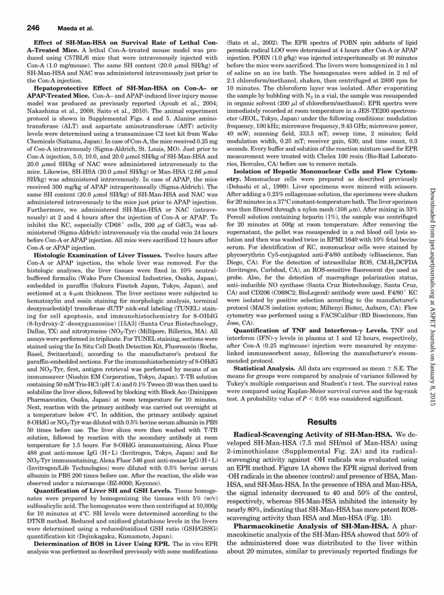
veloped SH-Man-HSA (7.5 mol SH/mol of Man-HSA) using2-iminothiolane (Supplemental Fig. 2A) and its radical-scavenging activity against ·OH radicals was evaluated usingan EPR method. Figure 1A shows the EPR signal derived from·OH radicals in the absence (control) and presence of HSA,Man-HSA, and SH-Man-HSA. In the presence of HSA andMan-HSA,the signal intensity decreased to 40 and 50% of the control,respectively, whereas SH-Man-HSA inhibited the intensity bynearly 80%, indicating that SH-Man-HSAhasmore potent ROS-scavenging activity than HSA and Man-HSA (Fig. 1B).Pharmacokinetic Analysis of SH-Man-HSA. A phar-
macokinetic analysis of the SH-Man-HSA showed that 50% ofthe administered dose was distributed to the liver withinabout 20 minutes, similar to previously reported findings for
246 Maeda et al.
at ASPE
T Journals on January 8, 2015
jpet.aspetjournals.orgD
ownloaded from
Man-HSA (Fig. 2A) (Hirata et al., 2010). Furthermore, thehepatic distribution of SH-Man-HSA was investigated bymeans of a fluorescence-imaging technique. In this procedure,the SHs of SH-Man-HSA were completely modified by thetreatment with bromobimane. Liver sections were observed at1 hour after the administration of bromobimane-labeled SH-Man-HSA (20.0 mmol SH/kg), and modified SH groups weredetected with a fluorescence probe in the form of a blueemission. As shown in Fig. 2B, substantial fluorescence wasobserved in the liver sections, especially near the sinusoidalregions. Such a fluorescence was largely inhibited by thepreadministration of a two times higher dose of mannan,a typical substrate for CD206, suggesting the importance ofthis receptor for distributing SH-Man-HSA to the liver.At the region near the sinusoid, there are several cells that
express CD206, such as KC, endothelial cells, and dendriticcells (Kogelberg et al., 2007). Our previous study revealed thatMan-HSA was preferentially distributed to KC, but not toendothelial cells (Hirata et al., 2010). It has been demon-strated that the C-type lectin–like domain binds complexcarbohydrates in which the chains are terminated withmannose,such as Man(n)GlcNAc2 (n, numbers of mannose residues). Forinstance, the carbohydrate-recognition domain of Dectin-2 isa C-type lectin with a very high affinity for high-mannosestructures, especially structures that contain more than sevenmannose residues, and its specificity is enhanced with an in-crease in the number of mannose residues (McGreal et al.,2006). Interestingly, Dectin-2 expression was predominantly
restricted to CD206-positive macrophages (Sun et al., 2013).Because SH-Man-HSA possesses an oligosaccharide chain withMan(12)GlcNAc2, it would be expected to be preferentiallyrecognized by Dectin-2 on KC that express CD206, but notendothelial cells.Evaluation of CD681/CD2061 KC Distribution of
SH-Man-HSA. To further characterize the distribution ofSH-Man-HSA to KC subtype, liver sections were subjectedto immunostaining with anti-HSA, CD68, and CD206 anti-bodies. The anti-HSA antibody used in this study did notcrossreact with the mouse species. At 1 hour after theadministration of bromobimane-labeled SH-Man-HSA,the fluorescence derived from bromobimane (blue) was mergedwith both the fluorescence of the anti-HSA antibody (red) andthe anti-CD68 antibody (green) as shown by white spots in theinset image (Fig. 3A). Similar white spots that hadmergedwiththe fluorescence of bromobimane, the anti-HSA antibody, andthe anti-CD206 antibody (green) were also observed (Fig. 3B).These data indicate that SH-Man-HSA was efficiently distrib-uted to CD681/CD2061 KC within 1 hour after the adminis-tration of bromobimane-labeled SH-Man-HSA depending on itsunique oligosaccharide moiety. However, the fluorescence ofbromobimane did not becomemergedwith both the fluorescencesof the anti-HSAantibody and the anti-CD68 antibody at 12 hoursafter the administration of bromobimane-labeled SH-Man-HSA(Fig. 3C, right panel), suggesting that SH-Man-HSA is suscep-tible to lysosomal degradation in CD681/CD2061 KC, and thatthemajority of the SHmoietieswere liberated fromSH-Man-HSAand moved to the outside of CD681/CD2061 KC.Hepatoprotective Effect of SH-Man-HSA on Con-
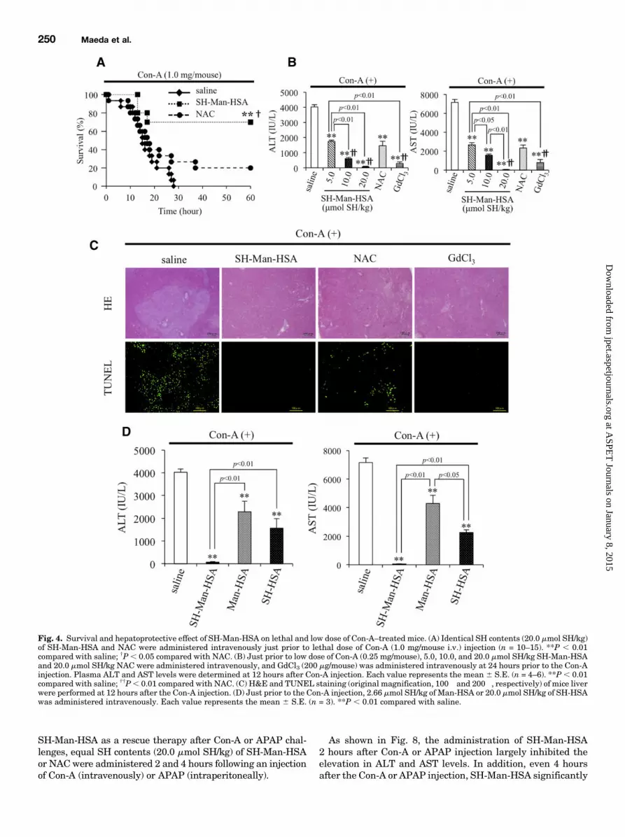
A–Treated Mice. To examine the effect of SH-Man-HSA onliver injury, we used a Con-A–induced hepatitis model, becauseit has been widely used as a model for liver injury, such asautoimmune and viral types. As shown in Fig. 4A, all of thesaline-treated mice died within 28 hours after a lethal dose(1.0 mg/mouse i.v.) of Con-A, whereas 20% of the NAC(20.0 mmol SH/kg)-treated mouse group survived. Interest-ingly, the administration of SH-Man-HSA (20.0 mmol SH/kg)resulted in a significant increase in survival rate, with 70%of the mice being alive at 60 hours after the Con-A injection.The hepatoprotective effects of SH-Man-HSA were evalu-
ated to monitor ALT and AST levels at 12 hours after theinjection of a low dose (0.25 mg/mouse i.v.) of Con-A becausethe plasma ALT and AST levels were at a maximum at12 hours post Con-A injection (Supplemental Fig. 6). SH-Man-HSA suppressed the elevation of plasma ALT and AST levelsthat had been induced by Con-A in a dose-dependent manner.In particular, 20.0 mmol SH/kg of SH-Man-HSA inhibited theelevation of ALT and AST plasma levels by 98 and 99% forCon-A–treated mice (Fig. 4B). Therefore, 20.0 mmol SH/kg ofSH-Man-HSA was adopted in the subsequent experiments(Supplemental Fig. 3).The elevation in levels of plasma ALT and AST in Con-
A–treated mice was also suppressed by 20.0 mmol SH/kg ofNAC, but the effect was much less than by SH-Man-HSA. Onthe other hand, pretreatment with GdCl3 at 24 hours beforethe Con-A injection resulted in a hepatoprotective effect thatwas equivalent to 20.0 mmol SH/kg of SH-Man-HSA (Fig. 4B).Histologic examinations of liver tissue in Con-A–induced
hepatitis were also performed with H&E and TUNELstaining (Fig. 4C). H&E staining of liver sections showedevidence of massive necrosis within the liver lobules in the
Fig. 1. Radical-scavenging activity of the SH-Man-HSA. (A) EPR spectraof ×OH and (B) the quantification of signal intensity were determinedusing an EPR method. The reaction solution contained 100 mM DTPA,9 mMDMPO, and 500 mMH2O2 in the absence or presence of 75 mMHSA,Man-HSA, or SH-Man-HSA in PBS (pH 7.4). Each value represents themean 6 S.E. (n = 3). *P , 0.05; **P , 0.01 compared with control.
SH-Man-HSA Attenuates Acute Liver Injury 247
at ASPE
T Journals on January 8, 2015
jpet.aspetjournals.orgD
ownloaded from

saline treatment group of the Con-A–challengedmice. Similarresults were obtained for TUNEL staining. In the salinetreatment groups, a number of TUNEL-positive hepatocyteswere observed for the Con-A–induced model, whereas noevidence for such positive cells was found in the case of theSH-Man-HSA andGdCl3 treatment groups. NAC also reducedTUNEL-positive cells, but to a lesser extent compared withSH-Man-HSA.The contribution of each functional component was exam-
ined in the Con-A–induced model using two HSA derivatives,namely, Man-HSA, which contains mannose residues but noadditional SH groups, and SH-HSA, which contains addi-tional SH moieties, but no mannose residues. As shown in Fig.4D,Man-HSA (2.66mmol SH/kg) and SH-HSA (20.0mmol SH/kg)significantly attenuated the Con-A–induced elevation in plasmaALT and AST levels. The extent of attenuation was in thefollowing order: Man-HSA , SH-HSA , SH-Man-HSA.Effect of SH-Man-HSA on Hepatic SH Level and
Oxidative Stress Induced by Con-A. SH levels in liverhomogenates were determined 12 hours after the injection ofCon-A (Fig. 5A) to evaluate the efficacy of SH delivery to theliver by SH-Man-HSA and NAC, both of which had the sameSH content (20.0 mmol SH/kg). The SH level of the saline-treated groups was decreased by Con-A injection, and itrecovered in the following treatment order: NAC,GdCl3, SH-Man-HSA. Furthermore, as shown in Fig. 5B, SH-Man-HSA
suppressed the decrease in GSH/GSSG, indicating thatSH-Man-HSA is superior to NAC in terms of delivering SH tothe liver.The effects of SH-Man-HSA or NAC on Con-A–induced free
radical formation in the liver were directly monitored in vivousing an EPR technique (Fig. 5C), in which LOO_ levels weredetermined in a liver homogenate. Radical production in-creased in a time-dependent manner, reaching a maximum at4 or 5 hours after the Con-A injection (Supplemental Fig. 7).As shown in Fig. 5D, more than 80% of the EPR signal inducedby the Con-A challenges was inhibited by both SH-Man-HSAand GdCl3, whereas less than 40% of the signal intensity wasdecreased by NAC at 4 hours after the Con-A injection. Similarantioxidative effects of SH-Man-HSA were also confirmed byimmunostaining of 8-OHdG and NO2-Tyr, markers for oxida-tive and nitrative stress, respectively, in the liver (Fig. 5E). Inaddition, Con-A markedly increased the fluorescence intensityof ROS derived from F4/801/ROS1–detecting probe (1686 27),whereas it was significantly inhibited by both SH-Man-HSA(57 6 6) and GdCl3 (70 6 5) (Fig. 5F). Such similar inhibitionsof ROS production in F4/801/ROS1 cells between SH-Man-HSA and GdCl3 could reflect comparable action against Con-A–induced hepatic damage.On the other hand, the possibility that the ROS-scavenging
activity of SH-Man-HSA might be mediated via the inhibitionof TNF or IFN-g that is closely associated with Con-A–induced
Fig. 2. Pharmacokinetic analysis of the SH-Man-HSA.(A) Plasma concentration curve and organ distribution ofSH-Man-HSA after 10.0 nmol SH/kg of 111In-labeled SH-Man-HSA was injected in tail vein of mice. Each valuerepresents the mean 6 S.E. (n = 3). (B) Furthermore, SH-Man-HSA distribution in the liver was observed in liversection using fluorescence image technique at 1 hour afterthe administration of bromobimane-labeled SH-Man-HSA(20.0 mmol SH/kg; original magnification, 400�). Thirtyminutes before the administration of bromobimane-labeledSH-Man-HSA, 6.0 mg/mouse of mannan was administered.
248 Maeda et al.
at ASPE
T Journals on January 8, 2015
jpet.aspetjournals.orgD
ownloaded from
hepatic damage cannot be completely excluded. However,SH-Man-HSA did not decrease to the levels of TNF or IFN-g(Fig. 5G).Hepatoprotective Effect of SH-Man-HSA on APAP-
Treated Mice. The hepatoprotective effect of SH-Man-HSAwas also examined against an APAP-induced hepatic injurymodel that was prepared by an intraperitoneal injection of300 mg/kg of APAP (Fig. 6). As in the case of Con-A–inducedhepatitis, equal SH contents (20.0 mmol SH/kg) of SH-Man-HSA or NAC were administered (intravenously) just beforeAPAP injection, and the plasma ALT and AST levels weremeasured at 12 hours after APAP injection (Supplemental Fig. 8).As a result, SH-Man-HSA largely inhibited APAP-induced
elevation of plasma ALT and AST levels by 96 and 94%,respectively. NAC also significantly suppressed the plasmaALT and AST levels by 61 and 57%, respectively, but this effectwas much less than that of SH-Man-HSA. Pretreatment withGdCl3 showed hepatoprotective effects similar to SH-Man-HSAadministration (Fig. 6A). This is consistent with the previousfinding that KC play an important role in the development ofAPAP-induced hepatic injury.H&E staining of liver sections indicated that the APAP-
induced massive necrosis within the centralobular region ofthe livers, as evidenced by the loss of basophilic staining,vacuolization, cell swelling, and karyolysis around the cen-trilobular veins, as previously reported (Fig. 6B, upper panel).However, liver sections from SH-Man-HSA administrationshowed little damage, and a nearly normal morphology waspreserved. Similar results were also obtained for a GdCl3pretreatment. H&E staining of tissue from the NAC admin-istration group showed some improvement in the integrity ofliver sections against APAP-induced hepatotoxicity, but itsefficacy was less than that of SH-Man-HSA and GdCl3. Similarresults were obtained for TUNEL staining (Fig. 6B, lowerpanel). In the saline treatment group, a number of TUNEL-positive hepatocytes were observed, whereas such positive cellscould not be found after SH-Man-HSA and GdCl3 administra-tion groups. NAC also reduced TUNEL-positive cells, but toa lesser extent compared with SH-Man-HSA.Effect of SH-Man-HSA on Hepatic SH Level and
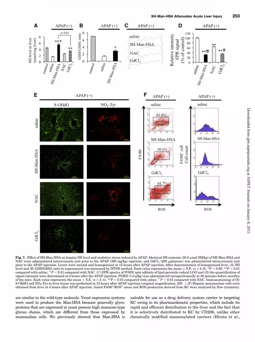
Oxidative Stress Induced by APAP. Figure 7A shows SHlevels in liver homogenates 12 hours after the injection of micewith APAP. The SH levels in liver were significantly decreasedas a result of the APAP injection, but the level was completelyrecovered by the subsequent SH-Man-HSA administration ora GaCl3 pretreatment. In contrast, the administration of NACwas ineffective in recovering APAP-induced SH depletion inthe liver. Furthermore, as shown in Fig. 7B, SH-Man-HSAsuppressed the decrease in GSH/GSSG, indicating thatSH-Man-HSA is superior to NAC in delivering SH to the liver.Figure 7, C and D, shows the effect of SH-Man-HSA on the
lipid peroxide radical in liver homogenate after APAP injectiontomice. As observed in the Con-A–induced hepatopathy model,a remarkable EPR signal was detected at 4 hours after APAPinjection (Supplemental Fig. 9). This EPR signal was signifi-cantly suppressed by the SH-Man-HSA or GdCl3 pretreatmentby approximately 70%, but NAC treatment only decreased thesignal intensity by approximately 40%.Previous findings revealed that oxidative and nitrative toxicity
in liver induced by APAP included lipid peroxidation and proteinnitration (Michael et al., 1999). As shown in Fig. 7E, our im-munostaining data against 8-OHdG and NO2-Tyr demonstratedthat SH-Man-HSA administration effectively inhibited theaccumulation of those oxidized products in the liver induced byAPAP, whereas NAC was less effective in inhibiting the ac-cumulation of those oxidative stress markers. Furthermore, toclarify the antioxidative activity of SH-Man-HSA in KC of APAP-induced hepatic injury mice, the F4/801/ROS1 KC populationwas estimated at 4 hours after APAP injection (300mg/kg i.p.) byflow cytometry (Fig. 7F). F4/801/ROS1 KC increased followingthe APAP injection, whereas it was decreased by SH-Man-HSAadministration. Pretreatment with GdCl3 also decreased thecontent of F4/801/ROS1 KC, to the lesser extent.Effect of Postadministration of SH-Man-HSA on Con-
A– or APAP-Treated Mice. To investigate the potential of
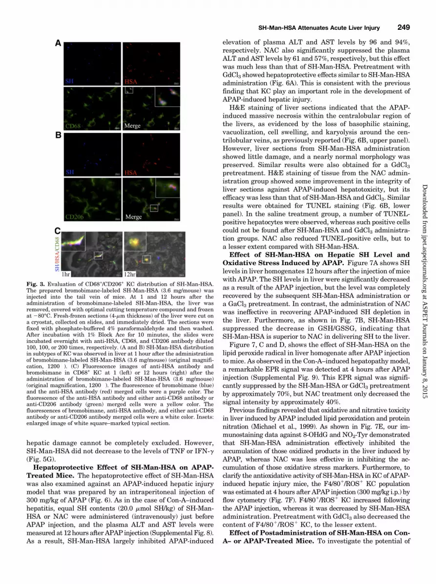
Fig. 3. Evaluation of CD68+/CD206+ KC distribution of SH-Man-HSA.The prepared bromobimane-labeled SH-Man-HSA (3.6 mg/mouse) wasinjected into the tail vein of mice. At 1 and 12 hours after theadministration of bromobimane-labeled SH-Man-HSA, the liver wasremoved, covered with optimal cutting temperature compound and frozenat 280°C. Fresh-frozen sections (4-mm thickness) of the liver were cut ona cryostat, collected on slides, and immediately dried. The sections werefixed with phosphate-buffered 4% paraformaldehyde and then washed.After incubation with 1% Block Ace for 10 minutes, the slides wereincubated overnight with anti-HSA, CD68, and CD206 antibody diluted100, 100, or 200 times, respectively. (A and B) SH-Man-HSA distributionin subtypes of KC was observed in liver at 1 hour after the administrationof bromobimane-labeled SH-Man-HSA (3.6 mg/mouse) (original magnifi-cation, 1200�). (C) Fluorescence images of anti-HSA antibody andbromobimane in CD68+ KC at 1 (left) or 12 hours (right) after theadministration of bromobimane-labeled SH-Man-HSA (3.6 mg/mouse)(original magnification, 1200�). The fluorescence of bromobimane (blue)and the anti-HSA antibody (red) merged cells were a purple color. Thefluorescence of the anti-HSA antibody and either anti-CD68 antibody oranti-CD206 antibody (green) merged cells were a yellow color. Thefluorescences of bromobimane, anti-HSA antibody, and either anti-CD68antibody or anti-CD206 antibody merged cells were a white color. Insets:enlarged image of white square–marked typical section.
SH-Man-HSA Attenuates Acute Liver Injury 249
at ASPE
T Journals on January 8, 2015
jpet.aspetjournals.orgD
ownloaded from
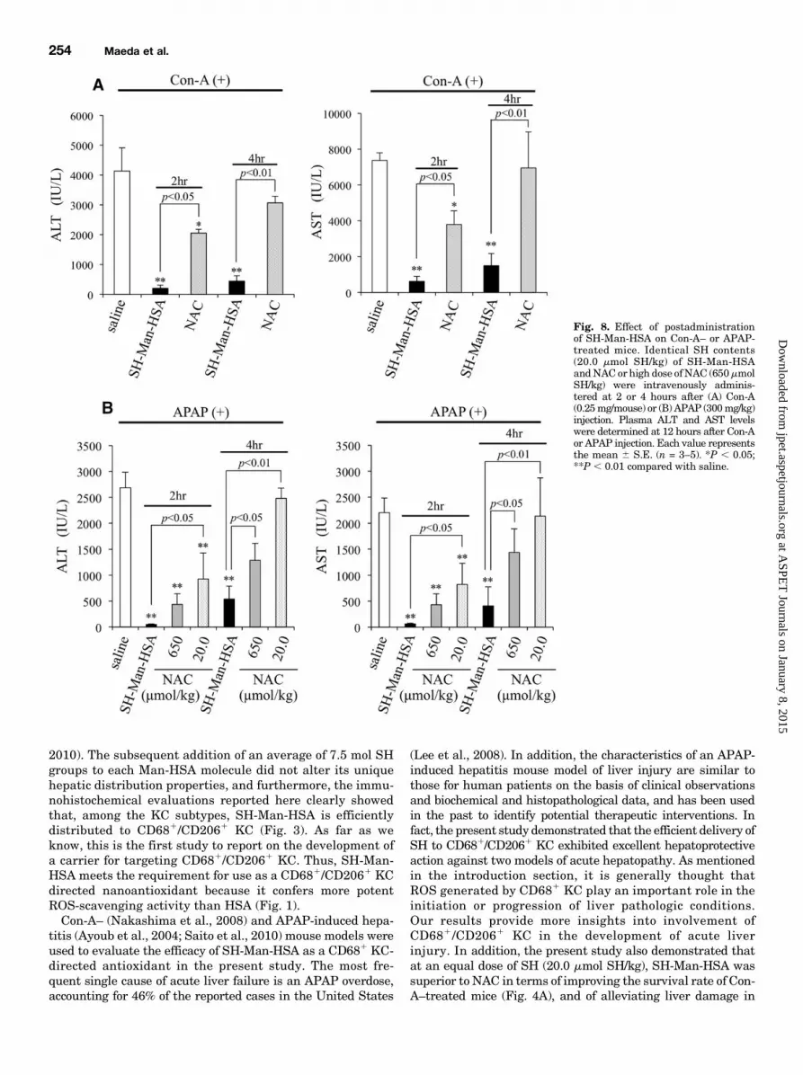
SH-Man-HSA as a rescue therapy after Con-A or APAP chal-lenges, equal SH contents (20.0 mmol SH/kg) of SH-Man-HSAor NACwere administered 2 and 4 hours following an injectionof Con-A (intravenously) or APAP (intraperitoneally).
As shown in Fig. 8, the administration of SH-Man-HSA2 hours after Con-A or APAP injection largely inhibited theelevation in ALT and AST levels. In addition, even 4 hoursafter the Con-A or APAP injection, SH-Man-HSA significantly
Fig. 4. Survival and hepatoprotective effect of SH-Man-HSA on lethal and low dose of Con-A–treated mice. (A) Identical SH contents (20.0 mmol SH/kg)of SH-Man-HSA and NAC were administered intravenously just prior to lethal dose of Con-A (1.0 mg/mouse i.v.) injection (n = 10–15). **P , 0.01compared with saline; †P, 0.05 compared with NAC. (B) Just prior to low dose of Con-A (0.25 mg/mouse), 5.0, 10.0, and 20.0 mmol SH/kg SH-Man-HSAand 20.0 mmol SH/kg NAC were administered intravenously, and GdCl3 (200 mg/mouse) was administered intravenously at 24 hours prior to the Con-Ainjection. Plasma ALT and AST levels were determined at 12 hours after Con-A injection. Each value represents the mean 6 S.E. (n = 4–6). **P , 0.01compared with saline; ††P, 0.01 compared with NAC. (C) H&E and TUNEL staining (original magnification, 100� and 200�, respectively) of mice liverwere performed at 12 hours after the Con-A injection. (D) Just prior to the Con-A injection, 2.66 mmol SH/kg of Man-HSA or 20.0 mmol SH/kg of SH-HSAwas administered intravenously. Each value represents the mean 6 S.E. (n = 3). **P , 0.01 compared with saline.
250 Maeda et al.
at ASPE
T Journals on January 8, 2015
jpet.aspetjournals.orgD
ownloaded from
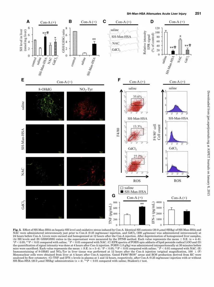
Fig. 5. Effect of SH-Man-HSA on hepatic SH level and oxidative stress induced by Con-A. Identical SH contents (20.0 mmol SH/kg) of SH-Man-HSA andNAC were administered intravenously just prior to Con-A (0.25 mg/mouse) injection, and GdCl3 (200 mg/mouse) was administered intravenously at24 hours before Con-A. Livers were excised and homogenized at 12 hours after the Con-A injection. After deproteination of homogenized liver samples,(A) SH levels and (B) GSH/GSSG ratios in the supernatant were measured by the DTNB method. Each value represents the mean 6 S.E. (n = 4–6).*P, 0.05; **P, 0.01 compared with saline; ††P, 0.01 compared with NAC. (C) EPR spectra of POBN spin adducts of lipid peroxide radical LOO_and (D)the quantification of signal intensity was done at 4 hours after Con-A injection. POBN (1.0 g/kg) was administered intraperitoneally at 30minutes beforemice were sacrificed. Each value represents the mean 6 S.E. (n = 3–4). *P, 0.05; **P, 0.01 compared with saline; ††P, 0.01 compared with NAC. (E)Immunostaining of 8-OHdG and NO2-Tyr in liver tissue was performed at 12 hours after the Con-A injection (original magnification, 100�). (F)Mononuclear cells were obtained from liver at 4 hours after Con-A injection. Gated F4/80+/ROS+ areas and ROS production derived from KC wereanalyzed by flow cytometry. (G) TNF and IFN-g levels in plasma at 1 and 12 hours, respectively, after Con-A (0.25 mg/mouse) injection with or withoutSH-Man-HSA (20.0 mmol SH/kg) administration (n = 4). **P , 0.01 compared with saline; Student’s t test.
SH-Man-HSA Attenuates Acute Liver Injury 251
at ASPE
T Journals on January 8, 2015
jpet.aspetjournals.orgD
ownloaded from

reduced the elevation of ALT and AST. In contrast, theadministration of NAC 2 or 4 hours later did not exhibitsufficient hepatoprotective action in either of the models.A previous study reported that the administration of a high
dose (650 mmol SH/kg) of NAC (intravenously) at 1.5 hoursafter an APAP injection (300mg/kg i.p.) exhibited a significanthepatoprotective action (Saito et al., 2010). Thus, the effect of650 mmol SH/kg of NAC on APAP-induced hepatic injury at2 or 4 hours after an APAP injection (300 mg/kg i.p.) was alsoexamined. At 2 hours following the APAP injection, a highdose of NAC showed a greater suppressive effect on ALT (84%)and AST (80%) elevation than a low dose (20.0 mmol SH/kg), asshown in Fig. 8B. However, a high dose of NAC showed onlya small inhibitory effect against ALT and AST elevations at4 hours after the APAP injection. These data indicate thatSH-Man-HSA was a more effective agent for treating theseexperimental acute liver injuries compared with NAC.To confirm whether the hepatoprotective action derived
from the postadministration of SH-Man-HSA resulted fromthe inhibition of hepatic oxidative stress as well as thepretreatment, we carried out histopathological and oxidativestress analyses after a post-treatment with SH-Man-HSA to
Con-A– and APAP-induced hepatopathy models. The liversections of H&E and the 8-OHdG staining of the SH-Man-HSA treatment at 2 hours after the Con-A and APAP injectionsclearly showed that SH-Man-HSA effectively inhibited liverdamage and hepatic oxidative stress (Supplemental Fig. 10),confirming that, even though a post-treatment, SH-Man-HSAshowed hepatoprotective action via the inhibition of hepaticoxidative stress similar to that observed for the pretreatment.
DiscussionThe main goal of the present study was to design a system
for delivering a nanoantioxidant containing SH groups to KC,especially CD681/CD2061, using mannose as a recognitionelement for use as a rescue therapeutic agent in treating variousacute liver injuries. HSA is a simple protein and contains nooligosaccharide chain structures. However, the insertion of aconsensus sequence for an oligosaccharide chain into the al-bumin gene (D63N, A320T, D494N) results in a protein thatcontains an oligosaccharide chain, as in some reported geneticvariants (Minchiotti et al., 2001). The pharmacokinetic proper-ties and biologic activities of these glycosylated mutants of HSA
Fig. 6. Hepatoprotective effect of SH-Man-HSA on APAP-treated mice. Just prior to APAP injection (300 mg/kg) 5.0, 10.0, and 20.0 mmol SH/kgSH-Man-HSA and 20.0 mmol SH/kg NAC were administered intravenously, and GdCl3 (200 mg/mouse) was administered intravenously at 24 hoursbefore APAP injection. (A) Plasma ALT and AST levels were determined at 12 hours after APAP injection. Each value represents the mean 6 S.E.(n = 4–6). **P , 0.01 compared with saline; ††P , 0.01 compared with NAC. (B) H&E and TUNEL staining (original magnification, 100� and 200�,respectively) of mouse liver tissue were performed at 12 hours after APAP injection.
252 Maeda et al.
at ASPE
T Journals on January 8, 2015
jpet.aspetjournals.orgD
ownloaded from
are similar to the wild-type molecule. Yeast expression systemswere used to produce the Man-HSA because generally glyco-proteins that are expressed in yeast possess high mannose-typeglycan chains, which are different from those expressed bymammalian cells. We previously showed that Man-HSA is
suitable for use as a drug delivery system carrier in targetingKC owing to its pharmacokinetic properties, which include itsrapid and efficient distribution to the liver and the fact thatit is selectively distributed to KC by CD206, unlike otherchemically modified mannosylated carriers (Hirata et al.,
Fig. 7. Effect of SH-Man-HSA on hepatic SH level and oxidative stress induced by APAP. Identical SH contents (20.0 mmol SH/kg) of SH-Man-HSA andNAC were administered intravenously just prior to the APAP (300 mg/kg) injection, and GdCl3 (200 mg/mouse) was administered intravenously justprior to the APAP injection. Livers were excised and homogenized at 12 hours after APAP injection. After deproteination of homogenized liver, (A) SHlevel and (B) GSH/GSSG ratio in supernatant was measured by DTNB method. Each value represents the mean6 S.E. (n = 4–6). *P, 0.05; **P, 0.01compared with saline; ††P, 0.01 compared with NAC. (C) EPR spectra of POBN spin adducts of lipid peroxide radical LOO_and (D) the quantification ofsignal intensity were determined at 4 hours after the APAP injection. POBN (1.0 g/kg) was administered intraperitoneally at 30 minutes before sacrificeof the mice. Each value represents the mean 6 S.E. (n = 3–4). **P , 0.01 compared with saline; ††P , 0.01 compared with NAC. Immunostaining of (E)8-OHdG and NO2-Tyr in liver tissue was performed at 12 hours after APAP injection (original magnification, 200�). (F) Hepatic mononuclear cells wereobtained from liver at 4 hours after APAP injection. Gated F4/80+/ROS+ areas and ROS production derived from KC were analyzed by flow cytometry.
SH-Man-HSA Attenuates Acute Liver Injury 253
at ASPE
T Journals on January 8, 2015
jpet.aspetjournals.orgD
ownloaded from
2010). The subsequent addition of an average of 7.5 mol SHgroups to each Man-HSA molecule did not alter its uniquehepatic distribution properties, and furthermore, the immu-nohistochemical evaluations reported here clearly showedthat, among the KC subtypes, SH-Man-HSA is efficientlydistributed to CD681/CD2061 KC (Fig. 3). As far as weknow, this is the first study to report on the development ofa carrier for targeting CD681/CD2061 KC. Thus, SH-Man-HSA meets the requirement for use as a CD681/CD2061 KCdirected nanoantioxidant because it confers more potentROS-scavenging activity than HSA (Fig. 1).Con-A– (Nakashima et al., 2008) and APAP-induced hepa-
titis (Ayoub et al., 2004; Saito et al., 2010) mouse models wereused to evaluate the efficacy of SH-Man-HSA as a CD681 KC-directed antioxidant in the present study. The most fre-quent single cause of acute liver failure is an APAP overdose,accounting for 46% of the reported cases in the United States
(Lee et al., 2008). In addition, the characteristics of an APAP-induced hepatitis mouse model of liver injury are similar tothose for human patients on the basis of clinical observationsand biochemical and histopathological data, and has been usedin the past to identify potential therapeutic interventions. Infact, the present study demonstrated that the efficient delivery ofSH to CD681/CD2061 KC exhibited excellent hepatoprotectiveaction against two models of acute hepatopathy. As mentionedin the introduction section, it is generally thought thatROS generated by CD681 KC play an important role in theinitiation or progression of liver pathologic conditions.Our results provide more insights into involvement ofCD681/CD2061 KC in the development of acute liverinjury. In addition, the present study also demonstrated thatat an equal dose of SH (20.0 mmol SH/kg), SH-Man-HSA wassuperior to NAC in terms of improving the survival rate of Con-A–treated mice (Fig. 4A), and of alleviating liver damage in
Fig. 8. Effect of postadministrationof SH-Man-HSA on Con-A– or APAP-treated mice. Identical SH contents(20.0 mmol SH/kg) of SH-Man-HSAandNAC or high dose ofNAC (650mmolSH/kg) were intravenously adminis-tered at 2 or 4 hours after (A) Con-A(0.25mg/mouse) or (B) APAP (300mg/kg)injection. Plasma ALT and AST levelswere determined at 12 hours after Con-Aor APAP injection. Each value representsthe mean 6 S.E. (n = 3–5). *P , 0.05;**P , 0.01 compared with saline.
254 Maeda et al.
at ASPE
T Journals on January 8, 2015
jpet.aspetjournals.orgD
ownloaded from
Con-A– and APAP-induced hepatitis models as evidenced by thesuppression of ALT and AST (Figs. 4B and 6A) and H&Estaining (Figs. 4C and 6B). Because the hepatoprotective effect ofMan-HSA and SH-HSAwere not comparable to that of SH-Man-HSA (Fig. 4D), this suggests that bothmannose residues and SHare essential for the therapeutic effect of SH-Man-HSA againsta hepatic injury. The hepatoprotective effect observed forSH-Man-HSAwas similar to that for aGdCl3 pretreatment (Figs.4B and 6A), indicating that SH-Man-HSA, when delivered toCD681KC, could efficiently scavenge ROS generated in responseto Con-A or APAP. In addition, excellent ROS-scavenging activitywas evident for SH-Man-HSA, as evidenced by in vivo EPR, flowcytometry, and oxidative or nitrative stress markers(8-OHdG, NO2-Tyr) (Figs. 5, C–F, and 7, C–F).On the other hand, it is well known that Con-A activates
T cells to produce IFN-g via binding to T-cell receptors. IFN-g,in turn, activates CD681 KC to produce TNF that interactswith the TNF receptor on the CD681 KC to produce ROS(Nakashima et al., 2008; Kinoshita et al., 2010). The possibilitythat the ROS-scavenging activity of SH-Man-HSA might bemediated via the inhibition of TNF or IFN-g cannot be com-pletely excluded. Interestingly, SH-Man-HSA levels did notdecrease to the levels of these inflammatory factors. This resultindicates that SH-Man-HSA attenuates Con-A–inducedhepatopathy regardless of the levels of these inflammatoryfactors (Fig. 5G). Therefore, SH-Man-HSA exhibited itshepatoprotective effect, not by influencing TNF or IFN-gproduction, but by suppressing ROS production producedby CD681/CD2061 KC, which occurs downstream from theproduction of these cytokines. Similar results were reportedfor other antioxidants, such as edarabone and lecithinizedsuperoxide dismutase, in Con-A–induced hepatitis (Nakashimaet al., 2008).There is a possibility that mannose residues or HSA mol-
ecules of SH-Man-HSA directly interact with the injectedCon-A molecules, thus leading to suppression of the toxicity ofCon-A. If this is correct, TNF and IFN-g, which are involvedin the development of Con-A–induced hepatopathy, shouldnot be increased after the coadministration of Con-A andSH-Man-HSA. However, we found similar elevations of theseinflammatory factors between Con-A injection and coadmin-istration of SH-Man-HSA and Con-A, indicating that Con-Astill preserved the potency that causes the induction of hepaticdamage when SH-Man-HSA and Con-A were coadministrated.This clearly excludes the possibility that mannose residues orHSA of SH-Man-HSA directly inactivate the hepatic toxicity ofCon-A. In fact, the postadministration of SH-Man-HSA alsoameliorated the Con-A–induced liver injury via the mechanismsimilar to that of the pretreatment (Supplemental Fig. 10).The role of ROS in APAP toxicity has been a subject of
debate for decades and is not without its controversies (Jameset al., 2003). However, the pathologic role of ROS has not beenfully understood because its effects can vary, depending onexperimental conditions. For example, in their comprehensivereview of that drug’s toxicity, Jaeschke et al. (2012) mentionedthat ROS can induce an inflammatory response in immunecells, including KC, and these extracellularly generated oxi-dants can diffuse into hepatocytes and trigger various types ofmitochondrial dysfunction and oxidant stress, which sub-sequently induce cell death. In addition, Botta et al. (2006)commented on the importance of glutathione synthesis byglutamate-cysteine ligase against APAP-induced liver injury
using glutamate-cysteine ligase transgenic mice. On the otherhand, Lewerenz et al. (2003) reported that antioxidantsprotect primary rat hepatocyte cultures against APAP-induced DNA strand breaks but not against APAP-inducedcytotoxicity. Moreover, the knockout of Cu, Zn-superoxidedismutase, a major intracellular antioxidant enzyme, wasreported to be resistant to APAP-induced hepatotoxicity (Leiet al., 2006). In this study, we demonstrated that both the pre-and postadministration of SH-Man-HSA that targets ROSderived from CD681/CD2061 KC significantly improve theCon-A– and APAP-induced hepatopathy mouse model via theinhibition of hepatic oxidative stress. This implies that ROSderived from CD681/CD2061 KC could be responsible for thedevelopment of liver damage in both disease models. Inaddition, we found that the SH-Man-HSA treatment did notsignificantly influence the early metabolic pathway of APAP-induced liver injury (data not shown). A similar result wasalso reported by Michael et al. (1999), that is, a pretreatmentof GdCl3 prevented APAP-induced hepatopathy without adecrease in the APAP-protein adducts. These findings indicatethat ROS derived from CD681/CD2061 KC are probably notinvolved in the early event of APAP toxicity and that SH-Man-HSA functions at the stage after the formation of N-acetyl-p-benzoquinone imine (NAPQI). Moreover, hypoxia-induciblefactor-1a (HIF-1a) is a critical transcription factor in responseto oxidative stress (James et al., 2006). It has been revealedthat ROS are important contributors to the early HIF-1astabilization that enhances APAP toxicity (Sparkenbaugh et al.,2011). Since NAC prevented HIF-1a accumulation via theinactivation of the NAPQI formation, it would be interesting toknow whether SH-Man-HSA also inhibits the stabilization ofHIF-1a or whether ROS derived from CD681/CD2061 KC areinvolved in the HIF-1a accumulation. As mentioned above,SH-Man-HSA does not significantly influence the early meta-bolic pathway of APAP toxicity, SH-Man-HSA or ROS derivedfrom KC may not be directly involved in the HIF-1a accumu-lation. These findings led us to conclude that, even though theearly events of the onset of acute liver injury are differentdepending on the disease, ROS derived fromCD681/CD2061KCmight be a common contributor for the development of acuteliver injury. Future investigations will be necessary to clarifythese interesting issues in terms of understanding the pathologicrole of ROS derived from CD681/CD2061 KC. In addition,CD206has been frequently used as amarker ofM2macrophagesthat are a type of anti-inflammatory immune cell (Choi et al.,2010). Thus, it is possible that SH-Man-HSA inhibits hepaticinjury via an increase in the population of M2 macrophages.However, SH-Man-HSA did not significantly influence thebalance of M1/M2 macrophages in both disease models(Supplemental Fig. 11). This indicates that the hepatopro-tective effect of SH-Man-HSA is not attributable to thechanges in the polarization of macrophages in liver.Clinically, NAC is only effective if given to patients the first
few hours after ingestion of an overdose of APAP. In the caseof the APAP hepatotoxicity mouse model, NAC was reportedto be the least beneficial if administered 3 or 4 hours after APAPinjection (Saito et al., 2010). Consistent with previous reports,we also found that, even at a high dose (650 mmol SH/kg), NACwas no longer effective at 4 hours after APAP injection (Fig. 8B).Surprisingly, 20.0 mmol SH/kg of SH-Man-HSA, a dose that wasapproximately 33 times less than theNACdose (650mmol SH/kg),exhibited a significant hepatoprotective effect, even though it
SH-Man-HSA Attenuates Acute Liver Injury 255
at ASPE
T Journals on January 8, 2015
jpet.aspetjournals.orgD
ownloaded from
was administered 4 hours later in both pathologic models. Invivo EPR data clearly showed that ROS were mainly generatedin the liver at 4 to 5 hours after the injection of Con-A or APAPand was completely inhibited by SH-Man-HSA or GdCl3.Taking into account the rapid and efficient delivery of SH intoCD681/CD2061 KC by SH-Man-HSA (Fig. 3) and the elevationin the hepatic SH content (Figs. 5A and 7A), these findingssuggest that the difference in the therapeutic effect betweenSH-Man-HSA and NAC given after an injection of Con-A orAPAP could be attributable to the difference in the rate and siteof distribution to the liver, especially CD681/CD2061 KC,between these two drugs.The issue of whether the recovery of the hepatic SH content
to control levels by SH-Man-HSA can be attributed to the SHmoieties in SH-Man-HSA or suppression of the consumptionof GSH, an intracellular substance with SH, or both, remainsunknown. In contrast to the hepatic SH content, the hepaticGSH/GSSG ratio was not recovered to the control level as theresult of the SH-Man-HSA treatment (Figs. 5B and 7B). Suchan inconsistency might explain why, initially, GSH is con-sumed as the result of a Con-A or APAP challenge, and then SHderived from SH-Man-HSA makes up for the deficit in hepaticSH levels. In fact, immunohistochemical analyses (Figs. 2 and 3)and the SH levels in liver homogenates (Figs. 5A and 7A)are supportive of the hypothesis that SH liberated fromSH-Man-HSA may still be retained within the liver, butnot CD681 KC, at 12 hours after SH-Man-HSA adminis-tration (Fig. 3C). SH-Man-HSA would be hydrolyzed in lyso-somes of CD681/CD2061KCafter endocytosis byCD206, and theSH groups would then be liberated from SH-Man-HSAbecause 2-iminothiolane, the SH labeling agent for HSAused in this study, is also hydrolyzed at a low pH, such aspH 5 (Singh et al., 1996). These SH groups then move to theoutside of the CD681/CD2061 KC and would contribute tothe elevation of the hepatic SH levels in disease models. There-fore, SH-Man-HSA is not only able to scavenge ROS in the livermore efficiently than NAC, but also ROS-scavenging activity isretained in the liver longer than NAC. It might be possible totreat a liver impairment with SH-Man-HSA at a lower dose anda lesser dosing frequency than is needed for NAC.In conclusion, SH-Man-HSA is a unique and powerful nano-
antioxidant that targetsROSderived fromCD681/CD2061KCbe-cause of its efficient and rapid delivery of SH toCD681/CD2061KC.SH-Man-HSA exhibited an excellent hepatoprotective actionagainst two experimental acute hepatitis models that wassuperior to NAC because of its sufficient suppression ofoxidative stress. Therefore, it has great potential for use as arescue therapy for acute hepatopathy.Moreover, the therapeuticimpact of SH-Man-HSA in chronic hepatic diseases in whichCD681 KC contribute to the onset and progression of diseases,such as in nonalcoholic steatohepatitis (Chedid et al., 2004)and alcoholic hepatitis (Gadd et al., 2014), deserves furtherexamination.
Authorship Contributions
Participated in research design:Maeda, Hirata, Watanabe, Ishima,Inatsu, Kinoshita, Otagiri, Maruyama.
Conducted experiments: Maeda, Hirata, Tanaka, Sasaki.Contributed new reagents or analytic tools: Maeda, Watanabe,
Ishima, Inatsu, Kinoshita, Tanaka, Sasaki, Maruyama.Performed data analysis: Maeda, Watanabe, Ishima, Inatsu,
Kinoshita, Tanaka, Sasaki, Maruyama.
Wrote or contributed to the writing of the manuscript: Maeda,Watanabe, Ishima, Chuang, Taguchi, Kinoshita, Tanaka, Sasaki,Otagiri, Maruyama.
References
Ayoub SS, Botting RM, Goorha S, Colville-Nash PR, Willoughby DA, and Ballou LR(2004) Acetaminophen-induced hypothermia in mice is mediated by a prostaglan-din endoperoxide synthase 1 gene-derived protein. Proc Natl Acad Sci USA 101:11165–11169.
Botta D, Shi S, White CC, Dabrowski MJ, Keener CL, Srinouanprachanh SL, FarinFM, Ware CB, Ladiges WC, Pierce RH, et al. (2006) Acetaminophen-induced liverinjury is attenuated in male glutamate-cysteine ligase transgenic mice. J BiolChem 281:28865–28875.
Chedid A, Arain S, Snyder A, Mathurin P, Capron F, and Naveau S (2004) Theimmunology of fibrogenesis in alcoholic liver disease. Arch Pathol Lab Med 128:1230–1238.
Choi KM, Kashyap PC, Dutta N, Stoltz GJ, Ordog T, Shea Donohue T, Bauer AJ,Linden DR, Szurszewski JH, Gibbons SJ, and Farrugia G (2010) CD206-positiveM2 macrophages that express heme oxygenase-1 protect against diabetic gastro-paresis in mice. Gastroenterology 138:2399–2409.
Dobashi H, Seki S, Habu Y, Ohkawa T, Takeshita S, Hiraide H, and Sekine I (1999)Activation of mouse liver natural killer cells and NK1.1(1) T cells by bacterialsuperantigen-primed Kupffer cells. Hepatology 30:430–436.
Gadd VL, Skoien R, Powell EE, Fagan KJ, Winterford C, Horsfall L, Irvine K,and Clouston AD (2014) The portal inflammatory infiltrate and ductular reactionin human non-alcoholic fatty liver disease. Hepatology 59:1393–1405.
Hirata K, Maruyama T, Watanabe H, Maeda H, Nakajou K, Iwao Y, Ishima Y,Katsumi H, Hashida M, and Otagiri M (2010) Genetically engineeredmannosylated-human serum albumin as a versatile carrier for liver-selectivetherapeutics. J Control Release 145:9–16.
Hnatowich DJ, Layne WW, and Childs RL (1982) The preparation and labeling ofDTPA-coupled albumin. Int J Appl Radiat Isot 33:327–332.
Hu W, Jiang Z, Zhang Y, Liu Q, Fan J, Luo N, Dong X, and Yu X (2012) Charac-terization of infiltrating macrophages in high glucose-induced peritoneal fibrosis inrats. Mol Med Rep 6:93–99.
Jaeschke H (2011) Reactive oxygen and mechanisms of inflammatory liver injury:Present concepts. J Gastroenterol Hepatol 26 (Suppl 1):173–179.
Jaeschke H, McGill MR, and Ramachandran A (2012) Oxidant stress, mitochondria,and cell death mechanisms in drug-induced liver injury: lessons learned fromacetaminophen hepatotoxicity. Drug Metab Rev 44:88–106.
James LP, Mayeux PR, and Hinson JA (2003) Acetaminophen-induced hepatotoxic-ity. Drug Metab Dispos 31:1499–1506.
James LP, Donahower B, Burke AS, McCullough S, and Hinson JA (2006) Inductionof the nuclear factor HIF-1alpha in acetaminophen toxicity: evidence for oxidativestress. Biochem Biophys Res Commun 343:171–176.
Katayama N, Nakajou K, Komori H, Uchida K, Yokoe J, Yasui N, Yamamoto H, KaiT, Sato M, Nakagawa T, et al. (2008) Design and evaluation of S-nitrosylatedhuman serum albumin as a novel anticancer drug. J Pharmacol Exp Ther 325:69–76.
Kinoshita M, Uchida T, Sato A, Nakashima M, Nakashima H, Shono S, Habu Y,Miyazaki H, Hiroi S, and Seki S (2010) Characterization of two F4/80-positiveKupffer cell subsets by their function and phenotype in mice. J Hepatol 53:903–910.
Kogelberg H, Tolner B, Sharma SK, Lowdell MW, Qureshi U, Robson M, Hillyer T,Pedley RB, Vervecken W, Contreras R, et al. (2007) Clearance mechanism ofa mannosylated antibody-enzyme fusion protein used in experimental cancertherapy. Glycobiology 17:36–45.
Korenaga M, Wang T, Li Y, Showalter LA, Chan T, Sun J, and Weinman SA (2005)Hepatitis C virus core protein inhibits mitochondrial electron transport andincreases reactive oxygen species (ROS) production. J Biol Chem 280:37481–37488.
Kragh-Hansen U, Donaldson D, and Jensen PH (2001) The glycan structure of al-bumin Redhill, a glycosylated variant of human serum albumin. Biochim BiophysActa 1550:20–26.
Laskin DL and Laskin JD (2001) Role of macrophages and inflammatory mediatorsin chemically induced toxicity. Toxicology 160:111–118.
Laskin DL, Gardner CR, Price VF, and Jollow DJ (1995) Modulation of macrophagefunctioning abrogates the acute hepatotoxicity of acetaminophen. Hepatology 21:1045–1050.
Lee WM, Squires RH, Jr, Nyberg SL, Doo E, and Hoofnagle JH (2008) Acute liverfailure: Summary of a workshop. Hepatology 47:1401–1415.
Lei XG, Zhu JH, McClung JP, Aregullin M, and Roneker CA (2006) Mice deficient inCu,Zn-superoxide dismutase are resistant to acetaminophen toxicity. Biochem J399:455–461.
Lewerenz V, Hanelt S, Nastevska C, El-Bahay C, Röhrdanz E, and Kahl R (2003)Antioxidants protect primary rat hepatocyte cultures against acetaminophen-induced DNA strand breaks but not against acetaminophen-induced cytotoxicity.Toxicology 191:179–187.
McGreal EP, Rosas M, Brown GD, Zamze S, Wong SY, Gordon S, Martinez-PomaresL, and Taylor PR (2006) The carbohydrate-recognition domain of Dectin-2 is aC-type lectin with specificity for high mannose. Glycobiology 16:422–430.
Michael SL, Pumford NR, Mayeux PR, Niesman MR, and Hinson JA (1999) Pre-treatment of mice with macrophage inactivators decreases acetaminophen hepa-totoxicity and the formation of reactive oxygen and nitrogen species. Hepatology30:186–195.
Minchiotti L, Campagnoli M, Rossi A, Cosulich ME, Monti M, Pucci P, Kragh-HansenU, Granel B, Disdier P, Weiller PJ, et al. (2001) A nucleotide insertion andframeshift cause albumin Kénitra, an extended and O-glycosylated mutant of
256 Maeda et al.
at ASPE
T Journals on January 8, 2015
jpet.aspetjournals.orgD
ownloaded from
human serum albumin with two additional disulfide bridges. Eur J Biochem 268:344–352.
Moreno-Otero R (2013) May oxidative stress contribute to autoimmune hepatitispathogenesis, and can antioxidants be of value as adjuvant therapy for refractorypatients? Dig Dis Sci 58:1440–1441.
Nakashima H, Kinoshita M, Nakashima M, Habu Y, Shono S, Uchida T, ShinomiyaN, and Seki S (2008) Superoxide produced by Kupffer cells is an essential effectorin concanavalin A-induced hepatitis in mice. Hepatology 48:1979–1988.
Roberts RA, Ganey PE, Ju C, Kamendulis LM, Rusyn I, and Klaunig JE (2007) Roleof the Kupffer cell in mediating hepatic toxicity and carcinogenesis. Toxicol Sci 96:2–15.
Sato K, Kadiiska MB, Ghio AJ, Corbett J, Fann YC, Holland SM, Thurman RG,and Mason RP (2002) In vivo lipid-derived free radical formation by NADPH oxi-dase in acute lung injury induced by lipopolysaccharide: a model for ARDS. FASEBJ 16:1713–1720.
Saito C, Zwingmann C, and Jaeschke H (2010) Novel mechanisms of protectionagainst acetaminophen hepatotoxicity in mice by glutathione and N-acetylcysteine.Hepatology 51:246–254.
Singh R, Kats L, Blättler WA, and Lambert JM (1996) Formation of N-substituted2-iminothiolanes when amino groups in proteins and peptides are modified by2-iminothiolane. Anal Biochem 236:114–125.
Sparkenbaugh EM, Saini Y, Greenwood KK, LaPres JJ, Luyendyk JP, Copple BL,Maddox JF, Ganey PE, and Roth RA (2011) The role of hypoxia-inducible factor-1ain acetaminophen hepatotoxicity. J Pharmacol Exp Ther 338:492–502.
Sun H, Xu XY, Shao HT, Su X, Wu XD, Wang Q, and Shi Y (2013) Dectin-2 ispredominately macrophage restricted and exhibits conspicuous expression duringAspergillus fumigatus invasion in human lung. Cell Immunol 284:60–67.
Tsukamoto H and Lu SC (2001) Current concepts in the pathogenesis of alcoholicliver injury. FASEB J 15:1335–1349.
Address correspondence to: Dr. Toru Maruyama, Department of Biophar-maceutics, Graduate School of Pharmaceutical Sciences, Kumamoto Univer-sity, 5-1 Oe-honmachi, Kumamoto 862-0973, Japan. E-mail: [email protected]
SH-Man-HSA Attenuates Acute Liver Injury 257
at ASPE
T Journals on January 8, 2015
jpet.aspetjournals.orgD
ownloaded from