
2 R )-2-Benzenesulfonamido-2-phenylethanoic acid: a new monoclinic polymorph
Upload
independentCategory
view
1download
0
Polymerized Complex SolGel Synthesis, Structural and OpticalProperties of Monoclinic Eu3+DopedKGd(WO4)2 Crystalline RedPhosphorsD. Thangaraju, A. Durairajan, S. Moorthy Babu, and Y. Hayakawa Citation: AIP Conf. Proc. 1391, 54 (2011); doi: 10.1063/1.3646778 View online: http://dx.doi.org/10.1063/1.3646778 View Table of Contents: http://proceedings.aip.org/dbt/dbt.jsp?KEY=APCPCS&Volume=1391&Issue=1 Published by the American Institute of Physics. Related ArticlesMechanical constraints enhance electrical energy densities of soft dielectrics Appl. Phys. Lett. 99, 171906 (2011) Liquid pearls Phys. Fluids 23, 091108 (2011) Non-positional cell microarray prepared by shape-coded polymeric microboards: A new microarray format formultiplex and high throughput cell-based assays Biomicrofluidics 5, 032001 (2011) The vibrational density of states of a disordered gel model J. Chem. Phys. 135, 104502 (2011) Effect of surface composition and roughness on the apparent surface free energy of silica aerogel materials Appl. Phys. Lett. 99, 104104 (2011) Additional information on AIP Conf. Proc.Journal Homepage: http://proceedings.aip.org/ Journal Information: http://proceedings.aip.org/about/about_the_proceedings Top downloads: http://proceedings.aip.org/dbt/most_downloaded.jsp?KEY=APCPCS Information for Authors: http://proceedings.aip.org/authors/information_for_authors
Downloaded 13 Nov 2011 to 117.240.242.163. Redistribution subject to AIP license or copyright; see http://proceedings.aip.org/about/rights_permissions
Polymerized Complex Sol-Gel Synthesis, Structural and Optical Properties of Monoclinic Eu3+ Doped KGd(WO4)2
Crystalline Red Phosphors
D. Thangaraju1, A. Durairajan1, S. Moorthy Babu1* and Y.Hayakawa2
1Crystal Growth Centre, Anna University, Chennai – 600 025, India 2 Research Institute of Electronics, Shizuoka University, 3-5-1 Johoku, Hamamatsu, Shizuoka 432-8011, Japan
*E-mail: [email protected], [email protected]
Abstract. 1% Eu3+ doped KGd(WO4)2 (KGW) was synthesized through Pechini sol-gel process and crystallized by subsequent annealing at high temperature. Potassium nitrate, gadolinium nitrate and ammonium para tungstate precursors were mixed with citric acid and ethylene glycol to synthesis the polymerizable complex gel. The gel was heated to 250 °C for decomposition of polymer, which after the brownish white powder was used to synthesis the pure form of 1% Eu:KGW. The pre-fired powder was further heated at high temperature’s (550, 600, 650 and 700 °C) for calcination. The properties of heat treated samples were characterized by powder XRD, FT-IR, Raman, FESEM and fluorescence analysis to understand the crystallinity, organic liberation, tungstate ribbon formation, surface morphology and emission nature, respectively. Phase evaluation from the amorphous pre-fired sample to well crystalline KGW powder formation was confirmed with powder XRD analysis. Powders calcined at 600 °C show the appearance of monoclinic phase of KGW. Crystalline peaks without intermediate compound peaks were observed for samples calcined at 700 °C. Gel degradation and formation of double tungstate was clearly seen in the FT-IR spectrum. FT-IR spectrum of synthesized gel also, confirms the citrate formation and etherification. FESEM analysis reveals the size and morphology of the powder. Double tungstate formation from the amorphous powder was analyzed using laser Raman spectral analysis. The emission property of the europium doped KGW was analyzed using fluorescence. Changes in emission intensity was observed for samples calcined at different temperatures. Keywords: Stimulated Raman scattering, sol-gel method, phosphors. PACS: 42.65.Dr, 81.20.Fw, 32.50.+d
INTRODUCTION
Double tungstates and molybtates were elaborately investigated due to its interesting luminescence properties and possible application in the solid state laser field. Monoclinic potassium based rare earth double tungstates KRE(WO4)2 (RE= Y, Gd, Lu) are well known laser host materials, its structural isotropy and biaxial nature have attached to the new applications. High third order non linear coefficient of these materials makes them as a good candidate for Stimulated Raman Scattering (SRS) applications [1]. Physical and chemical properties of these double tungstates draw additional attention towards industrial application. Possibility of doping of rare earth ions without substantial quenching in its skeleton extended its application in the phosphor field. Trivalent europium ion doped phosphors having numerous advantages. It is purely red emitting phosphor with allowed dipole transition from 5D0 to 7F1 and
informative hypersensitive transition 5D0 to 7F2. The decay time of luminescence in the emitting 5D0 level is longer [2]. The ionic radius of Eu3+ is comparable with Gd3+ based double tungstates. In these materials, KGd(WO4)2 (KGW) is a well known material for stimulated Raman scattering (SRS) because of its strong Raman interaction with two vibrational modes of different energies. Besides that, it also has good physical properties like high thermal conductivity, low thermal expansion and a high damage threshold. In addition, KGW is non-hygroscopic and has good mechanical and chemical stability [3]. The structure of the KGW is formed by the chains of WO4
2- ions along the c – axis and connected by oxygen atoms with two W–O bonds (WOW oxygen bridge bonds). The W2O8
4- dimmer ions are formed by two WO42- ions,
which are connected by four W-O bonds (WOOW double oxygen bridge bonds). The oxygen of the rest two W-O bonds are not involved in the chemical reaction with another tungstate atoms [4]. In this, the
Optics: Phenomena, Materials, Devices, and CharacterizationAIP Conf. Proc. 1391, 54-58 (2011); doi: 10.1063/1.3646778
© 2011 American Institute of Physics 978-0-7354-0960-6/$30.00
54
Downloaded 13 Nov 2011 to 117.240.242.163. Redistribution subject to AIP license or copyright; see http://proceedings.aip.org/about/rights_permissions
asymmetric stretching of the WOW bridges is responsible for intense Raman frequency shift at 901 and 768 cm-1. Third order non-linear coefficient of this material is very high and this makes it a suitable material for SRS applications [5]. These monoclinic KRE(WO4)2 (RE = Gd, Y and Lu) crystals are also excellent laser host materials. The advantages of the laser active elements doped in the Raman active crystalline materials are that it can be used to produce fundamental and Stokes lines simultaneously.
In this study, Eu3+ doped KGW nano crystals were synthesized by conventional polymeric sol-gel method and calcinations temperature depended luminescence character was analyzed. Derived powders have been characterized with structural, vibrational, morphological, luminescence analysis. The dependence of calcinations temperature on the quality and luminescence power of the derived powder was discussed.
EXPERIMENTAL PROCEDURE
This Conventional Pechini polymeric complex sol-gel method was one of the convenient methods to synthesize the multi component system with definitive homogeneity of reactants. Unlike ordinary solid state reaction, every individual reactant ions intimately mix together at atomic level with high homogeneity in this method. Hence, this method was used to synthesize monoclinic KGW which consists of three metal components (i.e. K, Gd, W).
The precursor materials taken in this synthesis were analytical grade. The starting precursors i.e. KNO3, Gd(NO3)3 and ammonium para tungstate were dissolved in 50 ml of deionised water separately. For doping, Eu(NO3)3 was synthesized from Eu2O3 with nitric acid. After the complete dissolution was achieved, the citric acid was mixed individually in the clear solution of nitrates and tungstates. Citric acid starts to react with metal ions and forms the citrate complex in the solution. For thorough mixing of individual citrates, the mixed solution was stirred for 45 minutes. Ethylene glycol was then added in the homogenously mixed citrate solution to polymerize the individual citrate complex. The total pH value of the solution was adjusted to 3.5 by adding ammonia solution. This mixed aqueous solution was heated at 80�C with uniform stirring for 4 hours to enhance the polyesterification reaction. The ratio between metal ions, citric acid and ethylene glycol was maintained as 0.9:1:1. The advantages of using lower citric acid ratio are that, the acidic pH was enough to ionize the total amount of [COOH]- for chelation. An increase in pH causes more citric acid to ionize and this lead to more carboxylic groups ready to chelate the metal ions.
High degree of chelation was responsible for high uniformity of metallic constituents in the solution [6,7]. Microwave heating was introduced for the fast evaporation of superfluous liquid molecules in the solution to control the degradation of polymer in a reversible reaction, which leads to the fast extraction of gel. Gel was found after complete evaporation of the liquid species present in the solution. The temperature was gradually increased to 110 �C for further drying and the sample was retained at this temperature for 5 hours. The obtained organic–inorganic gels were pre-fired to 250�C at a heating rate of 50�C/h, in resistive heating furnace in air. This temperature was maintained for 30 min. The pre-fired samples were further calcined at 550, 600, 650 and 700�C with a heating rate of 50�C/h. Calcination was carried out at different temperature’s to understand the luminescence nature of the samples.
RESULTS AND DISCUSSION
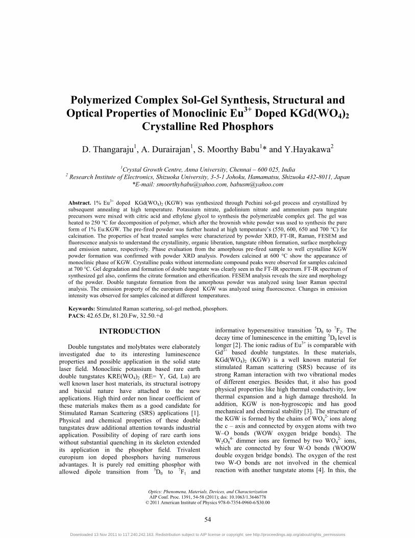
Phase analysis was carried out using Philips PW1710 diffractometer using CuKα radiation at room temperature. Powder XRD pattern of derived specimens at various calcinations temperatures were analyzed and compared in Fig. 1. Powders calcined at 700 �C shows a sharp and high intensity peaks.
FIGURE 1. Comparative Powder X-ray Diffraction patterns of gel, pre-fired and calcined samples.
The peaks observed for all 700 �C calcined powders were indexed (JCPDS Card PCPDF No: 89 – 6489) and they correspond to the low temperature phase of KGW. The evolution of monoclinic phase was observed after 600�C calcination, prefired samples at 250 and 550°C shows the presence of intermediate
55
Downloaded 13 Nov 2011 to 117.240.242.163. Redistribution subject to AIP license or copyright; see http://proceedings.aip.org/about/rights_permissions
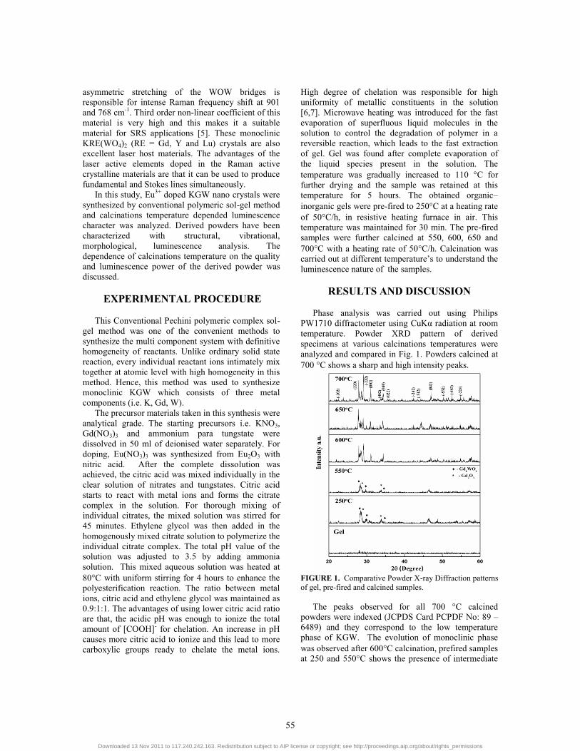
compound peaks. The pattern of dried samples shows amorphous nature. The lattice parameter values were calculated using these patterns for 700 �C calcined samples. Calculated lattice parameters are well matching with reported values. But in the case of pre-fired samples, the XRD pattern shows the high intense peaks corresponding to the intermediate oxides, like Gd2WO6 (JCPDS Card PCPDF No: 78-1708) and Gd2O3 (JCPDS Card PCPDF No: 86-2477). The peaks are indexed accordingly. These satellite peaks become weaker when the calcination temperature was increased. The FT-IR spectra for synthesized gel, pre-fired and calcined samples were recorded using JEOL WIN SPEC-50 FT-IR Spectrometer in the range of 400-4000 cm-1 (Fig. 2). Several absorption bands are observed for the polymeric resin and evaporation of polymer species was clearly observed at high temperature of calcination. The absorption peaks observed at 3000 – 3750 cm-1 belong to the stretching vibration of water and citric acid hydroxyl groups (O-H). This broad peak becomes weaker with increase in calcination temperature and the band disappears at higher temperature, (i.e) more than 700 ºC. The bands localized at 1500-1700 cm-1 and 1300-1485 cm-1 are assigned to asymmetrical and symmetrical stretching vibrations of carboxylate groups, respectively. The sharp band in the carboxylate region was observed in the spectrum. The four bands related to carboxylate stretching modes are observed at 1769, 1645, 1573 and 1389 cm-1 of the gel. These bands confirm the conversion of citric acid to citrate salt [8]. The band at 1769 cm-1 indicates the C=O stretching mode of ester which was formed between citric acid and ethylene glycol [9]. The asymmetric COO- stretching of sample appeared at 1645 and 1573 cm-1 which may be due to unidentate complex and a bridging complex (forms between the citric acid and metal ions) [8]. The band at 1389 cm-1 was attributed to the symmetric COO- stretching. This indicates the bridge formation between citric acid and metal ions by potassium and gadolinium ions [10]. Out of plane bending of the O-H appeared at 937 cm-1, which appears as little hump near the tungstate bands. The pre-fired samples show the presence of organics in the powder. All pre-fired samples also, indicate the carbonate oriented peaks at 1389 cm-1. The band at 1645 cm-1 was still present even after calcined at 700 �C. The reason for the band even at high temperature may be due to the strong chelating ability of citric acid that can stabilize the different metal ions within one molecule or complex [9]. The sample annealed at 700 ºC, show the absorption peaks corresponding to the appearance of (WO4)2-, which illustrates the formation of monoclinic KGW. The observed absorption bands at mid IR region 919, 842, 770, 627 and 473 cm-1 gives some information about tungstates. These absorption bands
agree well with the polycrystalline IR results [11, 12]. The band at 919 cm-1 was assigned for stretching of W-O and at 842 cm-1 for WOW. Stretching of double bridge WO
OW were observed at 770, 627 and 473 cm-1.
FIGURE 2. Comparative FT-IR spectrum of gel, pre-fired and calcined samples.
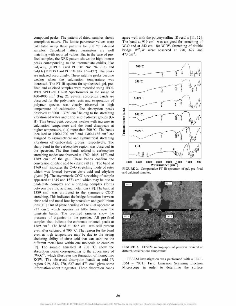
FIGURE 3. FESEM micrographs of powders derived at different calcinations temperaturs.
FESEM investigation was performed with a JEOL
JSM – 7001F Field Emission Scanning Electron Microscope in order to determine the surface
56
Downloaded 13 Nov 2011 to 117.240.242.163. Redistribution subject to AIP license or copyright; see http://proceedings.aip.org/about/rights_permissions
morphology and particle dimension. FESEM micrograph of samples 550°C pre-fired and calcined at 600, 650 and 700°C were shown in Fig 3. The FESEM image of pre-fired samples shows dimensionless spread of primary particles. There is no identification of particles with particular morphology. As mentioned in the powder XRD pattern, particle derived from 550��C may be primary particles of Gd2O3 and Gd2WO6. Powder calcined at 600�C shows the formation of shaped particles, the content of primary particles was decreased and Quadrangular shaped particles with angular edges were seen in the micro graph. The 650�C calcined sample shows further decrease of primary particles on the surface of the secondary particles and sub-angular edged particles with contorted rectangular shaped particles. High temperature calcinations lead the complete surface fusion of primary particles and formation of soft agglomerated KGW particles. The size of the particles was eventually increased at high temperatures. At high temperature calcined particles were appeared as contorted sphere with comparable size.
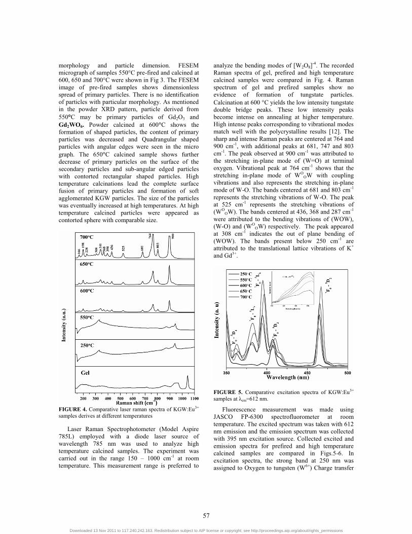
FIGURE 4. Comparative laser raman spectra of KGW:Eu3+ samples derives at different temperatures
Laser Raman Spectrophotometer (Model Aspire 785L) employed with a diode laser source of wavelength 785 nm was used to analyze high temperature calcined samples. The experiment was carried out in the range 150 – 1000 cm-1 at room temperature. This measurement range is preferred to
analyze the bending modes of [W2O8]-4. The recorded Raman spectra of gel, prefired and high temperature calcined samples were compared in Fig. 4. Raman spectrum of gel and prefired samples show no evidence of formation of tungstate particles. Calcination at 600 �C yields the low intensity tungstate double bridge peaks. These low intensity peaks become intense on annealing at higher temperature. High intense peaks corresponding to vibrational modes match well with the polycrystalline results [12]. The sharp and intense Raman peaks are centered at 764 and 900 cm-1, with additional peaks at 681, 747 and 803 cm-1. The peak observed at 900 cm-1 was attributed to the stretching in-plane mode of (W=O) at terminal oxygen. Vibrational peak at 764 cm-1 shows that the stretching in-plane mode of WO
OW with coupling vibrations and also represents the stretching in-plane mode of W-O. The bands centered at 681 and 803 cm-1 represents the stretching vibrations of W-O. The peak at 525 cm-1 represents the stretching vibrations of (WO
OW). The bands centered at 436, 368 and 287 cm-1 were attributed to the bending vibrations of (WOW), (W-O) and (WO
OW) respectively. The peak appeared at 308 cm-1 indicates the out of plane bending of (WOW). The bands present below 250 cm-1 are attributed to the translational lattice vibrations of K+ and Gd3+.
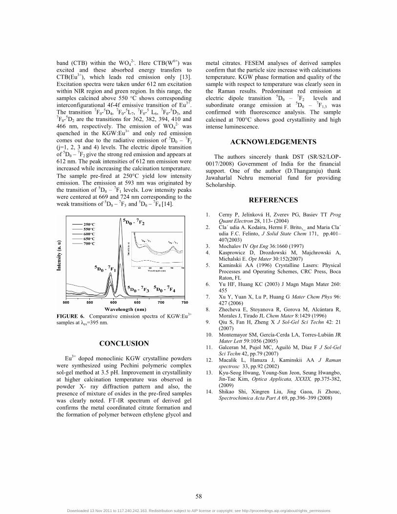
FIGURE 5. Comparative excitation spectra of KGW:Eu3+ samples at λem=612 nm.
Fluorescence measurement was made using JASCO FP-6300 spectrofluorometer at room temperature. The excited spectrum was taken with 612 nm emission and the emission spectrum was collected with 395 nm excitation source. Collected excited and emission spectra for prefired and high temperature calcined samples are compared in Figs.5-6. In excitation spectra, the strong band at 250 nm was assigned to Oxygen to tungsten (W6+) Charge transfer
57
Downloaded 13 Nov 2011 to 117.240.242.163. Redistribution subject to AIP license or copyright; see http://proceedings.aip.org/about/rights_permissions
band (CTB) within the WO42-. Here CTB(W6+) was
excited and these absorbed energy transfers to CTB(Eu3+), which leads red emission only [13]. Excitation spectra were taken under 612 nm excitation within NIR region and green region. In this range, the samples calcined above 550 °C shows corresponding interconfigurational 4f-4f emissive transition of Eu3+. The transition 7F0-5D4, 7F0-5L7, 7F0-5 L6, 7F0-5D3, and 7F0-5D2 are the transitions for 362, 382, 394, 410 and 466 nm, respectively. The emission of WO4
2- was quenched in the KGW:Eu3+ and only red emission comes out due to the radiative emission of 5D0 – 7Fj (j=1, 2, 3 and 4) levels. The electric dipole transition of 5D0 – 7F2 give the strong red emission and appears at 612 nm. The peak intensities of 612 nm emission were increased while increasing the calcination temperature. The sample pre-fired at 250�C yield low intensity emission. The emission at 593 nm was originated by the transition of 5D0 – 7F1 levels. Low intensity peaks were centered at 669 and 724 nm corresponding to the weak transitions of 5D0 – 7F3 and 5D0 – 7F4 [14].
FIGURE 6. Comparative emission spectra of KGW:Eu3+ samples at λex=395 nm.
CONCLUSION
Eu3+ doped monoclinic KGW crystalline powders were synthesized using Pechini polymeric complex sol-gel method at 3.5 pH. Improvement in crystallinity at higher calcination temperature was observed in powder X- ray diffraction pattern and also, the presence of mixture of oxides in the pre-fired samples was clearly noted. FT-IR spectrum of derived gel confirms the metal coordinated citrate formation and the formation of polymer between ethylene glycol and
metal citrates. FESEM analyses of derived samples confirm that the particle size increase with calcinations temperature. KGW phase formation and quality of the sample with respect to temperature was clearly seen in the Raman results. Predominant red emission at electric dipole transition 5D0 – 7F2 levels and subordinate orange emission at 5D0 – 7F1,3 was confirmed with fluorescence analysis. The sample calcined at 700�C shows good crystallinity and high intense luminescence.
ACKNOWLEDGEMENTS
The authors sincerely thank DST (SR/S2/LOP-0017/2008) Government of India for the financial support. One of the author (D.Thangaraju) thank Jawaharlal Nehru memorial fund for providing Scholarship.
REFERENCES
1. Cerny P, Jelínková H, Zverev PG, Basiev TT Prog Quant Electron 28, 113- (2004)
2. Cla´ udia A. Kodaira, Hermi F. Brito,_ and Maria Cla´ udia F.C. Felinto, J Solid State Chem 171, pp.401–407(2003)
3. Mochalov IV Opt Eng 36:1660 (1997) 4. Kasprowicz D, Drozdowski M, Majchrowski A,
Michalski E. Opt Mater 30:152(2007) 5. Kaminskii AA (1996) Crystalline Lasers: Physical
Processes and Operating Schemes, CRC Press, Boca Raton, FL
6. Yu HF, Huang KC (2003) J Magn Magn Mater 260: 455
7. Xu Y, Yuan X, Lu P, Huang G Mater Chem Phys 96: 427 (2006)
8. Zhecheva E, Stoyanova R, Gorova M, Alcántara R, Morales J, Tirado JL Chem Mater 8:1429 (1996)
9. Qiu S, Fan H, Zheng X J Sol-Gel Sci Techn 42: 21 (2007)
10. Montemayor SM, Gercía-Cerda LA, Torres-Lubián JR Mater Lett 59:1056 (2005)
11. Galceran M, Pujol MC, Aguiló M, Díaz F J Sol-Gel Sci Techn 42, pp.79 (2007)
12. Macalik L, Hanuza J, Kaminskii AA J Raman spectrosc 33, pp.92 (2002)
13. Kyu-Seog Hwang, Young-Sun Jeon, Seung Hwangbo, Jin-Tae Kim, Optica Applicata, XXXIX, pp.375-382, (2009)
14. Shikao Shi, Xingren Liu, Jing Gaoa, Ji Zhouc, Spectrochimica Acta Part A 69, pp.396–399 (2008)
58
Downloaded 13 Nov 2011 to 117.240.242.163. Redistribution subject to AIP license or copyright; see http://proceedings.aip.org/about/rights_permissions
Copyright © 2022 FDOKUMEN


![Phase Transitions: A Multinational Journal The structural transformation of monoclinic [(R)-C 5 H 14 N 2 ] [Cu(SO 4 ) 2 (H 2 O) 4 ]·2H 2 O into orthorhombic [(R)-C 5 H 14 N 2 ] 2](https://static.fdokumen.com/doc/165x107/6312e160fc260b71020ed6ea/phase-transitions-a-multinational-journal-the-structural-transformation-of-monoclinic.jpg)