
A second monoclinic polymorph of 2-amino-4,6-dichloropyrimidine
10
A second monoclinic polymorph of 2-amino-4,6-dichloropyrimidine Hoong-Kun Fun, a * Suchada Chantrapromma, b ‡ Subrata Jana, c Rinku Chakrabarty c and Shyamaprosad Goswami c a X-ray Crystallography Unit, School of Physics, Universiti Sains Malaysia, 11800 USM, Penang, Malaysia, b Crystal Materials Research Unit, Department of Chemistry, Faculty of Science, Prince of Songkla University, Hat-Yai, Songkhla 90112, Thailand, and c Department of Chemistry, Bengal Engineering and Science University’, Shibpur, Howrah, India 711 103 Correspondence e-mail: [email protected] Received 25 July 2008; accepted 27 July 2008 Key indicators: single-crystal X-ray study; T = 296 K; mean (C–C) = 0.003 A ˚; R factor = 0.041; wR factor = 0.098; data-to-parameter ratio = 15.4. The title chloro-substituted 2-aminopyrimidine, C 4 H 3 Cl 2 N 3 , is a second monoclinic polymorph of this compound which crystallizes in the space group C2/c. The structure was previously reported [Clews & Cochran (1948). Acta Cryst. 1, 4–11] in the space group P21/a. There are two crystal- lographically independent molecules in the asymmetric unit and each molecule is planar. The dihedral angle between the two pyrimidine rings is 30.71 (12) . In the crystal structure, molecules are linked via N—HN intermolecular hydrogen bonds, forming infinite one-dimensional chains along the a axis. These hydrogen bonds generate R 2 2 (8) ring motifs. The chains are stacked along the b axis. Related literature For bond-length data, see: Allen et al. (1987). For details of hydrogen-bond motifs, see: Bernstein et al. (1995). For related structures, see: the polymorph reported by Clews & Cochran (1948); Low et al. (2002). For applications of pyrimidine compounds and their supramolecular chemistry, see, for example: Blackburn & Gait (1996); Brown (1988); Hurst (1980); Goswami et al. (2008a,b); Ligthart et al. (2005); Sher- rington & Taskinen (2001). Experimental Crystal data C 4 H 3 Cl 2 N 3 M r = 163.99 Monoclinic, C2=c a = 32.060 (4) A ˚ b = 3.8045 (6) A ˚ c = 21.302 (3) A ˚ = 102.193 (7) V = 2539.6 (6) A ˚ 3 Z = 16 Mo K radiation = 0.92 mm 1 T = 296 (2) K 0.57 0.14 0.02 mm Data collection Bruker SMART APEX2 CCD area- detector diffractometer Absorption correction: multi-scan (SADABS; Bruker, 2005) T min = 0.620, T max = 0.985 12772 measured reflections 2886 independent reflections 1875 reflections with I >2(I) R int = 0.051 Refinement R[F 2 >2(F 2 )] = 0.040 wR(F 2 ) = 0.098 S = 1.02 2886 reflections 187 parameters All H-atom parameters refined Á max = 0.22 e A ˚ 3 Á min = 0.24 e A ˚ 3 Table 1 Hydrogen-bond geometry (A ˚ , ). D—HA D—H HA DA D—HA N3A—H2NAN1A i 0.75 (3) 2.43 (3) 3.172 (3) 176 (2) N3A—H1NAN2B i 0.87 (3) 2.33 (3) 3.201 (3) 172 (2) N3B—H1NBN2A i 0.87 (3) 2.39 (3) 3.253 (4) 174 (3) N3B—H2NBN1B ii 0.84 (3) 2.41 (3) 3.242 (3) 172 (3) Symmetry codes: (i) x þ 1; y þ 2; z þ 1; (ii) x þ 3 2 ; y þ 3 2 ; z þ 1. Data collection: APEX2 (Bruker, 2005); cell refinement: APEX2; data reduction: SAINT (Bruker, 2005); program(s) used to solve structure: SHELXTL (Sheldrick, 2008); program(s) used to refine structure: SHELXTL; molecular graphics: SHELXTL; software used to prepare material for publication: SHELXTL and PLATON (Spek, 2003). SJ, RC and SG acknowledge the DST [SR/S1/OC-13/2005] and CSIR [01(1913)/04/EMR-II], Government of India for financial support. SJ and RC thank the CSIR, Government of India, for research fellowships. The authors also thank Universiti Sains Malaysia for the Research University Golden Goose Grant No. 1001/PFIZIK/811012. Supplementary data and figures for this paper are available from the IUCr electronic archives (Reference: SJ2524). References Allen, F. H., Kennard, O., Watson, D. G., Brammer, L., Orpen, A. G. & Taylor, R. (1987). J. Chem. Soc. Perkin Trans. 2, pp. S1–S19. Bernstein, J., Davis, R. E., Shimoni, L. & Chang, N.-L. (1995). Angew. Chem. Int. Ed. Engl. 34, 1555–1573. Blackburn, G. M. & Gait, M. J. (1996). Nucleic Acids in Chemistry and Biology. Editors. Oxford University Press. Brown, D. J. (1988). Fused Pyrimidines The Chemistry of Heterocyclic Compounds, Vol. 24, pt. 3. New York: John Wiley & Sons. Bruker (2005). APEX2, SAINT and SADABS. Bruker AXS Inc., Madison, Wisconsin, USA. Clews, C. J. B. & Cochran, W. (1948). Acta Cryst. 1, 4–11. Goswami, S., Jana, S., Das, N. K., Fun, H.-K. & Chantrapromma, S. (2008a). J. Mol. Struct. 876, 313–321. organic compounds Acta Cryst. (2008). E64, o1659–o1660 doi:10.1107/S1600536808023714 Fun et al. o1659 Acta Crystallographica Section E Structure Reports Online ISSN 1600-5368 ‡ Additional correspondence author, email: [email protected].
-
Upload
independent -
Category
Documents
-
view
0 -
download
0
Transcript of A second monoclinic polymorph of 2-amino-4,6-dichloropyrimidine
A second monoclinic polymorph of2-amino-4,6-dichloropyrimidine
Hoong-Kun Fun,a* Suchada Chantrapromma,b‡ Subrata
Jana,c Rinku Chakrabartyc and Shyamaprosad Goswamic
aX-ray Crystallography Unit, School of Physics, Universiti Sains Malaysia, 11800
USM, Penang, Malaysia, bCrystal Materials Research Unit, Department of Chemistry,
Faculty of Science, Prince of Songkla University, Hat-Yai, Songkhla 90112, Thailand,
and cDepartment of Chemistry, Bengal Engineering and Science University’, Shibpur,
Howrah, India 711 103
Correspondence e-mail: [email protected]
Received 25 July 2008; accepted 27 July 2008
Key indicators: single-crystal X-ray study; T = 296 K; mean �(C–C) = 0.003 A;
R factor = 0.041; wR factor = 0.098; data-to-parameter ratio = 15.4.
The title chloro-substituted 2-aminopyrimidine, C4H3Cl2N3, is
a second monoclinic polymorph of this compound which
crystallizes in the space group C2/c. The structure was
previously reported [Clews & Cochran (1948). Acta Cryst. 1,
4–11] in the space group P21/a. There are two crystal-
lographically independent molecules in the asymmetric unit
and each molecule is planar. The dihedral angle between the
two pyrimidine rings is 30.71 (12)�. In the crystal structure,
molecules are linked via N—H� � �N intermolecular hydrogen
bonds, forming infinite one-dimensional chains along the a
axis. These hydrogen bonds generate R22(8) ring motifs. The
chains are stacked along the b axis.
Related literature
For bond-length data, see: Allen et al. (1987). For details of
hydrogen-bond motifs, see: Bernstein et al. (1995). For related
structures, see: the polymorph reported by Clews & Cochran
(1948); Low et al. (2002). For applications of pyrimidine
compounds and their supramolecular chemistry, see, for
example: Blackburn & Gait (1996); Brown (1988); Hurst
(1980); Goswami et al. (2008a,b); Ligthart et al. (2005); Sher-
rington & Taskinen (2001).
Experimental
Crystal data
C4H3Cl2N3
Mr = 163.99Monoclinic, C2=ca = 32.060 (4) Ab = 3.8045 (6) Ac = 21.302 (3) A� = 102.193 (7)�
V = 2539.6 (6) A3
Z = 16Mo K� radiation� = 0.92 mm�1
T = 296 (2) K0.57 � 0.14 � 0.02 mm
Data collection
Bruker SMART APEX2 CCD area-detector diffractometer
Absorption correction: multi-scan(SADABS; Bruker, 2005)Tmin = 0.620, Tmax = 0.985
12772 measured reflections2886 independent reflections1875 reflections with I > 2�(I)Rint = 0.051
Refinement
R[F 2 > 2�(F 2)] = 0.040wR(F 2) = 0.098S = 1.022886 reflections
187 parametersAll H-atom parameters refined��max = 0.22 e A�3
��min = �0.24 e A�3
Table 1Hydrogen-bond geometry (A, �).
D—H� � �A D—H H� � �A D� � �A D—H� � �A
N3A—H2NA� � �N1Ai 0.75 (3) 2.43 (3) 3.172 (3) 176 (2)N3A—H1NA� � �N2Bi 0.87 (3) 2.33 (3) 3.201 (3) 172 (2)N3B—H1NB� � �N2Ai 0.87 (3) 2.39 (3) 3.253 (4) 174 (3)N3B—H2NB� � �N1Bii 0.84 (3) 2.41 (3) 3.242 (3) 172 (3)
Symmetry codes: (i) �xþ 1;�yþ 2;�zþ 1; (ii) �xþ 32;�yþ 3
2;�zþ 1.
Data collection: APEX2 (Bruker, 2005); cell refinement: APEX2;
data reduction: SAINT (Bruker, 2005); program(s) used to solve
structure: SHELXTL (Sheldrick, 2008); program(s) used to refine
structure: SHELXTL; molecular graphics: SHELXTL; software used
to prepare material for publication: SHELXTL and PLATON (Spek,
2003).
SJ, RC and SG acknowledge the DST [SR/S1/OC-13/2005]
and CSIR [01(1913)/04/EMR-II], Government of India for
financial support. SJ and RC thank the CSIR, Government of
India, for research fellowships. The authors also thank
Universiti Sains Malaysia for the Research University Golden
Goose Grant No. 1001/PFIZIK/811012.
Supplementary data and figures for this paper are available from theIUCr electronic archives (Reference: SJ2524).
References
Allen, F. H., Kennard, O., Watson, D. G., Brammer, L., Orpen, A. G. & Taylor,R. (1987). J. Chem. Soc. Perkin Trans. 2, pp. S1–S19.
Bernstein, J., Davis, R. E., Shimoni, L. & Chang, N.-L. (1995). Angew. Chem.Int. Ed. Engl. 34, 1555–1573.
Blackburn, G. M. & Gait, M. J. (1996). Nucleic Acids in Chemistry and Biology.Editors. Oxford University Press.
Brown, D. J. (1988). Fused Pyrimidines The Chemistry of HeterocyclicCompounds, Vol. 24, pt. 3. New York: John Wiley & Sons.
Bruker (2005). APEX2, SAINT and SADABS. Bruker AXS Inc., Madison,Wisconsin, USA.
Clews, C. J. B. & Cochran, W. (1948). Acta Cryst. 1, 4–11.Goswami, S., Jana, S., Das, N. K., Fun, H.-K. & Chantrapromma, S. (2008a). J.
Mol. Struct. 876, 313–321.
organic compounds
Acta Cryst. (2008). E64, o1659–o1660 doi:10.1107/S1600536808023714 Fun et al. o1659
Acta Crystallographica Section E
Structure ReportsOnline
ISSN 1600-5368
‡ Additional correspondence author, email: [email protected].
Goswami, S., Jana, S., Hazra, A., Fun, H.-K. & Chantrapromma, S. (2008b).Supramol. Chem. 20, 495–500.
Hurst, D. T. (1980). Chemistry and Biochemistry of Pyrimidines, Purines,Pteridines. Chichester: Wiley.
Ligthart, G. B. W. L., Ohkawa, H., Sijbesma, R. P. & Meijer, E. W. (2005). J.Am. Chem. Soc. 127, 810–811.
Low, J. N., Quesada, A., Marchal, A., Melguizo, M., Nogueras, M. & Glidewell,C. (2002). Acta Cryst. C58, o289–o294.
Sheldrick, G. M. (2008). Acta Cryst. A64, 112–122.Sherrington, D. C. & Taskinen, K. A. (2001). Chem. Soc. Rev. 30, 83–93.Spek, A. L. (2003). J. Appl. Cryst. 36, 7–13.
organic compounds
o1660 Fun et al. � C4H3Cl2N3 Acta Cryst. (2008). E64, o1659–o1660

supplementary materials
supplementary materials
sup-1
Acta Cryst. (2008). E64, o1659-o1660 [ doi:10.1107/S1600536808023714 ]
A second monoclinic polymorph of 2-amino-4,6-dichloropyrimidine
H.-K. Fun, S. Chantrapromma, S. Jana, R. Chakrabarty and S. Goswami
Comment
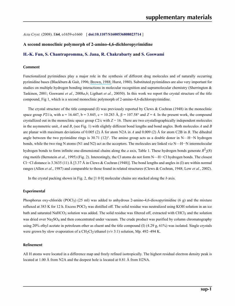
Functionalized pyrimidines play a major role in the synthesis of different drug molecules and of naturally occurringpyrimidine bases (Blackburn & Gait, 1996; Brown, 1988; Hurst, 1980). Substituted pyrimidines are also very important forstudies on multiple hydrogen bonding interactions in molecular recognition and supramolecular chemistry (Sherrington &Taskinen, 2001; Goswami et al., 2008a,b; Ligthart et al., 20050). In this work we report the crystal structure of the titlecompound, Fig 1, which is a second monoclinic polymorph of 2-amino-4,6-dichloropyrimidine.
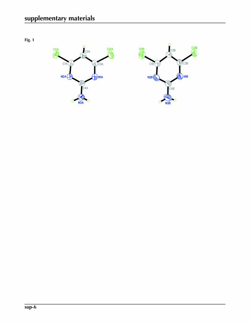
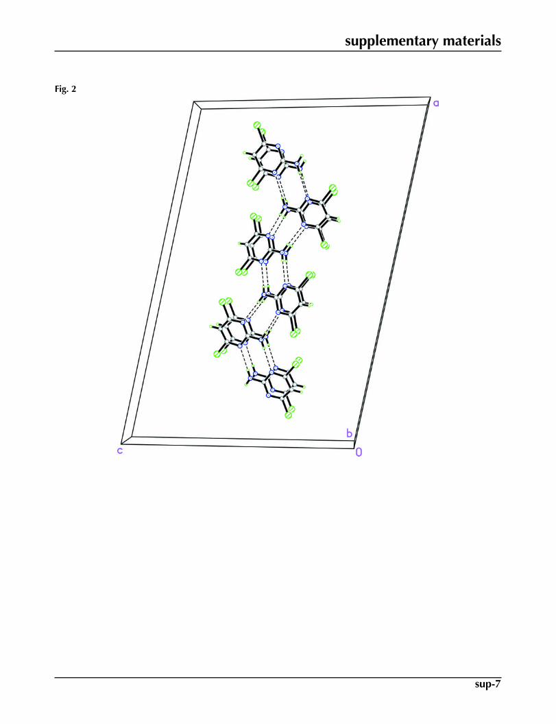
The crystal structure of the title compound (I) was previously reported by Clews & Cochran (1948) in the monoclinicspace group P21/a, with a = 16.447, b = 3.845, c = 10.283 Å, β = 107.58° and Z = 4. In the present work, the compoundcrystallized out in the monoclinic space group C2/c with Z = 16. There are two crystallographically independent moleculesin the asymmetric unit, A and B, (see Fig. 1) with slightly different bond lengths and bond angles. Both molecules A and Bare planar with maximum deviations of 0.005 (2) Å for atom N2A in A and 0.009 (2) Å for atom C2B in B. The dihedralangle between the two pyrimidine rings is 30.71 (12)°. The amino group acts as a double donor in N—H···N hydrogenbonds, while the two ring N atoms (N1 and N2) act as the acceptors. The molecules are linked via N—H···N intermolecular
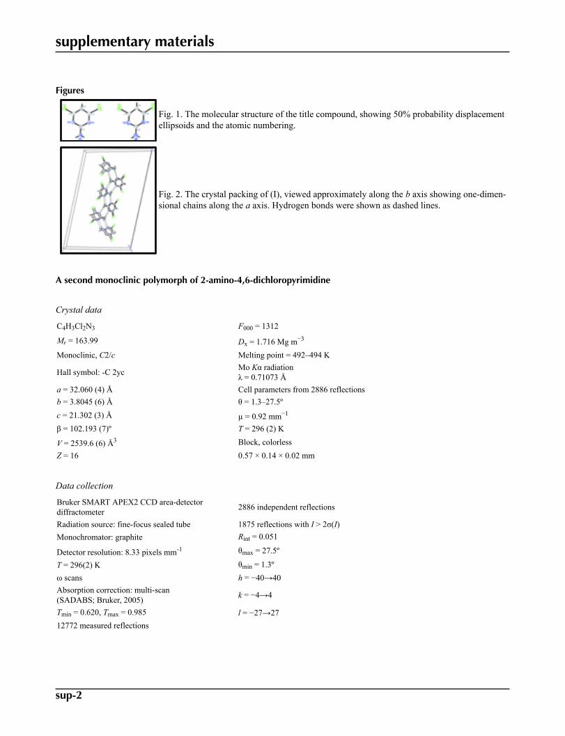
hydrogen bonds to form infinite one-dimensional chains along the a axis, Table 1. These hydrogen bonds generate R22(8)
ring motifs (Bernstein at al., 1995) (Fig. 2). Interestingly, the Cl atoms do not form N—H···Cl hydrogen bonds. The closestCl···Cl distance is 3.3635 (11) Å [3.37 Å in Clews & Cochran (1948)]. The bond lengths and angles in (I) are within normalranges (Allen et al., 1987) and comparable to those found in related structures (Clews & Cochran, 1948; Low et al., 2002).
In the crystal packing shown in Fig. 2, the [1 0 0] molecular chains are stacked along the b axis.
Experimental
Phosphorus oxy-chloride (POCl3) (25 ml) was added to anhydrous 2-amino-4,6-dioxopyrimidine (6 g) and the mixture
refluxed at 383 K for 12 h. Excess POCl3 was distilled off. The solid residue was neutralized using KOH solution in an ice
bath and saturated NaHCO3 solution was added. The solid residue was filtered off, extracted with CHCl3 and the solution
was dried over Na2SO4 and then concentrated under vacuum. The crude product was purified by column chromatography
using 20% ethyl acetate in petroleum ether as eluent and the title compound (I) (4.29 g, 61%) was isolated. Single crystalswere grown by slow evaporation of a CH2Cl2/ethanol (v/v 3:1) solution, Mp. 492–494 K.
Refinement
All H atoms were located in a difference map and freely refined isotropically. The highest residual electron density peak islocated at 1.00 Å from N2A and the deepest hole is located at 0.81 Å from H2NA.
supplementary materials
sup-2
Figures
Fig. 1. The molecular structure of the title compound, showing 50% probability displacementellipsoids and the atomic numbering.
Fig. 2. The crystal packing of (I), viewed approximately along the b axis showing one-dimen-sional chains along the a axis. Hydrogen bonds were shown as dashed lines.
A second monoclinic polymorph of 2-amino-4,6-dichloropyrimidine
Crystal data
C4H3Cl2N3 F000 = 1312
Mr = 163.99 Dx = 1.716 Mg m−3
Monoclinic, C2/c Melting point = 492–494 K
Hall symbol: -C 2yc Mo Kα radiationλ = 0.71073 Å
a = 32.060 (4) Å Cell parameters from 2886 reflectionsb = 3.8045 (6) Å θ = 1.3–27.5ºc = 21.302 (3) Å µ = 0.92 mm−1
β = 102.193 (7)º T = 296 (2) K
V = 2539.6 (6) Å3 Block, colorlessZ = 16 0.57 × 0.14 × 0.02 mm
Data collection
Bruker SMART APEX2 CCD area-detectordiffractometer 2886 independent reflections
Radiation source: fine-focus sealed tube 1875 reflections with I > 2σ(I)Monochromator: graphite Rint = 0.051
Detector resolution: 8.33 pixels mm-1 θmax = 27.5º
T = 296(2) K θmin = 1.3ºω scans h = −40→40Absorption correction: multi-scan(SADABS; Bruker, 2005) k = −4→4
Tmin = 0.620, Tmax = 0.985 l = −27→2712772 measured reflections
supplementary materials
sup-3
Refinement
Refinement on F2 Secondary atom site location: difference Fourier map
Least-squares matrix: full Hydrogen site location: inferred from neighbouringsites
R[F2 > 2σ(F2)] = 0.040 All H-atom parameters refined
wR(F2) = 0.098 w = 1/[σ2(Fo
2) + (0.0403P)2]where P = (Fo
2 + 2Fc2)/3
S = 1.02 (Δ/σ)max = 0.001
2886 reflections Δρmax = 0.22 e Å−3
187 parameters Δρmin = −0.24 e Å−3
Primary atom site location: structure-invariant directmethods Extinction correction: none
Special details
Geometry. All e.s.d.'s (except the e.s.d. in the dihedral angle between two l.s. planes) are estimated using the full covariance mat-rix. The cell e.s.d.'s are taken into account individually in the estimation of e.s.d.'s in distances, angles and torsion angles; correlationsbetween e.s.d.'s in cell parameters are only used when they are defined by crystal symmetry. An approximate (isotropic) treatment ofcell e.s.d.'s is used for estimating e.s.d.'s involving l.s. planes.
Refinement. Refinement of F2 against ALL reflections. The weighted R-factor wR and goodness of fit S are based on F2, convention-
al R-factors R are based on F, with F set to zero for negative F2. The threshold expression of F2 > σ(F2) is used only for calculating R-
factors(gt) etc. and is not relevant to the choice of reflections for refinement. R-factors based on F2 are statistically about twice as largeas those based on F, and R- factors based on ALL data will be even larger.
Fractional atomic coordinates and isotropic or equivalent isotropic displacement parameters (Å2)
x y z Uiso*/Ueq
Cl1A 0.330814 (19) 0.45738 (18) 0.35093 (3) 0.0475 (2)Cl2A 0.49584 (2) 0.5271 (2) 0.33992 (4) 0.0572 (2)N1A 0.46421 (6) 0.7708 (5) 0.43348 (9) 0.0371 (5)N2A 0.38986 (6) 0.7401 (5) 0.43849 (9) 0.0349 (5)N3A 0.43941 (8) 0.9927 (7) 0.51907 (11) 0.0490 (6)H2NA 0.4619 (8) 1.056 (7) 0.5293 (12) 0.033 (8)*H1NA 0.4174 (9) 1.026 (7) 0.5366 (14) 0.057 (9)*C1A 0.38306 (7) 0.5786 (6) 0.38271 (11) 0.0338 (6)C2A 0.41389 (8) 0.5008 (7) 0.34867 (12) 0.0377 (6)H2A 0.4102 (7) 0.386 (6) 0.3120 (11) 0.034 (7)*C3A 0.45423 (7) 0.6088 (7) 0.37766 (11) 0.0353 (6)C4A 0.43110 (7) 0.8296 (7) 0.46279 (11) 0.0350 (6)Cl1B 0.58263 (2) 1.02542 (19) 0.31034 (3) 0.0510 (2)Cl2B 0.73894 (2) 0.50031 (18) 0.32094 (3) 0.0474 (2)N1B 0.71199 (6) 0.7404 (6) 0.41918 (9) 0.0389 (5)N2B 0.64127 (6) 0.9690 (6) 0.41463 (9) 0.0406 (5)N3B 0.69119 (9) 0.9534 (8) 0.50908 (11) 0.0589 (7)H1NB 0.6706 (10) 1.049 (8) 0.5237 (16) 0.080 (12)*
supplementary materials
sup-4
H2NB 0.7156 (10) 0.881 (9) 0.5264 (16) 0.075 (11)*C1B 0.63348 (7) 0.9103 (6) 0.35235 (12) 0.0364 (6)C2B 0.66172 (7) 0.7709 (7) 0.31888 (11) 0.0374 (6)H2B 0.6550 (7) 0.753 (7) 0.2743 (12) 0.047 (7)*C3B 0.70066 (7) 0.6887 (7) 0.35702 (11) 0.0361 (6)C4B 0.68134 (8) 0.8851 (7) 0.44634 (11) 0.0403 (6)
Atomic displacement parameters (Å2)
U11 U22 U33 U12 U13 U23
Cl1A 0.0320 (3) 0.0586 (5) 0.0491 (4) −0.0065 (3) 0.0027 (3) −0.0029 (3)Cl2A 0.0442 (4) 0.0698 (5) 0.0658 (5) −0.0003 (4) 0.0298 (3) −0.0132 (4)N1A 0.0293 (10) 0.0422 (13) 0.0402 (11) 0.0003 (10) 0.0086 (9) −0.0014 (11)N2A 0.0295 (10) 0.0416 (13) 0.0339 (10) 0.0028 (10) 0.0073 (8) −0.0013 (10)N3A 0.0351 (14) 0.0707 (19) 0.0418 (13) −0.0073 (14) 0.0093 (11) −0.0161 (13)C1A 0.0293 (12) 0.0320 (14) 0.0385 (13) −0.0009 (11) 0.0040 (10) 0.0039 (11)C2A 0.0378 (14) 0.0402 (16) 0.0355 (13) −0.0012 (13) 0.0087 (11) −0.0057 (13)C3A 0.0338 (13) 0.0354 (14) 0.0396 (13) 0.0033 (12) 0.0141 (10) 0.0001 (12)C4A 0.0327 (13) 0.0404 (15) 0.0315 (12) −0.0020 (12) 0.0060 (10) 0.0013 (12)Cl1B 0.0344 (4) 0.0669 (5) 0.0514 (4) 0.0072 (4) 0.0083 (3) 0.0056 (4)Cl2B 0.0358 (3) 0.0566 (4) 0.0546 (4) −0.0001 (3) 0.0204 (3) −0.0103 (3)N1B 0.0355 (11) 0.0438 (13) 0.0386 (11) 0.0036 (11) 0.0109 (9) 0.0016 (10)N2B 0.0388 (12) 0.0481 (14) 0.0377 (11) 0.0044 (11) 0.0143 (9) 0.0001 (11)N3B 0.0534 (17) 0.087 (2) 0.0368 (13) 0.0187 (16) 0.0102 (12) −0.0034 (13)C1B 0.0315 (13) 0.0386 (15) 0.0407 (13) 0.0001 (12) 0.0112 (10) 0.0014 (12)C2B 0.0352 (14) 0.0465 (17) 0.0328 (13) −0.0025 (13) 0.0123 (11) −0.0036 (13)C3B 0.0329 (13) 0.0369 (15) 0.0416 (14) −0.0031 (12) 0.0151 (11) −0.0009 (12)C4B 0.0398 (15) 0.0477 (16) 0.0352 (13) 0.0043 (13) 0.0122 (11) 0.0023 (12)
Geometric parameters (Å, °)
Cl1A—C1A 1.731 (2) Cl1B—C1B 1.742 (2)Cl2A—C3A 1.725 (2) Cl2B—C3B 1.735 (2)N1A—C3A 1.317 (3) N1B—C3B 1.312 (3)N1A—C4A 1.359 (3) N1B—C4B 1.358 (3)N2A—C1A 1.314 (3) N2B—C1B 1.316 (3)N2A—C4A 1.357 (3) N2B—C4B 1.357 (3)N3A—C4A 1.326 (3) N3B—C4B 1.332 (3)N3A—H2NA 0.75 (2) N3B—H1NB 0.87 (3)N3A—H1NA 0.87 (3) N3B—H2NB 0.84 (3)C1A—C2A 1.376 (3) C1B—C2B 1.372 (3)C2A—C3A 1.373 (3) C2B—C3B 1.374 (3)C2A—H2A 0.88 (2) C2B—H2B 0.93 (2)
C3A—N1A—C4A 115.3 (2) C3B—N1B—C4B 114.8 (2)C1A—N2A—C4A 115.11 (19) C1B—N2B—C4B 114.9 (2)C4A—N3A—H2NA 114 (2) C4B—N3B—H1NB 114 (2)C4A—N3A—H1NA 115.4 (19) C4B—N3B—H2NB 112 (2)H2NA—N3A—H1NA 130 (3) H1NB—N3B—H2NB 134 (3)
supplementary materials
sup-5
N2A—C1A—C2A 125.2 (2) N2B—C1B—C2B 125.7 (2)N2A—C1A—Cl1A 116.12 (18) N2B—C1B—Cl1B 115.67 (18)C2A—C1A—Cl1A 118.68 (19) C2B—C1B—Cl1B 118.62 (19)C3A—C2A—C1A 114.3 (2) C1B—C2B—C3B 113.4 (2)C3A—C2A—H2A 118.9 (14) C1B—C2B—H2B 121.6 (15)C1A—C2A—H2A 126.7 (15) C3B—C2B—H2B 124.8 (14)N1A—C3A—C2A 124.9 (2) N1B—C3B—C2B 125.9 (2)N1A—C3A—Cl2A 116.14 (18) N1B—C3B—Cl2B 115.99 (17)C2A—C3A—Cl2A 118.95 (19) C2B—C3B—Cl2B 118.12 (18)N3A—C4A—N2A 117.1 (2) N3B—C4B—N2B 116.9 (2)N3A—C4A—N1A 117.7 (2) N3B—C4B—N1B 117.8 (2)N2A—C4A—N1A 125.2 (2) N2B—C4B—N1B 125.3 (2)
Hydrogen-bond geometry (Å, °)
D—H···A D—H H···A D···A D—H···A
N3A—H2NA···N1Ai 0.75 (3) 2.43 (3) 3.172 (3) 176 (2)
N3A—H1NA···N2Bi 0.87 (3) 2.33 (3) 3.201 (3) 172 (2)
N3B—H1NB···N2Ai 0.87 (3) 2.39 (3) 3.253 (4) 174 (3)
N3B—H2NB···N1Bii 0.84 (3) 2.41 (3) 3.242 (3) 172 (3)Symmetry codes: (i) −x+1, −y+2, −z+1; (ii) −x+3/2, −y+3/2, −z+1.
supplementary materials
sup-6
Fig. 1
supplementary materials
sup-7
Fig. 2


![Monoclinic polymorph of poly[aqua(μ 4 -hydrogen tartrato)sodium]](https://static.fdokumen.com/doc/165x107/63460bb1596bdb97a9093600/monoclinic-polymorph-of-polyaquam-4-hydrogen-tartratosodium.jpg)


![3-[(2-HYDROXYBENZYLIDENE) AMINO]PHENYL}IMINO)](https://static.fdokumen.com/doc/165x107/631c6e3f7051d371800f7901/3-2-hydroxybenzylidene-aminophenylimino.jpg)


