Matrix Isolation Studies of Carbonic Acid—The Vapor Phase above the β-Polymorph
Upload
independentCategory
view
0download
0
Intergrown New Zeolite Beta Polymorphs with Interconnected 12-Ring Channels Solved by Combining Electron Crystallography andSingle-Crystal X‑ray DiffractionZheng-Bao Yu,†,‡,∥ Yu Han,§ Lan Zhao,§ Shiliang Huang,† Qi-Yu Zheng,*,‡ Shuangzheng Lin,¶
Armando Cordova,¶ Xiaodong Zou,*,† and Junliang Sun*,†,∥
†Berzelii Center EXSELENT on Porous Materials, Department of Materials and Environmental Chemistry, Stockholm University,Stockholm SE−106 91, Sweden‡Beijing National Laboratory for Molecular Sciences, Institute of Chemistry, Chinese Academy of Sciences, Beijing 100190, People’sRepublic of China§Advanced Membranes and Porous Materials Center & Imaging and Characterization Core Lab, King Abdullah University of Scienceand Technology, Thuwal 21534, Saudi Arabia¶Department of Organic Chemistry, Arrhenius Laboratory, Stockholm University, Stockholm SE−106 91, Sweden∥College of Chemistry and Molecular Engineering, Peking University, Beijing 100871, People’s Republic of China
*S Supporting Information
ABSTRACT: Two new polymorphs of zeolite beta, denotedas SU-78A and SU-78B, were synthesized by employingdicyclohexylammonium hydroxides as organic structure-directing agents. The structure was solved by combiningtransmission electron microscopy and single-crystal X-raydiffraction. SU-78 is an intergrowth of SU-78A and SU-78Band contains interconnected 12-ring channels in threedirections. The two polymorphs are built from the samebuilding layer, similar to that for the zeolite beta family. The layer stacking in SU-78, however, is different from those in zeolitebeta polymorph A, B, and C, showing new zeolite framework topologies. SU-78 is thermally stable up to 600 °C.KEYWORDS: porous material, zeolite, zeolite beta, intergrowth, polymorph, crystal structure determination,transmission electron microscopy, electron crystallography
■ INTRODUCTION
Zeolites are crystalline microporous materials that have shownimportant applications as adsorbents, ion exchangers, andcatalysts.1 Beta is the first example of zeolites with high Si/Alratio; it was synthesized by Mobil Oil in 1967, usingtetraethylammonium hydroxide as an organic structure-directing agent (OSDA).2 The structure of beta containsthree-dimensional (3D) intersecting channels with large poreopenings defined by 12 (Si,Al)O4 tetrahedra (12-ring). Beta hasshown wide applications in catalysis such as hydrocarbonconversion,3 alkylation,4 and acylation5 of aromatics. Thestructure elucidation of zeolite beta has been very challenging,because of the intergrowth. Twenty years after its discovery,Newsam et al. and Higgins et al. independently proposed thestructure models of beta,6 which is an intergrowth of twoclosely related polymorphs: polymorph A and polymorph B.Another polymorph in the same beta familybeta polymorphC (BEC)was also proposed, but could only be synthesized12 years later by the Zou and Corma groups.7 The keyparameter for the success of synthesizing beta polymorph Cwas the incorporation of germanium in the framework tostabilize the double 4-rings (D4Rs) in the structure. The three
beta polymorphs are built from the same building layer, but thelayers are stacked in different ways.Zeolites with different channel systems can have different
properties. For example, zeolite beta polymorph A is chiral andmay display enantioselective properties in sorption, separation,and catalysis,8 while beta polymorph C can greatly enhance thediffusion and shape selectivity for acylation reactions.9 Greatefforts were made during the past decade to obtain zeolite beta-related materials or enrich a specific polymorph.10 Materialswith intergrowths of zeolite beta polymorph A, B, and C (ITQ-16)10e and polymorph B and CH (SSZ-63)10g have beenreported, which were synthesized using designed OSDAs.Herein, we report two new members of the zeolite beta family:SU-78A and SU-78B, synthesized using OSDAs with twodicyclohexyl groups. SU-78 is an intergrowth of SU-78A andSU-78B and consists of complex twinning and disorder, makingthe structure solution challenging. Recently, we reported thestructures of several novel zeolitesITQ-37,11a ITQ-38,11b and
Received: May 29, 2012Revised: September 12, 2012Published: September 14, 2012
Article
pubs.acs.org/cm
© 2012 American Chemical Society 3701 dx.doi.org/10.1021/cm301654d | Chem. Mater. 2012, 24, 3701−3706
ITQ-3911cdetermined by combining electron crystallographyand powder X-ray diffraction (PXRD). ITQ-39 is an inter-growth of three different polymorphs, and the structure wassolved from domains only a few nanometers in size.11c Here, forthe first time, we combine transmission electron microscopy(TEM) and single crystal X-ray diffraction (SXRD) to solve thecomplex intergrown structure of SU-78. In fact, SU-78A andSU-78B are the remaining two hypothetical polymorphs ofzeolite beta, polymorphs D and E, predicted by Burton and co-workers.10g To the best of our knowledge, what we present hereis the first report of a real material with the two polymorphs (Dand E) of zeolite beta.
■ EXPERIMENTAL SECTIONChemicals and Materials. The chemicals of dicyclohexyamine, N-
methyl-cyclohexylamine, iodomethane, iodoethane, 2-iodopropane,iodobutane, and tetraethyl orthosilicate (TEOS) were purchasedfrom Alfa Aesar and used without any modifications.Synthesis of OSDAs. Four different organic structure-directing
agents RnOH (n = 1−4) were tested (Figure 1). The synthesis of
R1OH is as follows. First of all, dicyclohexyamine and iodoethane weredissolved in methanol (MeOH) with a dicyclohexyamine:iodoethanemolar ratio of 1:1.2. The solution was kept stirring for 48 h at ambienttemperature. The solvent was then evaporated under reduced pressure,and the residual was washed with ethyl acetate (EtOAc) several timesto afford a white solid after drying. The solid powder was dissolved inwater and neutralized by excess of sodium hydroxide. Afterward, thesolution was extracted by CH2Cl2 for three times. The combinedorganic layer was dried by anhydrous MgSO4, and the solvent wasevaporated under reduced pressure to afford a slightly yellow liquid(N-ethyl dicyclohexylamine). In the second step, iodomethane wasadded to a solution of N-ethyl dicyclohexylamine in MeOH with a N-ethyl dicyclohexylamine:iodomethane molar ratio of 1:1.2 and themixture was stirred at ambient temperature for 48 h. After removal ofthe solvent, the residual was washed by EtOAc several times to afford awhite solid with a yield of ∼80%. The iodide was converted tohydroxide (R1OH) by using an ion-exchange resin in a batchovernight. The synthesis procedure of R2OH,
12 R3OH, and R4OH aresimilar to that of R1OH, except that iodomethane was used for R2OH;2-iodopropane and iodomethane were used, respectively, for R3OH;N-methyl-cyclohexylamine and iodobutane were used as the substratesfor R4OH.Synthesis of Zeolites Using RnOH as an OSDA. The synthesis
of SU-78 was carried out in an OH− media. Typically, GeO2 was firstadded into a solution of N-ethyl−N-methyl-dicyclohexylammoniumhydroxide (R1OH) in water under stirring. After the GeO2 wascompletely dissolved, a silica precursor tetraethyl orthosilicate (TEOS)was added. The mixture was kept stirring until the water and ethanol
were evaporated completely and a dry gel was formed. Quantitativewater then was added into the gel to give a batch composition of0.6SiO2:0.4GeO2:0.5R1OH:30H2O. The final mixture was heated in anautoclave at 175 °C under static conditions for 7 days. The solidproduct was recovered by filtration, washed with distilled water, anddried in air. Attempts were made to synthesize SU-78 using threeother RnOH OSDAs (n = 2−4) with structural motifs similar toR1OH. The synthesis batch composition and synthesis parameterswere kept the same as those using R1OH.
Characterization. Powder X-ray powder diffraction (PXRD) wasperformed on a PANalytical X’Pert PRO diffractometer equipped witha Pixel detector and using Cu Kα1 radiation (λ = 1.5406 Å). In situPXRD data were collected from room temperature (RT) to 600 °Cunder vacuum on a PANalytical X’Pert PRO MPD diffractometerequipped with an Anton-Parr XRK900 reaction chamber, using Cu Kαradiation (λ = 1.5418 Å). The heating rate was 2 °C/min and thetemperature was equilibrated for 5 min prior to each data collection.Thermogravimetric analysis (TGA) was performed in air from RT to780 °C with a heating rate of 8 °C/min, using a high-resolutionthermogravimetric analyzer (Perkin−Elmer, Model TGA 7). Thincross sections of SU-78 crystals were fabricated on an FEI HeliosModel NanoLab 400S FIB/SEM dual-beam system, using Pt/Cdeposition for sample protection. Scanning electron microscopy(SEM) was also performed on the same FIB/SEM dual-beam system.SAED patterns and HRTEM images were acquired on an FEI TitanST electron microscope (Cs = 1.2 mm; Cc = 1.5 mm; 300 kV).Synchrotron single-crystal X-ray diffraction (XRD) data were collectedat the beamline I19, Diamond Light Source, U.K. (λ = 0.68890 Å).The programs ELD13 and Trice14 were used for processing andindexing the SAED patterns, and for determining the unit cell,respectively. Crystallographic image processing on HRTEM imageswas carried out using the program CRISP.15
■ RESULTS AND DISCUSSION
Powder X-ray diffraction (PXRD) shows that pure SU-78 phasewas obtained using R1OH as the OSDA. We found that, whileR2OH was effective to direct the formation of SU-78, R3OHdecomposed during the synthesis and resulted in only GeO2.Using R4OH with only one cyclohexyl group led to ZSM-11.These results indicate that the synthesis of SU-78 may needstructure-directing agents (SDAs) with two dicyclohexylgroups.In situ PXRD shows that SU-78 was stable up to 600 °C (see
Figure S1 in the Supporting Information). Thermogravimetricanalysis (TGA) shows that the OSDA could be removed at 400°C (see Figure S2 in the Supporting Information). Theseresults indicate that permanent pores of SU-78 could begenerated by calcination. The framework was stable in amoisture-free environment after calcination, but collapsed afterbeing exposed in air for 1 h. The chemical formula of SU-78, ascalculated according to the results obtained from theinductively coupled plasma−mass spectrometry (ICP-MS)and TGA, is |(CH3)(C2H5)N(C6H12)2OH|·[Si0.71Ge0.29O2]16.Scanning electron microscopy (SEM) shows that all SU-78
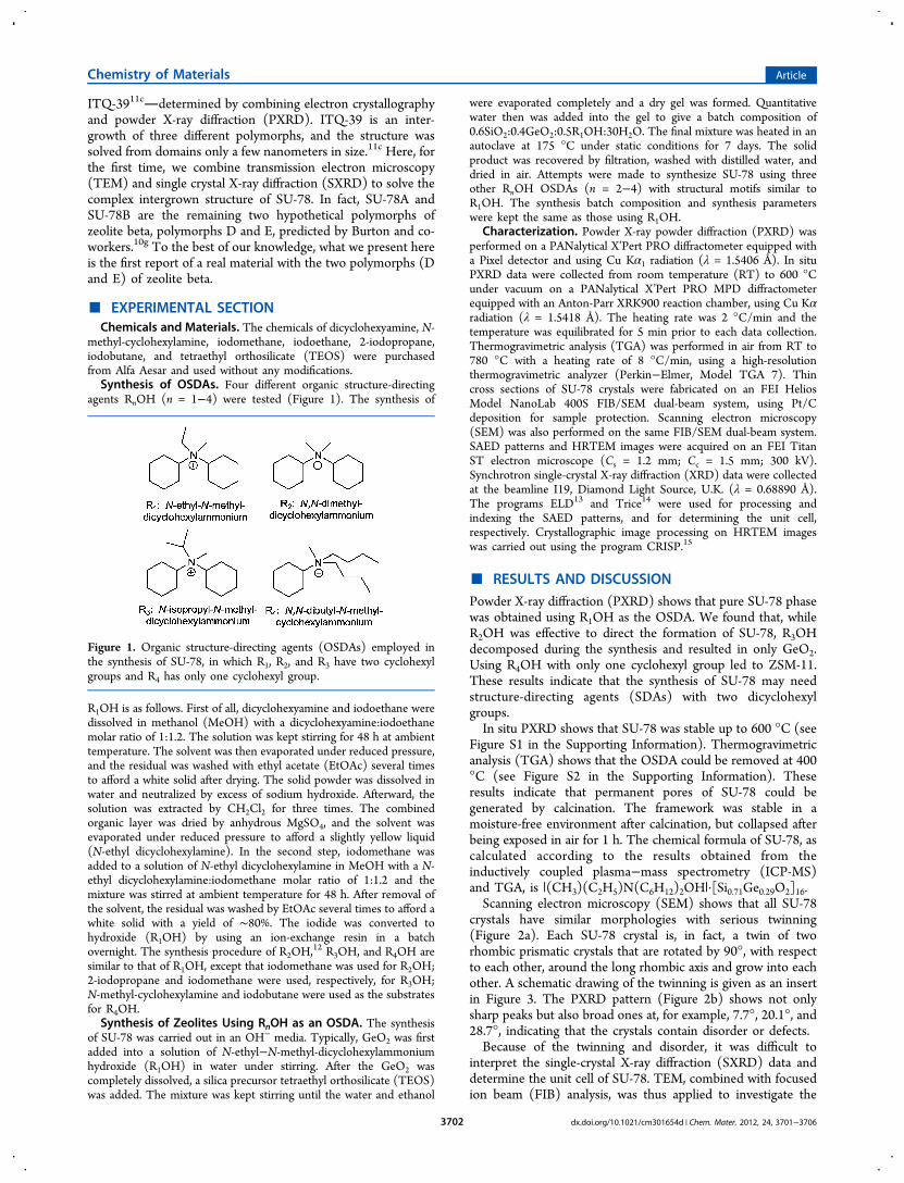
crystals have similar morphologies with serious twinning(Figure 2a). Each SU-78 crystal is, in fact, a twin of tworhombic prismatic crystals that are rotated by 90°, with respectto each other, around the long rhombic axis and grow into eachother. A schematic drawing of the twinning is given as an insertin Figure 3. The PXRD pattern (Figure 2b) shows not onlysharp peaks but also broad ones at, for example, 7.7°, 20.1°, and28.7°, indicating that the crystals contain disorder or defects.Because of the twinning and disorder, it was difficult to
interpret the single-crystal X-ray diffraction (SXRD) data anddetermine the unit cell of SU-78. TEM, combined with focusedion beam (FIB) analysis, was thus applied to investigate the
Figure 1. Organic structure-directing agents (OSDAs) employed inthe synthesis of SU-78, in which R1, R2, and R3 have two cyclohexylgroups and R4 has only one cyclohexyl group.
Chemistry of Materials Article
dx.doi.org/10.1021/cm301654d | Chem. Mater. 2012, 24, 3701−37063702
twinning and disorder in the crystal. We applied the FIBtechnique to fabricate thin cross sections in order to study theorientations in specific regions of the SU-78 crystals byselected-area electron diffraction (SAED). The schematicdrawing in Figure 3, together with Figure S3a in the SupportingInformation shows a thin cross section (thickness of <50 nm)cut from the center of a SU-78 crystal by FIB analysis. SAEDshowed that the cross section contained four crystal domains(see Figure S3a in the Supporting Information); all had thesame orientation and gave identical SAED patterns with sharpspots, as shown in Figure 3a. The boundaries between thedomains had a different orientation, giving diffraction patternswith both sharp spots and streaks (Figure 3b). The results fromSAED confirm that the SU-78 crystal contains two twincomponents, as observed by SEM. Further analysis of theSAED patterns shows that Figure 3a resembles the diffractionpattern of beta polymorph C along the a-axis, while Figure 3b issimilar to the diffraction pattern of zeolite beta with anintergrowth of polymorph A and polymorph B along the [110]direction.To determine the unit cell of SU-78, we collected a tilt series
of SAED patterns (see Figure S4 in the SupportingInformation) from a single domain (marked by the lower reddisk in Figure 3) starting from the pattern in Figure 3a (also seeFigure S4e in the Supporting Information). When the crystalwas tilted away from this orientation, streaks appeared in thefirst and second diffraction rows (see Figures S4b−4d and S4f−4h in the Supporting Information). After tilting by approx-imately ±45°, the third diffraction rows appeared as sharp spots(see Figures S4a and S4i in the Supporting Information). A
similar phenomenon was also observed in the disorderedzeolite beta. The space group and the unit-cell parameters ofSU-78B (one of the polymorphs of SU-78) were determinedfrom this tilt series to be P2/m and a = 12.6 Å, b = 12.8 Å, c =13.8 Å, β = 108°, using the programs ELD13 and Trice.14 All ofthe sharp peaks in both the PXRD and SAED patterns could beindexed as hkl with h = 3n using this unit cell (see Figure S4 inthe Supporting Information).After finding the twin laws and unit-cell parameters, it
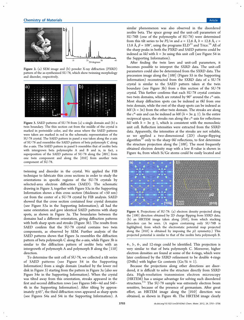
becomes possible to interpret the SXRD data. The unit-cellparameters could also be determined from the SXRD data. Theprecession image along the [100] (Figure S5 in the SupportingInformation) reconstructed from the SXRD data of a SU-78crystal is similar to the SAED pattern taken at the twinboundary (see Figure 3b) from a thin section of the SU-78crystal. This further confirms that each SU-78 crystal containstwo twin domains, which are rotated by 90° around the c*-axis.Most sharp diffraction spots can be indexed as 0kl from onetwin domain, while the rest of the sharp spots can be indexed ash0l (h = 3n) from the other twin domain. The streaks are alongthe c*-axis and can be indexed as h0l (h = 3n ± 1). In the entirereciprocal space, the streaks run along the c*-axis for reflectionshkl with h = 3n ± 1, which is consistent with the monoclinicunit cell. Reflection intensities were extracted from the SXRDdata. Apparently, the intensities at the streaks are not reliable,so we applied a two-dimensional (2D) charge-flippingalgorithm16 only to the sharp 0kl reflections, to first determinethe structure projection along the [100]. The most frequentlyobtained electron density map with a low R-value is shown inFigure 4a, from which Si/Ge atoms could be easily located and
4-, 5-, 6-, and 12-rings could be identified. This projection isvery similar to that of beta polymorph C. Moreover, higherelectron densities are found at some of the 4-rings, which werelater confirmed by the SXRD refinement to be double 4-rings(D4Rs) with higher Ge contents (Ge/Si ≈ 1).Because the projections along other directions are disor-
dered, it is difficult to solve the structure directly from SXRDdata. High-resolution transmission electron microscopy(HRTEM) has a unique advantage for solving such disorderedstructures.11c The SU-78 sample was extremely electron beamsensitive, because of the presence of germanium. After greateffort, an HRTEM image along the [010] direction wasobtained, as shown in Figure 4b. The HRTEM image clearly
Figure 2. (a) SEM image and (b) powder X-ray diffraction (PXRD)pattern of the as-synthesized SU-78, which show twinning morphologyand disorder, respectively.
Figure 3. SAED patterns of SU-78 from (a) a single domain and (b) atwin boundary. The thin section cut from the middle of the crystal ismarked in periwinkle color, and the areas where the SAED patternswere taken are marked in red in the schematic representation of theSU-78 crystal. The SAED pattern in panel a was taken along the a-axisof SU-78 and resembles the SAED pattern of beta polymorph C alongthe a-axis. The SAED pattern in panel b resembles that of zeolite betawith intergrown beta polymorphs A and B and is, in fact, asuperposition of the SAED patterns of SU-78 along the [001] fromone twin component and along the [010] from another twincomponent of SU-78.
Figure 4. Projections of SU-78: (a) electron density projected alongthe [100] direction obtained by 2D charge flipping from SXRD data;(b) an HRTEM image taken along [010], from which stackingdisorders can be seen. (A region containing only SU-78B ishighlighted, from which the electrostatic potential map projectedalong the [010] is obtained by imposing the p2 symmetry.) Thisprojected potential is similar to that of the zeolite beta polymorph B.
Chemistry of Materials Article
dx.doi.org/10.1021/cm301654d | Chem. Mater. 2012, 24, 3701−37063703

shows severe twinning in the ab-plane or stacking disordersalong the c*-axis. An electrostatic potential map projected alongthe [010] was obtained by applying crystallographic imageprocessing,17 using the program CRISP15 on a small orderedregion of SU-78B, as marked in Figure 4b. Combining thestructure projection along the [100] obtained from SXRD andthe projected potential along the [010] obtained fromHRTEM, a three-dimensional (3D) structure model could bebuilt. The resulting structure model of SU-78B shows thestructure feature of beta polymorph C along the [100], whilethat of beta polymorph B is shown along the [010]. All Siatoms are four-coordinated, as confirmed by the solid-state 29SiNMR (see Figure S7 in the Supporting Information). Despitethe serious disorders, the structure model could be refinedagainst the SXRD data by using only sharp reflections (hkl withh = 3n) and considering four twin components of SU-78B. Thisis not surprising, since the intensities of these sharp reflectionscould be estimated accurately. The refinement was convergedat R1 = 0.1139, with the highest residue peak being observed at0.67 e/Å3. The crystallographic data and structure refinementdetails are given in Table 1.
Based on the structure model of SU-78B and the stackingsequences observed in Figure 4b, the structure model ofanother polymorph SU-78A could be built, as shown in FigureS8 in the Supporting Information. SU-78A has the space groupPcmm. Its unit cell and atomic coordinates were optimized bydistant least-squares refinement by DLS-7618 and listed in
Table S1 in the Supporting Information. The structure of a realSU-78 material is an intergrowth of SU-78A and SU-78B, whichis presented in Figure 5. There are straight 12-ring channels
along the a-axis, stacked on top of each other as in betapolymorph C (BEC). While along the b-axis, the straight 12-ring channels are stacked with a shift of ±1/3a, similar to that inzeolite beta. These 12-ring channels intersect to form zigzag 12-ring channels along the c-axis. The SU-78 material consists of a3D 12-ring channel system. A series of PXRD patterns weresimulated with different SU-78A/SU-78B ratios using DIF-FaX19 (see Figure S10 in the Supporting Information). Thepattern with 50% SU-78A fits best the experimental one (seeFigure S11 in the Supporting Information) (Rp = 0.217),confirming our structure model.SU-78A and SU-78B, together with zeolite beta polymorphs
A, B, and C, are built from the same building layer containing4-, 5-, 6-, and 12-rings, as shown in Figure 6a. The differenceamong the different structures is the relative shifts betweenadjacent layers. There is no shift between the adjacent layers inbeta polymorph C (Figure 6a). In beta polymorph A, the shiftsof the consecutive layers are in the sequence of (0, 0), (1/3, 0),(1/3,
1/3) and (0, 1/3) (Figure 6b). In beta polymorph B, thecorresponding shifts are in the sequence of (0, 0), (1/3, 0), (
1/3,1/3), (
2/3,1/3), (
2/3,2/3) and (3/3,
2/3) (see Figure 6c). In SU-78A, the shifts of the consecutive layers are (0, 0) and (1/3, 0)(see Figure 6d), while in SU-78B, they are (0, 0), (1/3, 0) and(2/3, 0) (see Figure 6e). These show that the structures of SU-78A and SU-78B differ significantly from those of beta; thelayers shift only in one direction along the [100] in SU-78, butin two directions along the [100] and [010] in beta.Consequently, SU-78A and SU-78B have novel topologiesand the detailed information is given in Table 2.
■ CONCLUSIONSIn conclusion, by employing designed organic structure-directing agents (OSDAs) with two cyclohexyl groups, anovel germanosilicate zeolite (SU-78) was successfullysynthesized. Despite that, the crystals contain severe twinningand disorder and are electron-beam-sensitive; the structure ofSU-78 was solved using a new strategy, i.e., by combining TEM
Table 1. Crystallographic Data and Structure RefinementDetails of SU−78B Obtained from Synchrotron Single-Crystal X-ray Diffraction
parameter value/remark
chemical formula |(CH3)(C2H5)N(C6H12)2OH|[Si0.71Ge0.29O2]16crystal system monoclinicspace group P2/munit-cell parameters a = 12.590(2) Å
b = 12.852(2) Åc = 13.984(4) Åβ = 107.63(2)°
cell volume 2156.4 (8) Å3
Z 2density(calc) 1.755 g/cm3
wavelength 0.68890 Åradiation type synchrotrontemperature 150(2) KF(000) 1104crystal size 0.03 mm × 0.01 mm × 0.01 mmθmin, θmax 2.96°, 25.16°dataset (h; k; l) −15−15; −15−15; −17−17Tmin/Tmax 0.829/1.000refinement method full-matrix least-squares on F2
Nref, Npar 2625, 156data completeness 33.2%a
R1, wR2, S 0.1139, 0.2917, 0.974largest diff. peak and hole 0.663/−0.621(e/Å3)twin components (%) 29/25/16/28aTwo-thirds of the reflections are not included in the refinement dueto the disorder. Note that four twin components are included in therefinement: (1 0 0, 0 1 0, 0 0 1), (0 1 0, −1 0 0, 0.333−0.333 1), (−1 00, 0−1 0, 0.667 0 1) and (0−1 0, 1 0 0, 0.333 0.333 1) with 90°rotation between each other.
Figure 5. The channel systems in SU-78 superimposed with the 3Dstructure model viewed (a) along the a-axis showing the BEC-typechannels and (b) along the b-axis showing the beta-type channelsystems. The cyan surfaces are toward the pores while the bluesurfaces are toward the framework. The parts with SU-78A and SU-78B are marked.
Chemistry of Materials Article
dx.doi.org/10.1021/cm301654d | Chem. Mater. 2012, 24, 3701−37063704

with SXRD. SU-78 contains three-dimensional (3D) intersect-ing 12-ring channels and is an intergrowth of two polymorphs(SU-78A and SU-78B) with novel topologies. The frameworksof SU-78A and SU-78B are the same as the polymorphs E andD of zeolite beta that were proposed by Burton and co-workers.10g All polymorphs of zeolite beta are built from thesame building layers but with different stacking sequences. SU-78 is stable in a moisture-free environment after calcination.The discovery of the new family members in a zeolite family aswell-known as beta is important for understanding the zeoliteformation, designing novel zeolite structures, and exploringtheir applications.
■ ASSOCIATED CONTENT
*S Supporting InformationIn situ powder X-ray diffraction (PXRD), TGA curve, SEMimage showing the twinning of SU-78, a tilt series of SAEDpatterns, precession image from single-crystal X-ray diffraction(SXRD), simulated SXRD patterns, solid-state 29Si NMRspectrum, comparison between the simulated and experimentalPXRD patterns, 1H/13C NMR spectra of R3I and R4I, atomiccoordinates of SU-78A. This information is available free ofcharge via the Internet at http://pubs.acs.org.
■ AUTHOR INFORMATION
Corresponding Author*E-mail addresses: [email protected] (J.S.),[email protected] (Q.-Y.Z.), [email protected] (X.Z.).
NotesThe authors declare no competing financial interest.
■ ACKNOWLEDGMENTS
This work is supported financially by the Swedish ResearchCouncil (VR) and the Swedish Governmental Agency forInnovation Systems (VINNOVA) through the Berzelii CenterEXSELENT, the Major State Basic Research DevelopmentProgram of China (No. 2011CB932501), the National NaturalScience Foundation of China (No. 20932004).
Figure 6. Comparison of the stacking of the common building layer indifferent structures. (a) The building layer viewed along the c-axisshown as T−T connections (left) and as a simplified square lattice(right). It also resembles the packing in beta polymorph C (BEC),where all the layers are stacked exactly on top of each other. In betapolymorphs A and B, and SU-78A and SU-78B, the stacking of thebuilding layer is represented in panels (b), (c), (d), and (e),respectively. The colors in each figure indicate the different heights ofthe layers, changing gradually from dark green to light blue. Thebuilding layers in beta are shifted in both the x- and y-directions, whilethey are only shifted along the x-direction in SU-78. Note that the 90°rotation around the c*-axis of the adjacent layers is not shown in thisillustration.
Table 2. Topology of the Underlying Nets of SU-78A and SU-78B
property SU-78A SU-78B
space group of thenet embedding
Pcmm P2/m
transitivity 9(22)(25)(12) 9(20)(22)(11)natural tilingsignature
4[6.122]+[62.122]+2[4.52.122]+2[52.123]+[42.124]+[42.54.122]+[46]+2[54]+2[43.54]+3[42.54.62] 3[62.122]+2[4.52.122]+2[52.123]+[42.124]+[42.54.122]-2[43.54]+3[42.54.62]+2[54]+[46]
point symbol fornet
{4.53.62}{4.54.6}{42.52.6.7}{42.53.7}{43.5.6.7}2{53.62.9}{54.6.8} {4.53.62}{4.54.6}{42.52.6.7}{42.53.7}{43.5.6.7}2{53.62.9}{54.6.8}
coordinatessequencesvertex 1 4 10 19 32 50 72 99 132 170 203 4 9 18 32 51 74 101 131 166 201vertex 2 4 10 21 32 48 70 104 138 171 200 4 9 18 32 51 76 100 129 165 204vertex 3 4 11 18 29 46 76 104 131 160 197 4 12 17 30 51 74 102 125 161 208vertex 4 4 12 17 30 51 74 102 128 161 205 4 11 18 29 46 76 104 131 160 197vertex 5 4 9 18 32 51 76 102 130 164 202 4 11 20 28 43 75 102 125 156 197vertex 6 4 9 18 32 51 74 99 130 167 203 4 11 20 28 43 72 106 128 148 197vertex 7 4 12 18 31 48 73 105 130 159 201 4 12 18 31 48 73 105 133 159 196vertex 8 4 11 20 28 43 72 103 129 152 193 4 10 21 32 48 70 104 138 169 198vertex 9 4 11 20 28 43 75 105 124 152 201 4 10 19 32 50 72 99 132 170 206
Chemistry of Materials Article
dx.doi.org/10.1021/cm301654d | Chem. Mater. 2012, 24, 3701−37063705

■ REFERENCES(1) (a) Davis, M. E. Nature 2002, 417, 813. (b) Corma, A. J. Catal.2003, 216, 298.(2) Wadlinger, R. L.; Kerr, G. T.; Rosinski, E. J. (Mobil OilCorporation). U.S. Patent 3,308,069, 1967.(3) (a) Jansen, J. C.; Creyghton, E. J.; Njo, S. L.; Koningsveld, H. V.;Bekkum, H. V. Catal. Today 1997, 38, 205. (b) Bjørgen, M.; Kolboe, S.Appl. Catal., A 2002, 225, 285. (c) Bjørgen, M.; Olsbye, U.; Svelle, S.;Kolboe, S. Catal. Lett. 2004, 93, 37. (d) Liu, Y.; Guo, W.; Zhao, X. S.;Lian, J.; Dou, J.; Kooli, F. J. Porous Mater. 2006, 13, 359. (e) Taarning,E.; Osmundsen, C. M.; Yang, X.; Voss, B.; Andersen, S. I.; Christensen,C. H. Energy Environ. Sci. 2011, 4, 793. (f) Guo, W.; Xiong, C.; Huang,L.; Li, Q. J. Mater. Chem. 2011, 11, 1886.(4) (a) Reddy, K. S. N.; Rao, B. S.; Shiralkar, V. P. Appl. Catal., A1993, 95, 53. (b) Corma, A.; Gomez, V.; Martínez, A. Appl. Catal., A1994, 119, 83. (c) Meng, X. Y.; Qin, Z. F.; Zhang, Y.; Dong, M.;Wang, J. G. Catal. Lett. 2002, 83, 265. (d) Corma, A.; Martínez, A.; P.Arroyo, A.; Monteiro, J. L. F.; Sousa-Aguiar, E. F. Appl. Catal., A 1996,142, 139.(5) (a) Heinichen, H. K.; Holderich, W. F. In Proceedings of the 12thInternational Zeolite Conference; Treacy, M. M. J., Marcus, B. K., Bisher,M. E., Higgins, J. B., Eds.; Material Research Society: Warrendale, PA,1999; p 1085. (b) Andy, P.; Garcia-Martinez, J.; Lee, G.; Gonzalez, H.;Jones, C. W.; Davis, M. E. J. Catal. 2000, 192, 215. (c) Freese, U.;Heinrich, F.; Roessner, F. Catal. Today 1999, 49, 237. (d) Kennedy, E.M.; Lonyi, F.; Ballinger, T. H.; Rosynek, M. P.; Lunsford, J. H. EnergyFuels 1994, 8, 846. (e) García, R. A.; Serrano, D. P.; Vicente, G.;Otero, D.; Linares, M. Stud. Surf. Sci. Catal. 2008, 174, 1091.(6) (a) Treacy, M. M. J.; Newsam, J. M. Nature 1988, 332, 249.(b) Newsam, J. M.; Treacy, M. M. J.; Koetsier, W. T.; Gruyter, C. B.Proc. R. Soc. London A 1988, 420, 375. (c) Higgins, J. B.; LaPierre, R.B.; Schlenker, J. L.; Rohrman, A. C.; Wood, J. D.; Kerr, G. T.;Rohrbaugh, W. J. Zeolite 1988, 8, 446.(7) (a) Conradsson, T.; Dadachov, M. S.; Zou, X. D. MicroporousMesoporous Mater. 2000, 41, 183. (b) Corma, A.; Navarro, M. T.; Rey,F.; Rius, J.; Valencia, S. Angew. Chem., Int. Ed. 2001, 40, 2277.(c) Corma, A.; Navarro, M. T.; Rey, F.; Valencia, S. Chem. Commun.2001, 1486.(8) Davis, M. E.; Lobo, R. F. Chem. Mater. 1992, 4, 756.(9) Botella, P.; Corma, A.; Navarro, M. T.; Rey., F.; Sastre, G. J.Catal. 2003, 217, 406.(10) (a) Cantín, A.; Corma, A.; Díaz-Cabanas, M. J.; Jorda, J. L.;Moliner, M.; Rey, F. Angew. Chem., Int. Ed. 2006, 45, 8013. (b) Corma,A.; Moliner, M.; Cantín, A.; Díaz-Cabanas, M. J.; Jorda, J. L.; Zhang, D.L.; Sun, J. L.; Jansson, K.; Hovmoller, S.; Zou, X. D. Chem. Mater.2008, 20, 3218. (c) Kadgaonkar, M. D.; Kasture, M. W.; Bhange, D. S.;Joshi, P. N.; Ramasway, V.; Kumar, R. Microporous Mesoporous Mater.2007, 101, 108. (d) Kadgaonkar, M. D.; Kasture, M. W.; Bhange, D.S.; Joshi, P. N.; Ramasway, V.; Gupta, N. M.; Kumar, R. MicroporousMesoporous Mater. 2007, 105, 82. (e) Corma, A.; Navarro, M. T.; Rey,F.; Valencia, S. Chem. Commun. 2001, 1720. (f) Camblor, M. A.;Barret, P. A.; Díaz- Cabanas, M. J.; Villaescusa, L. A.; Puche, M.; Boix,T.; Perez, E.; Koller, H. Microporous Mesoporous Mater. 2001, 48, 11.(g) Burton, A. W.; Elomari, S.; Chan, I.; Pradhan, A.; Kibby, C. J. Phys.Chem. B 2005, 109, 20266.(11) (a) Sun, J; Bonneau, C.; Cantín, A; Corma, A.; Diaz-Cabanas,M. J.; Moliner, M.; Zhang, D.; Li, M.; Zou, X. D. Nature 2009, 458,1154. (b) Moliner, M.; Willhammar, T.; Wan, Gonzalez, J.; Rey, F.;Jorda, J. L.; Zou, X.; Corma, A. J. Am. Chem. Soc. 2012, 134, 6473.(c) Willhammar, T.; Sun, J.; Wan, W; Oleynikov, P.; Zhang, D.; Zou,X.; Moliner, M.; Gonzalez, J.; Martínez, C; Rey, F.; Corma, A. Nat.Chem. 2012, 4, 88.(12) (a) Cao, G.; Shah, M. J.; PCT Int. Appl. WO 2008033230 A220080320, 2008. (b) Cao, G. PCT Int. Appl. WO 2008033229 A220080320, 2008.(13) Zou, X. D.; Sukharev, Y.; Hovmoller, S. Ultramicroscopy 1993,49, 147.(14) Zou, X. D.; Hovmoller, A.; Hovmoller, S. Ultramicroscopy 2004,98, 187.
(15) Hovmoller, S. Ultramicroscopy 1992, 41, 121.(16) Xie, D.; McCusker, L. B. J. Am. Chem. Soc. 2011, 133, 20604.(17) (a) Sun, J. L.; He, Z. B.; Hovmoller, S.; Zou, X. D.; Gramm, F.;Baerlocher, C.; McCusker, L. B. Z. Kristallogr. 2010, 225, 77. (b) Zou,X. D.; Hovmoller, S.; Oleynikov, P. Electron Crystallography−ElectronMicroscopy and Electron Diffraction; Oxford University Press: 2011; p156.(18) Baerlocher, Ch.; Hepp, A.; Meier, W. M. DLS-76: Distanceleast-squares refinement program. ETH Zurich, Switzerland.(19) Treacy, M. M. J.; Deem, M. W.; Newsam, J. M. DIFFaX: AComputer Program for Calculating Diffraction from Faulted Crystals.Available via the Internet at http://www.public.asu.edu/mtreacy/DIFFaX.html.
Chemistry of Materials Article
dx.doi.org/10.1021/cm301654d | Chem. Mater. 2012, 24, 3701−37063706
Copyright © 2022 FDOKUMEN