Adhesion of dual-polymerized luting cement on superficial ...
14
Full Terms & Conditions of access and use can be found at http://www.tandfonline.com/action/journalInformation?journalCode=tast20 Journal of Adhesion Science and Technology ISSN: 0169-4243 (Print) 1568-5616 (Online) Journal homepage: http://www.tandfonline.com/loi/tast20 Adhesion of dual-polymerized luting cement on superficial and deep dentin using different one- step self-etch mild adhesive systems Larissa Blarer & Mutlu Özcan To cite this article: Larissa Blarer & Mutlu Özcan (2018): Adhesion of dual-polymerized luting cement on superficial and deep dentin using different one-step self-etch mild adhesive systems, Journal of Adhesion Science and Technology To link to this article: https://doi.org/10.1080/01694243.2018.1494399 Published online: 27 Oct 2018. Submit your article to this journal View Crossmark data
-
Upload
khangminh22 -
Category
Documents
-
view
2 -
download
0
Transcript of Adhesion of dual-polymerized luting cement on superficial ...
Full Terms & Conditions of access and use can be found athttp://www.tandfonline.com/action/journalInformation?journalCode=tast20
Journal of Adhesion Science and Technology
ISSN: 0169-4243 (Print) 1568-5616 (Online) Journal homepage: http://www.tandfonline.com/loi/tast20
Adhesion of dual-polymerized luting cement onsuperficial and deep dentin using different one-step self-etch mild adhesive systems
Larissa Blarer & Mutlu Özcan
To cite this article: Larissa Blarer & Mutlu Özcan (2018): Adhesion of dual-polymerized lutingcement on superficial and deep dentin using different one-step self-etch mild adhesive systems,Journal of Adhesion Science and Technology
To link to this article: https://doi.org/10.1080/01694243.2018.1494399
Published online: 27 Oct 2018.
Submit your article to this journal
View Crossmark data
Adhesion of dual-polymerized luting cement onsuperficial and deep dentin using different one-stepself-etch mild adhesive systems
Larissa Blarera and Mutlu €Ozcanb
aUniversity of Zurich, Center for Dental and Oral Medicine, Clinic for Fixed and RemovableProsthodontics and Dental Materials Science, Zurich, Switzerland; bUniversity of Zurich, DentalMaterials Unit, Center for Dental and Oral Medicine, Clinic for Fixed and Removable Prosthodonticsand Dental Materials Science, Zurich, Switzerland
ABSTRACTThis study evaluated adhesion of dual-polymerized resin cement tosuperficial dentin (SD) and deep dentin (DD) using one-step self-etch adhesives at varying pH. After smear layer was created onthird molars (N¼ 60, n¼ 15 per group), adhesive resins, 1- ClearfilS3 Bond Plus-CBP (Kuraray) (pH: 2.3), 2- Bisco All Bond Universal-BAU (Bisco) (pH: 3.2), 3- Single Bond Universal Adhesive-SBU (3MESPE) (pH: 2.7), 4- Nova Compo-B Plus-NCBP (Imicryl) (pH: 2.5–3),were applied on SD and DD. Resin cement (Variolink II, IvoclarVivadent) was adhered incrementally on the SD surfaces usingpolyethylene molds and photo-polymerized for 40 s from 5 direc-tions (output: 1200mw/cm2). After macroshear and microsheartest, in order to achieve DD specimens, SD were removed 1mm inthe pulp direction and the same bonding and test procedureswere performed. The specimens were kept at 37 �C for 24h. Theadhesion tests were conducted in the Universal Testing Machineand failure types were analyzed. The data were analyzed usingUnivariate ANOVA, Tukey�s, Kruskal-Wallis and Mann-Whitney tests(a¼ .05). Test method, dentin level and the adhesive resin signifi-cantly affected the results (MPa) (p< .05). After macroshear test,more incidences of cohesive failures in DD were observed withNCBP Plus. On SD, NCBP presented the highest results followed byBAU using macroshear test. On DD, NCBP presented the highestresults followed by SBU. Not only the pH but the chemical com-position affected adhesion especially to SD while in DD, the differ-ence between the adhesive resins was less significant.
ARTICLE HISTORYReceived 15 March 2018Accepted 19 June 2018
KEYWORDSAdhesion; adhesivecementation;bond strength test;dentin; macroshear test;microshear test;resin luting cement
Introduction
The longevity of indirect fixed-dental-prostheses (FDP) could be affected by multiplefactors including the cementation mode that is basically the final stage of consecutiveclinical procedures. In principle, the primary function of the cementation is to estab-lish reliable retention of the FDP, a durable seal of the space between the tooth and
CONTACT Mutlu €Ozcan [email protected] Plattenstrasse 11, CH-8032, Z€urich, Switzerland� 2018 Informa UK Limited, trading as Taylor & Francis Group
JOURNAL OF ADHESION SCIENCE AND TECHNOLOGYhttps://doi.org/10.1080/01694243.2018.1494399

the restoration and to provide adequate optical properties especially for ceramic orpolymeric FDPs [1]. Today, adhesive properties of dental cements are of importanceas it enables clinicians to perform minimal invasive restorations [2].
Current trends in dental research tend to simplify the clinical procedures, especiallyfor the applications of adhesive resins not only in restorative dentistry for direct appli-cations of resin-based materials but also in prosthetic dentistry in adhesive cementationof FDPs. Advances in these procedures primarily include the development of simplifiedbonding systems as a substitute for classical three-step systems [1]. On the other hand,traditional “etch and rinse” adhesive systems rely on the application of adhesive mono-mers to acid-etched dentin or the use of simplified self-etching, self-priming agents thatcontain hydrophilic and acidic monomers, acidic molecules, diluent monomers, photo-initiators and solvents usually at low pH. Such adhesives simultaneously etch the dentinand infiltrate the adhesive monomers into the dentin [3].
Different bonding strategies may affect the adhesion of resin cements to dentin[4–6]. In fact, long-term survival of FDPs may be very much dependent on the func-tion of the resin cement that adheres both to the restorative materials and the toothsubstance [7]. The reliable adhesion of the resin cements to both the dentin and theceramic becomes particularly important in the application of all-ceramics or surface-retained resin-bonded FDPs where mechanical retention does not dominate [1]. Also,when enamel margins do not exist after tooth preparation as a consequence of cariesremoval or tissue loss due to trauma, adhesion of the cement is dictated by dentinconditioning through etching and application of the bonding agents. While some pre-vious studies have shown that self-etch adhesives may result in lower bond strengthto dentin [7–9], others reported no significant difference with the use of mild univer-sal adhesives with etch-and rinse strategy [10]. In addition, due to the acidic natureof the self-etch adhesives and their permeability, their adhesion to dentin are compro-mised [9]. Typically, since etching dentin separately would result in higher interactionwith dentin [11] and due to their lesser hydrophilicity [12], “etch and rinse” adhesivesystems compensate for other factors and lead to higher bond strength of the resincements to dentin. However, this adds to the increased workflow and achieving bal-ance between dry but at the sometime sufficient humid dentin surface is consideredas a clinical challenge when “etch and rinse” adhesive systems are employed [13].
Resin cements, be it conventional or simplified, may perform differently dependingon their adhesive systems since the latter is primarily in contact with the dentin[14,15]. Contemporary adhesive systems used in dentistry interact with the enamel/dentin substrate either by removing the smear layer (etch-and-rinse technique) or bypartially dissolving the smear layer, decalcifying underlying intertubular dentin, andimpregnating any remaining smear layer for the bonding (self-etch technique) [16].While the etch-and-rinse bonding technique is initiated by a separate etching stepusing 35–37% phosphoric acid that is later rinsed away, the self-etch/primer agentcontaining acidic monomers are only air-dried, thus remaining within the modifiedsmear layer. The self-etch approach could also be called as “etch-and-dry” approach[17]. Such adhesives make the application less technique-sensitive for the clinicians[18]. Besides micromechanical interlocking through hybridization, specific functionalmonomers of ‘mild’ or ‘intermediate’ two-step self-etching adhesives were shown to
2 L. BLARER AND M. €OZCAN

interact chemically with residual hydroxyapatite crystals that remain available in thesubmicron hybrid layer [19]. While some studies reported higher bond strengths todentin with two-step self-etching adhesives compared to one-step ones [20], othersreported comparable [14] or lower bonding efficacy to dentin [21–23].
Resin-bonded FDPs are usually bonded to enamel or superficial dentin (SD).However, full-coverage FDPs, extensive overlays or endocrowns are cemented to deepdentin (DD) as such restorations require more room for the restorative material. Inprevious studies, bond strengths of resin composites to dental tissues were found to behigher in SD than in DD [24,25]. Since SD contains more intertubular dentin area andit is rich in collagen fibrils, it makes adhesion with resin-based materials favourable[11]. However, while impaired adhesion was reported on DD due to higher water con-tent compared to SD [24,25], other studies showed no significant difference on bothdentin substrates as a function of depth [26]. The controversial results could be in partdue to the difference in test methods or the pH effect of the adhesives on dentin [27].
Previous studies using stress distribution analyses have reported that some of thebond strength tests do not appropriately stress the interfacial zone [28,29]. Shear testshave been criticized for the development of non-homogeneous stress distributions atthe bonded interface, inducing either underestimation or misinterpretation of theresults, as the failure often starts in one of the substrates and not solely at the adhe-sive zone [28,29]. Conventional tensile tests also present some limitations, such as thedifficulty of specimen alignment and the tendency for heterogeneous stress distribu-tion at the adhesive interface. On the other hand, when specimens are aligned cor-rectly, the microtensile test shows more homogeneous distribution of stress, andthereby more sensitive comparison or evaluation of bond performances [22].However, small deviations in specimen alignment in the jig may cause increase bondstrength due to shear component being introduced during deboning bonded joints[30,31]. To the best of our knowledge, no study exists to date where both macro andmicroshear adhesion tests were employed in one study on both SD and DD.
The objectives of this study therefore were to evaluate the adhesion of conventionalresin-based luting cement together with self-etch adhesive resin systems to SD and DDusing both macroshear and microshear tests. The hypotheses tested were that a) adhesivestrength of resin cement to DD would be less than to SD, b) macroshear bond strengthresults would be less than microshear tests and c) self-etch adhesive resins with lower pHwould deliver higher bond strength results than those of the adhesives with higher pH.
Materials and Methods
Specimen preparation
The brands, types, main chemical compositions, manufacturers and batch numbers ofthe materials used for the experiments are listed in Table 1. This study comprisedfour self-etch adhesive resins and one conventional resin-based adhesive cement.Adhesive potential of the cement on SD and DD of third molar teeth was tested.
Human wisdom molars (N¼ 60, n¼ 15 per group), were collected and kept in dis-tilled water at 5 �C until the experiments. All teeth used in the present study wereextracted for reasons unrelated to this project. Written informed consent for research
JOURNAL OF ADHESION SCIENCE AND TECHNOLOGY 3

purpose of the extracted teeth was obtained by all donors prior to extraction accord-ing to the directives set by the National Federal Council. Ethical guidelines werestrictly followed and irreversible anonymization was performed in accordance withState and Federal Law [32–34]. After tissue remnants were removed with a scaler(H6/H7; Hu-Friedy, Chicago, IL), teeth were stored in .5% Chloramin T for 2 weeks.In order to determine the exact location of cusp tips, enamel, dentin and pulp hornsof the teeth, a previously describer method was used [22].
The teeth were embedded using polymethylmethacrylate (PMMA, Condular AG,Wager, Switzerland) with their occlusal surfaces exposed in polyethylene rings (diam-eter: 10mm, height: 12mm). The apices of the roots were shortened using a diamonddisc (IsoMet 1000, Buehler Ltd, Illinois, USA) under water-cooling when root lengthexceeded 12mm. Then, the cusps of the molars were removed in a trimmer(Isomet, Buehler Ltd., Lake Bluff, IL, USA) under water-cooling until flat dentin sur-faces were achieved. Dentin level after removal of cusp tips was considered as SDgroup. One mm below the SD level was indicated and considered as DD [25].
Dentin surfaces were exposed with a low speed disc under water-cooling and groundfinished using 600, 800 and 1000-grit, silicone carbide abrasive papers (EnglishAbrasives Ltd, London, United Kingdom) under water in sequence. In both SD andDD groups, flat dentin surfaces were polished with 400 grit silicon carbide papersunder water-cooling and then rinsed thoroughly in order to create bonding surfacescovered with smear layers [35,36]. Subsequently, bonding procedures were carried out.
Bonding procedures for adhesion tests
One calibrated operator carried out adhesive procedures throughout the experiments(Table 2). After conditioning the dentin surfaces, the adhesive luting cement
Table 1. The brands, abbreviations, manufacturers, chemical compositions and batch numbers ofthe materials used for the experiments.
Brand Manufacturer Chemical composition �pHBatch
numbers
Clearfil S3 BondPlus (CBP)
Kuraray,Kurashiki, Japan
bis-GMA, 2-HEMA, ethanol, sodiumfluoride,10-methacryloyloxydecyl dihydrogen-phosphate, hydrophilicaliphaticdimetha-crylate, hydrophobicaliphaticmethacry-late, colloidalsilica, dl-Campherquinone,accelerators, initiators, water
2.3 6E0012
Bisco All BondUniversal (BAU)
Bisco,Schaumburg, USA
Ethanol, bis-GMA 3.2 1300009232
Single BondUniversal (SBU)
3M ESPE AG,Seelfeld, Germany
MDP phosphate monomer, dimethacrylateresins, HEMA, Vitrobond copolymer, filler,ethanol, water, initiators, silane
2.7 497905
Nova Compo-BPlus (NCBP)
Imicryl Dis Malz.GmBH,Konya, Turkey
bis-GMA, 2-HEMA, ethanol,10-methacryloy-loxydecyl dihydrogenphosphate, 4-META,silanated high dispersed silica, dl-Campherquinone, accelerators, initia-tors, water
2.5–3 16224
Variolink II (Dualpolymerizedresin cement)
Ivoclar Vivadent,Schaan,Liechtenstein
UDMA, inorganic fillers, ytterbium trifluor-ide, initiators, stabilizers, pigments
S50635T24729
bis-GMA, bisphenol A glycol dimethacrylate; UDMA, Urethane dimethacrylate; HEMA, Hydroxyethyl methacrylate;MDP, 10- Methacryloyloxydecyl dihydrogen phosphate; 4-META, 4-methacryloxyethyl trimellitate anhydride. �pH val-ues of the adhesives are obtained from the manufacturers.
4 L. BLARER AND M. €OZCAN

(Variolink II, Ivoclar Vivadent, Schaan, Liechtenstein) were adhered incrementallywith a hand instrument on the dentin surfaces using translucent polytetrafluoroethyl-ene (Teflon) molds (DuPont, Saint-Gobain, France) (for macroshear bond test-SBT:height: 4mm, diameter: 2.9mm; for microshear bond test-mSBT: height: 4mm, diam-eter: .8mm) were stabilized on the dentin specimens in a custom made device. Themold was filled in two increments with the resin cement and polymerized for 40 sfrom 5 directions from a distance of 2mm (Bluephase, Ivoclar Vivadent) for 40 s(output: 1200mw/cm2). Oxygen inhibiting gel (Oxyguard, Kuraray, Tokyo, Japan)was applied at the bonded margins and rinsed with copious water after 1minute.
The bonded specimens were kept at 37 �C for 24 hours. After macro and micro-shear tests on the SD, the dentin surfaces were again ground finished using 600, 800and 1000-grit, silicone carbide abrasive papers under water in sequence inorder toachive DD and conditioned with the adhesive resins accordingly. The same resin lut-ing cement was adhered incrementally with a hand instrument on the dentin surfacesusing polyethylene moulds. The teeth were maintained wet at all times during all pro-cedures except during X-rays.
Macroshear and microshear tests
Specimens were mounted in the apparatus of the Universal Testing Machine (ZwickROELL Z2.5MA 18-1-3/7, Ulm, Germany) and the force was applied to the adhesiveinterface until failure occurred. The load was applied with a 50 kgf load cell to thesubstrate-adherend interface with the corresponding blades for each test method, asclose as possible to the surface of the substrate at a crosshead speed of 1mm/minand the stress-strain curve was analyzed with the software program (TestXpertVR ,Zwick ROELL, Ulm, Germany). Bond strength after each test (MPa) was calculatedby dividing the maximum load (N) by the bonding surface area of the resin cement.
Failure type evaluation
Failure sites were initially observed using an optical microscope (Stemi 2000-C, CarlZeiss, Gottingen, Germany) at �40 magnification and since no mixed failures were
Table 2. Application modes of the adhesive resins tested in this study according to eachmanufacturer�s instructions. See Table 1 for group abbreviations.CBP BAU SBU NCBP
Apply bond for 10 s Dispense 1–2 drops of adhe-sive resin into a well
Place one drop of of the adhe-sive resin and dual cureactivator in a mixing welland mix for 5 s
Dispense 1–2 drops of adhe-sive resin into a well
Dry with mild pressureair flow for 5 s
Apply two separate coats,scrubbing the preparationwith a microbrush for10–15 s per coat
Apply the adhesive mixture tothe prepared tooth and rubit in for 20 s
Apply two separate coats,scrubbing the preparationwith a microbrush for10–15 s per coat
Photo-polymerizefor 10 s
Photo-polymerize for 10 s Gently air dry the adhesiveresin for approximately 5 sto evaporate the solvent
Photo-polymerize for 10 s
Photo-polymerize for 10 s
JOURNAL OF ADHESION SCIENCE AND TECHNOLOGY 5
observed, the failure types were classified as follows: Score A¼ adhesive failurebetween the resin and the dentin; Score C¼ cohesive failure in dentin, Score CC:cohesive failure in the resin cement.
Statistical analysis
Statistical analysis was performed using Statistica 8.0 software for Windows (StatSoft,Inc., Tulsa, OK, USA). Kolmogorov-Smirnov and Shapiro-Wilk tests were used totest normal distribution of the data. The means of each group were analyzed byUnivariate analysis of variance (ANOVA), with bond strength as the dependent vari-able and adhesive resins (4 levels: CBP, BAU, SBU, NCBP) and dentin level (2 levels:superficial dentin-SD and deep dentin-DD) and test method (2 levels: macroshear vcmicroshear) (SPSS 11.0 software for Windows, SPSS Inc., Chicago, IL, USA). Multiplecomparisons were made by Tukey�s adjustment test, Kruskal-Wallis and Mann-Whitney tests. Maximum likelihood estimation without a correction factor was usedfor 2-parameter Weibull distribution, including the Weibull modulus, scale (m) andshape (0), to interpret predictability and reliability of adhesion (Minitab SoftwareV.16, State College, PA, USA) with different self-etch adhesive resins. P values lessthan .05 were considered to be statistically significant in all tests.
Results
Bond strength test method, depth of dentin and the adhesive resin material signifi-cantly affected bond strength (MPa) results (p< .05). Interaction terms were also sig-nificant (p< .05).
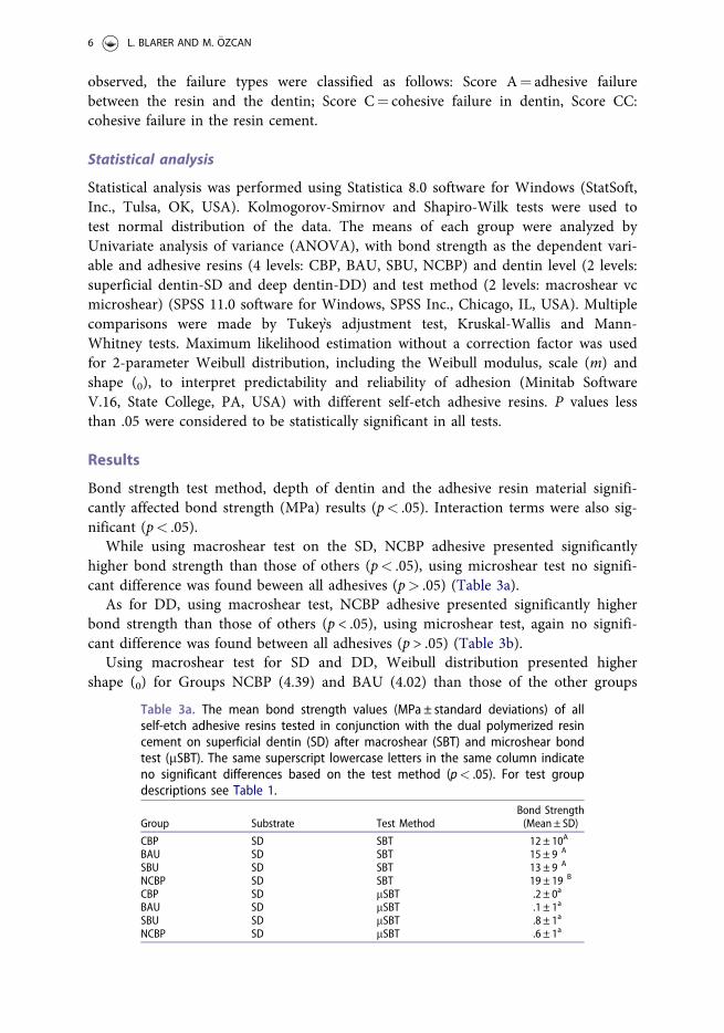
While using macroshear test on the SD, NCBP adhesive presented significantlyhigher bond strength than those of others (p< .05), using microshear test no signifi-cant difference was found beween all adhesives (p> .05) (Table 3a).
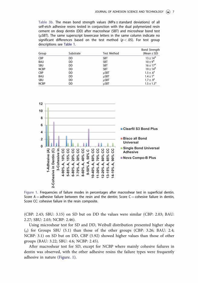
As for DD, using macroshear test, NCBP adhesive presented significantly higherbond strength than those of others (p < .05), using microshear test, again no signifi-cant difference was found between all adhesives (p > .05) (Table 3b).
Using macroshear test for SD and DD, Weibull distribution presented highershape (0) for Groups NCBP (4.39) and BAU (4.02) than those of the other groups
Table 3a. The mean bond strength values (MPa ± standard deviations) of allself-etch adhesive resins tested in conjunction with the dual polymerized resincement on superficial dentin (SD) after macroshear (SBT) and microshear bondtest (mSBT). The same superscript lowercase letters in the same column indicateno significant differences based on the test method (p< .05). For test groupdescriptions see Table 1.
Group Substrate Test MethodBond Strength(Mean ± SD)
CBP SD SBT 12 ± 10A
BAU SD SBT 15 ± 9 A
SBU SD SBT 13 ± 9 A
NCBP SD SBT 19 ± 19 B
CBP SD mSBT .2 ± 0a
BAU SD mSBT .1 ± 1a
SBU SD mSBT .8 ± 1a
NCBP SD mSBT .6 ± 1a
6 L. BLARER AND M. €OZCAN
(CBP: 2.43; SBU: 3.15) on SD but on DD the values were similar (CBP: 2.03; BAU:2.27; SBU: 2.03; NCBP: 2.46).
Using microshear test for SD and DD, Weibull distribution presented higher shape(0) for Groups SBU (5.1) than those of the other groups (CBP: 3.26; BAU: 2.4;NCBP: 3.1) on SD but on DD, CBP (5.92) showed higher values than those of othergroups (BAU: 3.22; SBU: 4.6; NCBP: 2.45).
After macroshear test for SD, except for NCBP where mainly cohesive failures indentin was observed, with the other adhesive resins the failure types were frequentlyadhesive in nature (Figure. 1).
Table 3b. The mean bond strength values (MPa ± standard deviations) of allself-etch adhesive resins tested in conjunction with the dual polymerized resincement on deep dentin (DD) after macroshear (SBT) and microshear bond test(mSBT). The same superscript lowercase letters in the same column indicate nosignificant differences based on the test method (p< .05). For test groupdescriptions see Table 1.
Group Substrate Test MethodBond Strength(Mean ± SD)
CBP DD SBT 13 ± 14A
BAU DD SBT 10 ± 9A
SBU DD SBT 16 ± 17A
NCBP DD SBT 19 ± 14B
CBP DD mSBT 1.5 ± .6a
BAU DD mSBT 1.4 ± 1a
SBU DD mSBT 1.7 ± .9a
NCBP DD mSBT 1.5 ± 1.3a
0
2
4
6
8
10
12
1-A
dh
esiv
e (A
)2-
Co
hes
ive
in D
enti
n (
C)
3-C
oh
esiv
e in
…4-
90%
A, 1
0% C
C5-
85%
A, 1
5% C
C6-
80%
A, 2
0% C
C7-
70%
A, 3
0% C
C8-
60%
A, 4
0% C
C9-
50%
A, 5
0% C
10-4
0% A
, 60%
CC
11-3
0% A
, 70%
CC
12-2
0% A
, 80%
CC
13-1
5% A
, 85%
CC
14-1
0% A
, 90%
CC
Clearfil S3 Bond Plus
Bisco all BondUniversal
Single Bond UniversalAdhesive
Nova Compo-B Plus
Figure 1. Frequencies of failure modes in percentages after macroshear test in superficial dentin.Score A¼ adhesive failure between the resin and the dentin; Score C¼ cohesive failure in dentin,Score CC: cohesive failure in the resin composite.
JOURNAL OF ADHESION SCIENCE AND TECHNOLOGY 7

On DD, with all adhesives, failure types were exclusively adhesive between thedentin and the resin material with both macro and microshear test methods (Score A).
Discussion
This study was undertaken in order to evaluate the adhesion of conventional resin-based luting cement in conjunction with self-etch adhesive resin systems to SD andDD using both macroshear and microshear tests. Based on the results of this study,the results were dependent on the test method, namely macroshear test resulted insignificantly higher results in DD compared to microshear yielding to rejection of thefirst hypothesis. Macroshear bond strength test resulted in significantly higher bondstrength results both on SD and DD compared to microshear test method. Thus, thesecond hypothesis could be rejected. Furthermore, self-etch adhesive resins with lesspH (2.3) did not deliver higher bond strength results than those of the adhesives withhigher pH (�2.5) resulting rejecting the third hypothesis.
Significantly higher results were obtained for bond strength of resin composite toSD and DD when macroshear test method was used. Interestingly, the smaller size ofthe bonded area did not necessarily resulted in higher bond strength. Althoughmicroshear offers bonded areas of 1 to 1.2mm2, application of the adhesive throughthe mould was complex and most possibly led to less good wettability of the adhesiveto the dentin surface yielding to practically no adhesion to the dentin substrate. Inthis study, adhesion procedures were performed on SD and DD in order to simulateclinical indications where restorations are bonded to both types of substrates. In thisregard, one important aspect in bonding to dentin is the density and orientation ofdentin tubuli. Bonding to DD has been expected to be more challenging than to SDmainly due to the reduced area of solid intertubular dentin associated with theincreased water content. There are controversial results supporting this statement. Inone study, tubular density and tubular cross-sectional area were not found to be sig-nificantly different in deep and coronal dentin [35]. This could be one reason for theinsignificant differences between SD and DD when macroshear test was used whenthe adhesive resins were compared one to one.
In order to measure the bond strength values between an adherent and a substrateaccurately, it is crucial that the bonding interface should be the most stressed region,regardless of the test methodology being employed. According to the Griffith’s theory[10], the tensile strength of the uniform materials decreases when the specimen sizeis increased. Overall, adhesion related studies in dentistry, bonded surface areas rangefrom 3mm2 to 1mm2 in macro- and micro-test methods, respectively [32]. Due tothe reduced bonded area and more homogeneous distribution of stresses, micro-testmethods tend to show significantly higher bond strength results than the macro-testmethods. This could eventually affect the ranking of materials being tested in onestudy [31]. However, microshear tests resulted in significantly lower values regardlessof SD and DD. Since this observation was made for all adhesives used in this study,most likely the wettability of the resin cement was not favourable which was placedthrough the .8mm2 diameter mold. Thus, not only the adhesives but also the
8 L. BLARER AND M. €OZCAN

rheological properties of the recin cement and in connection to that the technicallimitations might be a limitation of microshear tests in adhesion test methods.
One factor affecting the results of shear bond tests is the type of jig. Higher stressconcentration was reported with chisel followed by loop type of jigs but where thelatter was considered a better test in terms of stress distribution when compared tochisel type of jigs [37]. With the chisel jig, the knife blade may create shear andbending and thus the stress distribution at the interface is not the presumed as solelyshear bond strength. In addition, it is almost impossible to achieve homogenousforces at the interface that is in fact also the clinical situation in the majority of thecases. Nevertheless, in order to avoid possible deformation of the stainless steel stripduring the tests, a loop type of jig was not considered in this study.
The “etch-and-rinse” adhesives require a moist substrate for optimal bonding [38],making it highly sensitive since the collapse of over-dried, exposed collagen acts as adifficult substrate for the monomer infiltration. However, this also indicates that in thecase of increased dentin wetness, which occurs when dentin depth increases, a too wetcondition may be created [39]. This makes three-step “etch-and-rinse” adhesives moretechnique sensitive compared to self-etch adhesive types. Usually, 32–40% ortho-phos-phoric acid completely demineralizes 5–8lm into the intertubular dentin matrix andcreates nanometer-sized porosities around collagen fibrils [40]. It also partially deminer-alizes dentin to a greater depth [41]. Unfortunately, full infiltration of resin monomerswithin phosphoric acid etched dentin is a complex task. Excessive acid conditioningcauses deep demineralization that jeopardizes complete infiltration of resin monomers,thereby resulting in the formation of a weaker and unprotected demineralized dentinzone at the base of these hybrid layers [42]. Adhesives with milder acidity (pH around2.6) could contribute to the creation of a thinner partially demineralized collagen layercharacterized by the presence of mineral precipitation) [41] and less degradation underthe hybrid layer. Although the interaction of the etching agentes with dentin is limitedby the buffering effect of the mineral and organic phases, there is often a discrepancybetween the depth of dentin demineralization versus monomer penetration [43].Self-etching primers which contain non-rinsing, acidic, polymerizable monomers dis-solve the smear layer, or incorporate it into the bonding interface, as it demineralizesthe surface and covers collagen fibrils and hydroxyapatite crystals. Demineralizationand monomer infiltration occur simultaneously, preventing collagen from collapsingand avoiding the exposure of an unprotected collagen network [43].
The self-etching primers eliminate the technique-sensitive rinsing step to removethe phosphoric acid from dentin. However, the most efficient self-etching adhesivesare based on strongly acidic adhesive monomers. Most of the currently available self-etching adhesives are methacrylate-based with a pH value in the range of 1.5–2.5. Inthis study, two types of pH was of interest, namely pH of <2.3 and pH of 2.5 to 3.5.The results of this study indicated more favourable results with NCBP with pH of 2.5to 3 whereas BAU with similar pH resulted in lower bond strength values. Theseresults indicate that not only pH but the chemistry might have been influential onthe results. The presence of HEMA in NCBP might have increased wettability andthe MDP served for the chemical adhesion with the dentin. In addition to that4-META might have contributed to degree of polymerization [44].
JOURNAL OF ADHESION SCIENCE AND TECHNOLOGY 9

Bond strength results in adhesion studies should be also interpreted with failuretypes. Cohesive failures in the substrate and combination of adhesive and cohesive fail-ure types in the substrate and bonding agent indicate that bond strength of the adhe-sive system and the resin cement exceeds that of the cohesive strength of the substrate.After macroshear test for SD, except for NCBP where mainly cohesive failures in den-tin was observed, with the other adhesive resins the failure types were frequently adhe-sive. This finding shows that adhesion remains to be still less reliable on DD.
The presence of pulpal fluid under pressure in vital dentin could be an importantfactor influencing adhesive bonding to dentin [45]. One limitation of this study wasthe lack of such a simulation. In future studies, pulpal pressure should also be simu-lated when comparing the durability of adhesion of the adhesive resins tested [46,47].
Considering bond strength values and the failure types, self-etch adhesives, notonly the pH but the chemical composition of adhesive resins on dentin dictates thelevel of adhesion especially in SD. However, in DD, practically the difference betweenthe adhesive resins appears to be less of significance.
Conclusions
From this study, the following could be concluded:
1. All self-etch adhesives tested presented significantly higher bond strength onsuperficial dentin compared to deep dentin.
2. Macroshear test presented higher results for both superficial and deep dentincompared to microshear test.
3. Considering Weilbull parameters, characteristics of adhesion and thereby inter-facial strength seems to be more reliable with NCBP in macroshear but with SBUin microshear test.
4. After macroshear test, failure types were mainly cohesive in the resin but aftermicroshear test primarily adhesive failures between the material and dentinwere common.
Clinical Relevance
Considering bond strength values and the failure types, with the self-etch adhesivestested, not only the pH but the chemical composition dictates adhesion especially tosuperficial dentin. In deep dentin, the difference between the adhesive resins appearsto be less of significance.
Acknowledgement
We would like to acknowledge Imicryl, GmBH, Konya, Turkey for generous provision ofresin cements used in this study. The authors further acknowledge Mr. A. Trottmann,University of Z€urich, Center for Dental and Oral Medicine, Z€urich, Switzerland, for his assist-ance with the specimen preparation, Dr. M. Roos, from the Division of Biostatistics, Instituteof Social and Preventive Medicine, University of Zurich, Switzerland for her support with thestatistical analysis.
10 L. BLARER AND M. €OZCAN

Disclosure staement
The authors did not have any commercial interest in any of the materials used in this study.
References
[1] Edelhoff D, €Ozcan M. To what extend does the longevity of fixed dental prosthesesdepend on the function of cement? Clin Oral Implants Res. 2007;18:193–204.
[2] Manso AP, Carvallho RM. Dental cements for luting and bonding restorations:self-adhesive cements. Dent Clin North Am. 2017;61:821–834.
[3] Van Meerbeek B, De Munk J, Yoshida Y, et al. Adhesion to enamel and dentin: currentstatus and future challenges. Oper Dent. 2003;28:215–235.
[4] Tay FR, Pashley DH. Aggressiveness of contemporary self- etching systems 1: depth ofpenetration beyond smear layers. Dent Mater. 2001;17:296–308.
[5] Tay F, Pashley D, Yoshiyama M. Two modes of nanoleakage expression in single-stepadhesives. J Dent Res. 2002;81:47–476.
[6] Ferracane JL, Stansbury JW, Burke FJT. Self-adhesive resin cements- chemistry, proper-ties and clinical considerations. J Oral Rehabil. 2010;38:1–20.
[7] Armstrong SR, Vargas MA, Fang Q, et al. Microtensile bond strength of a total-etch3-step, total-etch 2-step, self-etch 2-step, and a self-etch 1-step dentin bonding systemthrough 15-month water storage. J Adhes Dent. 2003;5:47–56.
[8] Melo RM, €Ozcan M, Barbosa SH, et al. Bond strength of two resin cements on dentinusing different cementation strategies. J Esthet Restor Dent. 2010;22:262–268.
[9] Yoshida Y, Nagakane K, Fukuda R, et al. Comparative study on adhesive perform- anceof functional monomers. J Dent Res. 2004;83:454–458.
[10] Rosa WL, Piva E, Silva AF. Bond strength of universal adhesives: A systematic reviewand meta-analysis. J Dent. 2015;43:765–776.
[11] Suh BI, Feng L, Pashley DH, et al. Factors contributing to the incompatibility betweensimplified-step adhesives and chemically cured or dual-cured composites. Part III.Effect of acidic resin monomers. J Adhes Dent. 2003;5:267–282.
[12] Spencer P, Wang Y, Walker MP, et al. Molecular structure of acid-etched dentin smearlayers-in situ study. J Dent Res. 2007;80:1802–1807.
[13] Choi AN, Lee JH, Son SA, et al. Effect of dentin wetness on the bond strength ofuniversal adhesives. Materials. 2017;10:1224.
[14] Tay FR, Pashley DH, Suh BI, et al. Single-step adhesives are permeable membranes.J Dent. 2002;30:371–382.
[15] Chang J, Platt JA, Yi K, et al. Quantitative comparison of the water permeable zoneamong four types of dental adhesives used with a dual-cured composite. Oper Dent.2006;31:346–353.
[16] Tay FR, King NM, Suh BI, et al. Effect of delayed activation of light-cured resincomposites on bonding of all-in-one adhesives. J Adhes Dent. 2001;3:207–225.
[17] King NM, Tay FR, Pashley DH, et al. Conversion of one-step to two-step self-etchadhesives for improved efficacy and extended application. Am J Dent. 2005;18:126–134.
[18] Van Landuyt KL, Snauwaert J, De Munck J, et al. Origin of interfacial droplets withone-step adhesives. J Dent Res. 2007;86:739–744.
[19] Pereira PNR, Okuda M, Sano H, et al. Effect of intrinsic wetness and regional differ-ence on dentin bond strength. Dent Mater. 1999;15:46–53.
[20] Sensi L, Lopes G, Monteiro S, et al. Dentin bond strength of self-etching primers/adhe-sives. Oper Dent. 2005;30:63–68.
[21] Gale MS, Darvell BW. Thermal cycling procedures for laboratory testing of dentalrestorations. J Dent. 1999;27:89–99.
[22] Cheong C, King NM, Pashley DH, et al. Incompatibility of self-etch adhesives with chem-ical/dual-cured composites: two-step vs one-step systems. Oper Dent. 2003;28:747–755.
JOURNAL OF ADHESION SCIENCE AND TECHNOLOGY 11
[23] Bolhuis PB, de Gee AJ, Kleverlaan CJ, et al. Contraction stress and bond strength todentin for compatible and incompatible combinations of bonding systems and chemicaland light-cured core build-up resin composites. Dent Mater. 2006;22:223–233.
[24] Yoshiama M, Matsuo T, Ebisu S, et al. Regional bond strengths of self-etching/self-pri-ming adhesive systems. J Dent. 1998;26:609–616.
[25] €Ozcan M, Mese A. Adhesion of conventional and simplified resin-based luting cementsto superficial and deep dentin. Clin Oral Investig. 2012;16:1081–1088.
[26] Adebayo OA, Burrow, Tyas M. Bonding of one-step and two-step self-etchingprimer adhesives to dentin with different tubule orientations. Acta Odontol Scand.2008;66:159–168.
[27] Cardenas AFM, Siqueira FSF, Bandeca MC, et al. Impact of pH and application time ofmeta-phosphoric acid on resin-enamel and resin-dentin bonding. J Mech BehavBiomed Mater. 2018;78:352–361.
[28] Della Bona A, Van Noort R. Shear vs. tensile bond strength of resin composite bondedto ceramic. J Dent Res. 1995;74:1591–1596.
[29] Versluis A, Tantbirojn D, Douglas WH. Why do shear bond tests pull out dentin?J Dent Res. 1997;76:1298–1307.
[30] Betamar N, Cardew G, van Noort R. Influence of specimen designs on the microtensilebond strength to dentin. J Adhes Dent. 2007;9:159–168.
[31] Valandro LF, €Ozcan M, Amaral R, et al. Effect of testing methods on the bond strengthof resin to zirconia-alumina ceramic: microtensile versus shear test. Dent Mater J.2008;27:849–855.
[32] Human Research Ordinance (810.301), Art. 30.[33] World Medical Association (WMA): Declaration of Helsinki – Ethical Principles for
Medical Research Involving Human Subjects. 64th WMA General Assembly, Fortaleza,Brazil, 2013.
[34] Human Research Act (810.30), Art. 2 and 32, Human Research Ordinance (810.301),Art. 25.
[35] Lopes GC, Perdig~ao J, Lopes Mde F, et al. Dentin Bond strengths of simplified adhe-sives: effect of dentin depth. Compend Contin Educ Dent. 2006;27:340–346.
[36] Ulker M, €Ozcan M, Seng€un A, et al. Effect of artificial aging regimens on the perform-ance of self-etching adhesives. J Biomed Mater Res B Appl Biomater.2010;93:175–184.
[37] €Ozcan M, Kojima AN, Nishioka RS, et al. Effect of jig design and assessment of stressdistribution in testing metal-ceramic adhesion. J Prosthodont. 2016;25:665–669.
[38] Carvalho RM, Pegoraro TA, Tay FR, et al. Adhesive permeability affects coupling ofresin cements that utilize self-etching primers to dentine. J Dent. 2004;32:55–65.
[39] Paul SJ, Leach M, Rueggeberg FA, et al. Effect of water content on the physical proper-ties of model dentine primer and bonding resins. J Dent. 1999;27:209–214.
[40] Pashley DH, Tay FR, Breschi L. et al. State of the art etch-and-rinse adhesives. DentMater. 2011;27:1–16.
[41] Feitosa VP, Bazzocchi MG, Putignano A. et al. Dicalcium phosphate (CaHPO4.2H2O)precipitation through ortho- or meta-phosphoric acid-etching: effects on thedurability and nanoleakage/ultra-morphology of resin-dentine interfaces. J Dent.2013;41:068–1080.
[42] Hashimoto M, Ohno H, Endo K, et al. The effect of hybrid layer thickness onbond strength: demineralized dentin zone of the hybrid layer. Dent Mater.2000;16:406–411.
[43] Jacques P, Hebling J. Effect of dentin conditioners on the microtensile bond strengthof a conventional and a self-etching primer adhesive system. Dent Mater.2005;21:103–109.
[44] Hanabusa M, Yoshihara K, Yoshida Y, et al. Interference of functional monomers withpolymerization efficiency of adhesives. Eur J Oral Sci. 2016;124:204–209.
12 L. BLARER AND M. €OZCAN
[45] Caiado AC, de Goes MF, de Souza-Filho FJ, et al. The effect of acid etchant typeand dentin location on tubular density and dimension. J Prosthet Dent.2010;103:352–361.
[46] Griffith AA. The phenomena of rupture and flow in solids. Phil Trans R Soc London,Ser. A 221: 168–98, 1920.
[47] Van Meerbeek B, Peumans M, Poitevin A, et al. Relationship between bond strengthtests and clinical outcomes. Dent Mater. 2010;26:100–121.
JOURNAL OF ADHESION SCIENCE AND TECHNOLOGY 13