Present and future technology for simultaneous superficial thermoradiotherapy of breast cancer
18
Present and Future Technology for Simultaneous Superficial Thermoradiotherapy of Breast Cancer Eduardo G. Moros, Ph.D. 1 , Jose Peñagaricano, M.D. 1 , Petr Novàk, Ph.D. 1 , William L. Straube, M.S. 2 , and Robert J. Myerson, M.D. 2 1 Department of Radiation Oncology, University of Arkansas for Medical Sciences, Little Rock, AR 2 Radiation Oncology Department, Washington University School of Medicine, St. Louis, MO Abstract This paper reviews systems and techniques to deliver simultaneous thermoradiotherapy of breast cancer. It first covers the clinical implementation of simultaneous delivery of superficial (microwave or ultrasound) hyperthermia and external photon beam radiotherapy, first using a Co-60 teletherapy unit and later medical linear accelerators. The parallel development and related studies of the SURLAS, an advanced system specifically designed and developed for simultaneous thermoradiotherapy, follows. The performance characteristics of the SURLAS are reviewed and power limitation problems at high acoustic frequencies (> 3MHz) are discussed along with potential solutions. Next, the feasibility of simultaneous SURLAS hyperthermia and IMRT/IGRT is established based on published and newly presented studies. Finally, based on the encouraging clinical results thus far, it is concluded that new trials employing the latest technologies are warranted along with further developments in treatment planning. Keywords breast; cancer; chest wall; heat; hyperthermia; radiation therapy; radiotherapy; radiosensitization; recurrence; thermoradiotherapy; simultaneous; ultrasound; intensity modulated Hyperthermia and Cancer Conventional hyperthermia in cancer therapy can be defined as the elevation of tissue temperatures to 41~43°C for more than 30 minutes. Hyperthermia is typically used as an adjunct therapy to radiotherapy and/or chemotherapy [1–5]. Many well-conducted clinical trials, including phase III multi-institutional trials following quality assurance guidelines, have shown that hyperthermia can significantly increase both local tumor control rates and duration of local control in tumors that recur or persist after surgery, radiotherapy and/or chemotherapy [6–9]. Furthermore, hyperthermia has been shown beneficial in the treatment of residual microscopic disease in the management of local-regional breast cancer [10], in the treatment of soft tissue sarcomas preoperatively with radiation [11], and in the treatment of deep pelvic tumors [12,13]. A significant survival benefit was also reported when hyperthermia was combined with brachytherapy in the treatment of glioblastoma multiforme [14] and with external beam radiotherapy in the treatment of recurrent head and neck cancer [15]. Corresponding Author: Eduardo G. Moros, Ph.D., Department of Radiation Oncology, University of Arkansas for Medical Sciences, 4301 W. Markham St. # 771, Little Rock, AR 72205. NIH Public Access Author Manuscript Int J Hyperthermia. Author manuscript; available in PMC 2011 January 1. Published in final edited form as: Int J Hyperthermia. 2010 ; 26(7): 699–709. doi:10.3109/02656736.2010.493915. NIH-PA Author Manuscript NIH-PA Author Manuscript NIH-PA Author Manuscript
-
Upload
independent -
Category
Documents
-
view
0 -
download
0
Transcript of Present and future technology for simultaneous superficial thermoradiotherapy of breast cancer

Present and Future Technology for Simultaneous SuperficialThermoradiotherapy of Breast Cancer
Eduardo G. Moros, Ph.D.1, Jose Peñagaricano, M.D.1, Petr Novàk, Ph.D.1, William L. Straube,M.S.2, and Robert J. Myerson, M.D.21 Department of Radiation Oncology, University of Arkansas for Medical Sciences, Little Rock, AR2 Radiation Oncology Department, Washington University School of Medicine, St. Louis, MO
AbstractThis paper reviews systems and techniques to deliver simultaneous thermoradiotherapy of breastcancer. It first covers the clinical implementation of simultaneous delivery of superficial (microwaveor ultrasound) hyperthermia and external photon beam radiotherapy, first using a Co-60 teletherapyunit and later medical linear accelerators. The parallel development and related studies of theSURLAS, an advanced system specifically designed and developed for simultaneousthermoradiotherapy, follows. The performance characteristics of the SURLAS are reviewed andpower limitation problems at high acoustic frequencies (> 3MHz) are discussed along with potentialsolutions. Next, the feasibility of simultaneous SURLAS hyperthermia and IMRT/IGRT isestablished based on published and newly presented studies. Finally, based on the encouragingclinical results thus far, it is concluded that new trials employing the latest technologies are warrantedalong with further developments in treatment planning.
Keywordsbreast; cancer; chest wall; heat; hyperthermia; radiation therapy; radiotherapy; radiosensitization;recurrence; thermoradiotherapy; simultaneous; ultrasound; intensity modulated
Hyperthermia and CancerConventional hyperthermia in cancer therapy can be defined as the elevation of tissuetemperatures to 41~43°C for more than 30 minutes. Hyperthermia is typically used as anadjunct therapy to radiotherapy and/or chemotherapy [1–5]. Many well-conducted clinicaltrials, including phase III multi-institutional trials following quality assurance guidelines, haveshown that hyperthermia can significantly increase both local tumor control rates and durationof local control in tumors that recur or persist after surgery, radiotherapy and/or chemotherapy[6–9]. Furthermore, hyperthermia has been shown beneficial in the treatment of residualmicroscopic disease in the management of local-regional breast cancer [10], in the treatmentof soft tissue sarcomas preoperatively with radiation [11], and in the treatment of deep pelvictumors [12,13]. A significant survival benefit was also reported when hyperthermia wascombined with brachytherapy in the treatment of glioblastoma multiforme [14] and withexternal beam radiotherapy in the treatment of recurrent head and neck cancer [15].
Corresponding Author: Eduardo G. Moros, Ph.D., Department of Radiation Oncology, University of Arkansas for Medical Sciences,4301 W. Markham St. # 771, Little Rock, AR 72205.
NIH Public AccessAuthor ManuscriptInt J Hyperthermia. Author manuscript; available in PMC 2011 January 1.
Published in final edited form as:Int J Hyperthermia. 2010 ; 26(7): 699–709. doi:10.3109/02656736.2010.493915.
NIH
-PA Author Manuscript
NIH
-PA Author Manuscript
NIH
-PA Author Manuscript

In fact, when clinical trials have been conducted under widely accepted quality assuranceguidelines—which implies matching the heating technology to the target site along with theimplementation of a reasonable thermometry strategy—radiotherapy plus hyperthermia hasalways resulted in statistically better outcomes than radiotherapy alone [16].
Thermoradiotherapy of Breast CancerPatients with persistent and/or recurrent breast cancer and chest wall tumors have significantlybenefited when hyperthermia has been added to their radiotherapy and/or chemotherapyregimens [17–26]. One of the physical reasons for the success of hyperthermia in this groupof patients is the superficial location of the local disease. Superficial lesions are the leastdifficult to heat adequately because of their accessibility and proximity to external energysources. The composition of the local anatomy may also contribute to better heating [27,28].Another possible reason may be that superficial lesions are more amenable to invasivethermometry and thermal mapping of temperature sensors, thus providing more temperaturefeedback data during treatment, which can be used to improve heat delivery and ensuretreatment quality [29,30].
Clinical research efforts have demonstrated that the response of cancerous tumors to sequentialthermoradiotherapy (i.e., sequentially combined radiotherapy and hyperthermia) is wellcorrelated with power deposition coverage and/or thermal dose coverage [24,30–40]. Thesestudies have also point to the challenge of achieving consistently biologically meaningfulthermal doses in 100% of the target volume in routine clinical practice. Therefore, it is widelyaccepted that the benefits of hyperthermia as an adjunct to radiotherapy can be significantlyaugmented with improvements in treatment delivery techniques, better heating technology,advances in treatment planning and implementation of quality assurance guidelines [6,13,30,41,42]. Moreover, in vitro and animal studies have shown that when hyperthermia and radiationare administered simultaneously —rather than sequentially as it is conventionally done—heat-induced radiosensitization (HIR) is increased at thermal doses achievable in the clinic [43–47]. For instance, mild hyperthermia—41°C maintained for ~60 minutes—produces HIR ifradiation and heat are delivered simultaneously, but not sequentially. This is significant becausea minimum target temperature of 41°C is more clinically achievable than the > 42°C neededto produce HIR with sequential treatment [44,47,48]. The above facts support the developmentof clinical hyperthermia devices that permit simultaneous delivery of heat and ionizingradiation.
Clinically Implemented Approaches for Simultaneous Thermoradiotherapy ofBreast Cancer
In this section, a review is given of the systems and techniques clinically implemented for thedelivery of simultaneous thermoradiotherapy at Washington University in St. Louis. Thisresearch group has been the only one to complete several simultaneous thermoradiotherapyclinical trials with breast cancer being the site treated the most.
Microwave Hyperthermia and Co-60 TeletherapyAlthough it had been known from in vitro and in vivo experiments that simultaneous deliveryresulted in larger thermal enhancement ratios than sequential delivery, simultaneous treatmentshad not been tried with human patients mainly due to formidable logistical problems [46]. Theresearch group set out to overcome these problems and embark in a long-term clinical researcheffort. The first approach consisted of heating with 915 MHz microwave applicators andirradiating with a Co-60 teletherapy unit [49]. Both technologies enjoyed extensive clinicalexperience, were well-understood and had been used in sequential thermoradiotherapy [39,
Moros et al. Page 2
Int J Hyperthermia. Author manuscript; available in PMC 2011 January 1.
NIH
-PA Author Manuscript
NIH
-PA Author Manuscript
NIH
-PA Author Manuscript

40]. Therefore, any unusual response to the new therapy approach could be attributed to thesimultaneous delivery of heat and gamma radiation. Hyperthermia waveguide applicators wereused, one at the time. Two clinical setups were characterized. In one a microwave applicatorwas attached to a blocking tray and inserted into the tray slot in the head of the gantry of theteletherapy unit. Thus the gamma beam traveled through a mostly hollowed waveguide andinto a treatment volume after transversing a mineral oil coupling bolus. This was called the enface setup. The other setup was with the microwave propagation vector perpendicular to thegamma beam central axis. This was called the orthogonal setup. Using these setups a 400 cGyradiation fraction of 4 to 7 minutes duration could be delivered in the middle of a 60 minutehyperthermia treatment without interruptions. Temperatures and power level were remotelymonitored and recorded outside the teletherapy room. The microwave applicator was notmounted or in contact with the teletherapy unit in the orthogonal setup, so it did not affectedthe radiation dose distributions. However, for the en face setup the dose distributions wereaffected by the microwave applicator. Film measurements in a solid water phantom showeduniform dose within the radiation field, except for 10–18% attenuation under the metal tuningelectrodes inside the waveguide. This dose defect was clinically smoothed using featheringtechniques. This approach was used successfully without technical problems in a phase I/IIclinical trial. Importantly, an analysis showed that the temperature distributions achievedduring simultaneous delivery had the same general characteristics as those achieved duringsequential delivery, and that the steady state distributions were maintained during the time ofsimultaneous irradiation. This first experience demonstrated that simultaneous superficialmicrowave hyperthermia and external beam radiation was technically feasible and safe [49].
Ultrasound Hyperthermia and Co-60 TeletherapyThe waveguide microwave applicators limited the size of the lesions we could treat withhyperthermia simultaneously with gamma rays [39,40]. In order to be able to treat largerlesions, we decided to adapt a commercial planar ultrasound system to deliver simultaneoustreatments. This task was much more challenging because the ultrasound applicators were nothollowed like the microwave waveguides and degassed water was needed to couple the soundwaves into the treatment volume. The ultrasound applicators of the Labthermics 1000 systemwere also bulkier and heavier than the microwave applicators. An orthogonal setup similar tothe one used with microwaves was relatively simple to mimic, but many tumor sites wouldrequire an en face setup for proper treatment. Since ultrasound is reflected efficiently andspecularly from metal surfaces, we devised a reflecting system in which the ultrasound beam,initially directed perpendicularly to the gamma beam, was deflected 90° so that both beamstravel through the same window of entry into the tumor while the ultrasound source remainedoutside the radiation beam [50]. The reflecting system, which was mounted on the blockingtray, was filled with degassed water and made of water-equivalent materials, except for a 1mm sheet of polished brass used as the reflector. With this en face approach the absorbed powerpatterns generated with and without the reflecting system at the same extended distance fromthe transducer demonstrated that the hyperthermia system remained able to control heatingpattern s. The effect of the reflecting system on the gamma beam was minimal except foruniform attenuation, and the Co-60 beam had no effect on the performance of thethermocouples. Extensive testing followed by clinical treatments of patients on protocoldemonstrated that the modifications made did not impair the ability to deliver ultrasoundhyperthermia or teletherapy effectively. Again, this work showed that the implementation ofultrasound hyperthermia simultaneous with gamma irradiation was technically and clinicallyfeasible without complications or hazards to patients.
Moros et al. Page 3
Int J Hyperthermia. Author manuscript; available in PMC 2011 January 1.
NIH
-PA Author Manuscript
NIH
-PA Author Manuscript
NIH
-PA Author Manuscript

Superficial (Microwave or Ultrasound) Hyperthermia and Mega voltage Photon BeamRadiation using a Medical Linear Accelerator (Linac)
In 1995 the Co-60 teletherapy unit at the Mallinckrodt Institute of Radiology wasdecommissioned. The only option for external beam radiation was to develop the devices andtechniques to allow the use of a medical linear accelerator. Although excited about the use ofmodern radiation technology, the transfer to megavoltage photon beam necessitated additionaldevelopments and measurements. Both the orthogonal and the en face setups were successfullytransferred, but had to be adapted to the different geometry and dimensions of the linearaccelerator. Straube et al. (2001) reported on devices, techniques and dosimetry forsimultaneous thermoradiotherapy delivered with a linear accelerator including detailed day-to-day simulation and treatment procedures [51].
Summary of Simultaneous Thermoradiotherapy Clinical TrialsThe above described developments have made possible the conduct of clinical trials since 1992.The first phase I trial was ended successfully in 1995, a second phase I/II dose escalation trialclosed in 2001, and a third trial was initiated 1999. These clinical results have been summarizedpreviously by Myerson et al. (1999, 2004) [44,47]. Up-to-date, 119 patients have been enrolledon these three consecutive trials. The first two trials (60 cases) were for patients withmacroscopic disease, usually recurrent after prior therapies, including radiotherapy, andescalated the number of simultaneous treatments from 3 to 8 in a course of 30–32 Gy. The thirdtrial was to evaluate the short-and long -term effects and local control of simultaneousthermoradiotherapy in the treatment of high risk, but curative, subclinical (no visible diseaseafter surgery and/or chemotherapy) breast carcinoma in patients with no prior radiotherapy.The simultaneous administration of radiotherapy and hyperthermia is of particular potentialvalue for this patient population because tumor cells share normal tissue physiology. Fifty ninepatients were accrued in this trial. The number of simultaneous treatments was escalated from4 to 8 in a course of 60 Gy; most patients receiving 400 cGy per hyperthermia treatment—approximately 26% to 53% of the radiation dose was given under hyperthermic conditions.Elective hyperthermia was given to the primary tumor bed site plus an adjacent 6×12cm striprandomized to lie medial or lateral to the primary site. The adjacent strip served as an unheatedbut irradiated control. The first two studies demonstrated that the per treatment average thermaldose progressively improved with the number of hyperthermia treatments. In addition theproduct of radiation dose and total thermal dose was highly correlated with complete response[47]. In the third trial the overall local control was 97% with no significant morbidity differencebetween heated and control sectors and no increase in morbidity between 4 and 8 simultaneoustreatments.
Finally, the data clearly shows that chest wall lesions (recurrent breast cancer) have been themost responsive to simultaneous thermoradiotherapy with highly significant statisticalcorrelation with radio-thermal dose factors (p < 0.05) [47]. This exciting finding points to theimportance of developing thermoradiotherapy treatment planning systems and continuingefforts to improve clinical thermal dosimetry techniques.
The SURLAS: A Device Developed for Simultaneous ThermoradiotherapyIn parallel with the efforts to adapt existing commercially available hyperthermia technologyto deliver simultaneous hyperthermia and external photon beam radiation, ideas were beingdiscussed for the development of a system specifically designed for this purpose. During thedecade of the nineties, electron beams were used commonly in the treatment of superficiallesions due to their limited depth of penetration, which meant sparing of underlying normaltissues. However, the existing hyperthermia devices were too bulky/dense to permit the use ofelectron beams. This was the main reason only high energy photon beams had been used when
Moros et al. Page 4
Int J Hyperthermia. Author manuscript; available in PMC 2011 January 1.
NIH
-PA Author Manuscript
NIH
-PA Author Manuscript
NIH
-PA Author Manuscript

delivering simultaneous thermoradiotherapy. Eventually, the concept of the ScanningUltrasound Reflector Linear Arrays System, or SURLAS, was born [52,53]. By combiningultrasound linear arrays and scanning ultrasound reflectors, an applicator thin enough to permitthe passage of high energy electron beams could be made to work using an en face approach.Obviously, photon beams would also work as in the past. The device concept is illustrated inFigure 1, which shows a computer-aided solid model of the clinical grade applicator that wasdeveloped [54,55]. Figure 1 shows the basic components of a dual-frequency SURLASapplicator. The reflecting surfaces of a scanning double-faced V-shaped reflector make 45°angles with respect to the sound propagation coming from the (high and low frequency) arraysat the side ends of the applicator head (Figure 1 shows the windows for the arrays, not thearrays), thus both sound beams are deflected in parallel toward the treatment volume. As thereflector scans back and forth, the sound energy is distributed over the treatment volumeproducing therapeutic time-averaged temperature distributions with acceptable temperaturefluctuations [56,57]. Each array has several individually powered elements. Modulation ofpower input to the array elements as the reflector scans between the arrays provides control ofthe two-dimensional surface energy fluence pattern [55,58]. This capability is referred to asLateral Power Conformability or LPC. One of the arrays operates at a low frequency (1 ~ 2MHz) while the other operates at a high frequency (3 ~ 5 MHz). This arrangement allowsconcomitant dual-frequency insonation. Penetration depth control, or PDC, is thus achievedby varying the power input to one array relative to the other’s, i.e., frequency mixing [59,60].For example, for areas where the target volume is deeper, the low frequency can be weightedmore heavily than the high frequency. LPC plus PDC means that the SURLAS has 3D powerdeposition control, which can eventually be fully exploited with the implementation oftemperature feedback control strategies based on temperatures measured during treatment[61].
Several laboratory prototypes of the SURLAS were developed over the years in order todemonstrate feasibility of the overall concept and of particular features such as LPC and PDC.Finally, a clinical grade system was developed that could be used under approval of the FederalFood and Drug Administration (FDA) via the Investigational Device Exemption (IDE).However, manufacturing a clinical device proved to be a much more rigorous process thanassembling prototypes for laboratory studies. Important requirements concerning reliability,patient and operator safety, and quality control/assurance presented great challenges to theresearch group. In addition to these rigorous requirements, the following were specific designcriteria for the clinical SURLAS applicator:
1. Capable of heating superficial tumors extending up to 3 cm deep and up to 15 cm ×15 cm of lateral dimensions.
2. Capable of controlling power deposition in three dimensions.
3. No operational interference with radiotherapy linear accelerators.
4. Suitable for delivery of an external photon (en face or orthogonal setups) or electron(en face setup only) beam radiation during a hyperthermia treatment.
5. Significantly more compact and lighter than existing commercial ultrasoundapplicators.
These design criteria were met for the most part as reported in a paper published in early 2005that focused mainly on the hardware components of the clinical grade SURLAS [54]. A secondpaper published later that same year reported on the design, development, and testing of thepersonal computer based treatment delivery software that coordinated the interactions betweenthe operator, the SURLAS applicator and several peripheral devices [55]. There were twoimportant tasks in software development worth mentioning here. The first was the coordinationof the input power sequences to the elements of the high (4.9 MHz) and the low (1.9 MHz)
Moros et al. Page 5
Int J Hyperthermia. Author manuscript; available in PMC 2011 January 1.
NIH
-PA Author Manuscript
NIH
-PA Author Manuscript
NIH
-PA Author Manuscript

frequency arrays (eight 1.5 cm × 2.0 cm elements/array) with the position of the dual-facescanning reflector. This was achieved by dividing the treatment window in up to 64 sectors(minimum size of 2cm × 2 cm) and controlling the power delivery to each sector independentlyby adjusting the output power from the 16 channels of a programmable radio-frequencygenerator. Upon completion of the software development process, it was integrated with thehardware followed by extensive testing. The second important task was safety, which was aparamount concern and design criterion. To ensure safety, a failure mode and effects analysis,or FMEA, was applied to the entire system in order to identify safety issues and rank theirrelative importance. The FMEA analysis led to the implementation of a software structurewhere each peripheral device communicated independently and directly with the controllingpersonal computer, so that in case of a malfunction in any component of the system or anyviolation of a pre-defined safety criterion, the software can terminate treatment immediately.
Performance of the SURLASAs mentioned before the original motivation and goal was to develop an ultrasoundhyperthermia device that would enable simultaneous hyperthermia and electron beam radiation[52]. Consequently, we tried to make the profile (height) of the SURLAS as small as possiblein order that a high energy electron beam could travel through it and still be energetic enoughto penetrate into superficial lesions as deep as 3 cm (electron beams are attenuated in waterabout 2 MeV/cm and the 80 to 90% isodose cloud is commonly used for tumor coverage). Thismeant that we implemented the smallest arrays possible that our previous studies indicatedsuitable in terms of the required emittance (W/cm2) to induce adequate hyperthermia. Notethat the smaller the array the higher the required continuous wave (CW) emittance. This choiceand other factors discussed below, however, created problems in practice that required time tofigure out and resolve and de railed the clinical translation of the SURLAS.
The major problem was a decrease in electrical-to-acoustic efficiency with increasingtransducer-to-reflector distance [54]. This was particularly obvious for the high frequency arrayas shown in Figure 2, which presents radiation force balance measurements for the low (2a)and high (2b) linear arrays. This loss of ultrasonic power with distance was determined to benon -linear propagation, a subject of much research in recent years. Essentially, nonlinearpropagation is the conversion of ultrasonic energy from the fundamental frequency to higherfrequency harmonics as the waves are distorted while propagating from sinusoidal-like tosawtooth-like [62]. The wave distortion increases with the acoustic intensity, increasingdistance from the source and increasing frequency. It also depends on the characteristics of themedium. This phenomenon has been described by the shock parameter σ [63],
(1)
where β is the coefficient of nonlinearity of the medium, ε is the mach number, k is the wavenumber, and x the travel distance. Based on calculations using the shock parameter wedetermined that the high frequency array was suffering significant power loss due to non-linearpropagation. For the clinical SURLAS, the maximum travel distance of the waves from thearray to the distal water bolus membrane (including reflection) was about 23 cm. For the highfrequency waves(~ 4.9 MHz), nonlinear propagation was calculated to occur at acousticintensities as low as 0.7 W/cm 2at the maximum distance for σ =1 (onset of nonlinearpropagation). The acoustic intensities required for hyperthermia are higher than this value[53]. Moreover, note that scanning of the ultrasound energy over the treatment window requireshigher CW intensities in comparison to a single transducer of comparable size to the treatmentwindow [53,64]. When σ = 3, the waveforms become sawtooth causing intensity losses of 6dB with respect to the fundamental frequency. Assuming plane wave behavior, a series of
Moros et al. Page 6
Int J Hyperthermia. Author manuscript; available in PMC 2011 January 1.
NIH
-PA Author Manuscript
NIH
-PA Author Manuscript
NIH
-PA Author Manuscript

simulations were generated to determine 1) the CW intensity required for the clinical SURLASto induce and maintain hyperthermic temperatures, and 2) the maximum deliverable CWintensity for a shock parameter equal to3 (σ = 3). The frequency, the treatment widow length(~ scanning distance), and height of the linear array were variables. Selected results arepresented in Table 1, which shows the maximum fundamental frequency (fmax) for fourtreatment window lengths (WL) and three array heights (AH). The AH for the clinical SURLASis 1.5 cm and the thickness of the water coupling bolus is approximately2 cm. The developedclinical SURLAS has a maximum window length of 16 cm. Table 1 indicates that to utilize awindow this size fmax could not be higher than 1.89 MHz. The frequency of the high frequencyarray is ~ 4.9 MHz, hence significant nonlinear propagation effects are to be expected perequation (1) and as shown in Figure 2. We had seen this effect before with our commercialultrasound system for the en face setup at the frequency of ~3.5 MHz [50], but the source toskin distance there was over 50 cm, and despite the ~40% reduction of power adequatehyperthermia treatments were possible. This indicated that other factors were contributing tolow efficiencies. These were:
1. Reflectivity of a Styrofoam reflector relative to a brass reflector was estimated to beas 0.76 at 5.0 MHz [52].
2. The first radio frequency generator used had a drop in electrical power output forfrequencies > 3.5 MHz.
3. The array element size in the current arrays is 1.5 cm × 2 cm (the smallest ever used).This turned out to be too small due to: a) influence of the soldered spot and b) dampingby the mount at the edges.
4. Beam divergence at large reflector-to-source distance due to small misalignmentbetween the arrays and the reflector. Computer models had ruled out this possibility[53], but beam plot measurements of the clinical SURLAS suggested otherwise[54].
The message from Table 1 and the above listed factors is clear, to avoid nonlinear propagationand improve electrical-to-acoustic efficiency, a new SURLAS must be constructed consideringthe following: lower frequency for the high frequency array, larger surface areas arrays (largerAH), shorter treatment window length (smaller WL), reflecting areas > array projected areas,and a more powerful radiofrequency generator with flat frequency response. The lowerfrequency for the high frequency array means some reduction in PDC. The smaller treatmentwindow implies a proportional reduction in the size of the treatment area. A larger array heightmeans a higher profile applicator—less or not suitable for electron beams. And larger scanning(possibly metallic) reflector precludes electron beams. That leaves only photon beams for thedelivery of simultaneous thermoradiotherapy with the SURLAS. As discussed in the nextsection, this was actually welcome news due to advances in modern radiotherapy, especiallyintensity modulated radiation therapy or IMRT, as it is commonly called.
SURLAS and IMRT/IGRTIntensity modulated radiation therapy (IMRT) has become clinically widespread worldwide inthe last decade [65]. IMRT’s appeal is its ability to focus dose to the targeted volume(commonly called the planning treatment volume or PTV) while minimizing dose tosurrounding normal tissues. Our first working hypothesis was that IMRT can produce clinicallyacceptable dose distributions on a recurrent breast cancer tumor while that same tumor receivesultrasound hyperthermia using the SURLAS. To test this hypothesis two research projects wereinitiated: a radiation treatment planning study to compute dose distributions in recurrent breastcancer PTVs with the SURLAS applicator in place [66]; and an experimental study, where aradiation treatment plan was computed and delivered to a phantom with the SURLASapplicator on top of the phantom simulating a clinical setup, to evaluate the effects of the
Moros et al. Page 7
Int J Hyperthermia. Author manuscript; available in PMC 2011 January 1.
NIH
-PA Author Manuscript
NIH
-PA Author Manuscript
NIH
-PA Author Manuscript

presence and potential misplacement of the SURLAS applicator on measured dose distributions[67].
The above studies were carried out using the treatment planning station and treatment unit ofa Helical Tomotherapy (HT) system (Tomotherapy Inc., Madison, WI). This technology waschosen over conventional gantry-based medical linear accelerators (both available at ourinstitution), because we thought that the multiplicity of beam angles and the helical motionwould greatly minimize the perturbing effects of the SURLAS applicator on dose distributions.In addition, the mega voltage computed tomography (MVCT) feature of Tomotherapy, whichis used for image-guided radiotherapy (IGRT) to verify the patient’s position right beforetreatment delivery, could also be used for treatment planning purposes without major imagedistortions or artifacts due to the metallic parts of the SURLAS applicator[68]. Moreover, theMVCT images could be also used to verify the position of the SURLAS applicator with respectto the patient and from treatment to treatment. All these ideas were tested successfully [66,67].
The main conclusion from the treatment planning study was that simultaneous treatment withthe SURLAS and HT IMRT/IGRT is feasible as demonstrated by the clinically acceptableradiation treatment plans generated [66]. Likewise, the main conclusion of the experimentalstudy was that the delivered (measured) and planned radiation dose distributions were inexcellent agreement when the SURLAS applicator was positioned as it was planned.Misplacement of the applicator from its planned position was found to have small effects onthe measured dose distributions [67]. Potentially large discrepancies between planned anddelivered doses due to gross misplacement of the SURLAS can be further minimized by theimplementation of routine quality assurance procedures using a position-and-orientationmagnetic tracking device which was successful in precisely and reproducibly positioning ofthe applicator. Moreover, it was shown that there is great potential for the use of this trackingdevice for monitoring of the SURLAS applicator position and orientation during simultaneoustreatments, and not just for patient/SURLAS set-up prior to daily treatment using MVCT[67].
Do the above findings apply if a conventional medical linear accelerator is used? This isimportant question to answer because gantry-based linacs remain the most commonly usedtechnology in radiation therapy departments. Here we present new treatment planning resultsthat indicate that Linac-based IMRT can also deliver clinically acceptable radiation dosedistributions with the SURLAS applicator on the patient’s chest wall. The clinical setup andplanning MVCT can be seen on the axial plane of Figure 3. The PTV was the superficial casein Penagaricano et al. 2008, with maximum dimensions of 1 cm deep by 11.5 cm wide (lateral)by 11 cm long (superior-inferior) on the lower chest/upper abdominal wall mimicking a chestwall recurrence from breast cancer[66]. The results of Figures 3 and 4 were generated usingthe Pinnacle3 inverse IMRT treatment planning system (P3IMRT, Philips, Andover, MA) tomodel megavoltage photon beam dose deposition delivered by a Varian 2100 EX linearaccelerator with 120-leaf dynamic multi-leaf collimator (Varian Medical Systems, Palo Alto,CA). A 12 beam plan was used, 8 beams were coplanar (linac central axis on the axial plane)and 4 beams were non-coplanar (linac central axis on the sagittal plane). Of the 8 coplanarbeams, 6 were aimed from above the SURLAS applicator and 2 were aimed laterally andtangentially to the chest wall. The noncoplanar beams were all aimed from above. Theprescription was 50 Gy in 25 fractions to 95% of the PTV using 6 MV photons. It was assumedthat every fraction was given under hyperthermic conditions for the purpose of illustration.Cardiac toxicity would be solely dependent on radiation dose because none of the existing orforth coming devices would be able to induce significant hyperthermia in heart tissue due to1) the depth of heart tissue and 2) the very high cardiac perfusion rates.
Moros et al. Page 8
Int J Hyperthermia. Author manuscript; available in PMC 2011 January 1.
NIH
-PA Author Manuscript
NIH
-PA Author Manuscript
NIH
-PA Author Manuscript

Figure 3 shows computed dose distributions on the axial, sagittal and coronal planes throughthe PTV and SURLAS applicator. It can be readily seen that the mass was nicely covered bythe prescription dose with low doses to surrounding normal tissues. Dose volume histograms(DVH) for the PTV and organs at risk are shown in Figure 4. These DVHs are clinicallyacceptable for the PTV and surrounding organs and are comparable to the ones obtained usingthe Tomotherapy system [66]. The PTV mean dose, the PTV D95%, the heart, lung andabdomen mean doses, and the spinal cord maximum dose were 50.7, 50.4, 16.7, 3.7, 12.9 and10.0 Gy, respectively. These metrics are in good agreement with those reported byPenagaricano et al. 2008 for the Tomotherapy system (Table IV in [66]). In short, IMRT witheither radiation therapy system is suitable for simultaneous thermoradiotherapy using theSURLAS.
Besides more conformal radiation doses, there are other advantages of using IMRT with theSURLAS. First, IMRT is expected to be highly forgiving of the modifications needed to theSURLAS applicator to minimize non-linear propagation and improve electrical-to-acousticalefficiencies. Second, the clinical experience thus far has only used one radiation beamconcomitantly with hyperthermia; consequently the time of simultaneous delivery is shortcompared to the heating time. With IMRT and the SURLAS, the simultaneous time is expectedto be longer for hyperthermia can be given during an entire multi-beam IMRT fraction. Third,the application of IGRT and magnetic position-orientation tracking technologies, to ensurereproducible applicator positioning prior and during the treatments, may encourage physiciansto prescribe hyperthermia simultaneously with more if not every IMRT fraction. Finally, moresimultaneous fractions should maximize the radiosensitizing and physiological effects ofhyperthermia even if IMRT is delivered in shorter times using the emerging arc therapyparadigm [69].
Final RemarksThe clinical implementation of simultaneous thermoradiotherapy has been challenging butproven to be feasible and safe. Clinical trial results have been encouraging and breast cancersare thus far the most responsive, with statistically significant correlations (p < 0.05) with radio-thermal dosimetric factors [47]. Therefore, new technological developments such as combiningthe SURLAS and IMRT/IGRT with thermo-radio dose treatment planning should translate intoimprovements in radiothermal dose coverage and consequently in better treatment outcomes.Furthermore, several research groups have been developing advanced simultaneous heat andirradiation technologies that are moving toward clinical deployment while others haveattempted different clinical approaches using existing hyperthermia devices [70–76]. Inconclusion, new clinical protocols combining hypethermia and radiotherapy simultaneouslyare now possible and warranted.
AcknowledgmentsThe work reviewed in this paper was supported by NIH Grants CA 63121 and CA 71638, and by a grant from theWhitaker Foundation. We gratefully acknowledge partial support from the Central Arkansas Radiation TherapyInstitute (CARTI) and a Medical Research Endowment pilot award from College of Medicine of the University ofArkansas for Medical Sciences, Little Rock. We thank our cancer patients for their courage and participation in theclinical research. Finally, we gratefully acknowledge the contributions of many collaborators over the years as reflectedin the authorship of previously published papers and abstracts.
References1. Kampinga HH. Cell biological effects of hyperthermia alone or combined with radiation or drugs: a
short introduction to newcomers in the field. Int J Hyperthermia 2006;22:191–6. [PubMed: 16754338]2. Moyer HR, Delman KA. The role of hyperthermia in optimizing tumor response to regional therapy.
Int J Hyperthermia 2008;24:251–61. [PubMed: 18393003]
Moros et al. Page 9
Int J Hyperthermia. Author manuscript; available in PMC 2011 January 1.
NIH
-PA Author Manuscript
NIH
-PA Author Manuscript
NIH
-PA Author Manuscript

3. Hildebrandt B, Wust P. The biologic rationale of hyperthermia. Cancer Treat Res 2007;134:171–84.[PubMed: 17633053]
4. Dewhirst MW, Vujaskovic Z, Jones E, et al. Re-setting the biologic rationale for thermal therapy. IntJ Hyperthermia 2005;21:779–90. [PubMed: 16338861]
5. Bergs JW, Franken NA, Haveman J, et al. Hyperthermia, cisplatin and radiation trimodality treatment:a promising cancer treatment? A review from preclinical studies to clinical application. Int JHyperthermia 2007;23:329–41. [PubMed: 17558732]
6. Dewhirst MW, Prosnitz L, Thrall D, et al. Hyperthermic treatment of malignant diseases: current statusand a view toward the future. Semin Oncol 1997;24:616–25. [PubMed: 9422258]
7. Falk MH, Issels RD. Hyperthermia in oncology. Int J Hyperthermia 2001;17:1–18. [PubMed:11212876]
8. Hehr T, Wust P, Bamberg M, et al. Current and potential role of thermoradiotherapy for solid tumours.Onkologie 2003;26:295–302. [PubMed: 12845217]
9. Issels RD. Regional hyperthermia in high-risk soft tissue sarcomas. Curr Opin Oncol 2008;20:438–43. [PubMed: 18525341]
10. Kapp DS, Cox RS, Barnett TA, et al. Thermoradiotherapy for residual microscopic cancer: electiveor post-excisional hyperthermia and radiation therapy in the management of local-regional recurrentbreast cancer. Int J Radiat Oncol Biol Phys 1992;24:261–77. [PubMed: 1526865]
11. Prosnitz LR, Maguire P, Anderson JM, et al. The treatment of high-grade soft tissue sarcomas withpreoperative thermoradiotherapy. Int J Radiat Oncol Biol Phys 1999;45:941–9. [PubMed: 10571201]
12. van der Zee J, Gonzalez GD. The Dutch Deep Hyperthermia Trial: results in cervical cancer. Int JHyperthermia 2002;18:1–12. [PubMed: 11820462]
13. van der Zee J. Heating the patient: a promising approach? Ann Oncol 2002;13:1173–84. [PubMed:12181239]
14. Sneed PK, Stauffer PR, McDermott MW, et al. Survival benefit of hyperthermia in a prospectiverandomized trial of brachytherapy boost +/− hyperthermia for gliobla stoma multiforme. Int J RadiatOncol Biol Phys 1998;40:287–95. [PubMed: 9457811]
15. Valdagni R, Amichetti M. Report of long-term follow-up in a randomized trial comparing radiationtherapy and radiation therapy plus hyperthermia to metastatic lymph nodes in stage IV head and neckpatients. Int J Radiat Oncol Biol Phys 1994;28:163–9. [PubMed: 8270437]
16. Dewhirst MW, Sneed PK. Those in gene therapy should pay closer attention to lessons fromhyperthermia. Int J Radiat Oncol Biol Phys 2003;57:597–9. author reply 599–600. [PubMed:12957278]
17. Jones EL, Oleson JR, Prosnitz LR, et al. Randomized trial of hyperthermia and radiation for superficialtumors. J Clin Oncol 2005;23:3079–85. [PubMed: 15860867]
18. Jones EL, Prosnitz LR, Dewhirst MW, et al. Thermochemoradiotherapy improves oxygenation inlocally advanced breast cancer. Clin Cancer Res 2004;10:4287–93. [PubMed: 15240513]
19. Feyerabend T, Wiedemann GJ, Jager B, et al. Local hyperthermia, radiation, and chemotherapy inrecurrent breast cancer is feasible and effective except for inflammatory disease. Int J Radiat OncolBiol Phys 2001;49:1317–25. [PubMed: 11286840]
20. Hehr T, Lamprecht U, Glocker S, et al. Thermoradiotherapy for locally recurrent breast cancer withskin involvement. Int J Hyperthermia 2001;17:291–301. [PubMed: 11471981]
21. Jones EL, Marks LB, Prosnitz LR. Point: Hyperthermia with radiation for chest wall recurrences. JNatl Compr Canc Netw 2007;5:339–44. [PubMed: 17439762]
22. Wahl AO, Rademaker A, Kiel KD, et al. Multi-institutional review of repeat irradiation of chest walland breast for recurrent breast cancer. Int J Radiat Oncol Biol Phys 2008;70:477–84. [PubMed:17869019]
23. Welz S, Hehr T, Lamprecht U, et al. Thermoradiotherapy of the chest wall in locally advanced orrecurrent breast cancer with marginal resection. Int J Hyperthermia 2005;21:159–67. [PubMed:15764357]
24. Lee HK, Antell AG, Perez CA, et al. Superficial hyperthermia and irradiation for recurrent breastcarcinoma of the chest wall: prognostic factors in 196 tumors. Int J Radiat Oncol Biol Phys1998;40:365–75. [PubMed: 9457823]
Moros et al. Page 10
Int J Hyperthermia. Author manuscript; available in PMC 2011 January 1.
NIH
-PA Author Manuscript
NIH
-PA Author Manuscript
NIH
-PA Author Manuscript

25. Kapp DS, Barnett TA, Cox RS, et al. Hyperthermia and radiation therapy of local-regional recurrentbreast cancer: prognostic factors for response and local control of diffuse or nodular tumors. Int JRadiat Oncol Biol Phys 1991;20:1147–64. [PubMed: 2022519]
26. Vernon CC, Hand JW, Field SB, et al. Radiotherapy with or without hyperthermia in the treatmentof superficial localized breast cancer: results from five randomized controlled trials. InternationalCollaborative Hyperthermia Group. Int J Radiat Oncol Biol Phys 1996;35:731–44. [PubMed:8690639]
27. Leon SA, Asbell SO, Edelstein G, et al. Effects of hyperthermia on bone. I. Heating rate patternsinduced by microwave irradiation in bone and muscle phantoms. Int J Hyperthermia 1993;9:69–75.[PubMed: 8433027]
28. Moros EG, Novak P, Straube WL, et al. Thermal contribution of compact bone to intervening tissue-like media exposed to planar ultrasound. Phys Med Biol 2004;49:869–86. [PubMed: 15104313]
29. Hand JW. Guidelines for thermometry in clinical hyperthermia. Front Med Biol Eng 1992;4:99–104.[PubMed: 1510889]
30. Dewhirst MW, Phillips TL, Samulski TV, et al. RTOG quality assurance guidelines for clinical trialsusing hyperthermia. Int J Radiat Oncol Biol Phys 1990;18:1249–59. [PubMed: 2347733]
31. Cox RS, Kapp DS. Correlation of thermal parameters with outcome in combined radiation therapy-hyperthermia trials. Int J Hyperthermia 1992;8:719–32. [PubMed: 1479198]
32. Hand JW, Machin D, Vernon CC, et al. Analysis of thermal parameters obtained during phase IIItrials of hyperthermia as an adjunct to radiotherapy in the treatment of breast carcinoma. Int JHyperthermia 1997;13:343–64. [PubMed: 9278766]
33. Sherar M, Liu FF, Pintilie M, et al. Relationship between thermal dose and outcome inthermoradiotherapy treatments for superficial recurrences of breast cancer: data from a phase III trial.Int J Radiat Oncol Biol Phys 1997;39:371–80. [PubMed: 9308941]
34. Kapp DS, Cox RS. Thermal treatment parameters are most predictive of outcome in patients withsingle tumor nodules per treatment field in recurrent adenocarcinoma of the breast. Int J Radiat OncolBiol Phys 1995;33:887–99. [PubMed: 7591899]
35. Leopold KA, Dewhirst M, Samulski T, et al. Relationships among tumor temperature, treatment time,and histopathological outcome using preoperative hyperthermia with radiation in soft tissuesarcomas. Int J Radiat Oncol Biol Phys 1992;22:989–98. [PubMed: 1555991]
36. Leopold KA, Dewhirst MW, Samulski TV, et al. Cumulative minutes with T90 greater thanTempindex is predictive of response of superficial malignancies to hyperthermia and radiation. IntJ Radiat Oncol Biol Phys 1993;25:841–7. [PubMed: 8478235]
37. Oleson JR, Samulski TV, Leopold KA, et al. Sensitivity of hyperthermia trial outcomes to temperatureand time: implications for thermal goals of treatment. Int J Radiat Oncol Biol Phys 1993;25:289–97.[PubMed: 8420877]
38. Oleson JR, Dewhirst MW, Harrelson JM, et al. Tumor temperature distributions predict hyperthermiaeffect. Int J Radiat Oncol Biol Phys 1989;16:559–70. [PubMed: 2646258]
39. Myerson RJ, Perez CA, Emami B, et al. Tumor control in long-term survivors following superficialhyperthermia. Int J Radiat Oncol Biol Phys 1990;18:1123–9. [PubMed: 2347720]
40. Myerson RJ, Emami BN, Perez CA, et al. Equilibrium temperature distributions in uniform phantomsfor superficial microwave applicators: implications for temperature-based standards of applicatoradequacy. Int J Hyperthermia 1992;8:11–21. [PubMed: 1545156]
41. Wust P, Hildebrandt B, Sreenivasa G, et al. Hyperthermia in combined treatment of cancer. LancetOncol 2002;3:487–97. [PubMed: 12147435]
42. Roemer RB. Engineering aspects of hyperthermia therapy. Annu Rev Biomed Eng 1999;1:347–76.[PubMed: 11701493]
43. Dewey WC. Arrhenius relationships from the molecule and cell to the clinic. Int J Hyperthermia1994;10:457–83. [PubMed: 7963805]
44. Myerson RJ, Straube WL, Moros EG, et al. Simultaneous superficial hyperthermia and externalradiotherapy: report of thermal dosimetry and tolerance to treatment. Int J Hyperthermia1999;15:251–66. [PubMed: 10458566]
Moros et al. Page 11
Int J Hyperthermia. Author manuscript; available in PMC 2011 January 1.
NIH
-PA Author Manuscript
NIH
-PA Author Manuscript
NIH
-PA Author Manuscript
45. Steeves RA, Tompkins DT, Nash RN, et al. Thermoradiotherapy of intraocular tumors in an animalmodel: concurrent vs. sequential brachytherapy and ferromagnetic hyperthermia. Int J Radiat OncolBiol Phys 1995;33:659–62. [PubMed: 7558956]
46. Overgaard J. The current and potential role of hyperthermia in radiotherapy. Int J Radiat Oncol BiolPhys 1989;16:535–49. [PubMed: 2646256]
47. Myerson RJ, Roti Roti JL, Moros EG, et al. Modeling heat-induced radiosensitization: clinicalimplications. Int J Hyperthermia 2004;20:201–12. [PubMed: 15195514]
48. Xu M, Myerson RJ, Straube WL, et al. Radiosensitization of heat resistant human tumour cells by 1hour at 41.1 degrees C and its effect on DNA repair. Int J Hyperthermia 2002;18:385–403. [PubMed:12227926]
49. Moros EG, Straube WL, Klein EE, et al. Clinical system for simultaneous external superficialmicrowave hyperthermia and cobalt-60 radiation. Int J Hyperthermia 1995;11:11–26. [PubMed:7714365]
50. Straube WL, Moros EG, Low DA, et al. An ultrasound system for simultaneous ultrasoundhyperthermia and photon beam irradiation. Int J Radiat Oncol Biol Phys 1996;36:1189–200.[PubMed: 8985042]
51. Straube WL, Klein EE, Moros EG, et al. Dosimetry and techniques for simultaneous hyperthermiaand external beam radiation therapy. Int J Hyperthermia 2001;17:48–62. [PubMed: 11212880]
52. Moros EG, Straube WL, Klein EE, et al. Simultaneous delivery of electron beam therapy andultrasound hyperthermia using scanning reflectors: a feasibility study. Int J Radiat Oncol Biol Phys1995;31:893–904. [PubMed: 7860403]
53. Moros EG, Straube WL, Myerson RJ. A reflected-scanned ultrasound system for externalsimultaneous thermoradiotherapy. Ieee T Ultrason Ferr 1996;43:441–449.
54. Novak P, Moros EG, Straube WL, et al. SURLAS: a new clinical grade ultrasound system forsequential or concomitant thermoradiotherapy of superficial tumors: applicator description. MedPhys 2005;32:230–40. [PubMed: 15719974]
55. Novak P, Moros EG, Straube WL, et al. Treatment delivery software for a new clinical gradeultrasound system for thermoradiotherapy. Med Phys 2005;32:3246–56. [PubMed: 16372408]
56. Moros EG, Fan X, Straube WL, et al. Numerical and in vitro evaluation of temperature fluctuationsduring reflected-scanned planar ultrasound hyperthermia. Int J Hyperthermia 1998;14:367–82.[PubMed: 9690149]
57. Moros EG, Fan X, Straube WL. Ultrasound power deposition model for the chest wall. UltrasoundMed Biol 1999;25:1275–87. [PubMed: 10576270]
58. Moros EG, Straube WL, Myerson RJ. Potential for power deposition conformability using reflected-scanned planar ultrasound. Int J Hyperthermia 1996;12:723–36. [PubMed: 8950153]
59. Moros EG, Fan X, Straube WL. An investigation of penetration depth control using parallel opposedultrasound arrays and a scanning reflector. J Acoust Soc Am 1997;101:1734–41. [PubMed: 9069639]
60. Moros EG, Fan X, Straube WL. Experimental assessment of power and temperature penetration depthcontrol with a dual frequency ultrasonic system. Med Phys 1999;26:810–7. [PubMed: 10360546]
61. Moros EG, Straube WL, Myerson RJ, et al. The impact of ultrasonic parameters on chest wallhyperthermia. Int J Hyperthermia 2000;16:523–38. [PubMed: 11129263]
62. Harpen MD. Basic nonlinear acoustics: an introduction for radiological physicists. Med Phys2006;33:3241–7. [PubMed: 17022218]
63. Hamilton, MF.; Blackstock, DT. Nonlinear Acoustics. Academic Press; 1997.64. Moros EG, Myerson RJ, Straube WL. Aperture size to therapeutic volume relation for a multielement
ultrasound system: determination of applicator adequacy for superficial hyperthermia. Med Phys1993;20:1399–409. [PubMed: 8289722]
65. Veldeman L, Madani I, Hulstaert F, et al. Evidence behind use of intensity-modulated radiotherapy:a systematic review of comparative clinical studies. Lancet Oncology 2008;9:367–375. [PubMed:18374290]
66. Penagaricano JA, Moros E, Novak P, et al. Feasibility of concurrent treatment with the scanningultrasound reflector linear array system (SURLAS) and the helical tomotherapy system. Int JHyperthermia 2008;24:377–88. [PubMed: 18608592]
Moros et al. Page 12
Int J Hyperthermia. Author manuscript; available in PMC 2011 January 1.
NIH
-PA Author Manuscript
NIH
-PA Author Manuscript
NIH
-PA Author Manuscript

67. Novak P, Penagaricano JA, Nahirnyak V, et al. Influence of the SURLAS applicator on radiationdose distributions during simultaneous thermoradiotherapy with helical tomotherapy. Phys Med Biol2008;53:2509–22. [PubMed: 18424880]
68. Meeks SL, Harmon JF Jr, Langen KM, et al. Performance characterization of megavoltage computedtomography imaging on a helical tomotherapy unit. Med Phys 2005;32:2673–81. [PubMed:16193798]
69. Palma DA, Verbakel WF, Otto K, et al. New developments in arc radiation therapy: A review. CancerTreat Rev.
70. Armour EP, Raaphorst GP. Long duration mild temperature hyperthermia and brachytherapy. Int JHyperthermia 2004;20:175–89. [PubMed: 15195512]
71. Fuwa N, Nomoto Y, Shouji K, et al. Therapeutic effects of simultaneous intraluminal irradiation andintraluminal hyperthermia on oesophageal carcinoma. Br J Radiol 2001;74:709–14. [PubMed:11511495]
72. Geiger M, Strnad V, Lotter M, et al. Pulsed-dose rate brachytherapy with concomitant chemotherapyand interstitial hyperthermia in patients with recurrent head-and-neck cancer. Brachytherapy2002;1:149–53. [PubMed: 15090278]
73. Gelvich EA, Klimanov VA, Kramer-Ageev EA, et al. Computational evaluation of changes in ionizingradiation dose distribution in tissues caused by EM applicators when external radiation andhyperthermia act simultaneously. Int J Hyperthermia 2006;22:343–52. [PubMed: 16754354]
74. Juang T, Stauffer PR, Neuman DG, et al. Multilayer conformal applicator for microwave heating andbrachytherapy treatment of superficial tissue disease. Int J Hyperthermia 2006;22:527–44. [PubMed:17079212]
75. Sakurai H, Mitsuhashi N, Tamaki Y, et al. Clinical application of low dose-rate brachytherapycombined with simultaneous mild temperature hyperthermia. Anticancer Res 2001;21:679–84.[PubMed: 11299825]
76. Diederich CJ, Khalil IS, Stauffer PR, et al. Direct-coupled interstitial ultrasound applicators forsimultaneous thermobrachytherapy: a feasibility study. Int J Hyperthermia 1996;12:401–19.[PubMed: 9044908]
Moros et al. Page 13
Int J Hyperthermia. Author manuscript; available in PMC 2011 January 1.
NIH
-PA Author Manuscript
NIH
-PA Author Manuscript
NIH
-PA Author Manuscript

Figure 1.Three dimensional model of the SURLAS applicator showing basic components.
Moros et al. Page 14
Int J Hyperthermia. Author manuscript; available in PMC 2011 January 1.
NIH
-PA Author Manuscript
NIH
-PA Author Manuscript
NIH
-PA Author Manuscript

Figure 2.Force balance results for the low (a) and high (b) frequency arrays. The three curves in eachplot are for measurements for three different positions of the reflector with respect to an array.Distal was when the reflector was the furthest from then array in question and thus theultrasound traveled the longest before reaching the force balance detector. Each data value wasthe average of five measurements. Standard deviations were too small to plot as error bars.
Moros et al. Page 15
Int J Hyperthermia. Author manuscript; available in PMC 2011 January 1.
NIH
-PA Author Manuscript
NIH
-PA Author Manuscript
NIH
-PA Author Manuscript
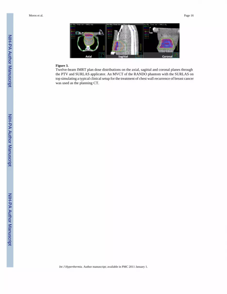
Figure 3.Twelve-beam IMRT plan dose distributions on the axial, sagittal and coronal planes throughthe PTV and SURLAS applicator. An MVCT of the RANDO phantom with the SURLAS ontop simulating a typical clinical setup for the treatment of chest wall recurrence of breast cancerwas used as the planning CT.
Moros et al. Page 16
Int J Hyperthermia. Author manuscript; available in PMC 2011 January 1.
NIH
-PA Author Manuscript
NIH
-PA Author Manuscript
NIH
-PA Author Manuscript

Figure 4.Dose volume histograms for the PTV and organs at risks for the treatment plan shown in Figure3.
Moros et al. Page 17
Int J Hyperthermia. Author manuscript; available in PMC 2011 January 1.
NIH
-PA Author Manuscript
NIH
-PA Author Manuscript
NIH
-PA Author Manuscript

NIH
-PA Author Manuscript
NIH
-PA Author Manuscript
NIH
-PA Author Manuscript
Moros et al. Page 18
Table 1
Maximum fundamental frequency (fmax) for four treatment window lengths (WL) and three array heights (AH).
WL (cm)
AH = 1.5 cm AH = 2.5 cm AH = 5 cm
fmax (MHz) fmax (MHz) fmax (MHz)
10 4.5 > 5.0 > 5.0
12 3.03 3.8 4.29
14 2.32 2.85 3.27
16 1.89 2.29 2.67
Int J Hyperthermia. Author manuscript; available in PMC 2011 January 1.