The effect of application sustained seating pressure on adhesive luting procedure
6
dental materials xxx ( 2 0 0 6 ) xxx–xxx available at www.sciencedirect.com journal homepage: www.intl.elsevierhealth.com/journals/dema The effect of application sustained seating pressure on adhesive luting procedure Nicoletta Chieffi a,∗ , Stefano Chersoni a,b , Federica Papacchini a , Michele Vano a,c , Cecilia Goracci a , Carel L. Davidson a , Franklin R. Tay a,d , Marco Ferrari a a Department of Dental Materials, University of Siena, Siena, Italy b Department of Dental Sciences, University of Bologna, Bologna, Italy c Department of Oral Surgery, University of Pisa, Pisa, Italy d Department of Oral Biology & Maxillofacial Pathology, Medical College of Georgia, Augusta, GA, USA article info Article history: Received 25 August 2005 Accepted 4 January 2006 Keywords: Seating pressure Resin cement Dentin adhesive system Bond strength abstract Objectives. To evaluate the effect of short versus long application seating pressure on the bond strength of resin blocks, luted with a dual-cured resin cement (Panavia F) to pre-coated or non pre-coated dentin with an hydrophobic light-cured adhesive (Clearfil Protect Bond). Methods. Sixteen non-carious human third molars were randomly divided into six Groups (four teeth each). Cylindrical composite blocks were luted with Panavia F (Group Ia) and with Clearfil Protect Bond with Panavia F (Group IIa) and seating pressure was applied for 5s. In Groups Ib and IIb, the two bonding procedures were respectively repeated, but the resin cylinder was maintained under constant pressure during the entire 3 min polymeriza- tion period for the resin cement. After storing in distilled water for 24 h, 0.9 mm × 0.9 mm sticks were produced from these luted specimens for microtensile bond testing and SEM examination. Results. The use of Clearfil Protect Bond with Panavia F produced higher bond strengths than the use of Panavia F (p < 0.05). Extending the time of pressure application up to 3 min increased the bond strength (p <0.001) and improved the integrity of the interfacial quality. Significance. The application of sustained seating pressure during luting procedures and the additional use of a hydrophobic light-cured adhesive both improve the final bond strength of the resin cement. © 2006 Elsevier Ltd and Academy of Dental Materials. All rights reserved. 1. Introduction It has been widely demonstrated that simplified self-etch adhesives can attract fluid from dentin after polymerization [1–6]. This fluid absorption is due to relatively high concentra- tions of ionic or acidic monomers in the self-etching primers (in the two-step systems) [1] or in adhesive (in the single- step systems) [2–6] essential to enable these systems to dif- fuse through the smear layer and demineralize the underlying dentin [7]. ∗ Corresponding author. Tel.: +39 0577 233131; fax: +39 0577 233117. E-mail addresses: nchieffi@libero.it (N. Chieffi), [email protected] (M. Ferrari). Recently, it was reported that incompatibility may be present when auto-cured or dual-cured resin cements were used in conjunction with simplified self-etch dentin adhe- sives [8,9]. Adverse chemical interaction and adhesive per- meability were identified as the two main factors responsible for the reduction in bond strength, when slow setting resin cements were coupled to bondings on hydrated dentin [10]. Even when the interaction between acidic simplified dentin adhesives and the basic amine catalysts in dual-curable resin cements may be eliminated by the use of sulphinate-type 0109-5641/$ – see front matter © 2006 Elsevier Ltd and Academy of Dental Materials. All rights reserved. doi:10.1016/j.dental.2006.01.006 DENTAL-904; No. of Pages 6
Transcript of The effect of application sustained seating pressure on adhesive luting procedure

d e n t a l m a t e r i a l s x x x ( 2 0 0 6 ) xxx–xxx
avai lab le at www.sc iencedi rec t .com
journa l homepage: www. int l .e lsev ierhea l th .com/ journa ls /dema
The effect of application sustained seating pressureon adhesive luting procedure
Nicoletta Chieffia,∗, Stefano Chersonia,b, Federica Papacchinia, Michele Vanoa,c,Cecilia Goraccia, Carel L. Davidsona, Franklin R. Taya,d, Marco Ferraria
a Department of Dental Materials, University of Siena, Siena, Italyb Department of Dental Sciences, University of Bologna, Bologna, Italyc Department of Oral Surgery, University of Pisa, Pisa, Italyd Department of Oral Biology & Maxillofacial Pathology, Medical College of Georgia, Augusta, GA, USA
a r t i c l e i n f o
A
R
A
K
S
R
D
B
a b s t r a c t
1
Ia[t(sfd
0d
rticle history:
eceived 25 August 2005
ccepted 4 January 2006
eywords:
eating pressure
esin cement
entin adhesive system
ond strength
Objectives. To evaluate the effect of short versus long application seating pressure on the bond
strength of resin blocks, luted with a dual-cured resin cement (Panavia F) to pre-coated or
non pre-coated dentin with an hydrophobic light-cured adhesive (Clearfil Protect Bond).
Methods. Sixteen non-carious human third molars were randomly divided into six Groups
(four teeth each). Cylindrical composite blocks were luted with Panavia F (Group Ia) and
with Clearfil Protect Bond with Panavia F (Group IIa) and seating pressure was applied for
5 s. In Groups Ib and IIb, the two bonding procedures were respectively repeated, but the
resin cylinder was maintained under constant pressure during the entire 3 min polymeriza-
tion period for the resin cement. After storing in distilled water for 24 h, 0.9 mm × 0.9 mm
sticks were produced from these luted specimens for microtensile bond testing and SEM
examination.
Results. The use of Clearfil Protect Bond with Panavia F produced higher bond strengths
than the use of Panavia F (p < 0.05). Extending the time of pressure application up to 3 min
increased the bond strength (p < 0.001) and improved the integrity of the interfacial quality.
Significance. The application of sustained seating pressure during luting procedures and the
additional use of a hydrophobic light-cured adhesive both improve the final bond strength
of the resin cement.
© 2006 Elsevier Ltd and Academy of Dental Materials. All rights reserved.
. Introduction
t has been widely demonstrated that simplified self-etchdhesives can attract fluid from dentin after polymerization1–6]. This fluid absorption is due to relatively high concentra-ions of ionic or acidic monomers in the self-etching primersin the two-step systems) [1] or in adhesive (in the single-tep systems) [2–6] essential to enable these systems to dif-use through the smear layer and demineralize the underlyingentin [7].
∗ Corresponding author. Tel.: +39 0577 233131; fax: +39 0577 233117.E-mail addresses: [email protected] (N. Chieffi), [email protected] (M. Ferrari).
Recently, it was reported that incompatibility may bepresent when auto-cured or dual-cured resin cements wereused in conjunction with simplified self-etch dentin adhe-sives [8,9]. Adverse chemical interaction and adhesive per-meability were identified as the two main factors responsiblefor the reduction in bond strength, when slow setting resincements were coupled to bondings on hydrated dentin [10].Even when the interaction between acidic simplified dentinadhesives and the basic amine catalysts in dual-curable resincements may be eliminated by the use of sulphinate-type
109-5641/$ – see front matter © 2006 Elsevier Ltd and Academy of Dental Materials. All rights reserved.oi:10.1016/j.dental.2006.01.006
DENTAL-904; No. of Pages 6
2 d e n t a l m a t e r i a l s x x x ( 2 0 0 6 ) xxx–xxx
Table 1 – Batch number (#), composition and manufacturer of the materials employed in this study
Materials Components Manufacturer
Panavia F (#41170) Primer a: HEMA, 10-MDP, 5-NMSA, water, accelerator Kuraray Medical Inc., Tokyo, JapanPrimer b: 5-NMSA, accelerator, water, sodium benzene sulfinate
Paste A: 10-MDP, hydrophobic aromatic dimethacrylate, hydrophobicaliphatic dimethacrylate, hydrophilic dimethacrylate, silanated silica,photoinitiator, benzoyl peroxidePaste B: hydrophobic aromatic dimethacrylate, hydrophobic aliphaticdimethacrylate, hydrophilic dimethacrylate, sodium aromaticsulfinate, accelerator, sodium fluoride, silanated barium glass
Clearfil Protect Bond (#41112) Primer: HEMA, 10-MDP, 12-MDPBa, hydrophilic dimethacrylate, water Kuraray Medical Inc., Tokyo, Japan
Bond: 10-MDP, Bis-GMA, HEMA, hydrophobic dimethacrylate,di-Camphoroquinone; N,N-diethanol-p-toluidine, silanated colloidalsilica, surface treated sodium fluoride
Abbreviations: HEMA: 2-hydroxyethyl methacrylate; 10-MDP: 10-methacryoloyloxydecyl dihydrogen phosphate; 5-NMSA: N-methacryloxyl-5-aminosalicylic acid; 12-MDPB: 12-methacryloyloxydodecylpyridinium bromide; Bis-GMA: bis-phenol A diglycidylmethacrylate.a MDPB has been included as for its antibacterial properties.
ternary catalysts in the self-etching primer and the resincement [11], adhesive permeability remains a possible causeof bond strength reduction. This is because auto/dual-curedresin cements have longer setting times than light-cured resincements and, in time, water can diffuse from the underlyingbonded dentin across the polymerized hydrophilic adhesivelayer via an osmotic gradient [6]. A previous study concludedthat the additional application of a more hydrophobic resinlayer resulted in improved coupling of the dual-cured resincement to dentin [12]. Another recent investigation [13] indi-cated that pressure application during the entire course ofthe setting of a dual-cured resin cement improves the bondstrength and reduces fluid interference from the underlyingdentin, with bonding covered dentin.
The objective of this study was to evaluate the effect of thesustained seating pressure on the integrity of the adhesivebond in resin blocks, in order to simulate indirect restora-tions, luted with a dual-cured resin cement. The effect of seat-ing pressure was examined by comparing the use of PanaviaF as recommended by the manufacturer (i.e., resin cementapplied to dentin primed with the one-step self-etch adhe-sive included in the kit), versus the application of Panavia F todentin that had first undergone a bonding procedure with thetwo-step self-etch adhesive Clearfil Protect Bond.
The null hypotheses tested were that: (a) the applicationof sustained seating pressure during the curing of the resin
cement has no influence on the microtensile bond strength ofresin blocks to dentin; and (b) the additional application of thetwo-step self-etch adhesive has no influence on the microten-sile bond strength of the dual-cured resin cement.
2. Materials and methods
Sixteen non-carious human third molars that had been storedin a 0.5% chloramine T solution at 4 ◦C were used within 1month following extraction. All the teeth were submitted tobonding in their normal hydrated state, as they were retrievedfrom the storage medium. The composition of the materials isshown in Table 1. The operating procedures for the four exper-imental and control Groups are summarized in Table 2.
2.1. Microtensile examination
2.1.1. Specimen preparationThe occlusal enamel was removed using a slow-speed sawwith a diamond-impregnated disk (Isomet, Buehler Ltd., LakeBluff, IL, USA) under water cooling. The roots of all teeth werecut at the same level, in order to obtained tooth samples witha standardized height (5 mm). A 180-grit silicon carbide paperwas used under running water to create a clinically relevantsmear layer on the dentin surface.
ir m
Sea
nt (P >
Table 2 – Procedures employed for the four Groups and the
Group designation
I (a and b) Panavia FII (a and b) Clearfil Protect Bond + Panavia F
Groups with the same superscripts (A–C) are not significantly differe∗ Values are means (standard deviations) in MPa.
icrotensile bond strengths
Microtensile bond strength (MPa)*
A Bting pressure: for 5 s Seating pressure: for 3 min
9.78 (5.66)C 20.71 (7.31)B
13.03 (7.01)B,C 31.11 (11.85)A
0.05).
d e n t a l m a t e r i a l s x x x ( 2 0 0 6 ) xxx–xxx 3
Composite cylinders (Tetric-Ceram, Ivoclar-Vivadent,Schaan, Liechtenstein) 10 mm in diameter and 6 mm inheight were prepared using a split aluminum mold, in orderto limit and standardize the bonding area. Prior to cementingprocedures, the bonding surface of each resin cylinder wassandblasted, etched with phosphoric acid, rinsed with waterand dried.
2.1.2. Cementing proceduresThe specimens were randomly divided in four Groups (N = 4).In Group Ia, the resin cement Panavia F (Kuraray Medical Inc.,Tokyo, Japan) was used according to manufacturer’s instruc-tions. ED Primer (A + B) was mixed and applied with a brush onthe dentin surface and left in place for 30 s. After drying theetched surface with gentle air flow, Panavia F resin cement(A + B) was mixed and applied to each of the freshly preparedcomposite cylinders.
In Group IIa, the light-curing self-etch adhesive ClearfilProtect Bond (Kuraray Medical Inc., Tokyo, Japan) wasused according to manufacturer’s instructions. This mate-rial also contains 12-methacryloyloxydodecylpyridinium bro-mide (MDPB) monomer to include antibacterial propertieswhich may be useful in cases of vital teeth. The self-etching primer was applied to the dentin surface with abrush and left in place for 20 s. After drying the etched sur-face with mild air flow, the bonding was applied on theetched-primed dentin, gently air flowed and light-cured for1ad
tTtrf
weimmTattidmM
2Afwtoica
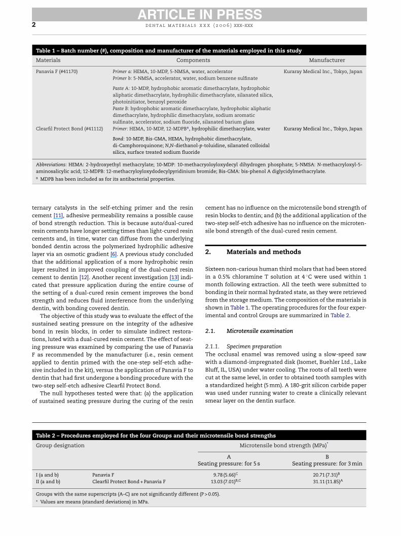
Fig. 1 – The metallic tool used to standardized the seatingpressure application. The tooth was positioned on the basalsurface of the tool and the composite cylinder wascemented, applying the weight on the upper surface of thetool.
DVA, Corona, CA, USA). The device was attached to a universaltesting machine (Triax digital 50, Controls, Milano, Italy) andloaded in tension at a crosshead speed of 0.5 mm/min untilfailure.
2.1.4. Statistical analysisThe population of bond strength values was normally dis-tributed according to the Kolmogorov–Smirnov test (p > 0.05).The two-way ANOVA was applied with bond strength as thedependent variable, material and pressure application time asfactors. Tukey’s test was used for post hoc comparisons.
2.2. Scanning electron microscopy (SEM)
2.2.1. Specimen preparationAfter microtensile bond testing, eight pairs (fractured com-posite and dentin sides) of specimens from each Group wererandomly selected for SEM examination. Each specimen wasmounted on metallic stubs, sputtered with gold/palladiumand observed with a SEM (JSM—6060LV, JEOL, Tokyo, Japan)operating at 10 or 15 kV.
Images of the two complementary detached interfaces foreach fractured specimen were taken at different standardizedmagnifications: 100–6000×.
0 s. Then Panavia F resin cement (A + B) was mixed andpplied on each of the freshly prepared composite cylin-ers.
In these two Groups, the composite block was cemented tohe dentin surface using a seating pressure of 1.25 MPa for 5 s.o ensure optimal polymerization of the resin cement alonghe exposed margins, a layer of Oxyguard was applied afteremoval of the excess cement with a probe and left in placeor at least 3 min.
In Group Ib and in Group IIb, the bonding proceduresere the same as described for Group Ia and Group IIa,
xcept for the application of the cementation pressure. Dur-ng the entire 3 min period necessary for complete poly-
erization of the resin cement, the resin cylinder wasaintained under a constant seating pressure of 1.25 MPa.
he excess cement was removed and the Oxiguard waspplied while the specimen was under pressure. In ordero standardize the applied pressure, a metallic tool (Fig. 1)hat delivered 10 kg was used. This resulted in a seat-ng force of 98.1 N. The pressure [N/m2] was calculated,ividing this force [N] by the surface area [m2] of theetal weight. The value obtained was finally converted intoPa.
.1.3. Bond strength evaluationfter cementation, all the specimens were stored in water
or 24 h at 37 ◦C. Each tooth was then sectioned verticallyith the Isomet saw into a series of slabs. The slabs were
hen sectioned vertically into 0.9 mm × 0.9 mm sticks, basedn the “non-trimming” version of the microtensile bond test-
ng technique [14]. Each stick was measured using a digitalaliper to determine the cross-sectional area. The sticks werettached to a testing device with cyanoacrylate glue (Zapit,
4 d e n t a l m a t e r i a l s x x x ( 2 0 0 6 ) xxx–xxx
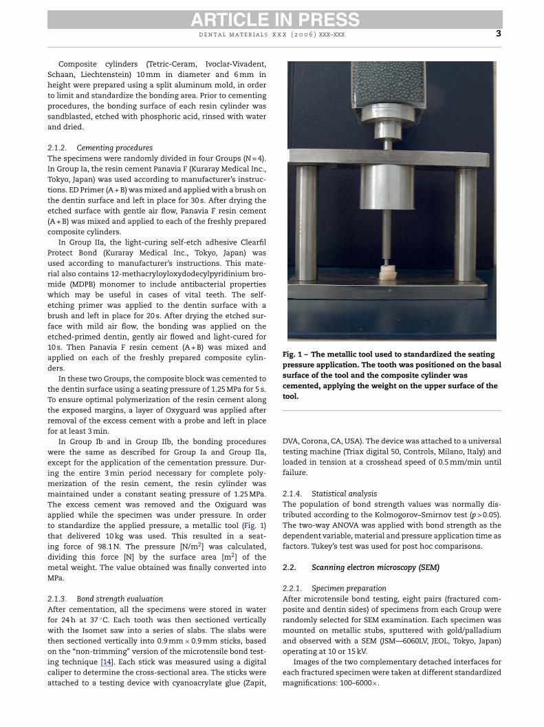
Fig. 2 – SEM imagine of fracture composite side of aspecimen from Group Ia, where the seating pressure wasapplied for 5 s. The micrograph (4000×) shows thestructural design of the defects observed.
3. Results
3.1. Microtensile bond strengths
Bond strength results of the four Groups are shown in Table 2.The selection of the luting material was a significant fac-
tor with regard to the bond strength obtained; with ClearfilProtect Bond + Panavia F measuring significantly higher bondstrengths than Panavia F (p < 0.05).
Pressure application time was also a significant factor.Extending the time of pressure application up to 3 min sig-nificantly increased the bond strength (p < 0.001).
The interaction between cement and time of pressureapplication was also significant to obtain the highest bondstrength. According to the Tukey’s test applied for post hoccomparisons, Group IIb achieved the highest bond strengthand the difference was statistically significant (p < 0.05). GroupIb and Group IIa were comparable (p > 0.05). The latter wascomparable to Group Ia, which had the lowest bond strength.
3.2. SEM observations
SEM analysis of the specimens from those Groups where theseating pressure was sustained for a few seconds, revealed thepresence of structural defects. These defects consisted of com-partmentalized structural entities that were nearly identical
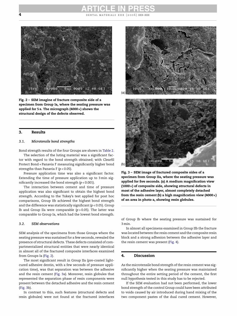
Fig. 3 – SEM image of fractured composite sides of aspecimen from Group IIa, where the seating pressure wasapplied for five seconds. (a) A medium magnification view(1000×) of composite side, showing structural defects inmost of the adhesive layer, almost completely detachedfrom the resin cement (b) a high magnification view (4000×)of an area in photo a, showing resin globules.
of Group Ib where the seating pressure was sustained for3 min.
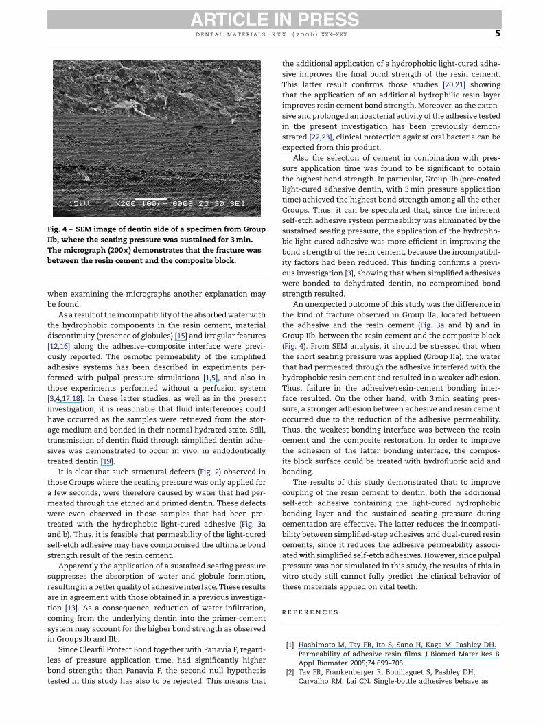
In almost all specimens examined in Group IIb the fracturewas located between the resin cement and the composite resinblock and a strong adhesion between the adhesive layer andthe resin cement was present (Fig. 4).
4. Discussion
As the microtensile bond strength of the resin cement was sig-nificantly higher when the seating pressure was maintainedthroughout the entire setting period of the cement, the firstnull hypothesis tested in this study has to be rejected.
If the SEM evaluation had not been performed, the lowerbond strength of the control Group could have been attributedto voids caused by air introduced during hand mixing of thetwo component pastes of the dual cured cement. However,
in almost all of the fractured composite interfaces examinedfrom Groups Ia (Fig. 2).
The most significant result in Group IIa (pre-coated light-cured adhesive dentin, with a few seconds of pressure appli-cation time), was that separation was between the adhesiveand the resin cement (Fig. 3a). Moreover, resin globules thatrepresented the separation phase of resin components werepresent between the detached adhesive and the resin cement(Fig. 3b).
In contrast to this, such features (structural defects andresin globules) were not found at the fractured interfaces
d e n t a l m a t e r i a l s x x x ( 2 0 0 6 ) xxx–xxx 5
Fig. 4 – SEM image of dentin side of a specimen from GroupIIb, where the seating pressure was sustained for 3 min.The micrograph (200×) demonstrates that the fracture wasbetween the resin cement and the composite block.
when examining the micrographs another explanation maybe found.
As a result of the incompatibility of the absorbed water withthe hydrophobic components in the resin cement, materialdiscontinuity (presence of globules) [15] and irregular features[12,16] along the adhesive–composite interface were previ-ously reported. The osmotic permeability of the simplifiedadhesive systems has been described in experiments per-formed with pulpal pressure simulations [1,5], and also inthose experiments performed without a perfusion system[3,4,17,18]. In these latter studies, as well as in the presentinvestigation, it is reasonable that fluid interferences couldhave occurred as the samples were retrieved from the stor-age medium and bonded in their normal hydrated state. Still,transmission of dentin fluid through simplified dentin adhe-sives was demonstrated to occur in vivo, in endodonticallytreated dentin [19].
It is clear that such structural defects (Fig. 2) observed inthose Groups where the seating pressure was only applied fora few seconds, were therefore caused by water that had per-meated through the etched and primed dentin. These defectswere even observed in those samples that had been pre-treated with the hydrophobic light-cured adhesive (Fig. 3aand b). Thus, it is feasible that permeability of the light-curedself-etch adhesive may have compromised the ultimate bondstrength result of the resin cement.
Apparently the application of a sustained seating pressuresratcsi
lbt
the additional application of a hydrophobic light-cured adhe-sive improves the final bond strength of the resin cement.This latter result confirms those studies [20,21] showingthat the application of an additional hydrophilic resin layerimproves resin cement bond strength. Moreover, as the exten-sive and prolonged antibacterial activity of the adhesive testedin the present investigation has been previously demon-strated [22,23], clinical protection against oral bacteria can beexpected from this product.
Also the selection of cement in combination with pres-sure application time was found to be significant to obtainthe highest bond strength. In particular, Group IIb (pre-coatedlight-cured adhesive dentin, with 3 min pressure applicationtime) achieved the highest bond strength among all the otherGroups. Thus, it can be speculated that, since the inherentself-etch adhesive system permeability was eliminated by thesustained seating pressure, the application of the hydropho-bic light-cured adhesive was more efficient in improving thebond strength of the resin cement, because the incompatibil-ity factors had been reduced. This finding confirms a previ-ous investigation [3], showing that when simplified adhesiveswere bonded to dehydrated dentin, no compromised bondstrength resulted.
An unexpected outcome of this study was the difference inthe kind of fracture observed in Group IIa, located betweenthe adhesive and the resin cement (Fig. 3a and b) and inGroup IIb, between the resin cement and the composite block
r
uppresses the absorption of water and globule formation,esulting in a better quality of adhesive interface. These resultsre in agreement with those obtained in a previous investiga-ion [13]. As a consequence, reduction of water infiltration,oming from the underlying dentin into the primer-cementystem may account for the higher bond strength as observedn Groups Ib and IIb.
Since Clearfil Protect Bond together with Panavia F, regard-ess of pressure application time, had significantly higherond strengths than Panavia F, the second null hypothesisested in this study has also to be rejected. This means that
(Fig. 4). From SEM analysis, it should be stressed that whenthe short seating pressure was applied (Group IIa), the waterthat had permeated through the adhesive interfered with thehydrophobic resin cement and resulted in a weaker adhesion.Thus, failure in the adhesive/resin-cement bonding inter-face resulted. On the other hand, with 3 min seating pres-sure, a stronger adhesion between adhesive and resin cementoccurred due to the reduction of the adhesive permeability.Thus, the weakest bonding interface was between the resincement and the composite restoration. In order to improvethe adhesion of the latter bonding interface, the compos-ite block surface could be treated with hydrofluoric acid andbonding.
The results of this study demonstrated that: to improvecoupling of the resin cement to dentin, both the additionalself-etch adhesive containing the light-cured hydrophobicbonding layer and the sustained seating pressure duringcementation are effective. The latter reduces the incompati-bility between simplified-step adhesives and dual-cured resincements, since it reduces the adhesive permeability associ-ated with simplified self-etch adhesives. However, since pulpalpressure was not simulated in this study, the results of this invitro study still cannot fully predict the clinical behavior ofthese materials applied on vital teeth.
e f e r e n c e s
[1] Hashimoto M, Tay FR, Ito S, Sano H, Kaga M, Pashley DH.Permeability of adhesive resin films. J Biomed Mater Res BAppl Biomater 2005;74:699–705.
[2] Tay FR, Frankenberger R, Bouillaguet S, Pashley DH,Carvalho RM, Lai CN. Single-bottle adhesives behave as
6 d e n t a l m a t e r i a l s x x x ( 2 0 0 6 ) xxx–xxx
permeable membranes after polymerization. I. In vivoevidence. J Dent 2004;32:611–21.
[3] Tay FR, Pashley DH, Suh B, Carvalho R, Miller M.Single-step, self-etch adhesives behave as permeable afterpolymerization. Part I. Bond strength and morphologicalevidence. Am J Dent 2004;17:271–8.
[4] Tay FR, Pashley DH, Garcia-Godoy F, Cynthia KYY.Single-step, self-etch adhesives behave as permeable afterpolymerization. Part II. Silver tracer penetration evidence.Am J Dent 2004;17:271–8.
[5] Itthagarun A, Tay FR, Pashley DH, Wefel JS, Garcia-GodoyF, Wei SHI. Single-step, self-etch adhesives behave aspermeable after polymerization. Part III. Evidence fromfluid conductance and artificial caries inhibition. Am JDent 2004;17:394–400.
[6] Tay FR, Pashley DH, Byoung IS, Carvalho R, Itthagarun A.Single-step adhesives are permeable membranes. J Dent2002;30:371–82.
[7] Yiu CK, Hiraishi N, Chersoni S, Breschi L, Ferrari M, PratiC, et al. Single-bottle adhesives behave as permeablemembranes after polymerisation. II. Differentialpermeability reduction with an oxalate desensitizer. J Dent2006;34:106–16.
[8] Ogawa T, Tanaka M, Matsuya S, Aizawa S, Koyano K.Setting characteristics of five autopolymerizing resinsmeasured by an oscillating rheometer. J Prosthet Dent2001;85:170–6.
[9] Sanares AM, Itthagarun A, King NM, Tay FR, Pashley DH.Adverse surface interactions between one-bottlelight-cured adhesives and chemical-cured composites.Dent Mater 2001;17:542–56.
[12] Carvalho RM, Pegoraro TA, Tay FR, Pegoraro LF, Silva NRFA,Pashley DH. Adhesive permeability affects coupling ofresin cements that utilize self-etching primers to dentin. JDent 2004;32:55–65.
[13] Chieffi N, Chersoni S, Papacchini F, Vano M, Goracci C,Davidson CL, et al. An in vitro study of the effect of theseating pressure on the adhesive bonding of indirectrestorations, Am J Dent, in press.
[14] Shono Y, Ogawa T, Terashita M, Carvalho RM, Pashley EL,Pashley DH. Regional measurement of resin-dentinbonding as an array. J Dent Res 1999;78:699–705.
[15] Tay F, Pashley DH. Have dentin adhesives become toohydrophilic? J Can Dent Assoc 2003;69:726–31.
[16] Mak YF, Lai SCN, Cheung GSP, Chan AWK, Tay FR, PashleyDH. Micro-tensile bond testing of resin cements to dentinand an indirect resin composite. Dent Mater2002;18:609–21.
[17] Chersoni S, Suppa P, Grandini S, Goracci C, Monticelli F,Yiu C, et al. In vivo and in vitro permeability of one-stepself-etch adhesives. J Dent Res 2004;83:459–64.
[18] Chersoni S, Suppa P, Breschi L, Ferrari M, Tay FR, PashleyDH, et al. Water movement in the hybrid layer afterdifferent dentin treatments. Dent Mater 2004;20:796–803.
[19] Chersoni S, Acquaviva GL, Prati C, Ferrari M, Grandini S,Pashley DH, et al. In vivo fluid movement through dentinadhesives in endodontically treated teeth. J Dent Res2005;84:223–7.
[20] Jayasooriya PR, Pereira PN, Nikaido T, Tagami J. Efficacy ofresin coating on bond strengths of resin cement to dentin.J Esthet Restor Dent 2003;15:105–13.
[21] Nikaido T, Cho E, Nakajima M, Tashiro H, Toba S, Burrow
[10] Tay FR, Pashley DH, Yiu CK, Sanares AM, Wei SH. Factorscontributing to the incompatibility between simplified-stepadhesives and chemically-cured or dual-cured composites.Part I. Single-step self-etching adhesive. J Adhes Dent2003;5:27–40.
[11] Nyunt NM, Imai Y. Adhesion to dentin with resin usingsulfinic acid initiator system. Dent Mater 1996;15:175–82.
MF, et al. Tensile bond strengths of resin cements tobovine dentin using resin coating. Am J Dent 2003;16:41–6.
[22] Imazato S, Tarumi H, Kato S, Ebisu S. Water sorption andcolour stability of composites containing the antibacterialmonomer MDPB. J Dent 1999;27:279–83.
[23] Imazato S, Ehara A, Torii M, Ebisu S. Antibacterial activityof dentine primer containing MDPB after curing. J Dent1998;26:267–71.