PhotoMEA: An opto-electronic biosensor for monitoring in vitro neuronal network activity
6
BioSystems 87 (2007) 150–155 PhotoMEA: An opto-electronic biosensor for monitoring in vitro neuronal network activity Diego Ghezzi a,∗ , Alessandra Pedrocchi a,1 , Andrea Menegon b,2 , Sara Mantero a,3 , Flavia Valtorta b,4 , Giancarlo Ferrigno a,5 a Bioengineering Department, Politecnico di Milano, p.zza Leonardo da Vinci 32, 20133 Milano, Italy b Department of Neuroscience, San Raffaele Scientific Institute and “Vita-Salute” University, via Olgettina 60, 20132 Milano, Italy Received 28 February 2005; received in revised form 8 July 2006; accepted 15 July 2006 Abstract PhotoMEA is a biosensor useful for the analysis of an in vitro neuronal network, fully based on optical methods. Its function is based on the stimulation of neurons with caged glutamate and the recording of neuronal activity by Voltage-Sensitive fluorescent Dyes (VSD). The main advantage is that it will be possible to stimulate even at sub-single neuron level and to record with high resolution the activity of the entire network in the culture. A large-scale view of neuronal intercommunications offers a unique opportunity for testing the ability of drugs to affect neuronal properties as well as alterations in the behaviour of the entire network. The concept and a prototype for validation is described here in detail. © 2006 Elsevier Ireland Ltd. All rights reserved. Keywords: Neuronal information processing; Technology for neuropharmacology; Neural-based biosensor; Bio-information processing system; Computational biology 1. Introduction The most important function of neurons and neuronal networks is to process, modulate and transmit informa- tion through electrical signals (i.e. spikes). Our ability in eliciting, modulating and recording this activity is fun- ∗ Corresponding author. Tel.: +39 02 23999506; fax: +39 02 23993360. E-mail addresses: [email protected] (D. Ghezzi), [email protected] (A. Pedrocchi), [email protected] (A. Menegon), [email protected] (S. Mantero), valtorta.fl[email protected] (F. Valtorta), [email protected] (G. Ferrigno). 1 Tel.: +39 02 23993371; fax: +39 02 23993360. 2 Tel.: +39 02 26434868; fax: +39 02 26434813. 3 Tel.: +39 02 23993376; fax: +39 02 23993360. 4 Tel.: +39 02 26434826; fax: +39 02 26434813. 5 Tel.: +39 02 23993371; fax: +39 02 23993360. damental for understanding the mechanisms that govern the central nervous system (CNS) functions. Therefore, one of the most relevant topic in neuroscience is the bet- ter understanding of the functional dynamics that govern the complex network properties. There are two possible approaches in studying neu- ronal functions. On one hand, a large-scale approach aims at understanding a more or less synchronized activ- ity of the whole neuronal system. On the other hand, a micro-scale approach studies detailed behavioural mod- els of the neuron, and also of the complex molecular systems which actively contribute to the generation and modulation of the whole neuronal activity. Brain functions originate from the coordinate activity of many neurons, acting in several regions involved in information processing. Thus, the study of the modula- tion of neuronal physiology that regulates neuronal activ- ity must necessarily be inserted into a large-scale view 0303-2647/$ – see front matter © 2006 Elsevier Ireland Ltd. All rights reserved. doi:10.1016/j.biosystems.2006.09.008
-
Upload
independent -
Category
Documents
-
view
0 -
download
0
Transcript of PhotoMEA: An opto-electronic biosensor for monitoring in vitro neuronal network activity
BioSystems 87 (2007) 150–155
PhotoMEA: An opto-electronic biosensor for monitoringin vitro neuronal network activity
Diego Ghezzi a,∗, Alessandra Pedrocchi a,1, Andrea Menegon b,2,Sara Mantero a,3, Flavia Valtorta b,4, Giancarlo Ferrigno a,5
a Bioengineering Department, Politecnico di Milano, p.zza Leonardo da Vinci 32, 20133 Milano, Italyb Department of Neuroscience, San Raffaele Scientific Institute and “Vita-Salute” University, via Olgettina 60, 20132 Milano, Italy
Received 28 February 2005; received in revised form 8 July 2006; accepted 15 July 2006
Abstract
PhotoMEA is a biosensor useful for the analysis of an in vitro neuronal network, fully based on optical methods. Its function isbased on the stimulation of neurons with caged glutamate and the recording of neuronal activity by Voltage-Sensitive fluorescentDyes (VSD). The main advantage is that it will be possible to stimulate even at sub-single neuron level and to record with high
resolution the activity of the entire network in the culture. A large-scale view of neuronal intercommunications offers a uniqueopportunity for testing the ability of drugs to affect neuronal properties as well as alterations in the behaviour of the entire network.The concept and a prototype for validation is described here in detail.© 2006 Elsevier Ireland Ltd. All rights reserved.harmac
Keywords: Neuronal information processing; Technology for neuropComputational biology1. Introduction
The most important function of neurons and neuronal
networks is to process, modulate and transmit informa-tion through electrical signals (i.e. spikes). Our ability ineliciting, modulating and recording this activity is fun-∗ Corresponding author. Tel.: +39 02 23999506;fax: +39 02 23993360.
E-mail addresses: [email protected](D. Ghezzi), [email protected] (A. Pedrocchi),[email protected] (A. Menegon), [email protected](S. Mantero), [email protected] (F. Valtorta),[email protected] (G. Ferrigno).
1 Tel.: +39 02 23993371; fax: +39 02 23993360.2 Tel.: +39 02 26434868; fax: +39 02 26434813.3 Tel.: +39 02 23993376; fax: +39 02 23993360.4 Tel.: +39 02 26434826; fax: +39 02 26434813.5 Tel.: +39 02 23993371; fax: +39 02 23993360.
0303-2647/$ – see front matter © 2006 Elsevier Ireland Ltd. All rights reservdoi:10.1016/j.biosystems.2006.09.008
ology; Neural-based biosensor; Bio-information processing system;
damental for understanding the mechanisms that governthe central nervous system (CNS) functions. Therefore,one of the most relevant topic in neuroscience is the bet-ter understanding of the functional dynamics that governthe complex network properties.
There are two possible approaches in studying neu-ronal functions. On one hand, a large-scale approachaims at understanding a more or less synchronized activ-ity of the whole neuronal system. On the other hand, amicro-scale approach studies detailed behavioural mod-els of the neuron, and also of the complex molecularsystems which actively contribute to the generation andmodulation of the whole neuronal activity.
Brain functions originate from the coordinate activity
of many neurons, acting in several regions involved ininformation processing. Thus, the study of the modula-tion of neuronal physiology that regulates neuronal activ-ity must necessarily be inserted into a large-scale viewed.
Systems
oimo
sanwa
iuwooibwcTwe(nrebed
obae
mftotcpon
lnbstn
D. Ghezzi et al. / Bio
f the neuronal network dynamics. A new breakthroughnto neuroscience will be the possibility to stimulate and
odulate a single neuron and study its response and thatf the entire network.
There is a growing need to observe smaller andmaller systems, kept alive in vitro, at a proper spatiallynd temporally resolution level. At the same time, it isecessary to monitor neuronal activity of the whole net-ork in order to understand how every single neuron is
ble to modulate the whole network activity.At the current state of the art, in most cases, these
nvestigations have been carried out by electrically stim-lating and recording the tissue of interest. Methodshich employ intra-cellular or extra-cellular electrodesr arrays have yielded important results in neurobiol-gy, but nowadays these technologies are showing somemportant limits. The stimulation of a single neuron cane traditionally performed by intra-cellular electrodesith the disadvantage of the mechanical damage of the
ell and the consequent alteration of the entire network.hus intra-cellular electrodes do not allow studies of thehole neuronal network. In contrast the extra-cellular
lectrodes, as Micro-Electrode Array devices (MEAs)Thomas et al., 1972; Gross, 1979; Pine, 1980), areot suitable for local stimulations and high-resolutionecordings, because of the interference generated bylectrical fields which spread in the medium. They cane used in order to monitor the electrical activity of thentire network, but with the limitation of recording someiscrete regions only.
Besides traditional electrophysiology, optical meth-ds for stimulating and recording neuronal activity haveeen used for a long time (Kotter et al., 1998; Kandler etl., 1998; Zecevic, 1996; Antic and Zecevic, 1995; Antict al., 1999).
The ability to use light provides a non-invasiveethod for precise temporal and spatial activation of dif-
erent regions of a neuronal network, which can be usedo stimulate single neurons as well as discrete regionsf the neuron itself. In addition, optical methods allowo monitor the entire network activity. However, at theurrent state of the art, optical methods are not yet inde-endent from electrical measurements, but they providenly a useful support for neurons stimulation, or alter-atively for recording membrane electrical activity.
Our work provides a technical solution for the stimu-ation and recording of the activity of an in vitro neuronaletwork entirely based on optical methods. The proposed
iosensor, PhotoMEA, is capable to combine an opticaltimulation of neural activity with high spatial resolu-ion in addition to recording the activity of the wholeetwork.87 (2007) 150–155 151
The PhotoMEA concept (Patent Pending numberMI2005A000114) is currently under testing.
2. Methods
2.1. Stimulation of neurons with light
Optical stimulation of neurons can be performed by differ-ent methods, including direct two-photon excitation (Hiraseet al., 2002), endogenous expression of molecules sensitive tolight (Zemelman et al., 2002) and caged glutamate activation(Callaway and Katz, 1993). These and other methods have beenrecently reviewed (Callaway and Yuste, 2002).
Since light can be focused with high spatial and temporalresolution, optical methods are the best solution to provide localand controlled stimulations to neurons or to any part thereof.In particular, the most physiological way used in order to cou-ple light and neuronal excitation is based on the use of thecaged neurotransmitter glutamate. Glutamate, the most impor-tant and widespread excitatory neurotransmitter in the brain,can be caged by a chemical group, which can be removed bylight absorption.
The basic approach is to switch caged glutamate into activeglutamate by ultraviolet light pulses with a single-photonmethod. In order to achieve high spatial resolution, light canbe focused by means of two-photon microscopy.
Currently, optical stimulations of neurons are matched withelectrical recording of neuronal activity performed by intra- orextra-cellular electrodes, with the consequence that the localstimulation performed by caged glutamate is usually limitedto the study of one single neuron (using an intracellular elec-trode for recording) or it is accompanied by a spread record-ing of a network activity with low resolution (using MEAdevices).
2.2. Optical recording of neuronal activity
Changes of neuronal membrane potential can be measuredoptically by different methods, either directly or by a varietyof molecular probes. Intrinsic optical signals are generated bynerve activity, which induces changes in light scattering. How-ever, such changes are usually small, therefore they are notapplicable to the study of in vitro neuronal networks (Grinvaldet al., 1988).
Using molecular probes, several optical properties, whichare sensitive to membrane potential, can be detected, e.g. flu-orescence, absorption, dichroism, birefringence, fluorescenceresonance energy transfer, non-linear second harmonic gen-eration, and resonance Raman absorption (Zochowski et al.,2000).
In particular, fluorescence is the most common property
used in optical imaging. Therefore, optical recording of neuralactivity is possible thanks to Voltage-Sensitive Dyes acting astransducers, which convert voltage changes into optical signalsrecorded by a proper optoelectronic sensor. Furthermore, VSDshave the ability to bind the membrane of neurons and change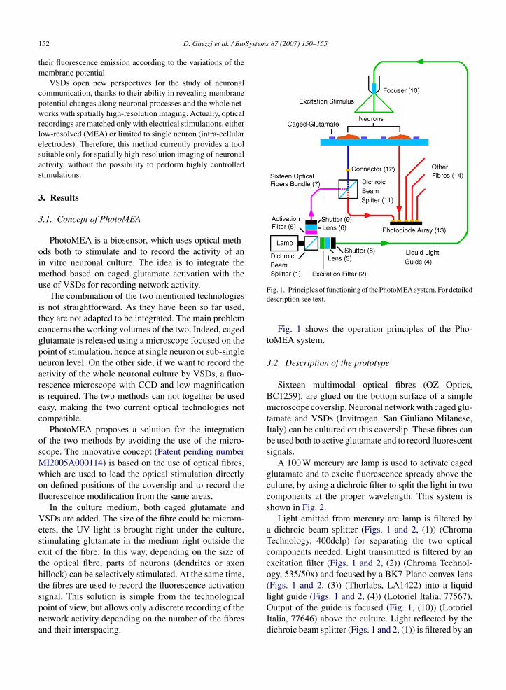
Systems 87 (2007) 150–155
152 D. Ghezzi et al. / Biotheir fluorescence emission according to the variations of themembrane potential.
VSDs open new perspectives for the study of neuronalcommunication, thanks to their ability in revealing membranepotential changes along neuronal processes and the whole net-works with spatially high-resolution imaging. Actually, opticalrecordings are matched only with electrical stimulations, eitherlow-resolved (MEA) or limited to single neuron (intra-cellularelectrodes). Therefore, this method currently provides a toolsuitable only for spatially high-resolution imaging of neuronalactivity, without the possibility to perform highly controlledstimulations.
3. Results
3.1. Concept of PhotoMEA
PhotoMEA is a biosensor, which uses optical meth-ods both to stimulate and to record the activity of anin vitro neuronal culture. The idea is to integrate themethod based on caged glutamate activation with theuse of VSDs for recording network activity.
The combination of the two mentioned technologiesis not straightforward. As they have been so far used,they are not adapted to be integrated. The main problemconcerns the working volumes of the two. Indeed, cagedglutamate is released using a microscope focused on thepoint of stimulation, hence at single neuron or sub-singleneuron level. On the other side, if we want to record theactivity of the whole neuronal culture by VSDs, a fluo-rescence microscope with CCD and low magnificationis required. The two methods can not together be usedeasy, making the two current optical technologies notcompatible.
PhotoMEA proposes a solution for the integrationof the two methods by avoiding the use of the micro-scope. The innovative concept (Patent pending numberMI2005A000114) is based on the use of optical fibres,which are used to lead the optical stimulation directlyon defined positions of the coverslip and to record thefluorescence modification from the same areas.
In the culture medium, both caged glutamate andVSDs are added. The size of the fibre could be microm-eters, the UV light is brought right under the culture,stimulating glutamate in the medium right outside theexit of the fibre. In this way, depending on the size ofthe optical fibre, parts of neurons (dendrites or axonhillock) can be selectively stimulated. At the same time,the fibres are used to record the fluorescence activation
signal. This solution is simple from the technologicalpoint of view, but allows only a discrete recording of thenetwork activity depending on the number of the fibresand their interspacing.Fig. 1. Principles of functioning of the PhotoMEA system. For detaileddescription see text.
Fig. 1 shows the operation principles of the Pho-toMEA system.
3.2. Description of the prototype
Sixteen multimodal optical fibres (OZ Optics,BC1259), are glued on the bottom surface of a simplemicroscope coverslip. Neuronal network with caged glu-tamate and VSDs (Invitrogen, San Giuliano Milanese,Italy) can be cultured on this coverslip. These fibres canbe used both to active glutamate and to record fluorescentsignals.
A 100 W mercury arc lamp is used to activate cagedglutamate and to excite fluorescence spready above theculture, by using a dichroic filter to split the light in twocomponents at the proper wavelength. This system isshown in Fig. 2.
Light emitted from mercury arc lamp is filtered bya dichroic beam splitter (Figs. 1 and 2, (1)) (ChromaTechnology, 400dclp) for separating the two opticalcomponents needed. Light transmitted is filtered by anexcitation filter (Figs. 1 and 2, (2)) (Chroma Technol-ogy, 535/50x) and focused by a BK7-Plano convex lens(Figs. 1 and 2, (3)) (Thorlabs, LA1422) into a liquid
light guide (Figs. 1 and 2, (4)) (Lotoriel Italia, 77567).Output of the guide is focused (Fig. 1, (10)) (LotorielItalia, 77646) above the culture. Light reflected by thedichroic beam splitter (Figs. 1 and 2, (1)) is filtered by an
D. Ghezzi et al. / BioSystems
Fcm
a3(O
asagwa
e
FtaF
ig. 2. First stage of the PhotoMEA system, which allows to activeaged-glutamate and to excite fluorescence by means of a single 100 Wercury arc lamp. For detailed description see text.
ctivation filter (Figs. 1 and 2, (5)) (Chroma Technology,60/80x) and focused by a UV fused silica-plano convexThorLabs, LA4725) into a 16 optical fibres bundle (OZptics, BC1259).Two electro-mechanical shutters (Figs. 1 and 2, (8
nd 9)) (Sunex, SHT934) have been used to generatehort pulses for glutamate uncaging and for fluorescencectivation, respectively. One shutter (Figs. 1 and 2, (9))enerates short pulses for uncaging caged glutamate,
hereas the other (Figs. 1 and 2, (8)) avoids optical dam-ge of the culture.By means of a second optical system, shown in Fig. 3,
ach of the sixteen optical fibres glued to the coverslip
ig. 3. Second stage of the PhotoMEA system, which allows to usehe same optical fibres glued on the bottom of coverslip both for toctive caged-glutamate and to record optical signals in fluorescence.or detailed description see text.
87 (2007) 150–155 153
can be used both to activate glutamate and to record flu-orescent signals.
Three pigtail style collimators (OZ Optics, LPC-04)have been used to couple optical fibres.
Light from one of the fibres of the bundle(Figs. 1 and 2, (7)), used to active caged glutamate, isreflected by a dichroic beam splitter (Figs. 2 and 3, (11))(Chroma Technology, 570dclp) into one of the opticalfibres glued on the bottom surface of the coverslip. Opti-cal signals from this same fibre are transmitted by thesame dichroic filter (Figs. 2 and 3, (11)) and lead to onephotodiode of the array (Fig. 1, (13)). This second stagehas been carried out for each of the sixteen fibres. Bymeans of sixteen connectors (Fig. 1, (12)), the sixteenoptical fibres can be used to stimulate and record or toonly record action potentials. Fibres (Fig. 1, (14)), whichare not linked to connectors (Fig. 1, (12)) can be directlyconnected to photodiode array (Fig. 1, (13)).
3.3. Prototype testing
In order to check the real effectiveness of the con-ceived method, a prototype has been built (Fig. 4). Itallows comparing the recordings of the PhotoMEA opti-cal system with those of standard MEA system, assumedas validating reference.
This is possible because, instead of the slide, wehave used a MEA under which optical fibres are glued.Besides, in order to get an optimal check of the system’sfunctioning, every optical fibre is glued exactly next to
a MEA electrode (Fig. 5), in such a way as to guaranteea high correspondence between recorded signals.For the first tests, we create neuronal networks ascontrolled as possible from a topological point of view,
Fig. 4. Prototype of the PhotoMEA system.

154 D. Ghezzi et al. / BioSystems
Fig. 5. Alignment of a copper mask of the MEA electrodes. Coppermask is used to glue optical fibres right next to MEA electrodes.
i.e. neuronal networks in which the distribution of cellsand the spatial development of neuronal processes areitalics determined parameters. This way, it is possible tosimplify considerably the network’s features in order tomake the correlation among recording signals easier forvalidation test of PhotoMEA functioning.
Lithographic methods of microtechnologies allow tocreate preferential areas of neuronal adhesion and con-nection leads on which they can develop their axonalprocesses (Wyart et al., 2002).
4. Discussion
PhotoMEA is a biosensor useful for the analysis ofan in vitro neuronal network, fully based on an opticalmethod. Its function is based on the stimulation of neu-rons with caged glutamate and the recording of neuronalactivity by fluorescence recording.
The methods for stimulating and recording the neuralactivity were well known in literature even if their com-bination was not straightforward. PhotoMEA is a try inthis direction.
The main advantages of PhotoMEA are the following:
(1) The possibility to stimulate a neuron or part of it withvery high resolution and in a physiological way byusing caged glutamate.
(2) The possibility of a simultaneous recording of theactivity of the whole neuronal network with high
resolution by using VSDs probes.(3) The possibility to perform acquisitions under thelaminar-flow cabinet, making the system totallyindependent from a microscope, hence simplifyingthe experimental setup.
87 (2007) 150–155
PhotoMEA is a prototype suitable for testing thefully optical method proposed. The testing prototypedescribed here allows a good validation of the Pho-toMEA concept proposed, thanks to its coupling witha MEA system. On the contrary, the current prototypingsolution does not overcome the main limit of the MEAdevices, which is the low resolution both in stimulationand in recording. Anyway, the concept of PhotoMEAtheoretically permits higher resolution and better stimu-lation and recording as well as, from a technical point ofyou, other improvement solutions are feasible.
The main step forward is in the choice of smalleroptical fibres (available off-the-shell down to 2 �m) andthe reduction of their interspacing. Of course, this solu-tion is applicable as well as the present prototype but itdoes not allow the validation by MEA. After the com-plete validation testing, currently ongoing in our lab-oratory, prototypes with smaller and denser fibres willfollow.
This novel device opens exciting perspectives in manyfields of neurobiology. Thus, it will be possible to analyzein great details the activity of the neurotransmitter glu-tamate in the stimulation of selected neuronal compart-ments and to reveal the extent of spreading of a localizedsignal. In addition, the possibility of studying the relativecontribution that an identified neuron has in the economyof an entire neuronal network may help in elucidatingwhether different sub-regions or neuronal subtypes playdistinct roles in the flux of electrical information flow-ing within the meshwork. Finally, a large-scale view ofneuronal intercommunications is of great interest in theelucidation of the mechanism of action of neurotropicdrugs and offers a unique opportunity for testing theirability to affect neuronal properties as well as alterationsin the behaviour of the entire network.
References
Antic, S., Zecevic, D., 1995. Optical signals from neurons with inter-nally applied voltage-sensitive dyes. J. Neurosci. 15, 1392–1405.
Antic, S., Major, G., Zecevic, D., 1999. Fast optical recordings ofmembrane potential changes from dendrites of pyramidal neurons.J. Neurophysiol. 82, 1615–1621.
Callaway, E.M., Katz, L.C., 1993. Photostimulation using caged glu-tamate reveals functional circuitry in living brain slices. Proc. Natl.Acad. Sci. U.S.A. 90, 7661–7665.
Callaway, E.M., Yuste, R., 2002. Stimulating neurons with light. Curr.Opin. Neurobiol. 12, 587–592.
Grinvald, A., Frosting, R.D., Lieke, E., Hildesheim, R., 1988. Opticalimaging of neuronal activity. Physiol. Rev. 68, 1285–1365.
Gross, G.W., 1979. Simultaneous single unit recording in vitro witha photoetched laser deinsulated gold multimicroelectrode surface.IEEE Trans. Biomed. Eng. 26, 273–279.
Hirase, H., Nikolenko, V., Goldberg, J.H., Yuste, R., 2002. Multiphotonstimulation of neurons. J. Neurobiol. 51, 237–247.

Systems
K
K
P
P
T
D. Ghezzi et al. / Bio
andler, K., Katz, L.C., Kauer, J.A., 1998. Focal photolysis of cagedglutamate produces long-term depression of hippocampal gluta-mate receptors. Nat. Neurosci. 1, 89–90.
otter, R., Staiger, J.F., Zilles, K., Luhmann, H.J., 1998. Analysingfunctional connectivity in brain slices by a combination of infraredvideo microscopy, flash photolysis of caged compounds and scan-ning methods. Neuroscience 86, 265–277.
atent pending number MI2005A000114, assigned to Politecnico diMilano, Technical University.
ine, J., 1980. Recording action potentials from cultured neuronswith extracellular microcircuit electrodes. J. Neurosci. Methods2, 19–31.
homas, C.A., Springer, P.A., Loeb, G.E., Berwald-Netter, Y., Okun,L.M., 1972. A miniature microelectrode array to monitor the
87 (2007) 150–155 155
bioelectric activity of cultured cells. Exp. Cell Res. 74, 61–66.
Wyart, C., Ybert, C., Bourdieu, L., Her, C., Prinz, C., Chatenay, D.,2002. Constrained synaptic connectivity in functional mammalianneuronal networks grown on pattened surfaces. J. Neurosci. Meth-ods 117, 123–131.
Zecevic, D., 1996. Multiple spike-initiation zones in single neuronsrevealed by voltage-sensitive dyes. Nature 23, 322–325.
Zemelman, B.V., Lee, G.A., Ng, M., Miesenbock, G., 2002. Selec-
tive photostimulation of genetically charged neurons. Neuron 33,15–22.Zochowski, M., Wachowiak, M., Falk, C.X., Cohen, L.B., Lam, Y.W.,Antic, S., Zecevic, D., 2000. Imaging membrane potential withvoltage-sensitive dyes. Biol. Bull. 198, 1–21.