On the role of Pb 0 atoms on the nucleation and growth of PbSe and PbTe nanoparticles
12
RESEARCH PAPER On the role of Pb 0 atoms on the nucleation and growth of PbSe and PbTe nanoparticles Domingo I. Garcia-Gutierrez • Lina M. De Leon-Covian • Diana F. Garcia-Gutierrez • M. Trevin ˜ o-Gonzalez • M. A. Garza-Navarro • S. Sepulveda-Guzman Received: 5 November 2012 / Accepted: 28 March 2013 Ó Springer Science+Business Media Dordrecht 2013 Abstract In this contribution, a nucleation and growth mechanism of PbSe and PbTe nanoparticles are proposed. The formation and growth of PbSe and PbTe nanoparticles during their reaction synthesis were studied and followed using transmission electron microscopy, and their related techniques. In the synthe- sis method, trioctylphosphine-selenide and telluride were used as the chalcogen precursors, while lead oleate was employed as the lead precursor. Different synthesis conditions were tested to assess the effect of varying the reaction time, lead to chalcogen ratio, reaction temper- ature, and lead oleate concentration. The synthesized nanoparticles were characterized by means of electron diffraction, energy dispersive X-ray spectroscopy, scanning transmission electron microscopy, and elec- tron energy loss spectroscopy, to obtain information related to their morphology, crystal structure, and composition. The experimental results suggest that the growth of the lead chalcogenide nanoparticles greatly relies on the reduction of Pb 2? ions to Pb 0 atoms at early reaction times; this reduction of the lead precursor is evidenced by the formation of Pb nanoparticles with sizes between 1 and 3 nm under certain synthesis conditions. These Pb nanoparticles gradually disappear as the reaction progresses, suggesting that the reduced Pb 0 atoms are able to contribute to the growth of the PbSe and PbTe nanoparticles, reaching sizes between 8 and 18 nm. The current results contribute to a better understanding of the nucleation and growth mecha- nisms of lead chalcogenide nanoparticles, which will enable the definition of more efficient synthesis routes of these types of nanostructures. Keywords Lead chalcogenides Á PbSe Á PbTe Á Nanoparticles Introduction The study of lead chalcogenide nanostructures has gained a lot of attention in recent years, due to their possible applications in the field of energy generation from alternate sources (Yan et al. 2008; Tai et al. 2008; Yang et al. 2010; Wang et al. 2011a, b; Huynh et al. 2002; Cui et al. 2006; Choi et al. 2009; Koleilat et al. 2008; Johnston et al. 2008). This interest has been emphasized by its low cost of production, and the D. I. Garcia-Gutierrez Á L. M. De Leon-Covian Á D. F. Garcia-Gutierrez Á M. A. Garza-Navarro Á S. Sepulveda-Guzman Facultad de Ingenierı ´a Meca ´nica y Ele ´ctrica, FIME, Universidad Auto ´noma de Nuevo Leo ´n, UANL, Av. Universidad S/N, Cd. Universitaria, C.P 66450 San Nicola ´s de los Garza, N L, Mexico D. I. Garcia-Gutierrez (&) Á L. M. De Leon-Covian Á D. F. Garcia-Gutierrez Á M. Trevin ˜o-Gonzalez Á M. A. Garza-Navarro Á S. Sepulveda-Guzman Centro de Innovacio ´n, Investigacio ´n y Desarrollo en Ingenierı ´a y Tecnologı ´a, CIIDIT, Universidad Auto ´noma de Nuevo Leo ´n, UANL, Km. 10 de la nueva carretera al Aeropuerto Internacional de Monterrey, PIIT Monterrey, C.P. 66600 Apodaca, N L, Mexico e-mail: [email protected] 123 J Nanopart Res (2013) 15:1620 DOI 10.1007/s11051-013-1620-7
Transcript of On the role of Pb 0 atoms on the nucleation and growth of PbSe and PbTe nanoparticles
RESEARCH PAPER
On the role of Pb0 atoms on the nucleation and growthof PbSe and PbTe nanoparticles
Domingo I. Garcia-Gutierrez • Lina M. De Leon-Covian •
Diana F. Garcia-Gutierrez • M. Trevino-Gonzalez •
M. A. Garza-Navarro • S. Sepulveda-Guzman
Received: 5 November 2012 / Accepted: 28 March 2013
� Springer Science+Business Media Dordrecht 2013
Abstract In this contribution, a nucleation and
growth mechanism of PbSe and PbTe nanoparticles
are proposed. The formation and growth of PbSe and
PbTe nanoparticles during their reaction synthesis were
studied and followed using transmission electron
microscopy, and their related techniques. In the synthe-
sis method, trioctylphosphine-selenide and telluride
were used as the chalcogen precursors, while lead oleate
was employed as the lead precursor. Different synthesis
conditions were tested to assess the effect of varying the
reaction time, lead to chalcogen ratio, reaction temper-
ature, and lead oleate concentration. The synthesized
nanoparticles were characterized by means of electron
diffraction, energy dispersive X-ray spectroscopy,
scanning transmission electron microscopy, and elec-
tron energy loss spectroscopy, to obtain information
related to their morphology, crystal structure, and
composition. The experimental results suggest that the
growth of the lead chalcogenide nanoparticles greatly
relies on the reduction of Pb2? ions to Pb0 atoms at early
reaction times; this reduction of the lead precursor is
evidenced by the formation of Pb nanoparticles with
sizes between 1 and 3 nm under certain synthesis
conditions. These Pb nanoparticles gradually disappear
as the reaction progresses, suggesting that the reduced
Pb0 atoms are able to contribute to the growth of the
PbSe and PbTe nanoparticles, reaching sizes between 8
and 18 nm. The current results contribute to a better
understanding of the nucleation and growth mecha-
nisms of lead chalcogenide nanoparticles, which will
enable the definition of more efficient synthesis routes of
these types of nanostructures.
Keywords Lead chalcogenides � PbSe � PbTe �Nanoparticles
Introduction
The study of lead chalcogenide nanostructures has
gained a lot of attention in recent years, due to their
possible applications in the field of energy generation
from alternate sources (Yan et al. 2008; Tai et al. 2008;
Yang et al. 2010; Wang et al. 2011a, b; Huynh et al.
2002; Cui et al. 2006; Choi et al. 2009; Koleilat et al.
2008; Johnston et al. 2008). This interest has been
emphasized by its low cost of production, and the
D. I. Garcia-Gutierrez � L. M. De Leon-Covian �D. F. Garcia-Gutierrez � M. A. Garza-Navarro �S. Sepulveda-Guzman
Facultad de Ingenierıa Mecanica y Electrica, FIME,
Universidad Autonoma de Nuevo Leon, UANL, Av.
Universidad S/N, Cd. Universitaria, C.P 66450 San
Nicolas de los Garza, N L, Mexico
D. I. Garcia-Gutierrez (&) � L. M. De Leon-Covian �D. F. Garcia-Gutierrez � M. Trevino-Gonzalez �M. A. Garza-Navarro � S. Sepulveda-Guzman
Centro de Innovacion, Investigacion y Desarrollo en
Ingenierıa y Tecnologıa, CIIDIT, Universidad Autonoma
de Nuevo Leon, UANL, Km. 10 de la nueva carretera al
Aeropuerto Internacional de Monterrey, PIIT Monterrey,
C.P. 66600 Apodaca, N L, Mexico
e-mail: [email protected]
123
J Nanopart Res (2013) 15:1620
DOI 10.1007/s11051-013-1620-7

flexibility offered by different synthesis routes (Yu et al.
2004; Murphy et al. 2006; Wise 2000). Several chemical
approaches have been proposed for the synthesis of lead
chalcogenide nanostructures, which have proven to be
effective on the control of its final size and shape. The
reported experimental evidence suggests that, depend-
ing on the synthesis parameters such as reaction
temperature, reaction time, molar ratio between the
lead precursor and the capping agent, molar ratio
between the lead precursor and the chalcogen (Te and
Se) precursor, reaction media, capping agent, among
others, it is possible to produce a wide variety of shapes
and morphologies: spherical nanoparticles (Yu et al.
2004; Murphy et al. 2006; Du et al. 2002; Lu et al. 2004),
cubic nanoparticles (Lu et al. 2004; Lifshitz et al. 2003;
Achimovicova et al. 2009; Kerner et al. 2001; Mokari
et al. 2007), nanorods (Murphy et al. 2006; Ziqubu et al.
2010), nanowires (Lifshitz et al. 2003; Cho et al. 2005),
cuboctahedral nanoparticles, octahedral nanoparticles
(Mokari et al. 2007), star-shaped nanoparticles (Houte-
pen et al. 2006), flower-shaped nanoparticles (Zhang
et al. 2007), etc. The control on the morphology, crystal
structure, and composition is strongly related to the
nucleation and growth mechanisms governing the
reaction synthesis. Nevertheless, the nucleation and
growth mechanisms of these chalcogenide systems are
not entirely understood. The information available in the
literature regarding the nucleation and growth mecha-
nisms in the lead chalcogenide systems is not extensive
enough to offer a full understanding of the pathways
governing these mechanisms. As a consequence, the
information used as a reference has been based,
primarily, in the experimental observations and
accepted nucleation and growth mechanisms in the
cadmium chalcogenide systems, due to their similarities
and to the fact that these systems have been more
extensively studied.
One of the recently accepted nucleation and growth
mechanism for cadmium selenide nanocrystals pro-
poses that phosphine selenides and anhydrous cadmium
octadecylphosphonate undergo a slow chemical reac-
tion (Owen et al. 2010). This reaction involves the
cleavage of the phosphorous chalcogenide double bond
(TOP=Se), proceeds by the nucleophilic attack of the
phosphonate or oleate on a (TOP=Se)—Cd (Liu et al.
2007), generating the initial CdSe species that will be
continuously liberated to the reaction medium, causing
supersaturation, nucleation, and nanocrystal growth
(Owen et al. 2010). Another recent study reports a
similar mechanism for the nucleation and growth of
PbSe nanoparticles (Evans et al. 2010). In this study, the
authors propose a similar reaction between the lead
oleate and a complex between a secondary phosphine,
present in the reaction, and the selenium. They propose
that this reaction provides the PbSe species that will
generate the nuclei, and will allow the growth of the
PbSe quantum dots (Evans et al. 2010). In this same
study, the authors report that the oxidation of a
secondary phosphine reduces the Pb2? ions on the lead
oleate to Pb0, after several hours of reaction and at high
temperatures (140 �C), and that this metallic product
seems to be inert to commonly used selenium sources,
hence, they believe, it is unlikely involved in the
quantum dot formation and growth.
In the current contribution, the formation and
growth mechanism of PbSe and PbTe nanoparticles
during their reaction synthesis were studied. Several
reaction times were assessed, considering different
synthesis conditions, in order to follow the nucleation
and growth processes of the synthesized PbTe and
PbSe nanoparticles, using transmission electron
microscopy and their related techniques. Based on
our experimental evidence, we propose a novel
nucleation and growth mechanism for the lead chal-
cogenide nanoparticles, which can be used as a basis to
improve the synthesis of these kinds of nanoparticles
systems, following analogous chemical approaches.
Experimental section
Materials
Lead oxide (PbO, 99.999 %), oleic acid (OA, 90 %),
selenium powder (99.5 %, 100 mesh), tellurium
powder (99.5 %, 100 mesh), trioctylphosphine (TOP,
90 %), 1-octadecene (ODE, 90 %), anhydrous tetra-
chloroethylene (TCE, 99 %), anhydrous hexane,
anhydrous chloroform, anhydrous ethanol, and anhy-
drous acetone were purchased from Sigma–Aldrich;
whereas dissolvent as anhydrous methanol was pur-
chased from J.T. Baker. All reagents were used as
received without further purification.
Synthesis of PbSe nanoparticles
A 1 M trioctylphosphine-selenium (TOP-Se) solution
was prepared by adding 1.974 g of Se to a 25-ml
Page 2 of 12 J Nanopart Res (2013) 15:1620
123

volumetric flask and it was diluted to the mark with
TOP, the mixture was heated at 50 �C and stirred until
it became completely transparent, and then it was
stirred at room temperature overnight and stored under
N2 atmosphere. The lead oleate precursor was prepared
by mixing PbO, oleic acid (OA), and 1-octadecene
(ODE) and heated at 170 �C for 30 min, under stirring.
Typically, for the synthesis of the PbSe nanoparticles
the temperature of the lead oleate precursor was
lowered to 150 �C and the TOP-Se solution was
quickly injected; immediately after adding the TOP-
Se, the PbSe nanoparticles began to form. Aliquots
were extracted at different reaction times: 1, 2, 5, 8, 15,
30, and 60 min. Once the selected reaction time was
reached, the reaction was quenched by adding anhy-
drous chloroform and placing the reaction in a cold
water bath. Several runs were carried out varying the
Pb:Se precursor’s ratio at 1:2, 3:1, 1:1, and 1:3. The
lead concentration in the lead precursor was also varied
at 0.40, 0.48, 0.32, and 0.104 M for Pb:Se precursor’s
ratio of 2:1, 3:1, 1:1, and 1:3, respectively. ODE was
used as the solvent, and the reaction volume was 50 ml.
The lead oleate preparation and the PbSe nanoparticles
synthesis were carried out in a N2 atmosphere.
Synthesis of PbTe nanoparticles
PbTe nanoparticles were synthesized in a three-neck-
round-bottom flask equipped with a condenser and a
hot plate with stirring. 1.56 g of lead oxide was added
to a mixture of 6.2 ml of OA and 18.8 ml of ODE,
giving a lead oleate with a molarity of 0.26 M; another
lead oleate solution was prepared with a molarity of
0.065 M. This mixture was heated to 170 �C and
stirred in a N2 atmosphere for 30 min; within this time,
the solution turned colorless, indicating the formation
of lead oleate. The trioctylphosphine-tellurium (TOP-
Te) precursor was prepared by adding 3.2 g of
tellurium powder to 50 ml of TOP. This solution was
stirred, in a N2 atmosphere at room temperature for
20 h. Afterwards, the solution was heated to 150 �C for
20 min. Once the temperature had stabilized, the TOP-
Te was quickly injected to the solution of lead oleate;
immediately after adding the TOP-Te, the PbTe
nanoparticles began to form. Two different reaction
temperatures were studied, 130 �C and 170 �C. Ali-
quots were extracted at different reaction times: 1, 2, 5,
8, 15, 30, and 60 min. Once the selected reaction time
was reached, the reaction was quenched by the addition
of anhydrous chloroform and by placing the reaction in
a cold water bath. Four experiments were carried out
varying the Pb:Te precursor’s ratio at 1.5:1, 3:1, 1:1,
and 1:3. For the Pb:1.5/Te:1 case, 10 ml of the TOP-Te
solution was quickly added to the lead oleate solution;
in the Pb:3/Te:1 case, 4.33 ml of the TOP-Te solution
was quickly added to the lead oleate; while for the Pb:1/
Te:1 case, 13 ml was added; and for the Pb:1/Te:3 case,
39 ml of the TOP-Te solution was added. Once the
selected reaction time was reached, the reaction was
placed in a cold water bath and anhydrous hexane was
added to the reaction. The PbTe nanoparticles synthe-
sis were carried out in a N2 atmosphere.
After the reaction was finished, the nanoparticles
were extracted from the reaction media by means of
centrifugation. The extraction was carried out using
anhydrous methanol and then precipitating the nano-
particles with anhydrous acetone. The precipitated
nanoparticles were redispersed in anhydrous hexane
(for PbTe) or anhydrous chloroform (for PbSe), as the
case; and the centrifugation and extraction process
were repeated several times.
Characterization
The synthesized nanoparticles were characterized by
means of transmission electron microscopy (TEM)
with the use of a FEI TEM Titan G2 80-300 operated at
300 kV, with scanning transmission electron micros-
copy (STEM) capabilities and equipped with a High
Angle Annular Dark Field (HAADF) detector from
Fishione, a Bright Field STEM detector from Gatan,
an Annular Dark Field STEM detector from Gatan,
and an EDAX energy dispersive X-ray spectroscopy
(EDXS) detector. Electron Energy Loss Spectroscopy
(EELS) spectra were acquired in a TEM JEOL JEM-
ARM 200F. The TEM samples were prepared casting
one drop of a solution containing the nanoparticles on
a carbon-coated copper grid.
Results and discussion
Morphological characteristics
In order to assess the variation of the morphological
characteristics of the synthesized nanoparticles sys-
tems, first we analyzed the samples at different
reaction times. Figure 1 resumes the morphological
J Nanopart Res (2013) 15:1620 Page 3 of 12
123

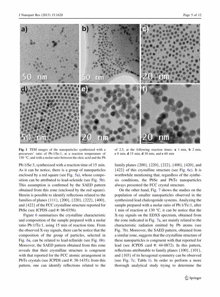
characteristics of the PbSe nanoparticles synthesized
with a precursors’ ratio of Pb:1/Se:1. Figure 1a shows,
at short reaction times (1 min), the size of the
nanoparticles can be grouped in two populations,
one composed of nanoparticles with sizes between 1
and 3 nm, and another with sizes between 5 and 7 nm.
After 2 min, these populations could still be observed,
although the population of the smaller nanoparticles
decreases in number, whereas the one of larger
nanoparticles increases (see Fig. 1b). After 8 min of
reaction, the population of smaller nanoparticles
almost disappears, and the resultant population seems
to be composed only of nanoparticles with sizes
between 6 and 8 nm (see Fig. 1c). At 15 min of
reaction, the population of the smaller nanoparticles
cannot be observed anymore; instead, the observed
nanoparticles display an average size of 10.6 nm (see
Fig. 1d). Moreover, after 30 and 60 min of reaction, it
can be observed that the particle size increases to 11
and 11.44 nm, respectively, and its morphology
changes from spherical to cubic, along with the
formation of nanoparticles agglomerates (see Fig. 1e, f).
It is worthwhile to mention that analogous features were
observed in both nanoparticles systems, PbSe and PbTe, at
all Pb/Se and Pb/Te precursors’ ratios.
On the other hand, the variation on the molar ratio
between the lead and the chalcogen precursors was
also considered to elucidate its influence on the final
characteristic of the synthesized nanoparticles. Fig-
ure 2 shows the TEM images obtained from nanopar-
ticles synthesized with molar ratios of Pb:1/Se:1, Pb:1/
Se:3, and Pb:3/Se:1, using 15 min as the reaction time.
As it can be observed, the nanoparticles synthesized
with a molar ratio of Pb:1/Se:1 have an average
particle size of 9.57 nm (see Fig. 2b, e), whereas those
prepared at a molar ratio of Pb:1/Se:3 have a size of
8.28 nm (see Fig. 2a, d). Moreover, in both cases, the
absence of populations of smaller nanoparticles can be
noticed. It is worthwhile to mention that for reactions
with excess of chalcogen precursor, or even with a
molar ratio of one between the precursors, only the
population of larger nanoparticles can be notice on
both synthesized systems, PbSe and PbTe. However,
Fig. 2c, f shows, in the cases when the lead precursor
was used in excess (i.e., Pb:3/Se:1), the synthesized
nanoparticles can be grouped in two populations
again; one of small nanoparticles, with sizes between 1
and 3 nm, and another with an average size of 8.8 nm,
even at reaction times of 15 min.
Next, we proceeded to evaluate the effect of the
reaction temperature on the morphology of the syn-
thesized nanoparticle systems. As an example, Fig. 3
resumes the morphological characteristic of the PbTe
nanoparticles synthesized at 130 and 170 �C, with a
molar ratio of Pb:1.5/Te:1, using a lead oleate solution
with concentration of 0.26 M, and considering 10 min
as reaction time. As it can be notice, at 130 �C the
population of smaller nanoparticles is not observed,
instead there is only a population composed of cubic
nanoparticles with an average size of 13 nm (see
Fig. 3a, c). Nonetheless, at 170 �C there can be
observed two populations of nanoparticles, one com-
posed of nanoparticles with size between 1 and 3 nm,
and the other composed of cubic nanoparticles with an
average size of 18 nm.
Finally, we evaluated the effect of the concentration
of lead on the lead precursor on the synthesis of the
nanoparticles. We synthesized PbTe nanoparticles at a
reaction temperature of 130 �C, considering 2 min of
reaction time and using a molar ratio of Pb:1/Te:1.
Figure 4a, c shows, after 2 min of reaction with a lead
concentration on the lead precursor of 0.065 M, the
population of smaller nanoparticles is not visible at all,
although there can be observed nanoparticles that have
an average size of 8 nm, as well as a narrow particle
size distribution. However, as it can be observed in
Fig. 4b, d, using a lead precursor with a higher
concentration of lead (0.26 M) leads to the appearance
of the aforementioned smaller particles population,
along with nanoparticles with analogous characteris-
tics to those observed in the previous experiment,
when a lead precursor with a lower concentration of
lead was used (see Fig. 4a, c).
From these results are safe to argue that at short
reaction times and using higher concentration of lead
on the reaction leads to the appearance of two different
particle size populations. However, in order to eluci-
date the crystalline characteristics and the composition
of the observed nanoparticles populations, we pro-
ceeded to analyze the synthesized samples by means
of HAADF-STEM, EDXS, EELS, and SAED
techniques.
Crystalline characteristics and composition
Figure 5 shows a HAADF-STEM image, correspond-
ing EDXS spectrum, as well as the SAED pattern
obtained from the sample prepared with molar ratio of
Page 4 of 12 J Nanopart Res (2013) 15:1620
123
Pb:1/Se:3, synthesized with a reaction time of 15 min.
As it can be notice, there is a group of nanoparticles
enclosed by a red square (see Fig. 5a), whose compo-
sition can be attributed to lead-selenide (see Fig. 5b).
This assumption is confirmed by the SAED pattern
obtained from this zone (enclosed by the red square).
Herein is possible to identify reflections related to the
families of planes {111}, {200}, {220}, {222}, {400},
and {422} of the FCC crystalline structure reported for
PbSe (see JCPDS card #: 06-0356).
Figure 6 summarizes the crystalline characteristic
and composition of the sample prepared with a molar
ratio Pb:1/Te:1, using 15 min of reaction time. From
the observed X-ray signals, there can be notice that the
composition of the group of particles, selected in
Fig. 6a, can be related to lead-telluride (see Fig. 6b).
Moreover, the SAED pattern obtained from this zone
reveals that their crystalline structure is congruent
with that reported for the FCC atomic arrangement in
PbTe crystals (see JCPDS card #: 38-1435); from this
pattern, one can identify reflections related to the
family planes {200}, {220}, {222}, {400}, {420}, and
{422} of this crystalline structure (see Fig. 6c). It is
worthwhile mentioning that, regardless of the synthe-
sis conditions, the PbSe and PbTe nanoparticles
always presented the FCC crystal structure.
On the other hand, Fig. 7 shows the studies on the
population of smaller nanoparticles observed in the
synthesized lead chalcogenide systems. Analyzing the
sample prepared with a molar ratio of Pb:1/Te:1, after
1 min of reaction at 130 �C, it can be notice that the
X-ray signals on the EDXS spectrum, obtained from
the zone indicated in Fig. 7a, are mainly related to the
characteristic radiation emitted by Pb atoms (see
Fig. 7b). Moreover, the SAED pattern, obtained from
a similar zone, suggests that the crystalline structure of
these nanoparticles is congruent with that reported for
lead (see JCPDS card #: 44-0872). In this pattern,
reflections attributable to family planes {100}, {101},
and {103} of its hexagonal symmetry can be observed
(see Fig. 7c; Table 1). In order to perform a more
thorough analytical study trying to determine the
Fig. 1 TEM images of the nanoparticles synthesized with a
precursors’ ratio of Pb:1/Se:1, at a reaction temperature of
150 �C, and with a molar ratio between the oleic acid and the Pb
of 2.5, at the following reaction times: a 1 min, b 2 min,
c 8 min, d 15 min, d 30 min, and e 60 min
J Nanopart Res (2013) 15:1620 Page 5 of 12
123

composition of this population of smaller nanoparti-
cles, we performed an EELS analysis, on similar
regions of this sample, trying to detect the character-
istic M4,5 EELS signal of Te, but no measurable peaks
attributable to such signal could be observed in the
acquired spectra.
This evidence suggests that the small nanoparticles
that were observed in the synthesized lead chalcogen-
ide systems are Pb nanoparticles. Furthermore, as it
was previously described, the appearance of these
nanoparticles occur at (1) short reaction times (1 min),
regardless of the other synthesis parameters (see
Fig. 1); (2) longer reaction times (even at 15 min)
with high concentration of lead precursor in the
reaction (see Fig. 2); (3) long reaction times (10 min)
and high reaction temperature (170 �C) (see Fig. 3);
and (4) short reaction times (2 min) with high
concentration of lead in the lead precursor (see Fig. 4).
However, when a low concentration of lead
precursor was used to synthesize whichever of the
lead chalcogen systems, the population of small
nanoparticles almost disappears after 8 min of reac-
tion. Nonetheless, at high concentration of lead
precursor, even after 15 min of reaction, the preva-
lence of a large number of small nanoparticles was
observed. These observations imply that a large
availability of lead precursor leads to the formation
of higher numbers of Pb nanoparticles, which seem to
disappear as the reaction progresses. Moreover, the
fact that they prevail even after 10 min, at a high
reaction temperature, when low concentration of lead
precursor is used, points to that fact that their
formation is thermally favored.
Nucleation and growth mechanism
As it was presented, our experimental results suggest
the reduction of Pb2? ions, from lead oleate, into Pb0
atoms at early reaction times, suggested by the
presence of Pb nanoparticles. In addition, their
Fig. 2 TEM images of the nanoparticles synthesized with a
reaction temperature of 150 �C, a reaction time of 15 min, with
a molar ratio between the oleic acid and the Pb of 2.5, and the
following ratios for the lead and selenium precursors: a and
d Pb:1/Se:3; b and e Pb:1/Se:1; and c and f Pb:3/Se:1
Page 6 of 12 J Nanopart Res (2013) 15:1620
123

formation and subsequent disappearance, as well as
the remarkable change on the sizes and morphology of
the PbSe and PbTe nanoparticles, as reaction pro-
gresses, imply that the reduced Pb0 atoms actually
contribute to the growth of the particles in both
systems. As has been reported elsewhere, the reduc-
tion of Pb2? ions into Pb0 atoms could be promoted by
the thermal oxidation of secondary phosphine (diphen-
ylphosphine, DPP) in the TOP reagent that we used in
our reaction (Evans et al. 2010). Nonetheless, in the
mentioned report, the reduction of Pb2? ions through
such mechanism requires several hours of reaction,
and occurs after the formation of the lead chalcogenide
nanoparticles, hence making unlikely their contribu-
tion to the formation and growth of PbSe and PbTe
nanoparticles. However, in the present study, Pb
nanoparticles were clearly observed at very short
reaction times, and it was also observed how these Pb
nanoparticles disappeared as the reaction time
increased. It is proposed that the observed Pb nano-
particles are not stable, since we believe they are
nucleating and re-dissolving in the reaction as time
elapses, sometimes the reduced Pb0 atoms contribute
to the formation of the lead nanoparticles, while in
other cases contribute to the growth of the lead
chalcogenide nanoparticles; but once the reaction is
stopped, the Pb nanoparticles that were formed at that
time remain in the final product. At the end of the
reaction, once all of the reagents have been consumed,
all of the Pb0 atoms are only forming part of the
chalcogenide nanoparticles. The time it takes for the
Pb nanoparticles to stop forming depends on the
concentration of lead on the reaction, as it was
observed on the experimental results, at higher
concentration of lead on the reaction, the Pb nanopar-
ticles can still be observed at longer reaction times
Fig. 3 TEM images of the nanoparticles synthesized with a molar ratio between the precursors of Pb:1.5/Te:1, a reaction time of
10 min, with a molar ratio between the oleic acid and the Pb of 3 and a reaction temperature of a and c 130 �C and b and d 170 �C
J Nanopart Res (2013) 15:1620 Page 7 of 12
123

(15 min); and at lower concentration of lead on the
reaction, the Pb nanoparticles stop to be observed at
shorter reaction times (2 min).
In an attempt to get a more detailed insight on the
manner those Pb0 atoms contributes to the growth of
the lead chalcogenide nanoparticles, elemental line
Fig. 4 TEM images of the nanoparticles synthesized with a molar ratio between the precursors of Pb:1/Te:1, a reaction temperature of
130 �C, a reaction time of 2 min, and a molarity of Pb in the lead precursor of a and c 0.065 M and b and d 0.26 M
Fig. 5 a HAADF-STEM image of a group of PbSe nanopar-
ticles analyzed; the red square marks the area analyzed for the
EDXS study and the SAED study. b EDXS spectrum acquired
for the analyzed nanoparticles. c SAED pattern recorded from
the group of nanoparticles analyzed. (Color figure online)
Page 8 of 12 J Nanopart Res (2013) 15:1620
123

profile analyses were performed on individual nano-
particles. Figure 8b shows the EDXS line profile
performed on a PbTe nanoparticle that is shown in
Fig. 8a; the red line observed in the image corresponds
to the region analyzed during the experiment. In the
elemental profiles, it can be seen that the Pb signal
starts to appear slightly before the Te signal does, and
it also ends slightly after. It can also be seen that the Pb
signal is slightly higher in both edges of the nanopar-
ticle, and closer to the center is comparable to that
from Te atoms. These results suggest a higher
concentration of lead atoms in the surface of the
nanoparticles. It is worthwhile mentioning that this
tendency was observed in both lead chalcogenide
nanoparticles systems. The presence of high concen-
tration of Pb atoms on the lead chalcogenide
Fig. 6 a HAADF-STEM image of a group of PbTe nanopar-
ticles analyzed; the red square marks the area analyzed for the
EDXS study and the SAED study. b EDXS spectrum acquired
for the analyzed nanoparticles. c SAED pattern recorded from
the group of nanoparticles studied. (Color figure online)
Fig. 7 a HAADF-STEM
image showing an area with
a high concentration of Pb
nanoparticles, the redrectangle shows the area
analyzed for the EDXS
analysis, EELS analysis, and
SAED study. b EDXS
spectrum showing a clear Pb
signal, but no clear Te signal
can be detected. c SAED
pattern obtained from a
group of Pb nanoparticles.
d EELS spectrum from the
analyzed region, no clear Te
M4,5 peak can be detected.
The sample studied
corresponds to the
nanoparticles synthesized
with a molar ratio between
the precursors of Pb:1/Te:1,
a reaction temperature of
130 �C, and a reaction time
of 1 min. (Color figure
online)
J Nanopart Res (2013) 15:1620 Page 9 of 12
123

nanoparticles has been previously reported by other
research groups (Moreels et al. 2008), and supports our
hypothesis about the contribution of Pb0 atoms on the
growth of the synthesized lead chalcogenide nanopar-
ticles, PbSe and PbTe. However, more detailed studies
based on EELS are underway to obtain a better insight
on the surface composition.
Based on the experimental evidence observed in the
current study, a formation and growth mechanism for
lead chalcogenide nanoparticles are proposed in
Fig. 9. It is proposed that the first units of PbX
(X=Se or Te) are formed by the mechanism described
by Evans et al. 2010, where a cleavage of the
phosphorous chalcogenide double bond (DPP=Se)
and proceeds by the nucleophilic attack of the oleate
on a (TOP=X)—Pb2?, generate the initial PbX species
(see Fig. 9a). However, a parallel process to the
formation of these PbX units or nuclei, is the reduction
of the Pb2? ions to Pb0 atoms by the oxidation of the
secondary phosphines, DPP, present in the reaction
(see Fig. 9b). These Pb0 atoms end up forming the Pb
nanoparticles that can be seen at early reaction times
and in the reactions with the excess of Pb. In addition,
another event that is taking place simultaneously is the
detachment of the X atoms from the ternary and/or
secondary phosphines, to end up in the reaction media
as X0 atoms. We have observed Te crystals (not
shown) in some of the reactions studied, in particular
in the reaction case Pb:1/Te:3, which suggest that not
all of the Te is interacting with the phosphines, leaving
some Te atoms available to contribute to the growth of
the nanoparticles along with the Pb0 reduced atoms.
The similarities observed in the results of both systems
suggest the availability of Se atoms in the reaction
media to contribute to the growth of the PbSe
nanoparticles along with the Pb0 atoms, just like in
the PbTe system. The Pb0 and the X0 atoms available
in the reaction media start to deposit on the surface of
the PbX nuclei previously formed, along with the PbX
species formed by the mechanism proposed by Evans
et al. 2010, contributing to the growth of the PbX
nanoparticles (see Fig. 9c). Figure 1 showed, it can be
seen that the concentration of Pb nanoparticles is high,
and the concentration of PbX nanoparticles is low at
short reaction times, but as the reaction time increases,
the amount of Pb nanoparticles observed decreases
considerably, and even at very high reaction times no
Pb nanoparticles can be observed, while the amount of
PbX nanoparticles increases considerably as the
reaction time increases. This observation supports
the argument previously stated that the Pb0 reduced
atoms and the X0 atoms start to deposit on the surface
of the previously formed PbX nuclei, and contribute to
the growth of the PbX nanoparticles. Finally, the
surface of the nanoparticles ends up with an excess of
lead, as it is suggested by the result presented in Fig. 8.
Table 1 Interplanar spacings measured on the electron dif-
fraction pattern acquired from the region shown in Fig. 7a
Electron diffraction
pattern
JCPDS 44-0872 Pb
Interplanar
spacing (A)
Interplanar
spacing (A)
Atomic
plane (hkl)
3.01 3.02 100
2.69 2.66 101
2.09 2.11
1.79 1.75 110
1.54 1.59 103
Interplanar spacings from JCPDS card # 44-0872
Fig. 8 a HAADF-STEM
image showing one PbTe
nanoparticle analyzed. The
red line indicates the region
where the linescan study
was performed, going from
left to right. b EDXS line
profiles for the Pb Ma and Te
La signals. The Pb Ma signal
can be observe to start and
finish, slightly before and
after the Ta La signal. (Color
figure online)
Page 10 of 12 J Nanopart Res (2013) 15:1620
123

Nonetheless, the lead signal observed on the surface of
the PbX nanoparticles could be related to the presence
of a capping layer at this surface. This capping layer on
the surface of the nanoparticles could have its origin
on the oxidation of the Pb0 atoms deposited on the last
layer of the nanoparticles, which are oxidized by the
oleic acid found in the reaction media, forming a lead
oleate capping layer. More detailed analyses are
underway to get a better insight on the capping layer
nature.
Conclusions
In conclusion, the variation of several synthesis
parameters, such as reaction time, lead to chalcogen
ratio, reaction temperature, and lead oleate concen-
tration, promoted the formation of two different
populations of nanoparticles, differentiated by their
size. The population of larger nanoparticles was the
one related to the chalcogenide nanoparticles (PbSe
and PbTe), whereas the population of smaller nano-
particles, with a size between 1 and 3 nm, was related
to the presence of Pb nanoparticles, as suggested by
EDXS, EELS, and electron diffraction results. The
observation of these Pb nanoparticles points to the
reduction of the Pb2? ions to Pb0 metallic atoms at
early stages of the reaction, and it was also observed
how these Pb0 atoms contribute to the growth of the
PbSe and PbTe nanoparticles. Based on the observa-
tions made, a growth mechanism for these types of
nanoparticles is proposed, where the first species of
PbX are formed by the routes currently accepted in the
literature, although, in a parallel process, the Pb2? ions
are reduced by the oxidation of a secondary phosphine
(DPP), present in the original TOP. Once the Pb0
atoms are formed, they are able to contribute to the
growth of the PbSe and PbTe nanoparticles; and the
lead chalcogenide nanoparticles end up with an excess
of Pb on its surface, as suggested by EDXS results.
Fig. 9 Growth mechanism proposed for the PbTe and PbSe nanoparticles, based on the experimental observations presented
J Nanopart Res (2013) 15:1620 Page 11 of 12
123

These results allow us to elucidate in better detail the
mechanisms of nucleation and growth of lead chalco-
genide nanoparticles, which will contribute to a better
understanding of the processes involved in the forma-
tion of this type of nanoparticles, the role played by the
different reagents in the reaction and eventually will
translate into synthesis methods more efficient, with
better control on the results obtained.
Acknowledgments This study was supported by the Mexican
Secretary of Education (SEP), PROMEP program, through the
project ‘‘Apoyo a la Incorporacion de Nuevos PTC’’, Project
Number PROMEP/103.5/10/3889, and by CONACYT Mexico
through Project Numbers 148391 and 154303. The authors also
thank the International Center for Nanotechnology and Advanced
Materials, ICNAM, at the University of Texas at San Antonio, for
the support provided for the use of their Advanced Microscopy
Center. LMDC and DFGG thank and acknowledge the financial
support received from CONACYT Mexico.
References
Achimovicova M, Daneu N, Recnik A, Durisin J, Balaz P,
Fabian M, Kovac J, Satka A (2009) Characterization of
mechanochemically synthesized lead selenide. Chem Pap
63(5): 562–567
Cho K-S, Talapin DV, Gaschler W, Murray CB (2005) Designing
PbSe nanowires and nanorings through oriented attachment
of nanoparticles. J Am Chem Soc 127:7140–7147
Choi JJ, Lim Y-F, Santiago-Berrios MB, Oh M, Hyun B-R, Sun
L, Bartnik AC, Goedhart A, Malliaras GG, Abruna HD,
Wise FW, Hanrath T (2009) PbSe nanocrystal excitonic
solar cells. Nano Lett 9(11):3749–3755
Cui D, Xu J, Zhu T, Paradee G, Ashok S (2006) Harvest of near
infrared light in PbSe nanocrystal-polymer hybrid photo-
voltaic cells. Appl Phys Lett 88:183111
Du H, Chen C, Krishnan R, Krauss TD, Harbold JM, Wise FW,
Thomas MG, Silcox J (2002) Optical properties of colloi-
dal PbSe nanocrystals. Nano Lett 2(11):1321–1324
Evans CM, Evans ME, Krauss TD (2010) Mysteries of TOPSe
revealed: insights into quantum dot nucleation. J Am Chem
Soc 132:10973–10975
Houtepen AJ, Koole R, Vanmaekelbergh D, Meeldijk J, Hickey
SGJ (2006) The hidden role of acetate in the PbSe nano-
crystal synthesis. J Am Chem Soc 128:6792–6793
Huynh WU, Dittmer JJ, Alivisatos AP (2002) Hybrid nanorod-
polymer solar cells. Science 295:2425–2427
Johnston KW, Pattantyus-Abraham AG, Clifford JP, Myrskog
SH, Hoogland S, Shukla H, Klem EJD, Levina L, Sargent
EH (2008) Schottky-quantum dot photovoltaics for effi-
cient infrared power conversion. Appl Phys Lett 92:122111
Kerner R, Palchik O, Gedanken A (2001) Sonochemical and
microwave-assisted preparations of PbTe and PbSe.A
comparative study. Chem Mater 13:1413–1419
Koleilat GI, Levina L, Shukla H, Myrskog SH, Hinds S, Pat-
tantyus-Abraham AG, Sargent EH (2008) Efficient, stable
infrared photovoltaics based on solution-cast colloidal
quantum dots. ACS Nano 2(5):833–840
Lifshitz E, Bashouti M, Kloper V, Kigel A, Eisen MS, Berger S
(2003) Synthesis and characterization of PbSe quantum
wires, multipods, quantum rods, and cubes. Nano Lett 3(6):
857–862
Liu H, Owen JS, Alivisatos P (2007) Mechanistic study of
precursor evolution in colloidal Group II–VI semicon-
ductor nanocrystal synthesis. J Am Chem Soc 129:305–312
Lu W, Fang J, Stokes KL, Lin JJ (2004) Shape evolution and self
assembly of monodisperse PbTe nanocrystals. J Am Chem
Soc 126:11798–11799
Mokari T, Zhang M, Yang PJ (2007) Shape, size, and assembly
control of PbTe nanocrystals. J Am Chem Soc 129:9864
Moreels I, Fritzinger B, Martins JC, Hens ZJ (2008) Surface
chemistry of colloidal PbSe nanocrystals. J Am Chem Soc
130:15081–15086
Murphy JE, Beard MC, Norman AG, Ahrenkiel SP, Johnson JC,
Yu P, Micic OI, Ellingson RJ, Nozik AJ (2006) PbTe col-
loidal nanocrystals: synthesis, characterization, and multiple
exciton generation. J Am Chem Soc 128:3241–3247
Owen JS, Chan EM, Liu H, Alivisatos P (2010) Precursor
conversion kinetics and the nucleation of cadmium sele-
nide nanocrystals. J Am Chem Soc 132:18206–18213
Tai G, Zhou B, Guo W (2008) Structural characterization and
thermoelectric transport properties of uniform single-
crystalline lead telluride nanowires. J Phys Chem C 112:
11314–11318
Wang Y, Cai K, Yao X (2011a) Facile fabrication and thermo-
electric properties of PbTe-modified poly(3,4-ethylenedi-
oxythiophene) nanotubes. ACS Appl Mater Interfaces 3:
1163–1166
Wang YY, Cai KF, Yin JL, An BJ, Du Y, Yao X (2011b) In situ
fabrication and thermoelectric properties of PbTe–polyan-
iline composite nanostructures. J Nanopart Res 13:533–539
Wise FW (2000) Lead salt quantum dots: the limit of strong
quantum confinement acc. Chem Res 33:773–780
Yan Q, Chen H, Zhou W, Hng HH, Chiang Boey FY, Ma J
(2008) A simple chemical approach for PbTe nanowires
with enhanced thermoelectric properties. Chem Mater
20:6298–6300
Yang Y, Taggart DK, Cheng MH, Hemminger JC, Penner RM
(2010) High-throughput measurement of the seebeck
coefficient and the electrical conductivity of lithographi-
cally patterned polycrystalline PbTe nanowires. J Phys
Chem Lett 1:3004–3011
Yu WW, Falkner JC, Shih BS, Colvin VL (2004) Preparation
and characterization of monodisperse PbSe semiconductor
nanocrystals in a noncoordinating solvent. Chem Mater
16:3318–3322
Zhang G, Lu X, Wang W, Li X (2007) Facile synthesis of a
hierarchical PbTe flower-like nanostructure and its shape
evolution process guided by a kinetically controlled
regime. Chem Mater 19:5207–5209
Ziqubu N, Ramasamy K, Rajasekhar PVSR, Revaprasadu N,
O’Brien P (2010) Simple route to dots and rods of PbTe
nanocrystals. Chem Mater 22:3817–3819
Page 12 of 12 J Nanopart Res (2013) 15:1620
123