EPITAXY and orientation control in chemical solution deposited PbS and PbSe monocrystalline films
Upload
independentCategory
view
6download
0
So
UVa
b
c
d
a
ARRA
KNSCM
1
PalaBs
gilbtiwbcuol
0d
Materials Chemistry and Physics 113 (2009) 107–114
Contents lists available at ScienceDirect
Materials Chemistry and Physics
journa l homepage: www.e lsev ier .com/ locate /matchemphys
ize- and shape-controlled synthesis and propertiesf colloidal PbSe nanocrystals
mesh Kumara,d, Shailesh N. Sharmaa,∗, Sukhvir Singhb, M. Kara,.N. Singhc, B.R. Mehtac, Rita Kakkard
Electronic Materials Division, National Physical Laboratory, Dr. K.S. Krishnan Marg, New Delhi 110 012, IndiaElectron Microscope Section, National Physical Laboratory, Dr. K.S. Krishnan Marg, New Delhi 110 012, IndiaDepartment of Physics, Thin Film Laboratory, Indian Institute of Technology (IIT), Hauz Khas, New Delhi 110 016, IndiaDepartment of Chemistry, University of Delhi, Delhi 110 007, India
r t i c l e i n f o
rticle history:eceived 20 March 2008
a b s t r a c t
In this work, it has been demonstrated that the co-ordinating tri-n-octylphosphine and oleic acid mixtureplays an essential role in the control of the growth, size distribution, colloidal stability, crystallinity and
eceived in revised form 3 June 2008ccepted 1 July 2008
eywords:anostructuresemiconductors
shape of the resulting PbSe nanocrystals. A systematic combination of the reaction and growth conditionsallows the possibility to produce high quality PbSe nanocrystals over a broad range of diameters withnarrow size distributions without any size selective precipitation techniques being applied. The cappingagent/ligands helps to control the grain growth of the nanocrystals during its formation and increases thedispersibility of the nanocrystals, which can easily be manipulated and used for various applications in
scopy
madteodqatnat
2
2
hemical synthesisicrostructure
optoelectronics and micro
. Introduction
Colloidal IV–VI semiconductor quantum dots such as PbS andbSe offer a unique possibility of strong quantum confinements compared with II–VI semiconductor nanoparticles due to theirarge Bohr radius ∼46 nm, about eight times larger than CdSe withnarrow direct bandgap (0.28 eV at 300 K) [1–3]. Due to its largeohr radius, PbSe has great potential for applications in NIR lasers,olar cells, optical switches, telecommunications, etc. [4–6].
However, all semiconductors often possess unsatisfied dan-ling bonds at the surfaces, which give rise to surface statesn the band gap. Due to the large surface-to-volume ratio inow dimensional systems, the properties are strongly influencedy their surface structure [7]. The preparation of semiconduc-or nanocrystals by the method of colloidal chemistry usuallynvolves capping of the nanocrystal surface with stabilizing ligands
hich fulfill various functions, including the passivation of danglingonds at the surfaces. Therefore, for the synthesis of high quality
apped-nanocrystals and to understand its properties, a detailednderstanding of the ligand surface interactions is necessary inrder to improve them for its better utilization in many techno-ogical applications [7–9]. Over the past decade, such work has∗ Corresponding author. Tel.: +91 11 45608652; fax: +91 11 25726938.E-mail address: [email protected] (S.N. Sharma).
adL2Tfwvtt
254-0584/$ – see front matter © 2008 Elsevier B.V. All rights reserved.oi:10.1016/j.matchemphys.2008.07.016
.© 2008 Elsevier B.V. All rights reserved.
ainly concentrated on II–VI and III–V semiconductor nanocrystalsnd little experimental information is available on IV–VI semicon-uctor [10,11]. Although recently, there has been few reports onhe synthesis of PbSe QD’s using various techniques, however, theffect of capping agents and growth temperature on the shapef the PbSe nanocrystals has received much less attention, tillate. This is partially due to the difficulty of synthesizing colloidaluantum dots with a well-controlled size, narrow size distributionnd well-passivated surface. The objective of this work is to syn-hesize colloidal PbSe quantum dots with a well-controlled size,arrow size distribution and passivated surface using TOP and Oleiccid/TOP as effective capping agents at different growth tempera-ures.
. Experimental
.1. Synthesis of TOP and oleic acid/TOP-capped PbSe nanocrystals
PbSe QDs were synthesized by chemical route method as described by Murray etl. [8]. All experiments were carried out under an inert argon atmosphere using stan-ard Schlenk type apparatus. In a typical synthesis of TOP-capped PbSe nanocrystals,ead acetate (1.5 g) in 5 mL TOP was heated at a temperature of 100–130 ◦C for–3 h followed by addition of TOPSe (12 mL, 1 M solution) at room temperature.
he reaction mixture was then stirred at a temperature between 80 ◦C and 150 ◦Cor 1–3 h to obtain a black suspension. The residue after centrifugation was washedith methanol to remove impurities. The final product was isolated from n-hexaneia centrifugation. For a typical synthesis of Oleic Acid/TOP-capped PbSe nanocrys-als, Oleic acid and Phenyl ether was used in addition to above chemicals and nearlyhe same procedure was adopted for the formation of molecular precursor solution.

108 U. Kumar et al. / Materials Chemistry and Physics 113 (2009) 107–114
; (c) T
Ta1wtSs(rn(
3
dowatdt
iaacfd
Ttoqagiad7cUtitPcfewasT
Fig. 1. FTIR transmittance spectra of (a) pure TOP; (b) pure oleic acid
he details of the synthesis procedure can be found in the work of Murray et al. [8]nd Du et al. [12]. The injection and growth temperature was varied from 80 ◦C to50 ◦C to obtain the desired particle size. Higher injection and growth temperaturesere used to prepare larger sized PbSe QDs. As in the case of TOP-capped PbSe,
he final product was isolated from n-hexane via centrifugation. TEM/HRTEM andAED (selected area electron diffraction) analysis for the estimation of crystalliteize, phase and crystalline planes was carried out using a JEOL electron microscopeJEM 2000EX) and Technai G2 operated at 80 kV and 200 kV accelerating voltageespectively. The surface morphology (SEM) was observed using a LEO 440 scan-ing electron microscope. FTIR spectra were recorded by PerkinElmer InstrumentSpectrum BX-500).
. Results and discussion
Upon variation of growth temperature from 80 ◦C to 150 ◦C forifferent capping agents TOP and oleic acid/TOP, PbSe nanocrystalsf different diameters were obtained. For the sake of convenience,e would designate PbSe nanocrystals produced using TOP alone
nd Oleic Acid/TOP capping agents as Tg and Og samples respec-ively. Here, the subscript g refers to the growth temperature useduring the synthesis of PbSe nanocrystals and it varies from 80 ◦Co 150 ◦C.
Fig. 1(a–h) shows FTIR spectra of Tg and Og samples correspond-ng to different growth temperatures of 80–150 ◦C, respectively,
nd for comparison, FTIR spectra of pure oleic acid and TOP arelso taken. In the FTIR spectra of pure TOP (Fig. 1(a)) and TOP-apped PbSe samples (Fig. 1(curves c, e and g)), the characteristiceatures of CH2 stretching and deformation modes are well evi-ent at ∼2840–2900 and ∼1500 cm−1, respectively [13,14]. In purebn
so
80; (d) O80; (e) T110; (f) O110; (g) T150 and (h) O150 PbSe nanocrystals.
OP FTIR spectra, a band between 1100 cm−1 and 1200 cm−1 showshe existence of P O stretching mode corresponding to phosphinexide [14] (Fig. 1(a)). However, this band at ∼1100–1200 cm−1 isuite weak for TOP-capped PbSe nanoparticles (Fig. 1(curves c, end g)) indicating inefficient capping of PbSe nanocrystals by TOProups. As shown in Fig. 1(b), pure oleic acid spectrum is character-zed by two bands at 2906 cm−1 and 2843 cm−1 attributed to thesymmetric CH2 and symmetric CH2 stretching modes, the CH2eformation vibration at 1410 cm−1, and the rocking vibration at00 cm−1, respectively [15]. The sharp band at 1700 cm−1 is theharacteristic stretching vibration of C O in carboxylic acid [13–15].pon comparison with Fig. 1(curves d, f and h), it was found that
he peaks at ∼2880–2990, 1440–1460 and 660–720 cm−1 also existn the IR spectra of OA-capped PbSe nanoparticles which indicatehe existence of C H group and long alkyl chain in the OA-cappedbSe nanoparticles [13–15]. This reveals that OA was successfullyapped on the surface of PbSe nanoparticles. In Fig. 1(curves, d,and h), peaks between 1570 cm−1 and 1590 cm−1 indicated thexistence of carboxylic acid salt in OA-capped PbSe nanoparticleshich was originally found at ∼1700 cm−1 in the case of pure oleic
cid as also reported by others [13–15]. Thus, OA-capped PbSeamples show appreciable signatures of oleic acid groups besidesOP groups which indicates efficient capping or perfect passivation
y oleic acid/TOP groups in contrast to that of TOP-capped PbSeanocrystallites.The effect of growth temperature and capping agents on thehape and morphology of PbSe nanocrystals are studied by meansf TEM/HRTEM and SAED. Fig. 2(a and b) shows a bright field TEM
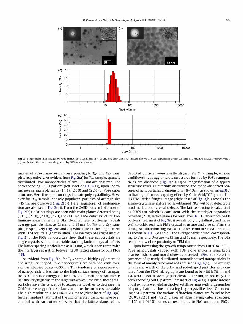
U. Kumar et al. / Materials Chemistry and Physics 113 (2009) 107–114 109
F d righ(
ipdcise∼tF(lapwFsTt[
aaotupGTfc
dctstiHssabpwsair
Pcptll1c
ig. 2. Bright field TEM images of PbSe nanocrystals; (a) and (b) T80 and O80 (left anc) and (d) are the corresponding sizes by DLS measurement.
mages of PbSe nanocrystals corresponding to T80 and O80 sam-les, respectively. As evident from Fig. 2(a) for T80 sample, sparselyistributed PbSe nanoparticles of size ∼20 nm are observed. Theorresponding SAED pattern (left inset of Fig. 2(a)), upon index-ng reveals main planes as (1 1 1), (2 0 0) and (2 2 0) of PbSe cubictructure. Here fine spots on rings indicate polycrystallinity. How-ver for O80 sample, densely populated particles of average size15 nm are observed (Fig. 2(b)). Here, signatures of agglomera-
ion are also seen (Fig. 2(b)). From the SAED pattern (left inset ofig. 2(b)), distinct rings are seen with main planes detected being1 1 1), (2 0 0), (2 1 0), (2 2 0) and (4 0 0) of PbSe cubic structure. Pre-iminary measurements of DLS (dynamic light scattering) revealsverage particle sizes as 21 nm and 13 nm for T80 and O80 sam-les, respectively (Fig. 2(c and d)) which are in close agreementith TEM results. High resolution TEM micrographs (right inset of
ig. 2) of the PbSe nanocrystals show that these nanocrystals areingle crystals without detectable stacking faults or crystal defects.he lattice spacing is calculated as 0.31 nm, which is consistent withhe interlayer separation between (2 0 0) lattice planes for bulk PbSe16].
As evident from Fig. 3(a) for T110 sample, highly agglomeratednd irregular shaped PbSe nanocrystals are obtained with aver-ge particle size being ∼300 nm. This tendency of agglomerationf nanoparticle arises due to the high surface energy of nanopar-icles. Gibb’s free energy of the surface of small nanoparticles issually very high due to the large surface-volume ratio, these small
articles have the tendency to aggregate together to decrease theibb’s free energy of the surface and make the surface-state stable.he high resolution TEM (HR-TEM) image (right inset of Fig. 3(a))urther implies that most of the agglomerated particles have beenoupled with each other showing that the lattice planes of theaoi((
t insets shows the corresponding SAED pattern and HRTEM images respectively);
epicted particles were mostly aligned. For O110 sample, variousauliflower-type agglomerate structures formed by PbSe nanopar-icles are observed (Fig. 3(b)). Upon magnification of a typicaltructure reveals uniformly distributed and mono-dispersed fea-ures of nanoparticles of dimensions ∼8–10 nm as shown in Fig. 3(c)ndicating enhanced capping effect by Oleic Acid/TOP group. TheRTEM lattice fringes image (right inset of Fig. 3(b)) reveals the
ingle-crystalline nature of as-obtained NCs without detectabletacking faults or crystal defects. The lattice spacing is calculateds 0.309 nm, which is consistent with the interlayer separationetween (2 0 0) lattice planes for bulk PbSe [16]. Furthermore, SAEDatterns (left inset of Fig. 3(b)) reveals poly-crystallinity and indexell to cubic rock salt PbSe crystal structure and also confirm the
trongest diffraction ring as (2 0 0) planes. From DLS measurementss shown in (Fig. 3(d and e)), the average particle sizes correspond-ng to T110 and O110 are ∼333 nm and 12 nm respectively. The DLSesults show close proximity to TEM data.
Upon increasing the growth temperature from 110 ◦C to 150 ◦C,bSe nanocrystals capped with TOP alone shows a remarkablehange in shape and morphology as observed in Fig. 4(a). Here, theresence of sparsely distributed, monodispersed nanoparticles inhe form of mainly cubes and rods are seen (Fig. 4(a)). The averageength and width of the cubic and rod-shaped particles as calcu-ated from the TEM micrographs are found to be ∼80 & 70 nm and70 & 40 nm so the average particle size ∼125 nm, respectively. Theorresponding SAED pattern (left inset of Fig. 4(a)) is quite intense
nd it exhibits well-defined polycrystalline rings with large numberf spotty features, thus indicating large crystallite sizes. On index-ng SAED pattern, the various diffraction planes are found to be:2 0 0), (2 2 0) and (4 2 2) planes of PbSe having cubic structure;1 1 3) and (4 0 0) planes corresponding to PbO-ortho and PbO2-
110 U. Kumar et al. / Materials Chemistry and Physics 113 (2009) 107–114
F nd rig( ment
ca2inla((cd
r(ha
p
F(
ig. 3. Bright field TEM images of PbSe nanocrystals; (a) and (b) T110 and O110 (left ac) Magnified image of O110; (d) and (e) are the corresponding sizes by DLS measure
ubic structures respectively. The diffraction peaks are indexedccording to the data from JCPDS file nos. 06–0354, 05–0570 and2–0389, respectively. The corresponding HRTEM image (right
nset of Fig. 4(a)) reveals the crystalline nature of these PbSeanoparticles. However, Oleic Acid/TOP capped PbSe nanocrystal-
ites exhibits higher monodispersity and polyhedral features with
verage particle size increased to ∼20 nm as compared to T110Fig. 4(b)). The results are supported by DLS measurements as wellFig. 4(c)). It is generally believed that for a spherical single phaserystal with average size ≤10–20 nm, its surface must be a polyhe-ron containing high-index crystallography planes which possiblysiodq
ig. 4. Bright field TEM images of PbSe nanocrystals; (a) and (b) T150 & O150 (left and righc) is the size (nm) of O150 by DLS measurement.
ht insets shows the corresponding SAED pattern and HRTEM images respectively);.
esult in a higher surface energy. The corresponding HRTEM imageright inset of Fig. 4(b)) reveal that these PbSe nanocrystals areighly crystalline and has rock salt structures that form orderedrrays.
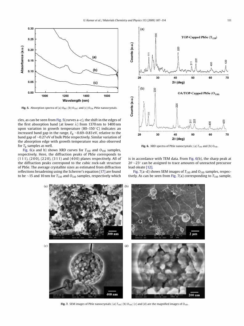
Fig. 5 shows the room temperature absorption spectra of Og sam-les prepared at different growth temperatures. Here, distinct and
harp absorption features are observed in these samples. With thencrease in growth temperature from 80 ◦C to 150 ◦C, the absorptionnset gradually shifts towards longer wavelength side (red-shift)ue to increase in PbSe nanocrystal dimensions in accordance withuantum confinement effects [3]. For these quantized nanoparti-t insets shows the corresponding SAED pattern and HRTEM images respectively);
U. Kumar et al. / Materials Chemistry and Physics 113 (2009) 107–114 111
ctuibtf
r(tort
i
Fig. 5. Absorption spectra of (a) O80; (b) O110; and (c) O150 PbSe nanocrystals.
les, as can be seen from Fig. 5(curves a–c), the shift in the edges ofhe first absorption band (at lower �) from 1370 nm to 1400 nmpon variation in growth temperature (80–150 ◦C) indicates an
ncreased band gap in the range, Eg ∼0.69–0.83 eV, relative to theand gap of ∼0.27 eV of bulk PbSe respectively. Similar variation ofhe absorption edge with growth temperature was also observedor Tg samples as well.
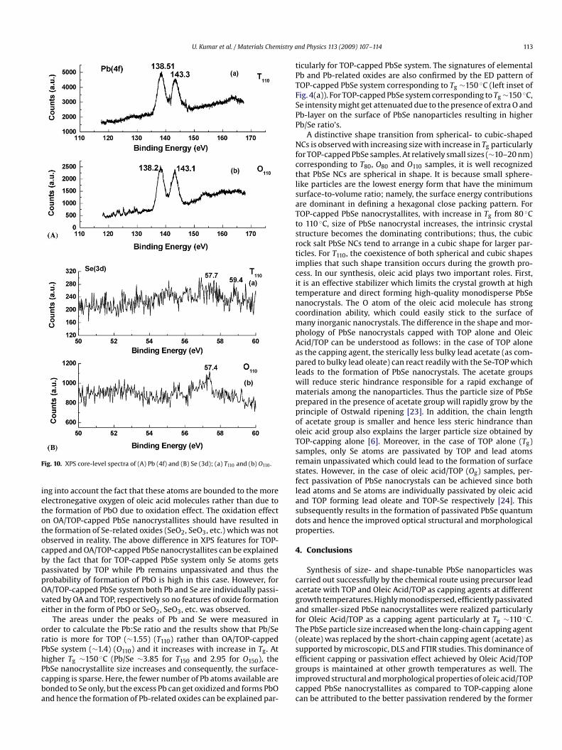
Fig. 6(a and b) shows XRD curves for T110 and O110 samples,espectively. Here, the diffraction peaks of PbSe corresponds to1 1 1), (2 0 0), (2 2 0), (3 1 1) and (4 0 0) planes respectively. All of
he diffraction peaks correspond to the cubic rock-salt structuref PbSe. The average crystallite sizes as estimated from diffractioneflections broadening using the Scherrer’s equation [17] are foundo be ∼15 and 10 nm for T110 and O110 samples, respectively which2l
t
Fig. 7. SEM images of PbSe nanocrystals; (a) T110; (b) O
Fig. 6. XRD spectra of PbSe nanocrystals; (a) T110 and (b) O110.
s in accordance with TEM data. From Fig. 6(b), the sharp peak at
� ∼23◦ can be assigned to trace amounts of unreacted precursoread oleate [12].Fig. 7(a–d) shows SEM images of T110 and O110 samples, respec-
ively. As can be seen from Fig. 7(a) corresponding to T110 sample,
110; (c) and (d) are the magnified images of O110.

112 U. Kumar et al. / Materials Chemistry and Physics 113 (2009) 107–114
anoc
i,Hicicgr
Of∼cdbaaf
avtt(naeilsta
as(
T1in(catively. For oleic acid/TOP-capped PbSe system, the presence of Pb(4f) binding energy at ∼138.2 eV and Se (3d) at ∼57.4 eV furtherconfirms the presence of only the PbSe phase [19,20]. Although, theobserved chemical shift of Pb (4f) core-levels is close to the valueof Pb O bonding (138.5 eV) but this shift can be explained by tak-
Fig. 8. AFM images of PbSe n
rregular shaped crystallites intermingled with the presence of rod-cubic- and needle-shaped nanoparticles in the debris are observed.ere particles are mostly agglomerated and presence of voids is also
ndicated (Fig. 7(a)). However for O110 sample, bunches of spheri-al shaped island of crystallites are observed (Fig. 7(b)). Upon closenspection of a typical island, presence of very small nanoparti-les of size ∼7–9 nm are observed (Fig. 7(c)). This feature of furtherrain growth is evident at higher magnification (Fig. 7(d)). The SEMesults commensurate well with TEM data.
Fig. 8(a and b) shows 2D- and 3D-view of AFM images of T110 and110 samples, respectively. For T110 sample, highly agglomerated
eatures are observed with surface Roughness: ZRMS ∼9.92 nm; ZAvg.8.10 nm and average size ∼300–400 nm, respectively. However, in
ontrast to this for O110 sample, the surface is relatively uniform andevoid of any highly agglomerated features with other parameterseing surface Roughness: ZRMS ∼3.36 nm; ZAvg. ∼2.23 nm and aver-ge size ∼8–10 nm, respectively. The low surface roughness valuesnd uniform distribution of PbSe nanoparticles of size ∼8–10 nmor O110 sample are in accordance with TEM and SEM data.
In order to examine the chemical composition, stoichiometricnd bonding changes in PbSe system with varied capping agentsiz. TOP and oleic acid/TOP (T110 and O110), the XPS survey spec-ra were obtained and are shown in Fig. 9. From Fig. 9(a and b),he signatures of Pb XPS signal (138–139 eV) [18–20], Se XPS signal57–58 eV) [18–20], (P XPS signal (∼133–134 eV) [18–20], C XPS sig-al (∼286–288 eV) [18–20] and O XPS signal (531–533 eV) [18–20]re the characteristic features of PbSe system respectively. The pres-nce of C, P and O comes from TOP and oleic acid/TOP groupsndicating the presence of TOP and oleic acid molecules as a capayer passivating the PbSe nanocrystals. The higher XPS peak inten-ity of C(1s) and O(1s) core-levels for Fig. 9(b) can be attributed tohe enhanced capping effect realized by oleic acid/TOP molecules
s compared to that of TOP-capped PbSe nanocrystallites.For PbSe system with different capping agents viz TOP alonend oleic acid/TOP are monitored by core-level XPS studies and arehown in Fig. 10(A and B). From Fig. 10(A), we can see that the Pb4f) core shows two peaks at 138.51 eV and 143.3 eV, respectively.
rystals; (a) T110 and (b) O110.
he observed chemical shift is close to the value of Pb-O bonding of38.4 eV [19,20] than with Pb Se bonding of 137.6 eV [19,20], thusndicating the presence of oxides on the surface of TOP-capped PbSeanoparticles. However, the corresponding Se (3d) XPS spectrumFig. 10(B)) shows the presence of Se (3d) binding energy by twoomponents, one at ∼57.7 eV (corresponding to PbSe) and anothert ∼59.4 eV (corresponding to SeO2, SeO3, etc.) [5,21,22], respec-
Fig. 9. XPS survey spectra of PbSe nanocrystallites; (a) T110 and (b) O110.
U. Kumar et al. / Materials Chemistry a
F
ietotocbppOve
orPhPcba
tPTFSPP
NfctlsaTtsrticitncmpAaplwmppooTsrsflasdp
4
cagafT(sefficient capping or passivation effect achieved by Oleic Acid/TOP
ig. 10. XPS core-level spectra of (A) Pb (4f) and (B) Se (3d); (a) T110 and (b) O110.
ng into account the fact that these atoms are bounded to the morelectronegative oxygen of oleic acid molecules rather than due tohe formation of PbO due to oxidation effect. The oxidation effectn OA/TOP-capped PbSe nanocrystallites should have resulted inhe formation of Se-related oxides (SeO2, SeO3, etc.) which was notbserved in reality. The above difference in XPS features for TOP-apped and OA/TOP-capped PbSe nanocrystallites can be explainedy the fact that for TOP-capped PbSe system only Se atoms getsassivated by TOP while Pb remains unpassivated and thus therobability of formation of PbO is high in this case. However, forA/TOP-capped PbSe system both Pb and Se are individually passi-ated by OA and TOP, respectively so no features of oxide formationither in the form of PbO or SeO2, SeO3, etc. was observed.
The areas under the peaks of Pb and Se were measured inrder to calculate the Pb:Se ratio and the results show that Pb/Seatio is more for TOP (∼1.55) (T110) rather than OA/TOP-cappedbSe system (∼1.4) (O110) and it increases with increase in Tg. Atigher Tg ∼150 ◦C (Pb/Se ∼3.85 for T150 and 2.95 for O150), the
bSe nanocrystallite size increases and consequently, the surface-apping is sparse. Here, the fewer number of Pb atoms available areonded to Se only, but the excess Pb can get oxidized and forms PbOnd hence the formation of Pb-related oxides can be explained par-gicc
nd Physics 113 (2009) 107–114 113
icularly for TOP-capped PbSe system. The signatures of elementalb and Pb-related oxides are also confirmed by the ED pattern ofOP-capped PbSe system corresponding to Tg ∼150 ◦C (left inset ofig. 4(a)). For TOP-capped PbSe system corresponding to Tg ∼150 ◦C,e intensity might get attenuated due to the presence of extra O andb-layer on the surface of PbSe nanoparticles resulting in higherb/Se ratio’s.
A distinctive shape transition from spherical- to cubic-shapedCs is observed with increasing size with increase in Tg particularly
or TOP-capped PbSe samples. At relatively small sizes (∼10–20 nm)orresponding to T80, O80 and O110 samples, it is well recognizedhat PbSe NCs are spherical in shape. It is because small sphere-ike particles are the lowest energy form that have the minimumurface-to-volume ratio; namely, the surface energy contributionsre dominant in defining a hexagonal close packing pattern. ForOP-capped PbSe nanocrystallites, with increase in Tg from 80 ◦Co 110 ◦C, size of PbSe nanocrystal increases, the intrinsic crystaltructure becomes the dominating contributions; thus, the cubicock salt PbSe NCs tend to arrange in a cubic shape for larger par-icles. For T110, the coexistence of both spherical and cubic shapesmplies that such shape transition occurs during the growth pro-ess. In our synthesis, oleic acid plays two important roles. First,t is an effective stabilizer which limits the crystal growth at highemperature and direct forming high-quality monodisperse PbSeanocrystals. The O atom of the oleic acid molecule has strongoordination ability, which could easily stick to the surface ofany inorganic nanocrystals. The difference in the shape and mor-
hology of PbSe nanocrystals capped with TOP alone and Oleiccid/TOP can be understood as follows: in the case of TOP alones the capping agent, the sterically less bulky lead acetate (as com-ared to bulky lead oleate) can react readily with the Se-TOP which
eads to the formation of PbSe nanocrystals. The acetate groupsill reduce steric hindrance responsible for a rapid exchange ofaterials among the nanoparticles. Thus the particle size of PbSe
repared in the presence of acetate group will rapidly grow by therinciple of Ostwald ripening [23]. In addition, the chain lengthf acetate group is smaller and hence less steric hindrance thanleic acid group also explains the larger particle size obtained byOP-capping alone [6]. Moreover, in the case of TOP alone (Tg)amples, only Se atoms are passivated by TOP and lead atomsemain unpassivated which could lead to the formation of surfacetates. However, in the case of oleic acid/TOP (Og) samples, per-ect passivation of PbSe nanocrystals can be achieved since bothead atoms and Se atoms are individually passivated by oleic acidnd TOP forming lead oleate and TOP-Se respectively [24]. Thisubsequently results in the formation of passivated PbSe quantumots and hence the improved optical structural and morphologicalroperties.
. Conclusions
Synthesis of size- and shape-tunable PbSe nanoparticles wasarried out successfully by the chemical route using precursor leadcetate with TOP and Oleic Acid/TOP as capping agents at differentrowth temperatures. Highly monodispersed, efficiently passivatednd smaller-sized PbSe nanocrystallites were realized particularlyor Oleic Acid/TOP as a capping agent particularly at Tg ∼110 ◦C.he PbSe particle size increased when the long-chain capping agentoleate) was replaced by the short-chain capping agent (acetate) asupported by microscopic, DLS and FTIR studies. This dominance of
roups is maintained at other growth temperatures as well. Themproved structural and morphological properties of oleic acid/TOPapped PbSe nanocrystallites as compared to TOP-capping alonean be attributed to the better passivation rendered by the former

1 istry a
ba
A
sN
R
[
[[
[[
[
[
[[
[
[[
14 U. Kumar et al. / Materials Chem
ecause of the slower reaction rate between lead oleate and Se-TOPs a result of the steric effect of the bulky oleate group.
cknowledgements
Authors thank Director, NPL for providing the facilities for theuccessful completion of this research work. UK acknowledges UGC,ew Delhi, for providing SRF fellowship.
eferences
[1] L.E. Brus, J. Phys. Chem. 90 (1986) 2555.[2] A. Heinglein, Chem. Rev. 89 (1989) 1861.[3] A.L. Efros, A.L. Efros, Sov. Phys. Semicond. 16 (1982) 772.[4] F.W. Wise, Acc. Chem. Res. 33 (2000) 773.[5] P.K. Khanna, N. Singh, S. Charan, A.K. Viswanath, K.R. Patil, Mater. Res. Bull. 42
(2007) 1414.
[6] I.C. Baek, S.I. Seok, N.C. Pramanik, S. Jana, M.A. Lim, B.Y. Ahn, C.J. Lee, Y.J. Jeong,J. Colloid Interface Sci. 310 (2007) 163.[7] J. Nanda, D.D. Sarma, J. Appl. Phys. 90 (2001) 2504.[8] C.B. Murray, S. Sun, W. Gaschler, H. Doyle, T.A. Betley, C.R. Kagan, IBM J. Res.
Dev. 45 (2001) 47.[9] W.W. Yu, J.C. Falkner, B.S. Shih, V.L. Colvin, Chem. Mater. 16 (2004) 3318.
[
[
[
nd Physics 113 (2009) 107–114
10] E. Lifshitz, M. Bashouti, V. Kloper, A. Kigel, M.S. Eisen, S. Berger, Nano Lett. 3(2003) 857.
11] B.L. Wehrenberg, C. Wang, P. Guyot-Sionnet, J. Phys. Chem. B 106 (2002) 10634.12] H. Du, C. Chen, R. Krishnan, T.D. Krauss, J.M. Harbold, F.W. Wise, M.G. Thomas,
J. Silcox, Nano Lett. 2 (2002) 1321.13] L. Zhang, R. He, H.C. Gu, Appl. Surf. Sci. 253 (2006) 2611.14] P.S. Kalsi, Spectroscopy of Organic Compounds, 5th ed., New Age International
(P) Ltd., Publishers, New Delhi, India, 2002.15] J.M. Luther, M. Law, Q. Song, C.L. Perkins, M.C. Beard, A.J. Nozik, ACS Nano 2
(2008) 271.16] W.F. McClune, Powder Diffraction File Alphabetical Index Inorganic Phase,
JCPDS, Swarthmore, PA, 1980.17] H.P. Klug, L.E. Alexander, X-ray Diffraction Procedures, Wiley, New York, 1954.18] C.D. Wagner, W.M. Riggs, L.E. Davis, J.F. Moulder, Handbook of X-ray Photoelec-
tron Spectroscopy, PerkinElmer, 1979.19] S. Sapra, J. Nanda, J.M. Pietryga, J.A. Hollingsworth, D.D. Sarma, J. Phys. Chem. B
110 (2006) 15244.20] D.J. Asunskis, L. Hanley, Surf. Sci. 601 (2007) 4648.21] H. Sharma, S.N. Sharma, S. Singh, R. Kishore, G. Singh, S.M. Shivaprasad, Appl.
Surf. Sci. 253 (2007) 5325.
22] H. Sharma, S.N. Sharma, S. Singh, R. Kishore, G. Singh, S.M. Shivaprasad, J. NanoSci. Nano Technol. 7 (2007) 1953.23] C.A. Leatherdale, W.K. Woo, F.V. Mikulee, M.G. Bawendi, J. Phys. Chem. B 106
(2002) 7619.24] A. Lobo, T. Moller, M. Nagel, H. Borchert, S.G. Hickey, H. Weller, J. Phys. Chem. B
109 (2005) 17422.
Copyright © 2022 FDOKUMEN