Neuroblast protuberances in the subventricular zone of the regenerative MRL/MpJ mouse
15
Neuroblast Protuberances in the Subventricular Zone of the Regenerative MRL/MpJ Mouse KASEY L. BAKER, 1,2 STEPHEN B. DANIELS, 2 JESSICA B. LENNINGTON, 1,2 THOMAS LARDARO, 1 ALEXANDRA CZAP, 1 RYAN Q. NOTTI, 1 OLIVER COOPER, 3 OLE ISACSON, 3 SALVATORE FRASCA JR., 4 AND JOANNE C. CONOVER 1,2 * 1 Center for Regenerative Biology, University of Connecticut, Storrs, Connecticut 06269 2 Department of Physiology and Neurobiology, University of Connecticut, Storrs, Connecticut 06269 3 Center for Neuroregenerative Research, McLean Hospital, Harvard Medical School, Belmont, Massachusetts 02478 4 Department of Pathobiology and Veterinary Science, University of Connecticut, Storrs, Connecticut 06269 ABSTRACT The MRL mouse is unique in its capacity for regenerative healing of wounds. This regener- ative ability includes complete closure, with little scarring, of wounds to the ear pinna and repair of cardiac muscle, without fibrosis, following cryoinjury. Here, we examine whether neurogenic zones within the MRL brain show enhanced regenerative capacity. The largest neurogenic zone in the adult brain, the subventricular zone (SVZ), lies adjacent to the lateral wall of the lateral ventricle and is responsible for replacement of interneuron populations within the olfactory bulb. Initial gross observation of the anterior forebrain in MRL mice revealed enlarged lateral ventri- cles; however, little neurodegeneration was detected within the SVZ or surrounding tissues. Instead, increased proliferation within the SVZ was observed, based on incorporation of the thymidine analogue bromodeoxyuridine. Closer examination using electron microscopy revealed that a significant number of SVZ astrocytes interpolated within the ependyma and established contact with the ventricle. In addition, subependymal, protuberant nests of cells, consisting primarily of neuroblasts, were found along the anterior SVZ of MRL mice. Whole mounts of the lateral wall of the lateral ventricle stained for the neuroblast marker doublecortin revealed normal formation of chains of migratory neuroblasts along the entire wall and introduction of enhanced green fluorescent protein-tagged retrovirus into the lateral ventricles confirmed that newly generated neuroblasts were able to track into the olfactory bulb. J. Comp. Neurol. 498: 747–761, 2006. © 2006 Wiley-Liss, Inc. Indexing terms: neurogenesis; neural stem cell; neuroblasts; regeneration; migration Neural stem cells (NSC) support neurogenesis in two major regions of the adult brain, the subgranular layers of the hippocampal dentate gyri and the subventricular zones (SVZ) along the lateral walls of the lateral ventricles (for reviews see Gage, 2000; Alvarez-Buylla et al., 2001; Peterson, 2002; Doetsch, 2003; Imura et al., 2003; Kem- permann et al., 2004; Wurmser et al., 2004; Alvarez- Buylla and Lim, 2004). SVZ neurogenesis generates new neurons that migrate as chains along the lateral wall of the lateral ventricle, culminating in the rostral migratory stream (RMS), where chains of neuroblasts transit the anterior forebrain in a restricted pathway and then enter the olfactory bulb (OB). In the OB, SVZ neuroblasts dif- ferentiate into either granule cell or periglomerular inter- neurons. To ensure continuous neurogenesis, the unique cytoarchitecture of the SVZ must support stem cell self- renewal, neuronal fate decisions, and tangential chain Grant sponsor: University of Connecticut Large Grant Fund. *Correspondence to: Joanne C. Conover, PhD, Center for Regenerative Biology, University of Connecticut, ATL Building, 1392 Storrs Road, Storrs, CT 06269-4243. E-mail: [email protected] Received 16 May 2005; Revised 19 August 2005; Accepted 9 May 2006 DOI 10.1002/cne.21090 Published online in Wiley InterScience (www.interscience.wiley.com). THE JOURNAL OF COMPARATIVE NEUROLOGY 498:747–761 (2006) © 2006 WILEY-LISS, INC.
-
Upload
independent -
Category
Documents
-
view
1 -
download
0
Transcript of Neuroblast protuberances in the subventricular zone of the regenerative MRL/MpJ mouse
Neuroblast Protuberances in theSubventricular Zone of the Regenerative
MRL/MpJ Mouse
KASEY L. BAKER,1,2 STEPHEN B. DANIELS,2 JESSICA B. LENNINGTON,1,2
THOMAS LARDARO,1 ALEXANDRA CZAP,1 RYAN Q. NOTTI,1 OLIVER COOPER,3
OLE ISACSON,3 SALVATORE FRASCA JR.,4 AND JOANNE C. CONOVER1,2*1Center for Regenerative Biology, University of Connecticut, Storrs, Connecticut 06269
2Department of Physiology and Neurobiology, University of Connecticut, Storrs,Connecticut 06269
3Center for Neuroregenerative Research, McLean Hospital, Harvard Medical School,Belmont, Massachusetts 02478
4Department of Pathobiology and Veterinary Science, University of Connecticut, Storrs,Connecticut 06269
ABSTRACTThe MRL mouse is unique in its capacity for regenerative healing of wounds. This regener-
ative ability includes complete closure, with little scarring, of wounds to the ear pinna and repairof cardiac muscle, without fibrosis, following cryoinjury. Here, we examine whether neurogeniczones within the MRL brain show enhanced regenerative capacity. The largest neurogenic zonein the adult brain, the subventricular zone (SVZ), lies adjacent to the lateral wall of the lateralventricle and is responsible for replacement of interneuron populations within the olfactory bulb.Initial gross observation of the anterior forebrain in MRL mice revealed enlarged lateral ventri-cles; however, little neurodegeneration was detected within the SVZ or surrounding tissues.Instead, increased proliferation within the SVZ was observed, based on incorporation of thethymidine analogue bromodeoxyuridine. Closer examination using electron microscopy revealedthat a significant number of SVZ astrocytes interpolated within the ependyma and establishedcontact with the ventricle. In addition, subependymal, protuberant nests of cells, consistingprimarily of neuroblasts, were found along the anterior SVZ of MRL mice. Whole mounts of thelateral wall of the lateral ventricle stained for the neuroblast marker doublecortin revealednormal formation of chains of migratory neuroblasts along the entire wall and introduction ofenhanced green fluorescent protein-tagged retrovirus into the lateral ventricles confirmed thatnewly generated neuroblasts were able to track into the olfactory bulb. J. Comp. Neurol. 498:747–761, 2006. © 2006 Wiley-Liss, Inc.
Indexing terms: neurogenesis; neural stem cell; neuroblasts; regeneration; migration
Neural stem cells (NSC) support neurogenesis in twomajor regions of the adult brain, the subgranular layers ofthe hippocampal dentate gyri and the subventricularzones (SVZ) along the lateral walls of the lateral ventricles(for reviews see Gage, 2000; Alvarez-Buylla et al., 2001;Peterson, 2002; Doetsch, 2003; Imura et al., 2003; Kem-permann et al., 2004; Wurmser et al., 2004; Alvarez-Buylla and Lim, 2004). SVZ neurogenesis generates newneurons that migrate as chains along the lateral wall ofthe lateral ventricle, culminating in the rostral migratorystream (RMS), where chains of neuroblasts transit theanterior forebrain in a restricted pathway and then enterthe olfactory bulb (OB). In the OB, SVZ neuroblasts dif-
ferentiate into either granule cell or periglomerular inter-neurons. To ensure continuous neurogenesis, the uniquecytoarchitecture of the SVZ must support stem cell self-renewal, neuronal fate decisions, and tangential chain
Grant sponsor: University of Connecticut Large Grant Fund.*Correspondence to: Joanne C. Conover, PhD, Center for Regenerative
Biology, University of Connecticut, ATL Building, 1392 Storrs Road,Storrs, CT 06269-4243. E-mail: [email protected]
Received 16 May 2005; Revised 19 August 2005; Accepted 9 May 2006DOI 10.1002/cne.21090Published online in Wiley InterScience (www.interscience.wiley.com).
THE JOURNAL OF COMPARATIVE NEUROLOGY 498:747–761 (2006)
© 2006 WILEY-LISS, INC.
migration. Studies have shown that interventions effect-ing normal regulatory signaling mechanisms within theSVZ can act either to diminish or to enhance neurogen-esis. For example, infusion of epidermal growth factor(EGF) into the lateral ventricles diminishes neurogenesisand supports gliogenesis (Doetsch et al., 2002). Also, in-troduction of truncated, tyrosine kinase-deficient EphB2into the lateral ventricles expands glial populationswithin the SVZ, although the mechanism for this expan-sion is not fully understood (Conover et al., 2000). Con-versely, transforming growth factor-! (TGF!; Cooper andIsacson, 2004) and the bone morphogenic protein (BMP)antagonist noggin promote SVZ neurogenesis (Lim et al.,2000), whereas the cytokines leukemia inhibitory factor(LIF) and ciliary neurotrophic factor (CNTF) act throughtheir gp130 receptor complex to promote NSC renewal(Shimazaki et al., 2001; Chojnacki et al., 2003). Althoughthese short-term perturbations of SVZ signaling mole-cules result in dramatic alterations to SVZ cytoarchitec-ture, it is unclear how the SVZ would compensate for andsupport long-term, sustained increases in neurogenesis.
The MRL/MpJ mouse (referred to here as MRL) is aninbred strain, with a composite genetic background fromfour different strains of mice: LG (75%), AKR (12.6%),C3H (12.1%), and C57BL/6 (0.3%; The Jackson Labora-tory, Mouse Genome Informatics). Unique to the MRLmouse is its potential for regenerative wound healing(Leferovich et al., 2001). After tissue removal by holepunch to the ear pinnae, MRL mice regenerate ear tissuewith little scarring and complete restoration of tissue ar-chitecture (Clark et al., 1998). In addition, cryoinjury tothe heart of MRL mice results in replacement of woundedtissue without fibrosis (Leferovich et al., 2001). Thesephenomena appear to be heightened responses to woundhealing, more reminiscent of regeneration seen in primi-tive vertebrates than the limited repair mechanismsfound in tissues of most mammals. Is there somethingunique about MRL stem cell niches and their capacity forregeneration?
To address this possibility, we examined the largestneurogenic region in the adult brain, the SVZ, in MRLmice. Previous, electron microscopic investigations of theSVZ have revealed the major cell types and cell–cell con-tacts along the lateral wall of the lateral ventricles(Doetsch et al., 1997, 1999; Conover et al., 2000). In com-parison with the outbred strain, CD-1, we found increasedproliferation and alterations in SVZ cytoarchitecture inMRL mice. Along the anterior SVZ, nests of neuroblastsbulged into the lateral ventricle. Whereas an intactependymal monolayer was maintained adjacent to theprotuberance, in other regions of the SVZ discontinuitiesin the ependyma were seen where astrocytic processescontacted the ventricle space.
MATERIALS AND METHODSAnimals
MRL/MpJ (abbreviated here as MRL), LG, andC57BL/6J mice were obtained from The Jackson Labora-tory. CD-1 mice were purchased from Charles River. Allstrains of mice were analyzed at 2 months of age. Animalprocedures were performed under protocols approved bythe Institutional Animal Care and Use Committee of theUniversity of Connecticut and conformed to National In-stitutes of Health guidelines.
ImmunocytochemistryMale mice were perfused transcardially with 0.9% sa-
line followed by 3% paraformaldehyde in phosphate-buffered saline (PBS; pH 7.4). Brains were removed andpostfixed overnight in 3% paraformaldehyde at 4°C.Brains were washed in PBS three times for 10 minuteseach, prior to cutting 50-"m sections with a vibratome(VT-1000S; Leica, Wetzlar, Germany). Free-floating sec-tions were washed in 0.1% Triton X-100 (Sigma, St. Louis,MO) in PBS three times for 10 minutes each, blocked in10% goat serum (Sigma) in PBS/0.1% Triton X-100 for 1hour, and incubated with primary antibody. The source,characterization, and controls are listed below for eachantibody used. Antibromodeoxyuridine (BrdU; catalog No.OBT0030; Accurate Chemical and Scientific Corporation,Westbury, NY) was raised in rat and reacts with BrdU insingle-stranded DNA or BrdU attached to protein carrieror free BrdU (manufacturer’s technical information). Themonoclonal anti-BrdU IgG2a antiserum was used at 5"g/ml. No staining was seen in cases in which animalswere not infused with BrdU. Anticaspase-3 (catalog No.AF835, lot CFZ32; R&D Systems, Minneapolis, MN) wasraised in rabbit against KLH coupled synthetic peptideCRGTELDCGIETD corresponding to amino acids 163–175 of human caspase-3. On Western blots, the antibodydetected the p17 subunit of caspase-3 and not the precur-sor form (manufacturer’s technical information and Paitelet al., 2003), and it was shown that this caspase-3 antiseralabels only apoptotic cells in the brain (Kuo et al., 2005).We used affinity-purified, polyclonal IgG caspase-3 anti-serum at 0.3 "g/ml. Antidoublecortin (DCX; catalog No.SC-8066; Santa Cruz Biotechnology, Santa Cruz, CA) wasraised in goat against an epitope of human doublecortinisoform a. This epitope is blocked by a peptide correspond-ing to residues 422–437 of human DCX (accession No. NP00054 of the NCBI database), and staining is abolished.Western blot analysis of fresh brain hippocampi revealeda single band at 50 kDa (Mizuguchi et al., 1999; Jin et al.,2004). Affinity-purified, polyclonal IgG antiserum wasused at a concentration of 1 "g/ml and stained cells withthe morphology and cell distribution of neuroblasts, aspreviously reported (Gleeson et al., 1999; Jin et al., 2004).Antiglial fibrillary acidic protein (GFAP; catalog No.Z0334, lot 096; Dako, Carpinteria, CA) was raised in rab-bit against cow GFAP isolated from spinal cord. A singlepolypeptide band was detected by Western blot at 50–53kDa (Toma et al., 2001). We used the polyclonal anti-GFAP IgG antiserum at 4.1 "g/ml and obtained stainingpatterns identical to those described in previous reports ofastrocyte cellular morphology and distribution in thebrain (Sawant et al., 1994). Anti-Ki67 (catalog No. NCL-Ki67p; Novocastra, Newcastle upon Tyne, United King-dom) was raised in rabbit against a prokaryotic recombi-nant fusion protein corresponding to a 1,086-bp Ki67 motifof the cDNA fragment (manufacturer’s technical informa-tion), and a double band at 345–395 kDa was detected byWestern blot analysis (Key et al., 1993). The polyclonalanti-Ki67 antibody was used at 24 ng/ml. After a 2-hourpulse with BrdU, all BrdU# cells also stained with Ki67antiserum. The doubly positive BrdU#, Ki67# populationwas a subpopulation of the total Ki67# population. Inaddition, as a positive control, the polyclonal Ki67 anti-sera was found to stain newborn cells that express cellcycle proteins in brain and tumor tissue (Quinones-
The Journal of Comparative Neurology. DOI 10.1002/cne
748 K.L. BAKER ET AL.
Hinojosa et al., 2006). Anti-PECAM-1 (also known as anti-CD31; catalog No. 553370; BD Biosciences/PharMingen,San Diego, CA) was raised in rat against 129/Sv mouse-derived endothelioma cell line tEnd.1. The monoclonalIgG2a antiserum was purified by affinity chromatographyand recognizes only the 130-kDa CD31 protein by Westernblot analysis (manufacturer’s technical information). An-tiserum to PECAM-1 was used at 1.7 "g/ml. Staining withthis antiserum was identical to that described in previousreports showing blood vessels in the adult mouse brain(van Praag et al., 2005). Antipolysialylated neural celladhesion molecule (PSA-NCAM; catalog No. AbC0019;AbCys, Paris, France) was raised in mouse against alpha2-8 linked neuraminic acid (n $ 10). By using hippocam-pal brain tissue, a 180-kDa band was recognized by West-ern blotting (Bouzioukh et al., 2001; Jin et al., 2004). Weused the monoclonal PSA-NCAM IgM antiserum at 0.125"g/ml and detected staining in cells with the morphologyand distribution of neuroblasts in the SVZ similar to thatin previous studies (Rousselot et al., 1995). Sections wereincubated with appropriate Alexa Fluor dye-conjugatedsecondary antibodies (Molecular Probes, Eugene, OR) for1 hour, washed three times for 10 minutes each in PBS(pH 7.4), and then incubated for 5 minutes in 2 "g/mlHoechst 33342 (Sigma). All tissue analyzed showed nolabeling when the primary antibody was omitted and thesecondary antibody alone was used as a control. Sectionswere washed for 5 minutes in PBS and coverslipped usingAquapolymount (Polysciences, Warrington, PA).
BrdU immunocytochemistyMice were injected with 300 mg BrdU/kg 2 hours prior
to perfusion (2-hour pulse; Cameron and McKay, 2001).BrdU immunostaining of 50-"m sections was conductedas described previously (Lie et al., 2002). Epifluorescenceimaging of BrdU# and Ki67# cells along the lateral wall ofthe lateral ventricle was performed with a Zeiss Axioskop2# microscope (Carl Zeiss MicroImaging, Inc., Thorn-wood, NY) and Retiga 1300 EX digital camera (Q-Imaging,Burnaby, British Columbia, Canada). BrdU# and Ki67#
cells along the lateral wall of the lateral ventricle werecounted in Openlab 3.1.5 imaging software (Improvision,Lexington, MA) in 18 anterior forebrain sections (50 "m),from coordinates 0.5–1.4 anterior, relative to bregma. Thisregion contains the protuberances found in MRL mice. Atleast three mice were used for each group, and statisticalanalyses were performed via Student’s t-test. BrdU/DCXand BrdU/GFAP colabeling was imaged with a Leica TCSSP2 confocal system together with a Leica DMIRE2 mi-croscope and Leica confocal software (version 2.61).
Cell deathAfter anticaspase-3 immunostaining, caspase-3-positive
cells within the SVZ were counted in 22 sections (50 "m)from coordinates 0.5–1.4 anterior/posterior, relative tobregma, as described above. For cell counts in the RMSand OB, caspase# cells were counted in every section (50"m) within the entire region of both structures. At leastthree mice were used for each group, and statistical anal-yses were performed via Student’s t-test.
Whole mountsWhole mounts of the entire lateral wall of the lateral
ventricle were prepared as previously described (Doetschand Alvarez-Buylla, 1996) and immunostained for DCX
(Santa Cruz Biotechnology). Whole mounts were placedonto glass slides, coverslipped with Aquapolymount (Poly-sciences), and imaged by epifluorescence microscopy.
Brain histologyMice were perfused and brains postfixed as described
above. To visualize Nissl substance, we followed standardprotocols. Briefly, anterior forebrain coronal sections areplaced onto Superfrost Plus slides and dried overnight.Tissue was then taken through a series of ethanol (70%,50%, 25%; 2 minutes each) and placed in 0.5% cresyl violetsolution for 2 minutes. Slides were then dehydrated in25%, 50%, and 75% ethanol and placed in a differentiator(acetic acid in 95% ethanol, 1:125) for 1.5 minutes. Dehy-dration steps continued with 95% and 100% ethanol, twiceeach for 2 minutes, followed by three rinses in xylene for2 minutes each. Slides were mounted with DPX and cov-erslipped. Coronal sections were imaged on a Zeiss ImagerZ1 microscope with an AxioCam MRm camera and Axio-Vision 4.5 software (Zeiss). Brain tissue was routinelyprocessed for paraffin embedding, and coronal slices (6"m) were cut, mounted onto slides, dried overnight,stained with hemotoxylin and eosin, and coverslippedwith DPX mountant.
Lateral ventricle volume, striatal volume,and RMS diameter measurements
Striatal and lateral ventricle volumes of CD-1 and MRLmice were calculated from cresyl violet-stained sectionswith a brightfield microscope (Zeiss Axioskop 2#) and astereology workstation (MicroBrightField, Williston, VT).The Cavalieri estimator was implemented on one-third ofthe 40-"m sections between #1.70 mm and %0.10 mm forthe striatum and between #1.54 mm and %0.10 mm forthe lateral ventricles, relative to Bregma. The coefficientof error was used to evaluate the precision of the calcu-lated volumes (P & 0.05).
To determine RMS diameters, 50-"m coronal sections(vibratome) of fixed brain were labeled for PSA-NCAMand GFAP. Fluorescent images were captured (Axioskop2#, Zeiss; and Retiga EX, Q-Imaging System, Burnaby,Canada) of the RMS. Measurements of the area of theRMS were performed in Openlab 3.1.5 (Improvision, Lex-ington, MA). Three RMS measurements were taken at3.67, 3.72, and 3.77 mm distal to the beginning of theolfactory bulbs, and mean values ' SEM were calculated.At least three mice were used for each group, and statis-tical analyses were performed via Student’s t-test.
Electron microscopyMale mice were perfused transcardially with 0.9% sa-
line, followed by 2% paraformaldehyde/2.5% glutaralde-hyde in 0.1 M PB (pH 7.4). Heads were postfixed by im-mersion overnight in 2% paraformaldehyde/2.5%glutaraldehyde in 0.1 M PB; brains were removed andwashed in PB three times for 40 minutes each. Three300-"m sections of the anterior forebrain were cut with avibratome, pinned to plastic dishes to prevent buckling,and processed as described previously (Conover et al.,2000). Briefly, sections were postfixed with 2% OsO4 in 0.1M PB for 1.25 hours, unpinned, and dehydrated through agraded EtOH series. Sections were en bloc stained in 2%uranyl acetate at the 70% EtOH step for 1.5 hours. Afterdehydration, whole sections were twice washed in pro-
The Journal of Comparative Neurology. DOI 10.1002/cne
749MRL SUBVENTRICULAR ZONE PROTUBERANCES
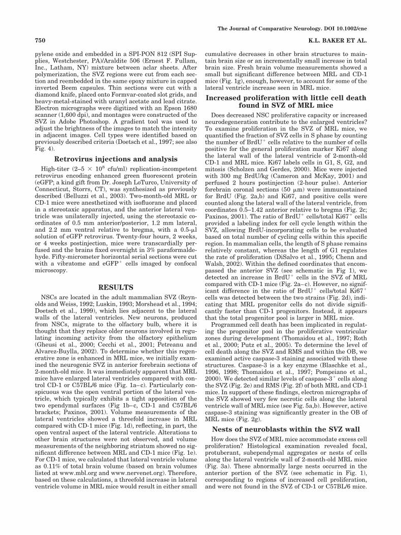
pylene oxide and embedded in a SPI-PON 812 (SPI Sup-plies, Westchester, PA)/Araldite 506 (Ernest F. Fullam,Inc., Latham, NY) mixture between aclar sheets. Afterpolymerization, the SVZ regions were cut from each sec-tion and reembedded in the same epoxy mixture in cappedinverted Beem capsules. Thin sections were cut with adiamond knife, placed onto Formvar-coated slot grids, andheavy-metal-stained with uranyl acetate and lead citrate.Electron micrographs were digitized with an Epson 1680scanner (1,600 dpi), and montages were constructed of theSVZ in Adobe Photoshop. A gradient tool was used toadjust the brightness of the images to match the intensityin adjacent images. Cell types were identified based onpreviously described criteria (Doetsch et al., 1997; see alsoFig. 4).
Retrovirus injections and analysisHigh-titer (2–5 ( 108 cfu/ml) replication-incompetent
retrovirus encoding enhanced green fluorescent protein(eGFP; a kind gift from Dr. Joseph LoTurco, University ofConnecticut, Storrs, CT), was synthesized as previouslydescribed (Belluzzi et al., 2003). Two-month-old MRL orCD-1 mice were anesthetized with isofluorane and placedin a stereotaxic apparatus, and the anterior lateral ven-tricle was unilaterally injected, using the stereotaxic co-ordinates of 0.5 mm anterior/posterior, 1.2 mm lateral,and 2.2 mm ventral relative to bregma, with a 0.5-"lsolution of eGFP retrovirus. Twenty-four hours, 2 weeks,or 4 weeks postinjection, mice were transcardially per-fused and the brains fixed overnight in 3% paraformalde-hyde. Fifty-micrometer horizontal serial sections were cutwith a vibratome and eGFP# cells imaged by confocalmicroscopy.
RESULTSNSCs are located in the adult mammalian SVZ (Reyn-
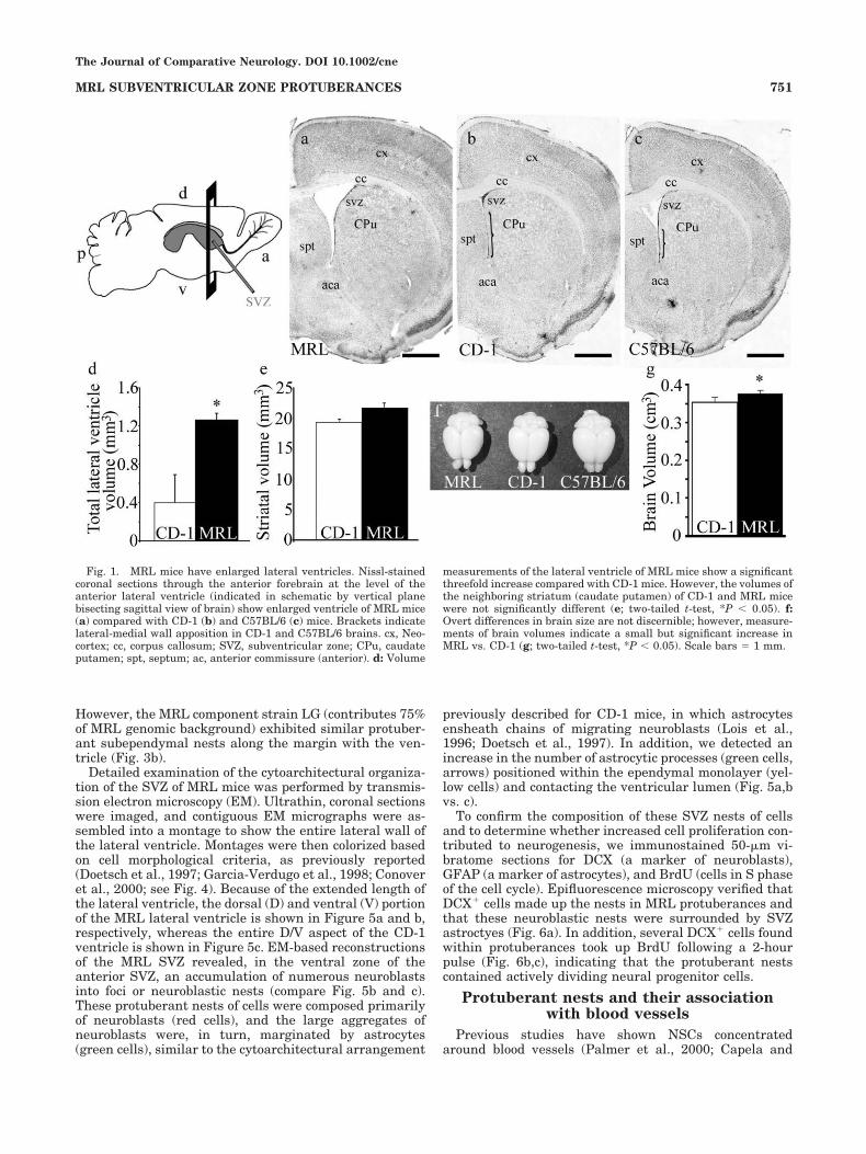
olds and Weiss, 1992; Luskin, 1993; Morshead et al., 1994;Doetsch et al., 1999), which lies adjacent to the lateralwalls of the lateral ventricles. New neurons, producedfrom NSCs, migrate to the olfactory bulb, where it isthought that they replace older neurons involved in regu-lating incoming activity from the olfactory epithelium(Gheusi et al., 2000; Cecchi et al., 2001; Petreanu andAlvarez-Buylla, 2002). To determine whether this regen-erative zone is enhanced in MRL mice, we initially exam-ined the neurogenic SVZ in anterior forebrain sections of2-month-old mice. It was immediately apparent that MRLmice have enlarged lateral ventricles compared with con-trol CD-1 or C57BL/6 mice (Fig. 1a–c). Particularly con-spicuous was the open ventral portion of the lateral ven-tricle, which typically exhibits a tight apposition of thetwo ependymal surfaces (Fig 1b–c, CD-1 and C57BL/6brackets; Paxinos, 2001). Volume measurements of thelateral ventricles showed a threefold increase in MRLcompared with CD-1 mice (Fig. 1d), reflecting, in part, theopen ventral aspect of the lateral ventricle. Alterations toother brain structures were not observed, and volumemeasurements of the neighboring striatum showed no sig-nificant difference between MRL and CD-1 mice (Fig. 1e).For CD-1 mice, we calculated that lateral ventricle volumeas 0.11% of total brain volume (based on brain volumeslisted at www.mbl.org and www.nervenet.org). Therefore,based on these calculations, a threefold increase in lateralventricle volume in MRL mice would result in either small
cumulative decreases in other brain structures to main-tain brain size or an incrementally small increase in totalbrain size. Fresh brain volume measurements showed asmall but significant difference between MRL and CD-1mice (Fig. 1g), enough, however, to account for some of thelateral ventricle increase seen in MRL mice.
Increased proliferation with little cell deathfound in SVZ of MRL mice
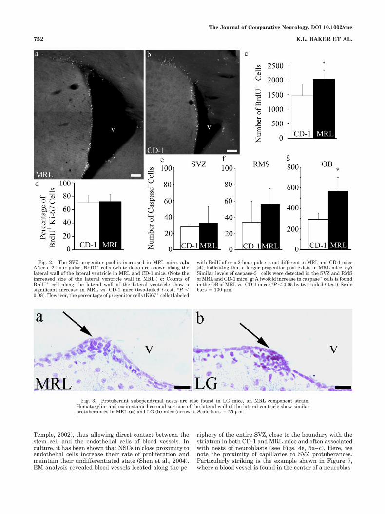
Does decreased NSC proliferative capacity or increasedneurodegeneration contribute to the enlarged ventricles?To examine proliferation in the SVZ of MRL mice, wequantified the fraction of SVZ cells in S phase by countingthe number of BrdU# cells relative to the number of cellspositive for the general proliferation marker Ki67 alongthe lateral wall of the lateral ventricle of 2-month-oldCD-1 and MRL mice. Ki67 labels cells in G1, S, G2, andmitosis (Scholzen and Gerdes, 2000). Mice were injectedwith 300 mg BrdU/kg (Cameron and McKay, 2001) andperfused 2 hours postinjection (2-hour pulse). Anteriorforebrain coronal sections (50 "m) were immunostainedfor BrdU (Fig. 2a,b) and Ki67, and positive cells werecounted along the lateral wall of the lateral ventricle, fromcoordinates 0.5–1.42 anterior relative to bregma (Fig. 2c;Paxinos, 2001). The ratio of BrdU# cells/total Ki67# cellsprovided a labeling index for cell cycle length within theSVZ, allowing BrdU-incorporating cells to be evaluatedbased on total number of cycling cells within this specificregion. In mammalian cells, the length of S phase remainsrelatively constant, whereas the length of G1 regulatesthe rate of proliferation (DiSalvo et al., 1995; Chenn andWalsh, 2002). Within the defined coordinates that encom-passed the anterior SVZ (see schematic in Fig 1), wedetected an increase in BrdU# cells in the SVZ of MRLcompared with CD-1 mice (Fig. 2a–c). However, no signif-icant difference in the ratio of BrdU# cells/total Ki67#
cells was detected between the two strains (Fig. 2d), indi-cating that MRL progenitor cells do not divide signifi-cantly faster than CD-1 progenitors. Instead, it appearsthat the total progenitor pool is larger in MRL mice.
Programmed cell death has been implicated in regulat-ing the progenitor pool in the proliferative ventricularzones during development (Thomaidou et al., 1997; Rothet al., 2000; Putz et al., 2005). To determine the level ofcell death along the SVZ and RMS and within the OB, weexamined active caspase-3 staining associated with thesestructures. Caspase-3 is a key enzyme (Blaschke et al.,1996, 1998; Thomaidou et al., 1997; Pompeiano et al.,2000). We detected similar levels of caspase-3# cells alongthe SVZ (Fig. 2e) and RMS (Fig. 2f) of both MRL and CD-1mice. In support of these findings, electron micrographs ofthe SVZ showed very few necrotic cells along the lateralventricle wall of MRL mice (see Fig. 5a,b). However, activecaspase-3 staining was significantly greater in the OB ofMRL mice (Fig. 2g).
Nests of neuroblasts within the SVZ wallHow does the SVZ of MRL mice accommodate excess cell
proliferation? Histological examination revealed focal,protuberant, subependymal aggregates or nests of cellsalong the lateral ventricle wall of 2-month-old MRL mice(Fig. 3a). These abnormally large nests occurred in theanterior portion of the SVZ (see schematic in Fig. 1),corresponding to regions of increased cell proliferation,and were not found in the SVZ of CD-1 or C57BL/6 mice.
The Journal of Comparative Neurology. DOI 10.1002/cne
750 K.L. BAKER ET AL.
However, the MRL component strain LG (contributes 75%of MRL genomic background) exhibited similar protuber-ant subependymal nests along the margin with the ven-tricle (Fig. 3b).
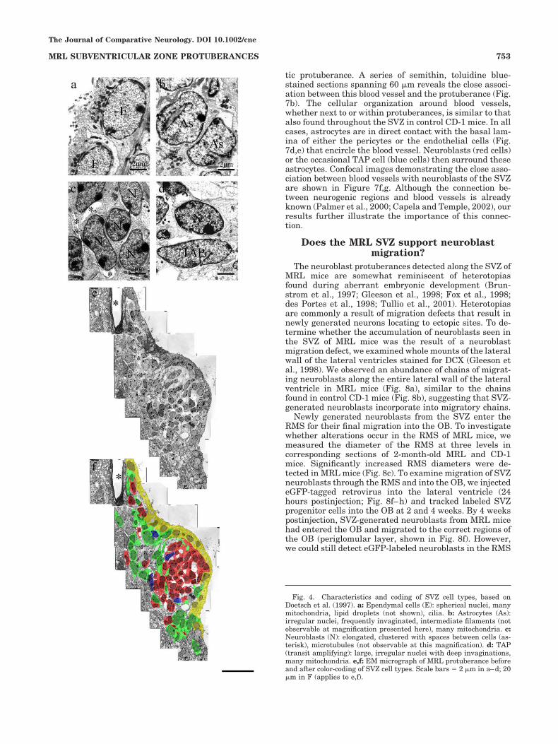
Detailed examination of the cytoarchitectural organiza-tion of the SVZ of MRL mice was performed by transmis-sion electron microscopy (EM). Ultrathin, coronal sectionswere imaged, and contiguous EM micrographs were as-sembled into a montage to show the entire lateral wall ofthe lateral ventricle. Montages were then colorized basedon cell morphological criteria, as previously reported(Doetsch et al., 1997; Garcia-Verdugo et al., 1998; Conoveret al., 2000; see Fig. 4). Because of the extended length ofthe lateral ventricle, the dorsal (D) and ventral (V) portionof the MRL lateral ventricle is shown in Figure 5a and b,respectively, whereas the entire D/V aspect of the CD-1ventricle is shown in Figure 5c. EM-based reconstructionsof the MRL SVZ revealed, in the ventral zone of theanterior SVZ, an accumulation of numerous neuroblastsinto foci or neuroblastic nests (compare Fig. 5b and c).These protuberant nests of cells were composed primarilyof neuroblasts (red cells), and the large aggregates ofneuroblasts were, in turn, marginated by astrocytes(green cells), similar to the cytoarchitectural arrangement
previously described for CD-1 mice, in which astrocytesensheath chains of migrating neuroblasts (Lois et al.,1996; Doetsch et al., 1997). In addition, we detected anincrease in the number of astrocytic processes (green cells,arrows) positioned within the ependymal monolayer (yel-low cells) and contacting the ventricular lumen (Fig. 5a,bvs. c).
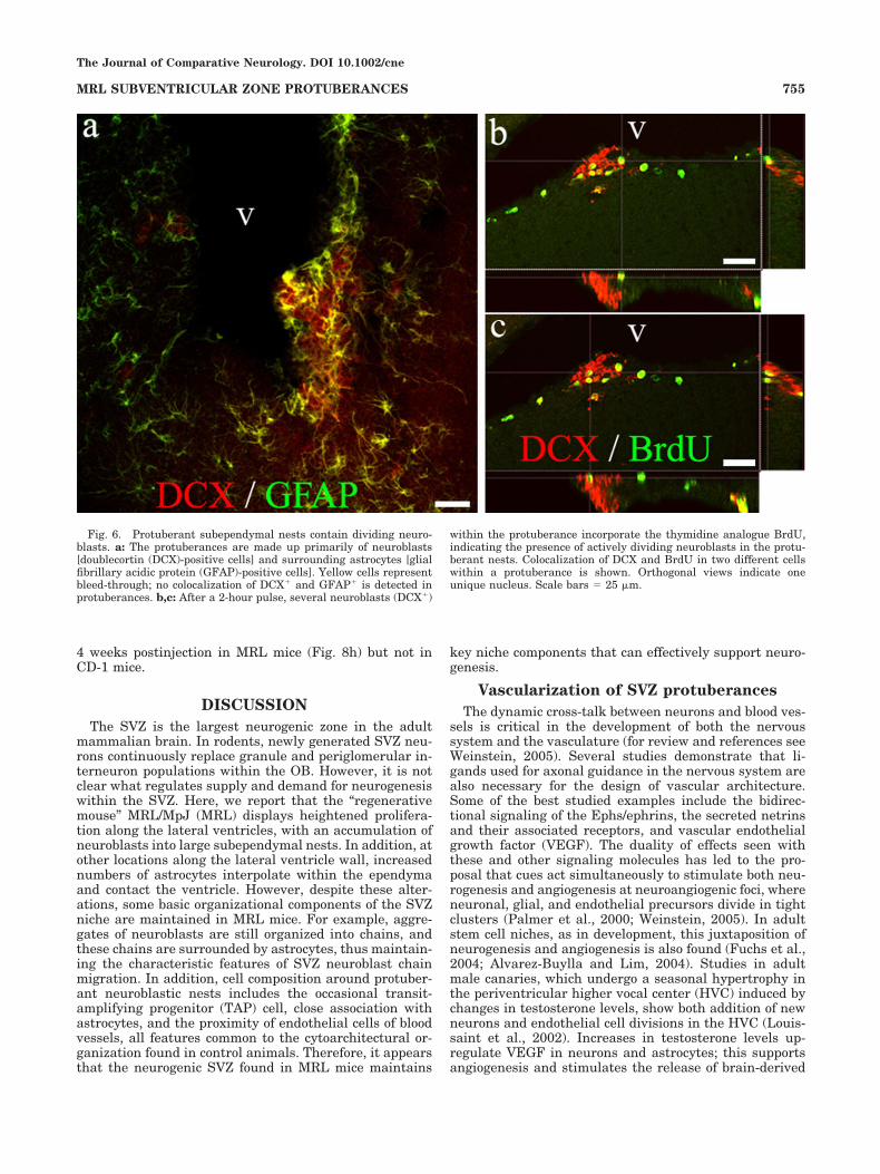
To confirm the composition of these SVZ nests of cellsand to determine whether increased cell proliferation con-tributed to neurogenesis, we immunostained 50-"m vi-bratome sections for DCX (a marker of neuroblasts),GFAP (a marker of astrocytes), and BrdU (cells in S phaseof the cell cycle). Epifluorescence microscopy verified thatDCX# cells made up the nests in MRL protuberances andthat these neuroblastic nests were surrounded by SVZastroctyes (Fig. 6a). In addition, several DCX# cells foundwithin protuberances took up BrdU following a 2-hourpulse (Fig. 6b,c), indicating that the protuberant nestscontained actively dividing neural progenitor cells.
Protuberant nests and their associationwith blood vessels
Previous studies have shown NSCs concentratedaround blood vessels (Palmer et al., 2000; Capela and
Fig. 1. MRL mice have enlarged lateral ventricles. Nissl-stainedcoronal sections through the anterior forebrain at the level of theanterior lateral ventricle (indicated in schematic by vertical planebisecting sagittal view of brain) show enlarged ventricle of MRL mice(a) compared with CD-1 (b) and C57BL/6 (c) mice. Brackets indicatelateral-medial wall apposition in CD-1 and C57BL/6 brains. cx, Neo-cortex; cc, corpus callosum; SVZ, subventricular zone; CPu, caudateputamen; spt, septum; ac, anterior commissure (anterior). d: Volume
measurements of the lateral ventricle of MRL mice show a significantthreefold increase compared with CD-1 mice. However, the volumes ofthe neighboring striatum (caudate putamen) of CD-1 and MRL micewere not significantly different (e; two-tailed t-test, *P & 0.05). f:Overt differences in brain size are not discernible; however, measure-ments of brain volumes indicate a small but significant increase inMRL vs. CD-1 (g; two-tailed t-test, *P & 0.05). Scale bars ) 1 mm.
The Journal of Comparative Neurology. DOI 10.1002/cne
751MRL SUBVENTRICULAR ZONE PROTUBERANCES
Temple, 2002), thus allowing direct contact between thestem cell and the endothelial cells of blood vessels. Inculture, it has been shown that NSCs in close proximity toendothelial cells increase their rate of proliferation andmaintain their undifferentiated state (Shen et al., 2004).EM analysis revealed blood vessels located along the pe-
riphery of the entire SVZ, close to the boundary with thestriatum in both CD-1 and MRL mice and often associatedwith nests of neuroblasts (see Figs. 4e, 5a–c). Here, wenote the proximity of capillaries to SVZ protuberances.Particularly striking is the example shown in Figure 7,where a blood vessel is found in the center of a neuroblas-
Fig. 2. The SVZ progenitor pool is increased in MRL mice. a,b:After a 2-hour pulse, BrdU# cells (white dots) are shown along thelateral wall of the lateral ventricle in MRL and CD-1 mice. (Note theincreased size of the lateral ventricle wall in MRL.) c: Counts ofBrdU# cell along the lateral wall of the lateral ventricle show asignificant increase in MRL vs. CD-1 mice (two-tailed t-test, *P &0.08). However, the percentage of progenitor cells (Ki67# cells) labeled
with BrdU after a 2-hour pulse is not different in MRL and CD-1 mice(d), indicating that a larger progenitor pool exists in MRL mice. e,f:Similar levels of caspase-3# cells were detected in the SVZ and RMSof MRL and CD-1 mice. g: A twofold increase in caspase# cells is foundin the OB of MRL vs. CD-1 mice (*P & 0.05 by two-tailed t-test). Scalebars ) 100 "m.
Fig. 3. Protuberant subependymal nests are also found in LG mice, an MRL component strain.Hematoxylin- and eosin-stained coronal sections of the lateral wall of the lateral ventricle show similarprotuberances in MRL (a) and LG (b) mice (arrows). Scale bars ) 25 "m.
The Journal of Comparative Neurology. DOI 10.1002/cne
752 K.L. BAKER ET AL.
tic protuberance. A series of semithin, toluidine blue-stained sections spanning 60 "m reveals the close associ-ation between this blood vessel and the protuberance (Fig.7b). The cellular organization around blood vessels,whether next to or within protuberances, is similar to thatalso found throughout the SVZ in control CD-1 mice. In allcases, astrocytes are in direct contact with the basal lam-ina of either the pericytes or the endothelial cells (Fig.7d,e) that encircle the blood vessel. Neuroblasts (red cells)or the occasional TAP cell (blue cells) then surround theseastrocytes. Confocal images demonstrating the close asso-ciation between blood vessels with neuroblasts of the SVZare shown in Figure 7f,g. Although the connection be-tween neurogenic regions and blood vessels is alreadyknown (Palmer et al., 2000; Capela and Temple, 2002), ourresults further illustrate the importance of this connec-tion.
Does the MRL SVZ support neuroblastmigration?
The neuroblast protuberances detected along the SVZ ofMRL mice are somewhat reminiscent of heterotopiasfound during aberrant embryonic development (Brun-strom et al., 1997; Gleeson et al., 1998; Fox et al., 1998;des Portes et al., 1998; Tullio et al., 2001). Heterotopiasare commonly a result of migration defects that result innewly generated neurons locating to ectopic sites. To de-termine whether the accumulation of neuroblasts seen inthe SVZ of MRL mice was the result of a neuroblastmigration defect, we examined whole mounts of the lateralwall of the lateral ventricles stained for DCX (Gleeson etal., 1998). We observed an abundance of chains of migrat-ing neuroblasts along the entire lateral wall of the lateralventricle in MRL mice (Fig. 8a), similar to the chainsfound in control CD-1 mice (Fig. 8b), suggesting that SVZ-generated neuroblasts incorporate into migratory chains.
Newly generated neuroblasts from the SVZ enter theRMS for their final migration into the OB. To investigatewhether alterations occur in the RMS of MRL mice, wemeasured the diameter of the RMS at three levels incorresponding sections of 2-month-old MRL and CD-1mice. Significantly increased RMS diameters were de-tected in MRL mice (Fig. 8c). To examine migration of SVZneuroblasts through the RMS and into the OB, we injectedeGFP-tagged retrovirus into the lateral ventricle (24hours postinjection; Fig. 8f–h) and tracked labeled SVZprogenitor cells into the OB at 2 and 4 weeks. By 4 weekspostinjection, SVZ-generated neuroblasts from MRL micehad entered the OB and migrated to the correct regions ofthe OB (periglomular layer, shown in Fig. 8f). However,we could still detect eGFP-labeled neuroblasts in the RMS
Fig. 4. Characteristics and coding of SVZ cell types, based onDoetsch et al. (1997). a: Ependymal cells (E): spherical nuclei, manymitochondria, lipid droplets (not shown), cilia. b: Astrocytes (As):irregular nuclei, frequently invaginated, intermediate filaments (notobservable at magnification presented here), many mitochondria. c:Neuroblasts (N): elongated, clustered with spaces between cells (as-terisk), microtubules (not observable at this magnification). d: TAP(transit amplifying): large, irregular nuclei with deep invaginations,many mitochondria. e,f: EM micrograph of MRL protuberance beforeand after color-coding of SVZ cell types. Scale bars ) 2 "m in a–d; 20"m in F (applies to e,f).
The Journal of Comparative Neurology. DOI 10.1002/cne
753MRL SUBVENTRICULAR ZONE PROTUBERANCES
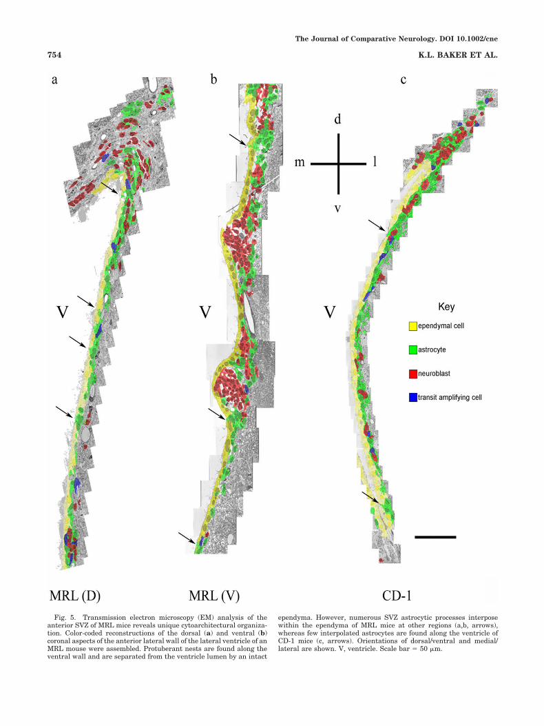
Fig. 5. Transmission electron microscopy (EM) analysis of theanterior SVZ of MRL mice reveals unique cytoarchitectural organiza-tion. Color-coded reconstructions of the dorsal (a) and ventral (b)coronal aspects of the anterior lateral wall of the lateral ventricle of anMRL mouse were assembled. Protuberant nests are found along theventral wall and are separated from the ventricle lumen by an intact
ependyma. However, numerous SVZ astrocytic processes interposewithin the ependyma of MRL mice at other regions (a,b, arrows),whereas few interpolated astrocytes are found along the ventricle ofCD-1 mice (c, arrows). Orientations of dorsal/ventral and medial/lateral are shown. V, ventricle. Scale bar ) 50 "m.
The Journal of Comparative Neurology. DOI 10.1002/cne
754 K.L. BAKER ET AL.
4 weeks postinjection in MRL mice (Fig. 8h) but not inCD-1 mice.
DISCUSSIONThe SVZ is the largest neurogenic zone in the adult
mammalian brain. In rodents, newly generated SVZ neu-rons continuously replace granule and periglomerular in-terneuron populations within the OB. However, it is notclear what regulates supply and demand for neurogenesiswithin the SVZ. Here, we report that the “regenerativemouse” MRL/MpJ (MRL) displays heightened prolifera-tion along the lateral ventricles, with an accumulation ofneuroblasts into large subependymal nests. In addition, atother locations along the lateral ventricle wall, increasednumbers of astrocytes interpolate within the ependymaand contact the ventricle. However, despite these alter-ations, some basic organizational components of the SVZniche are maintained in MRL mice. For example, aggre-gates of neuroblasts are still organized into chains, andthese chains are surrounded by astrocytes, thus maintain-ing the characteristic features of SVZ neuroblast chainmigration. In addition, cell composition around protuber-ant neuroblastic nests includes the occasional transit-amplifying progenitor (TAP) cell, close association withastrocytes, and the proximity of endothelial cells of bloodvessels, all features common to the cytoarchitectural or-ganization found in control animals. Therefore, it appearsthat the neurogenic SVZ found in MRL mice maintains
key niche components that can effectively support neuro-genesis.
Vascularization of SVZ protuberancesThe dynamic cross-talk between neurons and blood ves-
sels is critical in the development of both the nervoussystem and the vasculature (for review and references seeWeinstein, 2005). Several studies demonstrate that li-gands used for axonal guidance in the nervous system arealso necessary for the design of vascular architecture.Some of the best studied examples include the bidirec-tional signaling of the Ephs/ephrins, the secreted netrinsand their associated receptors, and vascular endothelialgrowth factor (VEGF). The duality of effects seen withthese and other signaling molecules has led to the pro-posal that cues act simultaneously to stimulate both neu-rogenesis and angiogenesis at neuroangiogenic foci, whereneuronal, glial, and endothelial precursors divide in tightclusters (Palmer et al., 2000; Weinstein, 2005). In adultstem cell niches, as in development, this juxtaposition ofneurogenesis and angiogenesis is also found (Fuchs et al.,2004; Alvarez-Buylla and Lim, 2004). Studies in adultmale canaries, which undergo a seasonal hypertrophy inthe periventricular higher vocal center (HVC) induced bychanges in testosterone levels, show both addition of newneurons and endothelial cell divisions in the HVC (Louis-saint et al., 2002). Increases in testosterone levels up-regulate VEGF in neurons and astrocytes; this supportsangiogenesis and stimulates the release of brain-derived
Fig. 6. Protuberant subependymal nests contain dividing neuro-blasts. a: The protuberances are made up primarily of neuroblasts[doublecortin (DCX)-positive cells] and surrounding astrocytes [glialfibrillary acidic protein (GFAP)-positive cells]. Yellow cells representbleed-through; no colocalization of DCX# and GFAP# is detected inprotuberances. b,c: After a 2-hour pulse, several neuroblasts (DCX#)
within the protuberance incorporate the thymidine analogue BrdU,indicating the presence of actively dividing neuroblasts in the protu-berant nests. Colocalization of DCX and BrdU in two different cellswithin a protuberance is shown. Orthogonal views indicate oneunique nucleus. Scale bars ) 25 "m.
The Journal of Comparative Neurology. DOI 10.1002/cne
755MRL SUBVENTRICULAR ZONE PROTUBERANCES
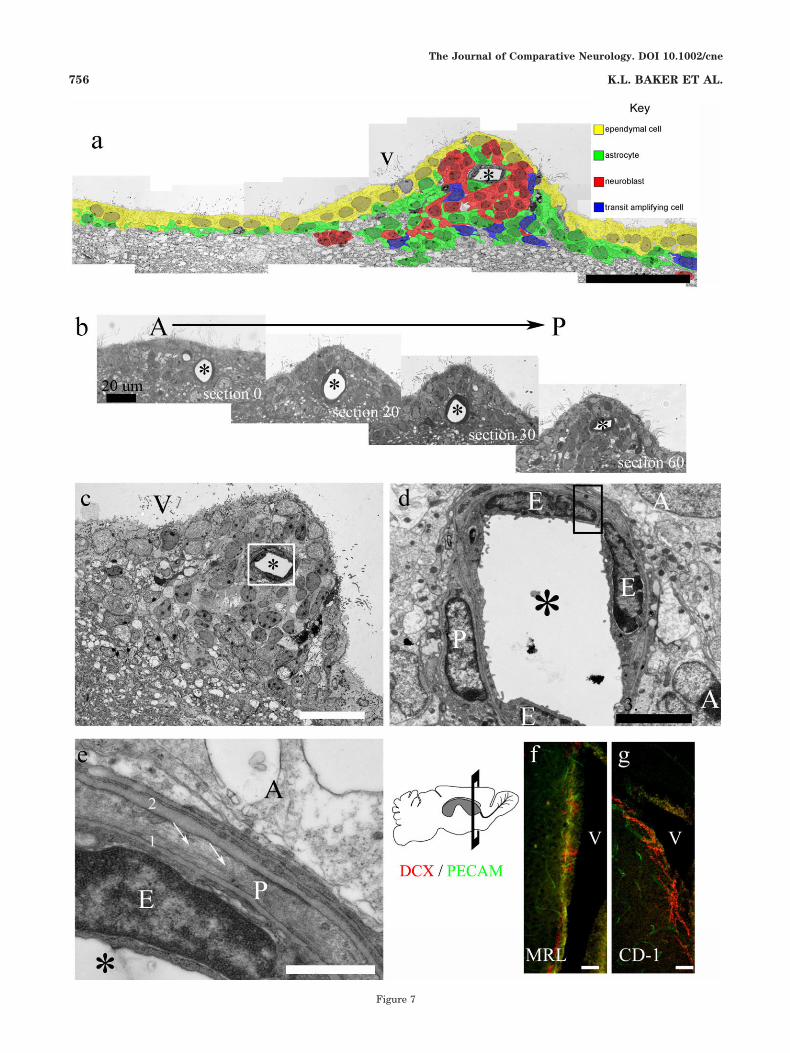
Figure 7
The Journal of Comparative Neurology. DOI 10.1002/cne
756 K.L. BAKER ET AL.
neurotrophic factor (BDNF) from endothelial cells. In-creased levels of BDNF then promote migration and re-cruitment of new neurons in the HVC ventricular zone(Louissaint et al., 2002).
In the MRL mouse, the SVZ also exhibits a close asso-ciation between blood vessels and areas of neurogenesis. Itis possible that SVZ blood vessels help support enhancedneurogenesis (Palmer et al., 2000; Louissaint et al., 2002;Capela and Temple, 2002; Shen et al., 2004); however,perivascular factors may also support the migration ofneurons (Kawamura et al., 1988; Wichterle et al., 1999;Louissaint et al., 2002; Weinstein, 2005). As found inbirds, it is likely that concordance between angiogenic andneurogenic factors acts to regulate both proliferation andmigration within the SVZ. The alterations that we de-tected in the SVZ of MRL mice may present a variation onneuroangiogenic regulation, allowing support of increasednumbers of neuroblasts in this neurogenic zone.
Enlarged ventricles and increased SVZproliferation in MRL mice
Enlarged ventricles are typically associated with neuro-degeneration and characteristically accompany aging.However, other factors, such as deregulation of stem cellself-renewal, can also contribute to enlarged ventricles.For example, Shi et al. (2004) found that mice lackingTLX, an orphan receptor that maintains NSCs in a prolif-erative, undifferentiated state, have expanded lateral ven-tricles. TLX mutant mice have no brain defects duringembryogenesis, but they show reduced cell proliferationand few nestin# cells in the neurogenic brain regions ofthe hippocampal dentate gyri and SVZ during adulthood.However, for MRL mice, we detect increased proliferationalong the lateral ventricles and aggregates of neuroblastsin subependymal outpocketings. This combination of en-larged ventricles and increased proliferation is more rem-iniscent of studies by Chenn and Walsh (2002, 2003), inwhich they observed a greatly expanded proliferativezone, with periventricular heterotopias, adjacent to anenlarged ventricular lumen in transgenic mice expressinga stabilized form of *-catenin in neural precursors. Sur-prisingly, these transgenic mice not only developed en-larged brains but the increased cerebral cortical surfacearea was arranged into folds resembling sulci and gyri ofhigher mammals. The increased proliferation we see in
MRL mice is on a much smaller scale and presents nogross cortical or OB alterations. Our observation that celldeath is significantly increased within the OB, but doesnot increase within either the SVZ or the RMS, suggeststhat cell numbers and thereby OB size are maintainedthrough regulated cell death. We speculate that the in-creases in proliferation and the accumulation of neuro-blasts found in the SVZ of MRL mice develop from anexpanded precursor pool that is maintained throughoutdevelopment. We hypothesize that the neighboring neuro-genic niche of the hippocampal dentate gyrus might alsohouse an enlarged precursor pool to support hippocampalneurogenesis. Similar findings of expanded precursorpools may be observed in other stem cell niches (e.g.,hematopoietic, epithelial, and intestinal). However, anal-ysis of enhanced hematopoiesis by means of a wide phe-notypic screen of mature hematopoietic components, pro-genitor populations, and hematopoietic stem cells isconfounded by the lack of an appropriate control for thecomposite genome of the MRL/MpJ mouse strain (unpub-lisdhed data from Leonardo Aguila, University of Con-necticut Health Center).
Chain migration in the SVZ and RMSof MRL mice
Newly generated neuroblasts migrate as chains alongthe lateral wall of the lateral ventricle and then travelanteriorly along the RMS into the OB. These chains ofneuroblasts are ensheathed by astrocytes. We observedneuroblast chains in whole-mount preparations of the lat-eral wall of the lateral ventricle in MRL mice. The chainsappeared robust and did not show any indications of ab-normal formation. In addition, although the RMS diame-ter was greater in MRL mice, the organization did notappear to be aberrant in any way, and retrovirus-labeledSVZ neuroblasts migrated through the RMS and into theOB. However, we did observe that labeled neuroblastsfrom the SVZ were still migrating through the RMS up to4 weeks postlabeling, whereas all labeled SVZ neuroblastsfrom CD-1 mice had exited the RMS and localized togranule or periglomerular layers at this time point.
Severe migration defects resulting from mutations inthe nonmuscle filamin, a protein that cross-links non-muscle actin, have been identified as a cause for the hu-man disease periventricular heterotopia (Fox et al., 1998),and ablation of nonmuscle myosin II-B heavy chain re-sults in similar periventricular heterotopias (Tullio et al.,2001). In both cases, neural cells fail to migrate to theirtarget destinations and proliferate in periventricular nod-ules. These migration defects are extreme and differ fromwhat we observe in MRL mice, i.e., normal neuroblastchain formation and the ability for SVZ neuroblasts tomigrate into the OB. Instead, our data support the hy-pothesis that an increased progenitor pool in MRL miceresults in more neuroblasts, creating a bottleneck in theanterior SVZ where neuroblasts enter the RMS, therebyslowing migration out of the SVZ. In this model, the for-mation of neuroblast protuberances is a consequence ofincreased SVZ proliferation generating increased num-bers of neuroblasts. It remains to be determined whetherextensive heterotopias develop as MRL mice age orwhether SVZ proliferation instead declines with age.
Fig. 7. Association between blood vessels and neuroblastic protu-berances. a: Colorized montage of assembled EM photomicrographsshowing a neuroblastic protuberance containing a capillary. b: Aseries of semithin sections, spanning 60 "m anterior to posterior,reveals the close association between the capillary and protuberanceshown in a. c: Slightly more anterior view of a reveals cell organiza-tion within the protuberance. d: Closeup of capillary (boxed area in c)shows a pericyte encircling endothelial cells that line the blood vessel.Astrocytes contact the pericyte and endothelial cells. e: Higher mag-nification of boxed area in d shows cell–cell interactions. The basallamina of an endothelial cell (1) contacts the basal lamina of thepericyte (2), which is in contact with a neighboring astrocyte. Notemicrofilaments within the cytoplasm of the pericyte (arrows). f,g:Coronal section of anterior forebrain of MRL and CD-1 mice showingarrangement of blood vessels (PECAM#) along the lateral wall of thelateral ventricle and their interaction with doublecortin-positive(DCX) neuroblasts. Asterisk, lumen of blood vessel; V, ventricle; E,endothelial cells; P, pericyte; A, astrocyte. Scale bars ) 50 "m in a,f,g;20 "m in b,c; 3 "m in d; 500 nm in e.
The Journal of Comparative Neurology. DOI 10.1002/cne
757MRL SUBVENTRICULAR ZONE PROTUBERANCES
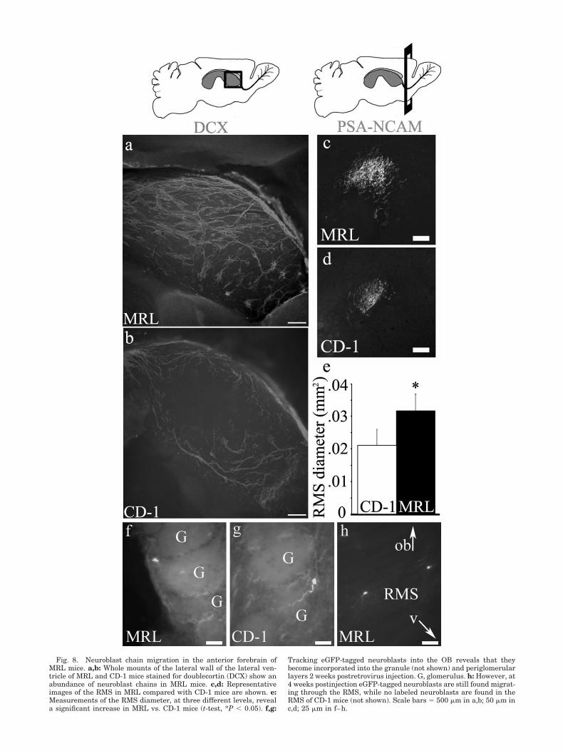
Fig. 8. Neuroblast chain migration in the anterior forebrain ofMRL mice. a,b: Whole mounts of the lateral wall of the lateral ven-tricle of MRL and CD-1 mice stained for doublecortin (DCX) show anabundance of neuroblast chains in MRL mice. c,d: Representativeimages of the RMS in MRL compared with CD-1 mice are shown. e:Measurements of the RMS diameter, at three different levels, reveala significant increase in MRL vs. CD-1 mice (t-test, *P & 0.05). f,g:
Tracking eGFP-tagged neuroblasts into the OB reveals that theybecome incorporated into the granule (not shown) and periglomerularlayers 2 weeks postretrovirus injection. G, glomerulus. h: However, at4 weeks postinjection eGFP-tagged neuroblasts are still found migrat-ing through the RMS, while no labeled neuroblasts are found in theRMS of CD-1 mice (not shown). Scale bars ) 500 "m in a,b; 50 "m inc,d; 25 "m in f–h.
The MRL backgroundThe MRL mouse is an inbred mouse with a genetic
background of LG (75%), AKR (12.6%), C3H (12.1%), andC57BL/6 (0.3%). In earlier studies, the MRL mouse andthe mutant MRL/lpr mouse were developed as congenicstrains that differed only in the lpr mutation, a loss offunction mutation of the Fas (CD95) gene (Theofilopoulosand Dixon, 1985). The lpr mutation leads to lymphadenop-athy and early-onset autoimmune systemic lupus ery-thematosus (SLE) within 3 months of age, with few micesurviving beyond 6 months of age (Dixon et al., 1978;Ballok et al., 2004). However, it is the inbred backgroundof the MRL mouse that leads to severe lupus, insofar asthe Fas-sufficient MRL (MRL/MpJ) strain also developslupus (late onset at 5–8 months of age, with a survival ofup to 2 years). To avoid complications of SLE in ouranalysis of the neurogenic SVZ, we used 2-month-old MRLmice, which do not show signs of autoimmune disease.
Subependymal neuroblast protuberances were foundnot only in MRL mice but also in the SVZ of 2-month-oldLG mice and the mutant MRL/lpr mice, whereas neither2-month-old CD-1 nor 2-month-old C57BL/6 mice exhib-ited these periventricular abnormalities. These results areconsistent with the studies of others showing enhancedwound healing in LG mice, but not in most other mousestrains, including CD-1 and C57BL/6 mice (Kench et al.,1999).
The finding that the MRL mouse has heightened woundhealing raises the possibility that wound healing andantigen-specific immune responses may be connected(Kench et al., 1999). Particularly noteworthy is the findingthat one antibody, detected in the serum of both MRL andMRL/lpr mice, is directed against Ki67, a cell proliferationmarker. The presence of anti-Ki67 antibody in MRL mice,but not other autoimmune mice, suggests that it is specificto the SLE that develops in these mice (Bloch et al., 1995).It is tantalizing to speculate that highly proliferativezones that provide the heightened regenerative capacityin these mice might also provide the targets for the auto-immune attack in SLE. However, there are several indi-cations that the wound healing ability in MRL mice isindependent of the development of autoimmunity (Kenchet al., 1999), and wound healing has been suggested to bea quantitative trait resulting from the contribution ofseveral MRL genes (Clark et al., 1998; McBrearty et al.,1998).
CONCLUSIONSOur findings demonstrate that the MRL mouse strain
supports increased proliferation, with the formation ofprotuberant nests of neuroblasts in the neurogenic nicheof the SVZ. It is interesting that this phenotype is similar,but on a much reduced scale, to that found in a model ofincreased neurogenesis produced through the stablizationof *-catenin (Chenn and Walsh, 2002, 2003). Although it istantalizing to speculate that the changes we observe in theneurogenic SVZ are part of a comprehensive regenerativeprogram unique to the MRL background, this is still notclear. The naturally occurring loss of OB neurons mayrepresent a requirement for ongoing physiological repair.Whether production of excess neuroblasts in the SVZ ac-tually supports increased incorporation of functional neu-rons in the OB remains to be determined. Additionally,
because we did not generate a specific brain injury in MRLmice that could potentially activate or utilize the newneurons produced by the SVZ, we do not know whetherinjury would result in redirection of neuroblasts to the siteof insult. Magavi et al. (2000) have shown that progenitorcells, possibly from the SVZ, can be recruited into theadult cerebral cortex following a targeted cortical lesion.Several forms of injury (i.e., cortical aspiration, corticaltransection, fimbria fornix lesions, inflammatory demyeli-nation, seizures, stroke, excitotoxic striatal lesions, andfluid precussion injury) increase cell proliferation in theSVZ (Lichtenwalner and Parent, 2006), but it is not clearwhether injury-induced neurogenesis results in proper in-tegration and function of newly generated neurons (forrecent reviews see Hallbergson et al., 2003; Lichtenwalnerand Parent, 2006). Further examination of the MRLmouse, and perhaps its component strain LG, in conjunc-tion with injury models, will provide insights into themechanisms regulating supply and demand for SVZ neu-rogenesis. In addition, examination of progenitor pools inother adult MRL stem cell niches will address a possiblelink between enhanced stem cell niches and regenerativecapacity.
LITERATURE CITEDAlvarez-Buylla A, Lim DA. 2004. For the long run: maintaining germinal
niches in the adult brain. Neuron 41:683–686.Alvarez-Buylla A, Garcia-Verdugo JM, Tramontin AD. 2001. A unified
hypothesis on the lineage of neural stem cells. Nat Rev Neurosci2:287–293.
Ballok DA, Earls AM, Krasnik C, Hoffman SA, Sakic B. 2004.Autoimmune-induced damage of the midbrain dopaminergic system inlupus-prone mice. J Neuroimmunol 152:83–97.
Belluzzi O, Benedusi M, Ackman J, LoTurco JJ. 2003. Electrophysiologicaldifferentiation of new neurons in the olfactory bulb. J Neurosci 23:10411–10418.
Blaschke AJ, Staley K, Chun J. 1996. Widespread programmed cell deathin proliferative and postmitotic regions of the fetal cerebral cortex.Development 122:1165–1174.
Blaschke AJ, Weiner JA, Chun J. 1998. Programmed cell death is auniversal feature of embryonic and postnatal neuroproliferative re-gions throughout the central nervous system. J Comp Neurol 396:39–50.
Bloch DB, Rabkina D, Bloch KD. 1995. The cell proliferation-associatedprotein Ki-67 is a target of autoantibodies in the serum of MRL mice.Lab Invest 73:366–371.
Bouzioukh F, Tell F, Jean A, Rougon G. 2001. NMDA receptor and nitricoxide synthase activation regulate polysialylated neural cell adhesionmolecule expression in adult brainstem synapses. J Neurosci 21:4721–4730.
Brunstrom JE, Gray-Swain MR, Osborne PA, Pearlman AL. 1997. Neuro-nal heterotopias in the developing cerebral cortex produced byneurotrophin-4. Neuron 18:505–517.
Cameron HA, McKay RD. 2001. Adult neurogenesis produces a large poolof new granule cells in the dentate gyrus. J Comp Neurol 435:406–417.
Capela A, Temple S. 2002. LeX/ssea-1 is expressed by adult mouse CNSstem cells, identifying them as nonependymal. Neuron 35:865–875.
Cecchi GA, Petreanu LT, Alvarez-Buylla A, Magnasco MO. 2001. Unsu-pervised learning and adaptation in a model of adult neurogenesis.J Comput Neurosci 11:175–182.
Chenn A, Walsh CA. 2002. Regulation of cerebral cortical size by control ofcell cycle exit in neural precursors. Science 297:365–369.
Chenn A, Walsh CA. 2003. Increased neuronal production, enlarged fore-brains and cytoarchitectural distortions in beta-catenin overexpressingtransgenic mice. Cereb Cortex 13:599–606.
Chojnacki A, Shimazaki T, Gregg C, Weinmaster G, Weiss S. 2003. Glyco-protein 130 signaling regulates Notch1 expression and activation in theself-renewal of mammalian forebrain neural stem cells. J Neurosci23:1730–1741.
Clark LD, Clark RK, Heber-Katz E. 1998. A new murine model for mam-
The Journal of Comparative Neurology. DOI 10.1002/cne
759MRL SUBVENTRICULAR ZONE PROTUBERANCES
malian wound repair and regeneration. Clin Immunol Immunopathol88:35–45.
Conover JC, Doetsch F, Garcia-Verdugo JM, Gale NW, Yancopoulos GD,Alvarez-Buylla A. 2000. Disruption of Eph/ephrin signaling affectsmigration and proliferation in the adult subventricular zone. Nat Neu-rosci 3:1091–1097.
Cooper O, Isacson O. 2004. Intrastriatal transforming growth factor alphadelivery to a model of Parkinson’s disease induces proliferation andmigration of endogenous adult neural progenitor cells without differ-entiation into dopaminergic neurons. J Neurosci 24:8924–8931.
des Portes V, Pinard JM, Billuart P, Vinet MC, Koulakoff A, Carrie A,Gelot A, Dupuis E, Motte J, Berwald-Netter Y, Catala M, Kahn A,Beldjord C, Chelly J. 1998. A novel CNS gene required for neuronalmigration and involved in X-linked subcortical laminar heterotopia andlissencephaly syndrome. Cell 92:51–61.
DiSalvo CV, Zhang D, Jacobberger JW. 1995. Regulation of NIH-3T3 cellG1 phase transit by serum during exponential growth. Cell Prolif28:511–524.
Dixon FJ, Andrews BS, Eisenberg RA, McConahey PJ, Theofilopoulos AN,Wilson CB. 1978. Etiology and pathogenesis of a spontaneous lupus-like syndrome in mice. Arthritis Rheum 21(Suppl):S64–S67.
Doetsch F. 2003. The glial identity of neural stem cells. Nat Neurosci6:1127–1134.
Doetsch F, Alvarez-Buylla A. 1996. Network of tangential pathways forneuronal migration in adult mammalian brain. Proc Natl Acad Sci U SA 93:14895–14900.
Doetsch F, Garcia-Verdugo JM, Alvarez-Buylla A. 1997. Cellular composi-tion and three-dimensional organization of the subventricular germi-nal zone in the adult mammalian brain. J Neurosci 17:5046–5061.
Doetsch F, Caille I, Lim DA, Garcia-Verdugo JM, Alvarez-Buylla A. 1999.Subventricular zone astrocytes are neural stem cells in the adult mam-malian brain. Cell 97:703–716.
Doetsch F, Petreanu L, Caille I, Garcia-Verdugo JM, Alvarez-Buylla A.2002. EGF converts transit-amplifying neurogenic precursors in theadult brain into multipotent stem cells. Neuron 36:1021–1034.
Fox JW, Lamperti ED, Eksioglu YZ, Hong SE, Feng Y, Graham DA,Scheffer IE, Dobyns WB, Hirsch BA, Radtke RA, Berkovic SF, Hutten-locher PR, Walsh CA. 1998. Mutations in filamin 1 prevent migrationof cerebral cortical neurons in human periventricular heterotopia. Neu-ron 21:1315–1325.
Fuchs E, Tumbar T, Guasch G. 2004. Socializing with the neighbors: stemcells and their niche. Cell 116:769–778.
Gage FH. 2000. Mammalian neural stem cells. Science 287:1433–1438.Garcia-Verdugo JM, Doetsch F, Wichterle H, Lim DA, Alvarez-Buylla A.
1998. Architecture and cell types of the adult subventricular zone: insearch of the stem cells. J Neurobiol 36:234–248.
Gheusi G, Cremer H, McLean H, Chazal G, Vincent JD, Lledo PM. 2000.Importance of newly generated neurons in the adult olfactory bulb forodor discrimination. Proc Natl Acad Sci U S A 97:1823–1828.
Gleeson JG, Allen KM, Fox JW, Lamperti ED, Berkovic S, Scheffer I,Cooper EC, Dobyns WB, Minnerath SR, Ross ME, Walsh CA. 1998.Doublecortin, a brain-specific gene mutated in human X-linked lissen-cephaly and double cortex syndrome, encodes a putative signalingprotein. Cell 92:63–72.
Gleeson JG, Lin PT, Flanagan LA, Walsh CA. 1999. Doublecortin is amicrotubule-associated protein and is expressed widely by migratingneurons. Neuron 23:257–271.
Hallbergson AF, Gnatenco C, Peterson DA. 2003. Neurogenesis and braininjury: managing a renewable resource for repair. J Clin Invest 112:1128–1133.
Imura T, Kornblum HI, Sofroniew MV. 2003. The predominant neuralstem cell isolated from postnatal and adult forebrain but not earlyembryonic forebrain expresses GFAP. J Neurosci 23:2824–2832.
Jin K, Peel AL, Mao XO, Xie L, Cottrell BA, Henshall DC, Greenberg DA.2004. Increased hippocampal neurogenesis in Alzheimer’s disease. ProcNatl Acad Sci U S A 101:343–347.
Kawamura K, Nanami T, Kikuchi Y, Kitakami A. 1988. Grafted granuleand Purkinje cells can migrate into the mature cerebellum of normaladult rats. Exp Brain Res 70:477–484.
Kempermann G, Wiskott L, Gage FH. 2004. Functional significance ofadult neurogenesis. Curr Opin Neurobiol 14:186–191.
Kench JA, Russell DM, Fadok VA, Young SK, Worthen GS, Jones-CarsonJ, Henson JE, Henson PM, Nemazee D. 1999. Aberrant wound healingand TGF-beta production in the autoimmune-prone MRL/# mouse.Clin Immunol 92:300–310.
Key G, Becker MH, Baron B, Duchrow M, Schluter C, Flad HD, Gerdes J.1993. New Ki-67-equivalent murine monoclonal antibodies (MIB 1–3)generated against bacterially expressed parts of the Ki-67 cDNA con-taining three 62 base pair repetitive elements encoding for the Ki-67epitope. Lab Invest 68:629–636.
Kuo LT, Simpson A, Schanzer A, Tse J, An SF, Scaravilli F, Groves MJ.2005. Effects of systemically administered NT-3 on sensory neuron lossand nestin expression following axotomy. J Comp Neurol 482:320–332.
Leferovich JM, Bedelbaeva K, Samulewicz S, Zhang XM, Zwas D, LankfordEB, Heber-Katz E. 2001. Heart regeneration in adult MRL mice. ProcNatl Acad Sci U S A 98:9830–9835.
Lichtenwalner RJ, Parent JM. 2006. Adult neurogenesis and the ischemicforebrain. J Cereb Blood Flow Metab 26:1–20.
Lie DC, Dziewczapolski G, Willhoite AR, Kaspar BK, Shults CW, Gage FH.2002. The adult substantia nigra contains progenitor cells with neuro-genic potential. J Neurosci 22:6639–6649.
Lim DA, Tramontin AD, Trevejo JM, Herrera DG, Garcia-Verdugo JM,Alvarez-Buylla A. 2000. Noggin antagonizes BMP signaling to create aniche for adult neurogenesis. Neuron 28:713–726.
Lois C, Garcia-Verdugo JM, Alvarez-Buylla A. 1996. Chain migration ofneuronal precursors. Science 271:978–981.
Louissaint A Jr, Rao S, Leventhal C, Goldman SA. 2002. Coordinatedinteraction of neurogenesis and angiogenesis in the adult songbirdbrain. Neuron 34:945–960.
Luskin MB. 1993. Restricted proliferation and migration of postnatallygenerated neurons derived from the forebrain subventricular zone.Neuron 11:173–189.
Magavi SS, Leavitt BR, Macklis JD. 2000. Induction of neurogenesis in theneocortex of adult mice. Nature 405:951–955.
McBrearty BA, Clark LD, Zhang XM, Blankenhorn EP, Heber-Katz E.1998. Genetic analysis of a mammalian wound-healing trait. Proc NatlAcad Sci U S A 95:11792–11797.
Mizuguchi M, Qin J, Yamada M, Ikeda K, Takashima S. 1999. Highexpression of doublecortin and KIAA0369 protein in fetal brain sug-gests their specific role in neuronal migration. Am J Pathol 155:1713–1721.
Morshead CM, Reynolds BA, Craig CG, McBurney MW, Staines WA,Morassutti D, Weiss S, van der Kooy D. 1994. Neural stem cells in theadult mammalian forebrain: a relatively quiescent subpopulation ofsubependymal cells. Neuron 13:1071–1082.
Paitel E, Fahraeus R, Checler F. 2003. Cellular prion protein sensitizesneurons to apoptotic stimuli through Mdm2-regulated and p53-dependent caspase 3-like activation. J Biol Chem 278:10061–10066.
Palmer TD, Willhoite AR, Gage FH. 2000. Vascular niche for adult hip-pocampal neurogenesis. J Comp Neurol 425:479–494.
Paxinos GF. 2001. The mouse brain. San Diego: Academic Press.Peterson DA. 2002. Stem cells in brain plasticity and repair. Curr Opin
Pharmacol 2:34–42.Petreanu L, Alvarez-Buylla A. 2002. Maturation and death of adult-born
olfactory bulb granule neurons: role of olfaction. J Neurosci 22:6106–6113.
Pompeiano M, Blaschke AJ, Flavell RA, Srinivasan A, Chun J. 2000.Decreased apoptosis in proliferative and postmitotic regions of thecaspase 3-deficient embryonic central nervous system. J Comp Neurol423:1–12.
Putz U, Harwell C, Nedivi E. 2005. Soluble CPG15 expressed during earlydevelopment rescues cortical progenitors from apoptosis. Nat Neurosci8:322–331.
Quinones-Hinojosa A, Sanai N, Soriano-Navarro M, Gonzalez-Perez O,Mirzadeh Z, Gil-Perotin S, Romero-Rodriguez R, Berger MS, Garcia-Verdugo JM, Alvarez-Buylla A. 2006. Cellular composition and cytoar-chitecture of the adult human subventricular zone: a niche of neuralstem cells. J Comp Neurol 494:415–434.
Reynolds BA, Weiss S. 1992. Generation of neurons and astrocytes fromisolated cells of the adult mammalian central nervous system. Science255:1707–1710.
Roth KA, Kuan C, Haydar TF, D’Sa-Eipper C, Shindler KS, Zheng TS,Kuida K, Flavell RA, Rakic P. 2000. Epistatic and independent func-tions of caspase-3 and Bcl-X(L) in developmental programmed celldeath. Proc Natl Acad Sci U S A 97:466–471.
Rousselot P, Lois C, Alvarez-Buylla A. 1995. Embryonic (PSA) N-CAMreveals chains of migrating neuroblasts between the lateral ventricleand the olfactory bulb of adult mice. J Comp Neurol 351:51–61.
Sawant LA, Hasgekar NN, Vyasarayani LS. 1994. Developmental expres-
The Journal of Comparative Neurology. DOI 10.1002/cne
760 K.L. BAKER ET AL.
sion of neurofilament and glial filament proteins in rat cerebellum. IntJ Dev Biol 38:429–437.
Scholzen T and Gerdes J. 2000. The Ki-67 protein: from the known and theunknown. J Cell Physiol 182(3): 311–22.
Shen Q, Goderie SK, Jin L, Karanth N, Sun Y, Abramova N, Vincent P,Pumiglia K, Temple S. 2004. Endothelial cells stimulate self-renewal and expand neurogenesis of neural stem cells. Science 304:1338 –1340.
Shi Y, Chichung Lie D, Taupin P, Nakashima K, Ray J, Yu RT, Gage FH,Evans RM. 2004. Expression and function of orphan nuclear receptorTLX in adult neural stem cells. Nature 427:78–83.
Shimazaki T, Shingo T, Weiss S. 2001. The ciliary neurotrophic factor/leukemia inhibitory factor/gp130 receptor complex operates in themaintenance of mammalian forebrain neural stem cells. J Neurosci21:7642–7653.
Theofilopoulos AN, Dixon FJ. 1985. Murine models of systemic lupuserythematosus. Adv Immunol 37:269–390.
Thomaidou D, Mione MC, Cavanagh JF, Parnavelas JG. 1997. Apoptosis
and its relation to the cell cycle in the developing cerebral cortex.J Neurosci 17:1075–1085.
Toma JG, Akhavan M, Fernandes KJ, Barnabe-Heider F, Sadikot A,Kaplan DR, Miller FD. 2001. Isolation of multipotent adult stem cellsfrom the dermis of mammalian skin. Nat Cell Biol 3:778–784.
Tullio AN, Bridgman PC, Tresser NJ, Chan CC, Conti MA, Adelstein RS,Hara Y. 2001. Structural abnormalities develop in the brain afterablation of the gene encoding nonmuscle myosin II-B heavy chain.J Comp Neurol 433:62–74.
van Praag H, Shubert T, Zhao C, Gage FH. 2005. Exercise enhanceslearning and hippocampal neurogenesis in aged mice. J Neurosci 25:8680–8685.
Weinstein BM. 2005. Vessels and nerves: marching to the same tune. Cell120:299–302.
Wichterle H, Garcia-Verdugo JM, Herrera DG, Alvarez-Buylla A. 1999.Young neurons from medial ganglionic eminence disperse in adult andembryonic brain. Nat Neurosci 2:461–466.
Wurmser AE, Palmer TD, Gage FH. 2004. Neuroscience. Cellular interac-tions in the stem cell niche. Science 304:1253–1255.
The Journal of Comparative Neurology. DOI 10.1002/cne
761MRL SUBVENTRICULAR ZONE PROTUBERANCES