
Multivalent Ligand-Receptor Binding Interactions in the Fibroblast Growth Factor System Produce a...
20
Biochemistry 1994,33, 10229-10248 10229 Multivalent Ligand-Receptor Binding Interactions in the Fibroblast Growth Factor System Produce a Cooperative Growth Factor and Heparin Mechanism for Receptor Dimerization Michael W. Pantoliano,' Robert A. Horlick, Barry A. Springer, Drew E. Van Dyk, Timothy Tobery,t Diana R. Wetmore, James D. Lear,§ Ara T. Nahapetian, Jodi D. Bradley, and William P. Siskll Crystallography and Biophysical Chemistry Group, The Du Pont Merck Pharmaceutical Company, Du Pont Experimental Station, Wilmington, Delaware 19880 Received October 20, 1993: Revised Manuscript Received June 4, 1994" ABSTRACT: The binding interactions for the three primary reactants of the fibroblast growth factor (FGF) system, basic FGF (bFGF), an FGF receptor, FGFR1, and the cofactor heparin/heparan sulfate (HS), were explored by isothermal titrating calorimetry, ultracentrifugation, and molecular modeling. The binding reactions were first dissected into three binary reactions: (1) FGFRl + bFGF - FGFRl/bFGF, K1 = 41 (f12) nM; (2) FGFRl + HS - FGFRl/HS, KZ = 104 (f17) pM; and (3) bFGF + HS - bFGF/HS, K3 = 470 (f20) nM, where HS = low MW heparin, -3 kDa. The first, binding of bFGF to FGFRl in the absence of HS, was found to be a simple binary binding reaction that is enthalpy dominated and characterized by a single equilibrium constant, K1. The conditional reactions of bFGF and FGFRl in the presence of heparin were then examined under conditions that saturate only the bFGF heparin site (1.5 equiv of HS/bFGF) or saturate the HS binding sites of both bFGF and FGFRl (1.0 mM HS). Both 3- and 5-kDa low MW heparins increased the affinity for FGFRl binding to bFGF by - 10-fold (Kd = 4.9 f 2.0 nM), relative to the reaction with no HS. In addition, HS, at a minimum of 1.5 equiv/bFGF, induced a second FGFRl molecule to bind to another lower affinity secondary site on bFGF (K4 = 1.9 f 0.7 pM) in an entropy-dominated reaction to yield a quaternary complex containing two FGFRl , one bFGF, and at least one HS. Molecular weight estimates by analytical ultracentrifugation of such fully bound complexes were consistent with this proposed composition. To understand these binding reactions in terms of structural components of FGFRl, a three-dimensional model of FGFRl was constructed using segment match modeling. Electrostatic potential calculations confirmed that an elongated cluster, - 15 X 35 A, of nine cationic residues focused positive potential (+2ks7') to the solvent-exposed @-sheet A, B, E, C' surface of the D(I1) domain model, strongly implicating this locus as the HS binding region of FGFR1. Structural models for HS binding to FGFRl, and HS binding to bFGF, were built individually and then assembled to juxtapose adjacent binding sites for receptor and HS on bFGF, against matching proposed growth factor and HS binding sites on FGFRl . The calorimetric binding results and the molecular modeling exercises suggest that bFGF and HS participate in a concerted bridge mechanism for the dimerization of FGFRl in vitro and presumably for mitogenic signal transduction in vivo. The thermodynamic driving force for receptor dimerization can be explained in terms of allosteric multivalent binding reactions that allow for the cooperative energetic coupling of heparin binding reactions on FGFRl and bFGF, reactions 2 and 3, with growth factor/receptor binding events, reactions 1 and 4. Finally, the observation that pentosan polysulfate binds to FGFRl with a 10-fold higher affinity than HS [Kd = 10.9 (f3.5) pM] may provide an opportunity to reexamine the mechanism of action of this sulfated oligosaccharide and other related inhibitors of angiogenesis currently under investigation for the treatment of breast cancer and AIDS-related Kaposi sarcoma. There are currently known to be at least nine members of the fibroblast growth factor (FGF)' family including acidic FGF (aFGF or FGF-l), basic FGF (bFGF or FGF-2) (Gospodarowiczet al., 1986), keratinocyte growth factor (KGF or FGF-7) (Finch et al., 1989), and the oncogene products int-2 or FGF-3 (Moore et al., 1986), hstlKaposi-FGF (K- FGF or FGF-4) (Delli Bovi et al., 1988; Taira et al., 1987), FGF-5 (Zhan et al., 1988), and FGF-6 (Marics et al., 1989). All of these proteins bind heparin and share 30-55% identity at the primary amino acid sequence level (Dionne et al., 1990). Various members of the FGFfamily have been shown to exhibit * Author to whom correspondence should be addressed at 3-Dimen- sional Pharmaceuticals Inc., 3700 Market St., Philadelphia, PA 19104. Present address: Johns Hopkins University, Baltimore, MD 21218. Present address: University of Pennsylvania,Philadelphia,PA 19 104. 11 Present address: Ares Advanced Technology, Randolph, MA 02368. @Abstractpublished in Advance ACS Abstracts, July 15, 1994. 0006-2960/94/0433- 10229$04.50/0 potent mitogenic activity toward cells of mesenchymal, neuronal, and epithelial origin and to display potent angiogenic properties and are thought to play an important role in I Abbreviations: D(I), D(II), and D(III), three extracellular immu- noglobulin-like domains of the FGF receptor, where the roman numerals refer to their position relative to the amino terminus; DTT, dithiothreitol; EDTA, disodium salt of ethylenediaminetetraacetic acid; FGF, fibroblast growth factor, where bFGF and aFGF are basic and acidic FGF, respectively;FGFRl, human flg FGF receptor, unless otherwise noted; D(I1)-D(II1) FGFR1, the two extracellular immunoglobulin-like domain truncated form of this receptor; HBGF, heparin binding growth factor; HS, heparin or heparan sulfate-like oligosaccharides;HSPG, heparan sulfate proteoglycan; Hepes, N-(2-hydroxyethyl)piperazine-N'-2-ethane- sulfonic acid; IgSF, immunoglobulin superfamily; ITC, isothermal titrating calorimetry; k~ = Boltzmann constant; kDa, kilodaltons;NaOAc, sodium acetate; PCR, polymerization chain reaction; PMSF, phenylmethane- sulfonyl fluoride; PPS, pentosan polysulfate; RT-PCR, reverse tran- scriptase polymerization chain reaction; SD, standard deviation; Tris, tris(hydroxymethy1)aminomethane. 0 1994 American Chemical Society
Transcript of Multivalent Ligand-Receptor Binding Interactions in the Fibroblast Growth Factor System Produce a...

Biochemistry 1994,33, 10229-10248 10229
Multivalent Ligand-Receptor Binding Interactions in the Fibroblast Growth Factor System Produce a Cooperative Growth Factor and Heparin Mechanism for
Receptor Dimerization Michael W. Pantoliano,' Robert A. Horlick, Barry A. Springer, Drew E. Van Dyk, Timothy Tobery,t
Diana R. Wetmore, James D. Lear,§ Ara T. Nahapetian, Jodi D. Bradley, and William P. Siskll
Crystallography and Biophysical Chemistry Group, The Du Pont Merck Pharmaceutical Company, Du Pont Experimental Station, Wilmington, Delaware 19880
Received October 20, 1993: Revised Manuscript Received June 4, 1994"
ABSTRACT: The binding interactions for the three primary reactants of the fibroblast growth factor (FGF) system, basic FGF (bFGF), an FGF receptor, FGFR1, and the cofactor heparin/heparan sulfate (HS), were explored by isothermal titrating calorimetry, ultracentrifugation, and molecular modeling. The binding reactions were first dissected into three binary reactions: (1) FGFRl + bFGF - FGFRl /bFGF, K1 = 41 (f12) nM; (2) FGFRl + H S - FGFRl /HS, KZ = 104 (f17) pM; and (3) bFGF + HS - bFGF/HS, K3 = 470 (f20) nM, where HS = low MW heparin, -3 kDa. The first, binding of bFGF to FGFRl in the absence of HS, was found to be a simple binary binding reaction that is enthalpy dominated and characterized by a single equilibrium constant, K1. The conditional reactions of bFGF and FGFRl in the presence of heparin were then examined under conditions that saturate only the bFGF heparin site (1.5 equiv of HS/bFGF) or saturate the HS binding sites of both bFGF and FGFRl (1.0 mM HS). Both 3- and 5-kDa low MW heparins increased the affinity for FGFRl binding to bFGF by - 10-fold (Kd = 4.9 f 2.0 nM), relative to the reaction with no HS. In addition, HS, a t a minimum of 1.5 equiv/bFGF, induced a second FGFRl molecule to bind to another lower affinity secondary site on bFGF (K4 = 1.9 f 0.7 pM) in an entropy-dominated reaction to yield a quaternary complex containing two FGFRl , one bFGF, and at least one HS. Molecular weight estimates by analytical ultracentrifugation of such fully bound complexes were consistent with this proposed composition. To understand these binding reactions in terms of structural components of FGFRl , a three-dimensional model of FGFRl was constructed using segment match modeling. Electrostatic potential calculations confirmed that an elongated cluster, - 15 X 35 A, of nine cationic residues focused positive potential (+2ks7') to the solvent-exposed @-sheet A, B, E, C' surface of the D(I1) domain model, strongly implicating this locus as the HS binding region of FGFR1. Structural models for HS binding to FGFRl , and HS binding to bFGF, were built individually and then assembled to juxtapose adjacent binding sites for receptor and H S on bFGF, against matching proposed growth factor and HS binding sites on FGFRl . The calorimetric binding results and the molecular modeling exercises suggest that bFGF and H S participate in a concerted bridge mechanism for the dimerization of FGFRl in vitro and presumably for mitogenic signal transduction in vivo. The thermodynamic driving force for receptor dimerization can be explained in terms of allosteric multivalent binding reactions that allow for the cooperative energetic coupling of heparin binding reactions on FGFRl and bFGF, reactions 2 and 3, with growth factor/receptor binding events, reactions 1 and 4. Finally, the observation that pentosan polysulfate binds to FGFRl with a 10-fold higher affinity than HS [Kd = 10.9 ( f3 .5) pM] may provide an opportunity to reexamine the mechanism of action of this sulfated oligosaccharide and other related inhibitors of angiogenesis currently under investigation for the treatment of breast cancer and AIDS-related Kaposi sarcoma.
There are currently known to be at least nine members of the fibroblast growth factor (FGF)' family including acidic FGF (aFGF or FGF-l), basic FGF (bFGF or FGF-2) (Gospodarowicz et al., 1986), keratinocyte growth factor (KGF or FGF-7) (Finch et al., 1989), and the oncogene products int-2 or FGF-3 (Moore et al., 1986), hstlKaposi-FGF (K- FGF or FGF-4) (Delli Bovi et al., 1988; Taira et al., 1987), FGF-5 (Zhan et al., 1988), and FGF-6 (Marics et al., 1989). All of these proteins bind heparin and share 30-55% identity at the primary amino acid sequence level (Dionne et al., 1990). Various members of the FGFfamily have been shown to exhibit
* Author to whom correspondence should be addressed at 3-Dimen- sional Pharmaceuticals Inc., 3700 Market St., Philadelphia, PA 19104.
Present address: Johns Hopkins University, Baltimore, MD 21218. Present address: University of Pennsylvania, Philadelphia, PA 19 104.
11 Present address: Ares Advanced Technology, Randolph, MA 02368. @Abstract published in Advance ACS Abstracts, July 15, 1994.
0006-2960/94/0433- 10229$04.50/0
potent mitogenic activity toward cells of mesenchymal, neuronal, and epithelial origin and to display potent angiogenic properties and are thought to play an important role in
I Abbreviations: D(I), D(II), and D(III), three extracellular immu- noglobulin-like domains of the FGF receptor, where the roman numerals refer to their position relative to the amino terminus; DTT, dithiothreitol; EDTA, disodium salt of ethylenediaminetetraacetic acid; FGF, fibroblast growth factor, where bFGF and aFGF are basic and acidic FGF, respectively; FGFRl, human f l g FGF receptor, unless otherwise noted; D(I1)-D(II1) FGFR1, the two extracellular immunoglobulin-like domain truncated form of this receptor; HBGF, heparin binding growth factor; HS, heparin or heparan sulfate-like oligosaccharides; HSPG, heparan sulfate proteoglycan; Hepes, N-(2-hydroxyethyl)piperazine-N'-2-ethane- sulfonic acid; IgSF, immunoglobulin superfamily; ITC, isothermal titrating calorimetry; k~ = Boltzmann constant; kDa, kilodaltons; NaOAc, sodium acetate; PCR, polymerization chain reaction; PMSF, phenylmethane- sulfonyl fluoride; PPS, pentosan polysulfate; RT-PCR, reverse tran- scriptase polymerization chain reaction; SD, standard deviation; Tris, tris(hydroxymethy1)aminomethane.
0 1994 American Chemical Society

10230 Biochemistry, Vol.
A i
51
101
151
201
251
B 1
51 (39)
101 (89)
151 (139)
33, No. 34, 1994
MWSWKCLLFW AVLVTATLCT
ETDNTKPNPV APYWTSPEKM
KNGKGFKPDH RIGGYKVRYA
TYQLDVVERS PHRPILQAGL
HIEUGSKIG PDNLPYVQIL
LAGNSIGLSH HSAWLTVLEA
Pantoliano et al.
EO D DDDDSSSE
EKKLHAVPAA KTVKFKCPSS GTPPTLRWL
TWSIIMDSW PSDKGHYTCI VENEYGSIBH
P m T V A L G S NVEFMCKVYS DPQPHIQWLK
KTAGUTTDK E M E V L H L W SFEDAGEYTC
LEERPAVMT
Dfi -%-%-
-$-si-
(1) TLPALPEDGG SGAFPPGHFK DPKRLYCKNG GFFLRIHPDG
RVDGVREKSD PHIKLQLQAE ERGWSIKGV CANRYLAMKE DGRLLASKCV
TDECFFFERL ESNNYNTYRS RKYTSWYVAL KRTGQYKLGS KTGPGQKAIL
FLPMSAKS FIGURE 1: (A) Primary amino acid sequences of the extracellular binding domain, D(I1)-D(III), of human FGFRl receptor. The seven potential Asn-linked glycosylation sites (Asn-X-Ser/Thr) are underlined and in bold type. The sequence in italic type refers to the 21 amino acid signal peptide. Amino acids determined by N-terminal sequence analysis (Edman & Begg, 1967) of the purified protein are underlined and are consistent with proper cleavage of the signal sequence in vivo. A second minor sequence, NHz-SXTLXEQDAL-, indicated the presence of a small amount of a truncated receptor protein missing two amino acids at the N-terminus, possibly lost during signal sequence cleavage. The rest of the sequence was translated from the DNA sequence. The boxed sequence refers to the acid box region of FGFR1, an unusual string of acidic residues of unknown function. The conserved cysteines of the IgSF are shown in shadow type. This receptor is similar to the
form isolated from human hepatoma cells but with the site deletion -CTAGCT- (-RM-) previously identified with the y form of the human liver FGFRl (Hou et al., 1991). (B) Primary amino acid sequence of human bFGF. The numbers in parentheses correspond to the residues of bFGF numbered according to the scheme of the X-ray crystal structures of Zhang et al. (1991) and Eriksson et al. (1991). Amino acids determined by N-terminal sequence analysis of the purified protein are underlined and were found to be in agreement with that expected from the translated DNA sequence with the initiation Met removed during expression.
embryogenesis (Folkman & Klagsbrun, 1987; Gospodarowicz et al., 1987; Burgess & Maciag, 1989). In adult organisms, the FGFs are thought to play a role in wound healing (Folkman & Klagsbrun, 1987) and in the pathology of tumor growth (Folkman, 1985), rheumatoid arthritis (Melnyket al., 1990), diabetic retinopathy (Sivalingam et al., 1990), and psoriasis (Sharpe et al., 1989).
The receptors that bind the FGFs also belong to a multigene family. Recently, four closely related genes encoding the FGF receptor subtypes, FGFRl orfrg (Lee et al., 1989; Isacchi et al., 1990; Pasquale & Singer, 1989), FGFR2 or bek (Dionne et al., 1990; Houssaint et al., 1990), FGFR3 or cek2 (Pasquale, 1990), and FGFR4 (Partanen et al., 1991; Horlick et al., 1992), have been cloned and characterized. In addition, the mRNAs encoding FGFRl and FGFRZ are known to undergo several typesofalternativesplicing (Johnsonet al., 1990,1991). We have cloned a two immunoglobulin-like domain form of the FGFRl receptor subtype from A1 59 human endometrial carcinoma cells in culture that is similar to the 0 form isolated from human hepatoma cells (Hou et al., 1991). The portion of the FGFRl receptor cDNA that includes the amino- terminal residues comprising the two membrane proximal immunoglobulin superfamily (IgSF) domains of FGFR 1 has been cloned and expressed in Sf9 insect cells using a baculovirus expression vector (Figure 1).
To understand the binding interactions of bFGF and its receptor, FGFRl, in molecular detail, a biophysical examina- tion of the energetics of the binding reactions of the FGF system was initiated through isothermal titrating calorimetry
(ITC), analytical ultracentrifugation, and molecular modeling. The extent to which the binding energetics for growth factor/ receptor interactions are understood and correlated with the available X-ray structures of acidic and basic FGF (Zhang et al., 1991; Eriksson et al., 1991; Zhuet al., 1991; Agoet al., 199 1) will largely determine the success of individual efforts to design growth factor antagonists by protein engineering or through structure-based drug design strategies (Kuntz, 1992; Weber et al., 1992,1994). New antagonistsof bFGF/FGFRl interactions will find applications for the control of angiogenesis and disease states that are neovascularization dependent such as tumor growth and metastasis in addition to several eye diseases (Moses & Langer, 1991; Folkman & Shing, 1992; Harris et al., 1992; Lippman, 1993).
MATERIALS AND METHODS Chemicals and Reagents. Hepes and dithiothreitol (DTT)
were purchased from Research Organics Inc. and used without further purification. Low MW heparin (av 3 kDa) (depoly- merized by peroxidolysis of bovine intestinal mucosa), pentosan polysulfate (4 kDa), and PMSF were purchased from Sigma (St. Louis, MO) and used without further purification. Low M W heparin (4800 kDa) which was depolymerized by nitrous acid treatment (lot 01 1291) was purchased from Calbiochem (San Diego, CA) and is identical to RD Heparin 5000 from Hepar Industries (Franklin, OH). N-Glycosidase F was purchased from Boehringer Mannheim (Indianapolis, IN). Prewashed dialysis bags (1 4-kDa cutoff) were purchased from BRL/Life Technologies Inc. (Gaithersburg, MD). Doubly

Binding Interactions in the FGF System
distilled and deionized water was used throughout for the preparation of buffers.
Isolationof FGFRl cDNA. Poly(A+) mRNA was isolated from cultured A1 59 human endometrial carcinoma cells using Invitrogen’s FastTrack mRNA isolation kit, and cDNA was synthesized from this using the Red Module kit (Invitrogen). Oligonucleotides CTGCATATGTGGAGCTGGAAGTGC (sense) and TTTGGATCCTCAGGTCATCACTGCCG- GCCTCTCTTC (antisense) were used in a PCR reaction to amplify the putative extracellular (binding) domain of the FGFRl subtype. The reaction was carried out at 94 OC X 1 min, 55 OC X 30 s, and 72 OC X 1 min for 29 cycles, and the resulting 861-bp fragment representing the D(I1)-D(II1) form of FGFRl was subcloned into pGEM5 for sequencing. The resulting construct was designated pGEM-FGFR.
Construction of D(II)-D(III) FGFRl Recombinant Bacu- lovirus. The extracellular domain of FGFRl was amplified from the initial cDNA construct pGEM-FGFR by PCR. The amplified fragment from pGEM-FGFR encoding amino acids 1-279 (including the putative signal peptide) was gel purified and subsequently ligated into the baculovirus transfer vector pJVPlOZ (Vialard et al., 1990). The PCR primers were designed with flanking NheI sites to facilitate cloning into the NheI site of the transfer vector. In addition, the 3’ antisense primer contained a translation termination codon (TAA) directly after amino acid residue 279. The resulting transfer vector was designated pJVP-FGFR. Sf9 cells were cotrans- fected with wild-type AcMNPV viral DNA and the recom- binant transfer vector pJVP-FGFR using lipofectin according to the manufacturer’s specifications (Life Technologies- Gibco). Lac+ recombinant baculoviruses were visually screened and plaque purified as described (Vialard et al., 1990; Summers & Smith, 1987). The resulting recombinant baculovirus was designated AcFGFR.
Expression of D(II)-D(III) FGFRl. Sf9 insect cells were infected with the recombinant baculovirus AcFGFR at an MOI of - 5 . The cells were grown in Grace’s supplemented insect cell media containing 3% calf serum to a density of -lo9 cells using a CelliGen bioreactor. After -72 h the cells were removed by centrifugation (6000g for 20 min), and the culture supernatant containing the soluble form of the FGFRl was stored at -20 OC.
Cloning, Expression, and Purification of bFGF. Poly(A+) mRNA was isolated from Fogarty’s glioma cells and subjected to RT-PCR (Sambrook et al., 1989). The resulting amplified DNA included the coding region from the ATG start codon through the stop codon. The resulting cDNA was cloned and expressed in Escherichia coli as described by Squires et al. (1988) and purified using heparin-Sepharose (Shing et al., 1984; Lobb & Fett, 1984; Gospodarowicz et al., 1984) in the presence of 20 mM DTT. Amino-terminal analysis of the purified bFGF indicated the expected sequence and that the initiating Met had been removed by the E. coli (Figure 1).
Isothermal Titrating Calorimetry. Samples of purified bFGF and the extracellular D(I1)-D(II1) FGFRl were prepared for calorimetry experiments by first concentrating the purified proteins to 20-500 pM using a Centriprep (Amicon, Beverly, MA) centrifugal micro-ultrafiltration device (YMlO membrane; 10-kDa cutoff). The two proteins were placed in separatedialysis bags and then dialyzed against 2 X 1 L of the same buffer solution. In most cases, DTT was present at 1.0 mM to keep the surface cysteines of bFGF reduced during the course of the calorimetry experiments. After dialysis the protein solutions were filtered (0.2 pm) and analyzed by UV-vis absorption spectroscopy to determine the protein concentration (see below).
Biochemistry, Vol. 33, No. 34, 1994 10231
A MicroCal omega titrating microcalorimeter (MicroCal Inc., Northampton, MA) interfaced with an IBM (386) computer was used to measure the incremental reaction heats for the titration of the primary reactants: bFGF, FGFR1, and low MW heparin. The instrument design and experi- mental application have been described elsewhere (Wiseman et al., 1989; Brandts et al., 1990: Connelly et al., 1990; Lin et al., 1991). Concentrated D(I1)-D(I1I) FGFRl or bFGF solutions (20-400 pM) were equilibrated in the reaction cell at 26 OC with stirring (400 rpm) until the baseline noise was 0.005 pcal/s or lower. The solution was then titrated by addition of 20 X 14- or 30 X 9.3-pL aliquots of bFGF, receptor, or HS solutions at 7-min intervals. Measurements were corrected for heats of dilution of ligand into buffer.
The experimental observable measured by the calorimeter is the difference in heat, AQ,,,, between the i and i - 1 injections: AQm = QP-1,. This reaction heat observed for each ith step for a simple association reaction, M + X CJ
MX, can be expressed as AQfn = Qfj, - Q(i-1) = AH [MXfi, - MXpl,], where M is the macromolecule in the calorimetric cell, X is the ligand in the syringe, MX is the product,2 MXfO is the amount of product present at each step, i, and A H is the molar enthalpy of binding. The [MXfi, ] will be a function of the [XItotal, in the cell at step i, the [MItotal, and the association constant, K,. A mathematical model that relates the change in the reaction heat with changes in the [XItotal, AQfi,/A[X]tota~, to the parameters AH, [Mltotal, and n (stoi- chiometry) and the association constant, K,, was previously described (Wiseman et al., 1989; Connelly et al., 1990). This model was used to simulate the binding isotherms, and an iterative nonlinear least squares fitting program, ORIGIN (MicroCal), was used to fit the simulated binding isotherms to the experimental AQfi, by floating n, K,, and AH as fitting parameters in a standard Marquardt fashion for the mini- mization of the sum of the squared residuals (Wiseman et al., 1989).
A mathematical model for two independent sets of sites and the methods used for the curve fitting were also previously described by Lin et al. (1991). Briefly, the heat content, Q, of the solution within the calorimetric cell of volume VO can be described as Q = [M]tota~Vo(nlOIAH1 + n282AH2), where 01 and02arethefractionalsaturationquotients, [MX]/[MJt0,1 for the two independent binding sites, and are related to the association constants in the following way: K I = el/( 1 - 01) [XI and K2 = 02/( 1 - 0,) [XI. It is possible to simulate the binding isotherms as described above by making initial guesses for the six fitting parameters, nl, K I , AHI, n2, K2, and AH2, and calculating AQ(i,calc for comparison with the experimentally observed values, AQ,i,, for each ith step, where AQfi, = Qw - Qfi-l, . The nonlinear least squares curve fitting was then iterated by allowing the fitting parameters to “float” while utilizing Marquardt methods for the minimization of the sum of the squared residuals (Lin et al., 1991).
Sedimentation Equilibrium Studies. Equilibrium sedi- mentation of the D(I1)-D(II1) FGFRl /bFGF complex, in the presence of 1 .O mM low MW heparin, was examined using a Beckman XLA analytical ultracentrifuge. Concentrated solutions of FGFRl and bFGF were dialyzed separately against 50 mM Hepes, 0.1 M NaCl, and 1.0 mM DTT, as described above for the ITC experiments. Samples were prepared by mixing the reactants in specific mole fractions in the presence of 1 .O mM low M W heparin (- 3 kDa). Samples
*The convention for titrating calorimetry (Wiseman et al., 1989; Lin et al., 1991; Connelly et al., 1990) is to call whichever reactant in the calorimetric cell the macromolecule, M, and whichever reactant in the syringe the ligand, X.

10232 Biochemistry, Vol. 33, No. 34, 1994 Pantoliano et al.
of 110 pL were loaded along with 10 pL of FC-310 fluorocarbon oil into the standard carbon-filled Epon resin, six channel centerpieces with fused silica windows. Buffer solutions alone were loaded into the corresponding reference compartments. Centrifugation at each noted speed was carried out for at least 20 h (20 "C), a time sufficient to attain equilibrium. Data were analyzed using procedures for non- linear least squares curve fitting of analytical ultracentrifuge data that we modified from Brooks et al. (1994) to use the Marquart-Levenberg algorithm internal to IgorPro (Wave- metrics, Lake Oswego, OR). Single ideal species curve fits were used toobtain values for the "buoyant" molecular weights, defined as Mb = M , (1 - vp) , where Mb = buoyant molecular weight, M , = gram-atomic molecular weight, v = sedimenting species partial specific volume, and p = solvent density in the same units as v. These values do not depend on assumptions of protein-specific volume (v), and since exact v's are not known for the various species, it is convenient to define all molecular weights in this fashion. Conversion of Mb to M , can be done using estimates of the partial specific volume and solvent density together using the above equation.
Curve-fitting functions were defined to allow simultaneous, weighted least squares fitting of different data sets with buoyant molecular weight (Mb) and baseline absorbance as global parameters. To reduce the number of fitting param- eters, baseline absorbance for each cell compartment was determined experimentally by a final centrifugation at 48 000 rpm to deplete protein from the meniscus regions. In concordance with usual practice, data and curve fits were analyzed as optical density-radial distance curves where the fitting function was constrained to intersect the data within the data domain.
Molecular Modeling. Modeling of D(I1)-D(II1) FGFRl was performed using the segment match modeling program, SEGMOD, described by Levitt (1992). This software uses a database of highly refined known X-ray structures, such as the IgSF, to build unknown target structures, such as D(I1)- D(II1) FGFRI, from primary sequence homology. This program matched the D(I1) FGFRl sequence with the coordinates of the cH2 domain of the Ig Fc fragment (Deisenhofer, 1981). Similarly, the D(II1) FGFRl sequence was matched with the coordinates of the VH domain of the HYHEL-5 Fab structure of Sheriff et al. (1987). The molecular mechanics program ENCAD (Levitt, 1983) was used for energy minimization of the structures constructed with SEGMOD. The inter-&sheet disulfide bonds of each domain were formed from the freecysteines using INSIGHTII software (Biosym Corp., San Diego, CA) followed by further energy minimization using the conjugate gradient (2000 steps) subroutine within DISCOVER (v.6.8) and the Consistent valence force field (CVFF) atom potentials within INSIGHTII (v.2.1.1). The simulations were performed on a Silicon Graphics ESX480 workstation. A more comprehensive description of these molecular modeling exercises is in preparation (Pantoliano et al., unpublished results).
RESULTS
Affinity Chromatographic Purification of D(II)-D(III) FGFRI. The strategy employed for the purification of D( 11)- D(II1) FGFRl was to first bind 2.0 pmol of bFGF (35 mg) to heparin-Sepharose (Hi-Trap 5-mL column, Pharmacia) under conditions where it is known to bind to this matrix, Le., 50 mM Hepes, pH 7.5,0.50 M NaCI, and 20 mM DTT. After the column was washed with the same buffer (no DTT), the resulting bFGF/heparinSepharose affinity matrix was equili- brated with the baculovirus-infected insect cell culture media
kDa A B C
66.2 1
31.0 21.5 14.4 '
0 0 30 60 90
Time (min) FIGURE 2: Affinity chromatography of D(I1)-D(II1) FGFRl using bFGF/heparin-sepharose. Culture media containing secreted receptor was equilibrated with loading buffer (50 mM Hepes, 0.5 M NaCl, pH 7.4) and applied to a 1.5 X 2.5 cm column of bFGF/heparin- sepharose (1 .O mL/min) similarly equilibrated. The column was washed with loading buffer until the absorbance returned to baseline (2-4 h), followed (at arrow) by 6 column volumes of elution buffer A (20 mM NaOAc, pH 5.0, 0.1 M NaCI). A linear gradient of elution buffer B (20 mM NaOAc, pH 5.0, 3.0 M NaCl) was used to develop the profile. The gradient was held at the point where the absorbance peaked (21.0% buffer B or 0.71 M NaCl) until the UV absorbance returned to baseline. Elution was run at 2.0 mL/min at ambient temperature (23 "C). All of these steps were accomplished using a Pharmacia FPLC system. Fractions eluted between 32 and 80 min were pooled, dialyzed against 50 mM Hepes, pH 7.4, and analyzed by SDS-PAGE as shown in the inset. (Inset) SDS-PAGE analysis. The procedure is essentially that of Laemmli (1970) employing prepoured polyacrylamide gradient gels (8-1 6%) pur- chased from Novex (Encinitas, CA). Lanes: A, M W standard (kDa) (Bio-Rad low MW markers); B, concentrated baculovirus culture media before being loaded onto the affinity column (major impurity is serum albumin); C, affinity-purified D( 11)-D(II1) FGFRl eluted with the pH 5.0 salt gradient. Each lane was loaded with -20 pg of protein.
containing the secreted D(I1)-D(II1) FGFRI, washed with starting buffer (no DTT), and selectively eluted at pH 5.0, as described in the legend for Figure 2. The selective elution takes advantage of the reported observation that low pH reduces affinity of bFGF to high-affinity receptors (Moscatelli, 1987) and our observation that bFGF will not elute from heparin-sepharose at pH 5.0 until [NaCI] 1 1 .O M, thereby ensuring that the bFGF remains bound under conditions that elute D( 11)-D( 111) FGFRl . Control experiments conducted at pH 7.5 and 0.50 M NaCI, but in the absence of bFGF, show that the D(I1)-D(II1) FGFRl does not bind to heparin- Sepharose under these condition^.^
Preparation of the baculovirus culture media for affinity chromatography was accomplished by simply adjusting the pH of the culture media to 7.5 and [NaCI] to 0.50 M by slow addition of solid Tris base and NaCI, respectively, and loading the resulting s ~ l u t i o n . ~ Recoveries were - 15 mg of D( 11)-
However, when solutionsof D(I1)-D(II1) FGFRl were equilibrated with heparin-sepharose (no bFGF) under conditions where the ionic strength was lower (0.1 M NaCI), the binding of D(I1)-D(II1) FGFRl was observed, in spite of a pl -5.2 for this protein. Elution of receptor bound to heparin-Sepharose (no bFGF) could be effected by increasing the salt concentration to 0.50 M NaCI. This was an early indication that FGFRl by itself binds heparin and is consistent with a relatively weak heparin binding site on D(I1)-D(II1) FGFRl (see text).
4PMSF was present at 0.3 mM in all solutions prior to affinity purification.

Binding Interactions in the FGF System
D(II1) FGFRl/L of culture media with an -100-fold purification, as judged from A280 measurements that employed the E280 for this protein (see below). The affinity-purified receptor was estimated to be 93-99% pure, as determined by the observed stoichiometry of the bFGF + FGFRl bFGF/ FGFRl binding reaction (see below) and from the SDS- PAGE analysis (Figure 2).
The affinity chromatographic method of Figure 2 exploits the specific growth factor/receptor binding interactions unique to this protein and has advantages over lectin/carbohydrate methods (Kiefer et al., 1991). For example, this scheme can be used to purify deglycosylated forms of this protein prepared by enzymatic deglycosylation (see below) or through expression in E. coli and refolding using procedures for reversible folding of other members of IgSF (Pantoliano et al., 1991). In this latter application the scheme of Figure 2 has the important ability to discriminate between active and inactive forms of FGFR1. Furthermore, this strategy may be adapted for the purification of FGFR2, FGFR3, FGFR4, KGFR, etc. and their variant alternatively spliced forms (Johnson et al., 1990, 1991; Miki et al., 1992; Jaye et al., 1992) with minor modification^.^ This affinity chromatography strategy may also be useful for other receptors that bind heparin binding proteins that are outside the FGF family.6
Extinction Coefficient for D(II)-D(III) FGFRl. The extinction coefficient at 280 nm was determined for this protein by first measuring the UV absorption spectrum of purified (filtered) protein solutions and then submitting the samples to acid hydrolysis and amino acid composition analysis. The [protein] was then calculated on the basis of the observed quantities of Asx, Glx, Ala, Pro, Leu, and Lys released upon acid hydrolysis and their known frequency in the primary sequence translated from the gene. The molar extinction coefficient for D(I1)-D(II1) FGFRl calculated in this way was found to be e280 = 46732 (f279) M-' cm-l for five determinations. It was also possible to calculate an Eo.1% = 1.17 (*0.01) after measuring the FGFRl molecular weight. A MW of -40 000 was determined by D(I1)-D(II1) FGFRl migration upon SDS-PAGE analysis (see Figure 2), which was found to be in agreement with a MW = 39 900 (*3000) determined from a size-exclusion HPLC column (G3000 PWXL) under native conditions. The e280 was also estimated by the frequency of Trp and Tyr residues in the translated primary amino acid sequence of the gene by employing the individual molar extinction coefficients (e = 1413 M-I cm-l for Tyr; e = 63 10 M-l cm-l for Trp) near 280 nm (Handbook of Chemistry and Physics, 55th ed.). The calculated e280 = 44 267 M-l cm-1 ( 5 Trp and 9 Tyr) was in close agreement with that measured by amino acid composition.
Extinction Coefficient f o r bFGF. A similar analysis for bFGF revealed E280 = 16 766 (*239) M-l cm-l (Eo,1% = 0.96 f 0.02 using MW = 17 400) for six determinations of bFGF amino acid composition. The MW for bFGF was based on
The best growth factor to use as the heparin-bound ligand will depend on the known differences in binding selectivity. For example, KGF or aFGF would be preferable to bFGF for the purification of KGFR because of the poor binding of bFGF to KGFR (Miki et al., 1992). Other modifications may be needed to accommodate the differences in pH and salt dependence of the heparin/HBGF/FGFR interactions likely to be encountered.
The HBGF family now includes platelet-derived growth factor (PDGF), vascular endothelial cell growth factor (VEGF), heparin binding epidermal growth factor (HB-EGF; Higashiyama et al., 1992), granu- locyte macrophage colony stimulating factor (GM-CSF; Roberts et al., 1988), interleukin-3 (Roberts et al., 1988), osteogenin (Luyten et ai., 1989; Wozney et al., 1988), hepatocyte growth factor (HGF), Schwannoma-derived growth factor, and pleiotrophin (Klagsbrun & Baird, 1991; Chauhan et al., 1993).
Biochemistry, Vol. 33, No. 34, 1994 10233
Tim (min) 0.5 .J 0 50 100
1
1 I I I 1 0.0 0.5 1 .o 1.5 2.0
[bFGF]t / [FGFRlIt FIGURE 3: Binding isotherm for the bFGF titration of D(I1)-D(II1) FGFRl in the absence of heparin a t 26 "C and pH 7.5. (Top) An 18.2 pM solution of D(I1)-D(II1) FGFRl (M) was equilibrated in the calorimetric cell and titrated with 20 14-bL injections (14 WL delivered over a 12-s duration) of 110 pM bFGF (X) by employing an injection schedule that separated the additions by 7 min. The rate of this reaction was sufficiently fast under these conditions (ken = 2.5 X lo* M-l min-I; Nugent & Edelman, 1992) that this injection schedule allowed complete equilibration between additions. (Bottom) The area under each injection signal, A@,,, was integrated and plotted in the bottom panel as a function of the mole ratio of the reactants, after subtraction of the blank. The solid line represents a nonlinear least squares fit of the reaction heat for each injection (A@,,) with the assumption of a single binding site comprised of the three fitting parameters, n, K,, and AH, which were all allowed to Voat" during computer iterations (see Materials and Methods). This experiment was carried out a number of times under various conditions, and the results are summarized in Table 1.
the translated primary amino acid sequence for its gene. The calculated e280 = 16 201 M-l cm-l (1 Trp and 7 Tyr) was also in close agreement with that determined by amino acid composition.
N - Terminal Sequence Analysis of D(II)-D(III) FGFRl and bFGF. Results for N-terminal sequence analysis for purified samples of FGFRl and bFGF appear in Figure 1 and are consistent with the sequence expected from the cDNA expression constructs.
Deglycosylation of D(II)-D(III) FGFRI. The extent of glycosylation of the receptor was examined by treating the purified protein with N-glycosidase F7 (Tarentino et al., 1985) for - 17 h at 23 "C, followed by repurification of the D(I1)- D(II1) FGFRl by affinity chromatography. The deglyco- sylated receptor could be bound and eluted from the affinity
N-Glycosidase F cleaves the N-glycan linkage of glycoproteins between asparagine and the carbohydrate chain. The enzymatic cleavage reaction was conducted under native and denaturing conditions to examine whether native conditions were sufficient for the effectual removal of the carbohydrate chains. Analysis by SDS-PAGE confirmed that heat denaturation and detergents gave the same extent ofcarbohydrate removal.

10234 Biochemistry, Vol. 33, No. 34, 1994 Pantoliano et al.
Table 1
ligand bFGF bFGF bFGF bFGF bFGF bFGF bFGF
(A) Binding Parameters for Titration of bFGF into D(I1)-D(II1) FGFRl Solutions' ' [ F G F R I ~
[DTT] (mM) in cell (wM) 0.0 4.1 0.0 4.1 1 .o 18.2 1 .o 18.2 1 .o 20.1 1 .o 20.1 1 .o 32.2
Kdb (nM) 27.0 (f7.3) 55.6 (f21.6) 43.5 (f17.0) 52.6 (f55.4) 32.3 (f20.8) 50.0 (f25.0) 26.3 (f20.8) 41.0 (f12.4)
n 0.98 (f0.02) 1.01 (f0.03) 0.93 (10.01) 0.95 (f0.03) 0.84 (f0.02) 0.82 (fO.O1) 0.98 (fO.01) 0.93 (f0.07)
AGO (kcal/mol) AH (kcal/mol) Ths (kcal/mol) -10.4 -12.2 (f0.3) -1.8
-9.9 -12.0 ( i 0 . 4 ) -2.1 -10.1 -10.7 (f0.2) -0.6 -10.0 -10.7 ( f0 .6 ) -0.7 -10.3 -10.5 (f0.3) -0.2 -10.0 -1 1.0 (k0.3) -1 .o -10.4 - 1 1 . 1 (f0.3) -0.7 -10.2 (f0.2) -11.2 (f0.7) -1.0 (f0.7)
(B) Binding Parameters for Titration of D(I1)-D(II1) FGFRl into bFGF Solutionsc ligand [bFGF] in cell (pM) Kdb (nM) n AGO (kcal/mol) AH (kcal/mol) Ths (kcal/mol)
FGFRl 18.9 47.6 (f20.4) 0.93 (fO.01) -10.0 -15.0 (f0.4) -5.0 FGFRl 18.9 50.0 (f17.5) 0.90 (fO.01) -10.0 -13.8 (f0.3) -3.8
48.8 (f2.0) 0.92 (f0.02) -10.0 (f0.02) -14.4 (f0.8) -4.4 (i0.8) a All experiments were performed at 26 O C in 50 mM Hepes, pH 7.5, and 100 mM NaCI. The ligand, bFGF, was titrated into the reaction cell
which contained D(I1)-D(II1) FGFRl solutions at the concentrations shown. The data were analyzed with the assumption of one set of sites. b The numbers within parentheses are the uncertainty for the fitting parameters found for individual experiments. The numbers in parentheses that appear next to the mean value (in bold type) represent f l SD for the group of individual experiments used in the calculation of the mean (seven experiments). Calculations of thermodynamic state functions were determined from AGO = -RT In K,,, AGO = LW - T U , and Ka = 1/Kd; 1.0 kcal/mol = 4.184 kJ/mol. The ligand, D(I1)-D(II1) FGFR1, was titrated into the reaction cell which contained bFGF solutions at the concentrations shown.
matrix in the same way as the untreated protein. SDS-PAGE analysis revealed that the diffuse doublet band at -40 000 for the untreated FGFRl was changed to a sharp single band at -33 000. The lower MW is consistent with the MW = 28 700 expected from the amino acid sequence (Figure 1). Thus, 20 to 40% of the MW of baculovirus-derived D(I1)- D(II1) FGFRl can be attributed to Asn-linked glycosylation, accordant with the seven potential Asn-linked glycosylation sites (Figure 1A).
The similar characteristics of the glycosylated and degly- cosylated forms of FGFRl for binding to and elution from the bFGF/heparin-Sepharose affinity matrix suggest that the carbohydrate moieties do not play a significant role in the binding interactions to the growth factor. This was also confirmed through competition assays of the glycosylated and deglycosylated forms of FGFRl that employed BHK cells or rat lung membranes where ICs0 values were found to be the same within experimental error. These observations are consistent with those of Kiefer et al. (1991) and Bergonzoni et al. (1992) for the threedomainversion,D(I)-D(I1)-D(III), of FGFR1.
Isothermal Titrating Calorimetry. The binding reactions of the primary reactants, D(I1)-D(II1) FGFR1, bFGF, and low MW heparin, were investigated by isothermal titrating calorimetry (ITC). The three binary binding reactions for these reactants were first investigated individually and then conditionally in the presence of low MW heparin.
Binary Binding Reactions of bFGF, D(II)-D(III) FGFRI, and HS. (A) bFGF Titration of D(II)-D(III) FGFRl in the Absence of HS. In the absence of HS, bFGF bound to D(I1)- D(II1) FGFRl in an exothermic manner with a K d = 41 f 12 nM and a stoichiometry of nearly 1 : 1, as shown in Figure 3 and Table 1. This binary binding reaction was studied under a variety of conditions which included (a) reversing the order of addition of reactants, (b) altering the buffer present during the reaction (Hepes-NaOH, or Tris-HC1, at 50 mM and pH 7 . 9 , (c) varying theconcentrationoftheD(I1)-D(II1) FGFRl while titrating with bFGF, and (d) adjusting the [DTT] present during the reaction.
The results for the titration experiments performed by addition of bFGF to solutions of D(I1)-D(II1) FGFRl in the calorimetric cell at T = 26 OC , pH 7.5 (50 mM Hepes-
NaOH), 0.10 M NaCl, and 1.0 mM DTT are presented in Table 1. The titration experiments were performed at different [FGFRl] in the cell (4-32 pM) to examine the molecularity of the reaction. For each experiment the data could be fit to a simple single-site reaction model with the same observed fitting parameters within experimental error. The lack of evidence for a [protein] dependence for this binding reaction suggests that there is no significant change in the aggregation state of either protein in this concentration range or upon binding under the conditions employed for these experiments. The results obtained in 0 and 1 .O mM DTT are not significantly different (Table l) , suggesting that intermolecular disulfide bond formation for bFGF does not occur under these conditions.
(B) D(II)-D(1II) FGFRl Titration of bFGF in the Absence of HS. The reversibility of the order of addition of binding reactants was explored by carrying out the titration in the reverse direction where the reaction progressed from low to high receptor:FGF ratios rather than from high to low, as discussed above. The results for those experiments are presented in Table 1 and show that the binding parameters are essentially the same. This degree of reversibility for the order of addition of reactants further supports the 1: 1 binary binding mode of growth factor/receptor with K d = 48.8 nM, in the absence of HS.
(C) Effect of Buffers on the bFGF Titration of D(II)-D(III) FGFRl in the Absence of HS. In order to test the effect of buffers on the binary reaction of bFGF and FGFRl and to examine the role of protons in this reaction, the ITC experiments were also performed using Tris-HC1 under the same conditions (pH 7 .5 , 50 mM, and 0.10 M NaCl) as described above for Hepes-NaOH. The results for these experiments are summarized in Table 2 and show that the binding parameters are slightly different (& = 20 f 12 nM in Tris-HC1 buffer compared with K d = 41 f 12 nM) but not necessarily significant given the observed error bars. More- over, there is a small decrease in the observed Afzobind that correlates with the enthalpy of ionization, AHoion, of the two buffers tested: +3.9 and +11.3 kcal/mol for Hepes and Tris, respectively (Grimes, 1985). If protons participate in the binding reaction, then AHoion of the buffer would be superimposed on the enthalpy of bFGF binding to FGFRl in

Binding Interactions in the FGF System Biochemistry, Vol. 33, No. 34, 1994 10235 ~ ~~~
Table 2: Effect of Tris-HC1 Buffer on the Titration of bFGF into D(I1)-D(II1) FGFRl Solutions'
[FGFRl] ligand in cell (wM) K d b (nM) n AGO (kcal/mol) AH (kcal/mol) TAS (kcal/mol) bFGF 22.9 15.7 (k7.4) 1.04 (kO.01) -10.7 -13.0 (f0.4) -2.3 bFGF 22.9 32.3 (f10.4) 0.98 (kO.01) -10.3 -13.9 (f0.2) -3.6
20.0 (f11.7) 1.01 (f0.04) -10.5 (k0.3) -13.5 (f0.6) -3.0 (k0.9) These experiments were performed at 26 OC in 50 mM Tris-HCl bluffer, pH 7.5, 100 mM NaCl, and 1.0 mM DTT. The data were analyzed
as described in the text. See footnote b of Table 1 for a description of the binding parameters.
the data of Tables 1 and 2. This can be analyzed by plotting the AHoion VS AH'bind due to the linear relationship AHObind = ~ o I C a C t -b NH(AHoion ), where AHobind is the observed enthalpy under any given condition, AHor,ac, 0, intercept) is the enthalpy, independent of buffer and pH effects, and NH (slope) is the number of protons released during the reaction which consequently bind to the buffer (Murphy et al., 1993). Plotting the data obtained in Hepes and Tris-HC1 buffers in this way suggested that -0.30 protons are released upon binding of bFGF to D(I1)-D(II1) FGFRl at pH 7.5 and that the proton-independent enthalpy can be extrapolated to AHo,,,, = -10.0 kcal/mol. Additional experiments in other buffers are needed to confirm this result.
(0) Heparin and Pentosan Polysulfate Binding to D(II)- D(III) FGFRl in the Absence of bFGF. The reaction heat titration data and the resultant binding isotherm for the titration of D(I1)-D(II1) FGFRl with low MW heparin (3 kDa) are shown in Figure 4. The rate of this reaction was found to be faster than the reaction of bFGF with FGFRl in Figure 3 ( k o n = 2.5 X lo8 M-l min-l; Nugent & Edelman, 1992), so that the equal volumes of injected HS solution (14 pL delivered over a 12-s duration) spaced at 7-min intervals were sufficient to allow complete equilibration between injections. The experiment was conducted three times to yield Kd = 104 ( f17 ) p M , n = 1.39 (f0.06), and AH = -5.63 (f0.07) kcal/mol. Similarly, the data for the titration of D(I1)-D(II1) FGFRl with low MW heparin (5 kDa), RD Heparin 5000, is shown in Figure 5. In this case the binding parameters were found to be Kd = 85 (f7) pM, n = 1.17 (f0.02), and AH = -7.48 (f0.2) kcal/mol.
The titration of D(I1)-D(II1) FGFRl with pentosan polysulfate (PPS) was also examined. This sulfated oligosac- charide was found to bind to D(I1)-D(II1) FGFRl with - 10- fold higher binding affinity: Kd = 10.9 (f3.5) pM, n = 0.83 (f0.06), and AH = -12.1 (f0.95) kcal/mol (Figure6). These values represent the mean for two independent experiments.
The titrations of FGFRl with low MW heparin (3 and 5 kDa) and PPS fit to single binding equilibria with stoichi- ometries of - 1 : 1, and the closeness of the computer-generated fits could be judged from the plots of the reaction heats vs aliquot injectionvalues in Figures 4-6. Testing the reversibility of these reactions, where FGFRl is titrated into HS, was not feasible due to the millimolar concentrations of FGFRl required. Notwithstanding this inspection of reversibility, these reactions appear to be specific binary binding reactions with thermodynamic parameters that approximate those expected for two homogeneous reactants: saturation binding with -1:l stoichiometry over a wide range of fractional saturation, B = [FGFR1/HS]/[FGFR1],o,a~ = 0.09-0.82, in the two cases of low MW heparin (3 or 5 kDa), and B = 0.09-0.90, in the case of PPS. The homogeneity of FGFRl could be judged to be -93-99% pure from Figure 2 as discussed above. The homogeneity of the H S derivatives, however, should not be assumed due to the commonly observed variation in degree of sulfation and range of MW which are variables related to the synthesis of HS by cells (Gallagher & Turnbull, 1992; Yanagishita & Hascall, 1992) and the
Time (min)
0 50 100 150
1 I I I I 0-
8 -4
-6
0.0 0.5 1.0 1.5 2.0 2.5 3.0
[Heparin 3kDalt / FGFRlIt FIGURE 4: Low MW heparin (3 kDa) binding to D(1I)-D(II1) FGFRl. A 3.56 mM solution of low M W heparin (-3 kDa) was titrated into a 333 wM D(I1)-D(II1) FGFRl solution using a 20 X 14-pL injection schedule spaced at 7-min intervals ( T = 25.6 "C). The D(I1)-D(II1) FGFRl solution had been dialyzed vs 2 X 1 L of 50 mM Hepes and 0.10 M NaC1, and the heparin solution was prepared gravimetrically by dissolving solid low MW heparin in the same buffer. (Top) Raw titration data for D(I1)-D(II1) FGFRl showing the 20individual changes in Q(,, after each injection ofheparin. (Bottom) Resultant binding isotherm for low MW heparin titration of D(I1)-D(II1) FGFRl after integration of the area under each injection peak and subtraction of the blank. The experimental data were fit in the same manner as described for Figure 3. This experiment was carried out three times, and a summary of the binding parameters are Kd = 104 (h17) pM, n = 1.36 (&0.14), and AH = -5.57 kcal/ mol.
method of depolymerization by commercial sources. The low MW heparin samples, 3 kDa and RD Heparin 5000 (5 kDa), are from two different commercial sources and were prepared by two different depolymerization methods, peroxidolysis and nitrous acid, respectively. Nevertheless, each was found to bind to FGFRl with very similar binding parameters (Figures 4 and 5), suggesting that the microheterogeneity of each sample either is too small to detect or does not result in large changes in the binding affinity to FGFRl.* One would expect to be able to detect by ITC a subspecies of low MW heparin that
8 Similar results were observed upon binding of 3- and 5-kDa low MW HS samples to bFGF (Thompson et al., 1994).

10236 Biochemistry, Vol. 33, No. 34, 1994 Time (min)
100 150
Pantoliano et al.
Time (min) 0 50 100 150
I I I I I
0.0 0.5 1.0 1.5 2.0 2.5
[RD Heparin 50001t / [FGFRlIt FIGURE 5 : Low MW heparin ( 5 kDa) binding to D(I1)-D(II1) FGFR1. A 3.46 mM solution of low MW heparin ( - 5 kDa), RD Heparin 5000, was titrated into a 342 pM D(I1)-D(II1) FGFRl solution using a 20 X 14-pL injection schedule spaced at 7-min intervals ( T = 25.5 "C). The D(I1)-D(II1) FGFRl solution had been dialyzed vs 2 X 1 L of 50 mM Hepes and 0.10 M NaC1, and the heparin solution was prepared gravimetrically by dissolving solid low MW heparin (RD Heparin 5000) in the same buffer. (Top) Raw titration data for the 20 individual changes in Q,,) after each injection of heparin. (Bottom) Binding isotherm for low MW ( 5 kDa) heparin titration of D(I1)-D(II1) FGFR1. The experimental data were fit in the same manner as described for Figure 3. This experiment was carried out twice, and a summary of the binding parameters are Kd = 85.3 (*lo) pM, n = 1.17 (f0.14), and AH = -7.48 kcal/mol. comprised as little as 0.15 mole fraction of the sample but with LS-fold increase in binding affinity than the majority of the sample. The absence of bi- or multicomponent binding isotherms, together with the indifference to the method of preparation or commercial source of the two heparin deriva- tives, suggests that the average binding characteristics of low MW heparin to FGFRl can be approximated by the single binding equilibria reported in Figures 4 and 5. The results for FGFRl /PPS binding in Figure 6 support this supposition since the - 10-fold increase in binding affinity of this sulfated oligosaccharide permitted a higher degree of fractional saturation at the end of the titration (e = 0.90), yet retaining good fidelity to a single binding equilibrium throughout the binding isotherm. Confirmation of these results must wait until sufficient quantities of homogeneous low MW HS derivatives (Tyrrell et al., 1993) become more widely available.
tion for HS binding to FGFRl with modest binding affinity comes from the binding of D(I1)-D(II1) FGFRl to heparin- Sepharose when the ionic strength is low (0.1 M NaCl, pH = 7.5). The elution of FGFRl from heparinSepharosecould be effected as a single relatively sharp peak by applying a linear salt gradient from 0.1 to 1.0 M NaCl (FGFR1 elutes near 0.37 M NaCl at pH 7.5; data not shown). A collection of many different species of immobilized heparin due to
Additional support for the single binding equilibria descrip-,
x -8 E i .{ - 1 0 4 j 4 3 j /
-12 1 I I 1 I 0.0 0.5 1 .o 1.5 2.0 2.5
[PPSIt / [FGFRlIt FIGURE 6: Pentosan polysulfate binding to D(I1)-D(II1) FGFRl. A 1.0 mM solution of PPS (4 kDa) was titrated into a 105 pM D(I1)-D(II1) FGFRl solutionusing a 20 X 14-rL injection schedule spaced at 7-min intervals (T = 25.6 "C). The D(I1)-D(II1) FGFRl solution had been dialyzed vs 2 X 1 L of 50 mM Hepes and 0.10 M NaCl, and the PPS solution was prepared gravimetrically by dissolving solid PPS in the same buffer. (Top) Raw titration data for D(I1)- D(II1) FGFRl showing the 20 individual changes in after each injection of PPS. (Bottom) Resultant binding isotherm for PPS titration of D(I1)-D(II1) FGFRl after integration of the area under each injection peak and subtraction of the blank. The experimental data were fit in the same manner as described for Figure 3. This experiment was carried out twice, and a summary of the binding parameters are Kd = 10.9 (f3.5) pM, n = 0.83 (f0.06), and AZ-Z = -12.1 (f0.95) kcal/mol.
microheterogeneity would be expected to yield a much more complicated elution profile, depending on the number and relative affinities of the individual components.
Conditional Reactions of bFGF and D(II)-D(III) FGFRl in thePresenceofLowMWHeparin. (A) D(II)-D(III) FGFRl Titrations of bFGF While Saturating the HS Binding Sites of both FGFRl and bFGF with 1 .O mM Low MW Heparin. In order to examine the binding parameters of bFGF and FGFRl while saturating the heparin binding site of FGFR1, the ITC experiments of Table 1 were repeated in the presence of 1 .O mM heparin. This concentration of heparin, which is - 10-fold above the observed Kd for heparin/FGFRl binding (see above), will also ensure that the heparin binding site of bFGF is saturated as well (Kd = 470 f 20 nM; Thompson et al., 1994). The binding isotherm for the titration of bFGF with D(I1)-D(II1) FGFRl under these conditions is shown in Figure 7. The binding reaction can be divided into two parts: (1) the growth factor is in excess for the early additions of receptor, and at this stage of the reaction the data can be fit to one high-affinity binding reaction of K1 = 5.3 f 4.5 nM with - 1:l bindingstoichiometryandAH=-13.9 f0 .7 kcal/ mol; (2) following addition of 1.0 mol equiv of receptor to bFGF, the data were found to be consistent with a second but weaker binding reaction with K2 = 1.2 f 0.4 /IM, also of 1:l

Binding Interactions in the FGF System
Time (min)
0 50 loo 150 200
0.5
i O'O -0.5
Biochemistry, Vol. 33, No. 34, 1994 10237
bFGF reactions, AH1 and AH2, are more negative by -2.4 kcal/mol. This result is similar to that described for the reaction in the absence of HS (Table 2) and is again consistent with the release of -0.3 proton per receptor binding event at pH 7.5. A proton-independent AH extrapolates to AHo,,,, = -12.5 kcal/mol for site 1 and +9.5 kcal/mol for site 2.
The - 10-fold potentiation of binding affinity for KI in the presence of low MW heparin brings the affinity of the bFGF/ FGFRl reaction to an approach to the limit of measurement of Kd using ITC (Wiseman et al., 1989). The reaction, as observed in Figure 7, is beginning to approximate an end- point titration since the number of points in the transition region used for estimates of Kd are becoming fewer with each increase of the bFGF/FGFRI binding affinity. This is reflected in the larger error bars obtained for the nonlinear least squares fits and reduces the ability to distinguish between Kd = 5 and 1 nM. Therefore, the K1 values reported in Tables 3-5 should be considered apparent Kd's and use the error estimates as an indication of the uncertainty inherent for these measurements.
(B) bFGF Titrations of D(II)-D(III) FGFRl While Satu- rating the Heparin Binding Sites of both FGFRl and bFGF with Low M W Heparin. The reversibility of the FGFRI/ bFGF binding reaction was examined by performing the titration in the reverse direction by progressing from high to low receptor:FGF ratios rather than from low to high, as discussed above. A representative binding isotherm for bFGF titration of D(I1)-D(II1) FGFRl in the presence of 0.62 mM low MW heparin is shown in Figure 8. This binding isotherm can also be divided into two parts: an early phase that occurs between 0 and -0.5 equiv of bFGF and a second phase that occurs between [bFGF],/[FGFRlIt = -0.5 and 1.0. After addition of 1 .O equiv of bFGF the incremental reaction heats return to baseline. This result is also consistent with two binding sites on bFGF for D(I1)-D(II1) FGFRl as observed in Figure 7, because in this case the receptor is in excess, and the early additions of growth factor will result in two simultaneous receptor binding reactions on bFGF with a reaction heat of AH1 + AH2. This analysis anticipates that once sufficient bFGF has been added ([bFGF],/ [FGFRl], = -0.5) to engage all of the receptor in a 2 to 1 complex; then further additions of bFGF will result in receptor displacement from the lower affinity site in the 2 to 1 complex in favor of binding to the -250-fold higher affinity binding site on newly added bFGF. The heat change for the second phase of the reaction is A H 1 - A H 2 * since site 1 has a somewhat higher affinity than site 2. Under these conditions, all of the FGFRl is in the 1:l complex when the molar ratio reaches - 1 .O, and additional injections of bFGF result in no further reaction heat, Le., return to baseline.
This reaction was repeated six times using different concentrations of 3-kDa low MW HS (0.64-0.1 1 mM), and the results were found to be essentially the same in each case, as shown in Table 4. The binding isotherms were resolved into their composite binding site parameters using a two-site fitting strategy to simulate the simultaneous binding events using the data for site 1 and site 2 obtained for the reverse direction of reactant additions (Figure 7) as a starting point for curve fitting. The larger variability for these binding parameters, when compared to the results reported in Tables 1-3, reflects the uncertainty involved in attempting to fit two bFGF/FGFRl binding reactions that are not sequentially isolated, as they are for the reverse reactions in Figure 7. However, there is a strong agreement between the two collective sets of data since both implicate 2.0 mol equiv of FGFRl binding per 1 .O mol equiv of bFGF. The first receptor
.g -10 M 2
-15 -5i 0.0 0.5 1.0 1.5 2.0 2.5 3.0 3.5
[FGFRlIt / [bFGF]t FIGURE 7: D(I1)-D(II1) FGFRl titration of bFGF while saturating both receptor and bFGF with 1.0 mM low MW heparin. This experiment was conducted by first dialyzing stock solutions of bFGF and D(I1)-D(II1) FGFRl in separate dialysis bags against 50 mM Hepes, pH 7.5, 0.10 M NaCI, and 1.0 mM DTT, at 4 OC (2 X 1 L changes). The bFGF was diluted to 23.5 pM (5.0 mL) and made 1.0 mM in low MW heparin (-3kDa) by adding 135 pL of a 37 mM HS stock solution (prepared in dialysate buffer). (Top) The resulting bFGF/heparin solution was then equilibrated in the calorimetric cell at 25.9 OC and titrated with 30 X 9.3-pL injections of 322pM D(I1)- D(II1) FGFRl (similarlymade 1.OmMinlow MW HS) byemploying an injection schedule that separated the additions by 7 min. The raw data for this titration are shown in the top panel. Control experiments were conducted by titrating FGFRl (1.0 mM heparin) into buffer solutions (1.0 mM heparin) with no bFGF present. The raw data for these control injections were similar to the last four or five injections near the end of the sample titration. (Bottom) Resultant binding isotherm for D(I1)-D(II1) FGFRl titration of bFGF in the presence of 1 .O mM HS after integration of the area under each injection peak and subtraction of the blank (titration of FGFRl into buffer, 1.0 mM in HS). The solid line represents a nonlinear least squares fit of the reaction heat for each injection (AQ,,,) with the assumption of two independent sites comprised of the six fitting parameters, nl, K I , AH,, n2, K2, and A H 2 , which were all allowed to float during computer iterations (see Materials and Methods). The convergent best fit for this experiment was found for nl = 0.97 (*O.Ol), K I = 1.3 (h1.0) nM, AH1 = -12.9 (* 0.30) kcal/mol, n2 = 0.97 (*0.06), K2 = 0.91 (f0.43) pM, and A H 2 = +8.5 (h0.75) kcal/mol. The error estimates (f1.0 SD) in parentheses for the fitting parameters correspond to the nonlinear least squares fit for the experimental data points for this particular experiment. The experiment was repeated three times in Hepes and once in Tris-HC1 buffer, and the results are summarized in Table 3.
stoichiometry, and AH = +8.3 f 0.1 kcal/mol. This experiment was repeated three times in Hepes and once in Tris-HC1 buffer (50 mM , pH 7.5, 0.1 M NaCI), and the results are summarized in Table 3. Similar results were obtained when 1 .O mM RD Heparin 5000 was substituted for low MW heparin (3 kDa) in this conditional reaction (data not shown).
The results in Tris-HC1 buffer are very similar to those in Hepes except that in this case the AHvalues for both receptor/
10238 Biochemistry, Vol. 33, No. 34, 1994 Pantoliano et al.
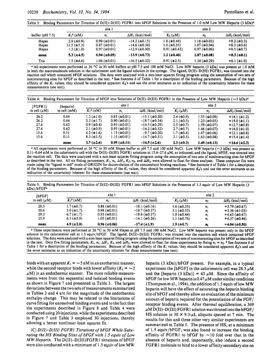
Table 3: Bindinn Parameters for Titration of D(II)-D(III) FGFRl into bFGF Solutions in the Presence of 1.0 mM Low MW Heparin (3 kDaP site 1 site 2
buffer (OH 7.5) Klb (nM) nl AH1 (kcalhol) K2 (uM) n2 A H 2 (kcalhol) Hepes 2.9 (f0.9) 0.99 (fO.O1) -14.2 (fO.15) 1.8 (f0.44) 1.16 (f0.03) +8.2 (f0.3) Hepes 11.5 (f5.3) 0.87 (fO.O1) -14.6 (f0.34) 1.0 (f0.35) 1.07 (f0.04) +8.3 (f0.6) Hepes 1.3 (fl.O) 0.97 (f0.01) -12.9 (f0.30) 0.91 (f0.43) 0.97 (f0.06) +8.5 (f0.7) mean 5.3 (f4.5) 0.94 (f0.05) -13.9 (f0.73) 1.2 (f0.40) 1.07 (f0.08) +8.3 (f0.1) Tris 1.5 (f4.6) 1.06 (fO.01) -16.5 (f0.32) 0.91 (12.5) 1.34 (f0.29) +6.1 (f1.0)
All experiments were performed at 26 OC in 50 mM buffers at pH 7.5 and 100 mM NaCI. Low MW heparin (3 kDa) was present at 1.0 mM in both the macromolecule solution in the calorimeter and the ligand solution in the syringe. The ligand, D(I1)-D(II1) FGFR1, was titrated into the reaction cell which contained bFGF solutions. The data were analyzed with a non-least squares fitting program using the assumption of two sets of noninteracting sites for bFGF as described in the text. * See footnote b of Table 1 for a description of the binding parameters. Because of the high affinity of the K1 values, they should be considered apparent Kd'S and use the error estimates as an indication of the uncertainty inherent for these measurements (see text).
Table 4: Binding Parameters for Titration of bFGF into Solutions of D(I1)-D(II1) FGFRl in the Presence of Low MW Heparin (-3 kDa)" [ FGFR 11 [heparin] site 1 site 2
in cell (pM) in cell (mM) Klb (nM) nl AH1 (kcal/mol) K2 (pM) n2 A H 2 (kcal/mol) 26.2 0.64 1.3 (f1.0) 0.85 (fO.O1) -15.1 (f0.50) 2.4 (f0.9) 1.55 (f0.09) +14.1 (f1.2) 26.2 0.64 5.3 (f1.7) 0.90 (fO.O1) -18.7 (f0.34) 2.1 (f0.5) 1.23 (f0.05) +19.0 ( i l . l ) 27.6 0.62 0.9 (f0.4) 0.93 (fO.O1) -14.5 (f0.29) 2.5 (f0.7) 1.59 (f0.07) +12.7 (f0.8) 27.6 0.62 2.1 (f0.9) 0.95 (fO.O1) -14.2 (f0.32) 2.7 (f0.7) 1.46 (f0.07) +14.0 (f1.0) 15.6 0.11 4.2 (f1.6) 1.13 (fO.O1) -9.7 (f0.20) 1.7 (f0.4) 1.47 (f0.06) +12.1 (f0.8) 15.6 0.11 8.3 (f3.5) 1.15 (fO.01) -14.7 (f0.31) 2.1 (f0.5) 1.63 (f0.08) +15.4 (f1.4)
mean 3.7 (f2.6) 0.99 (f0.11) -14.5 (f2.6) 2.3 (f0.3) 1.49 (f0.13) +14.6 (f2.3) "All experiments were performed at 26 OC in 50 mM Hepes buffer at pH 7.5 and 100 mM NaCI. Low MW heparin (-3 kDa) was present at
0.1 1-0.64 mM in the calorimetric cell with the D(I1)-D(II1) FGFRl solutions at 15.6-27.6 pM, as indicated, and the ligand, bFGF, was titrated into the reaction cell. The data were analyzed with a non-least squares fitting program using the assumption of two sets of noninteracting sites for bFGF as described in the text. All six fitting parameters, K1, nl, AHI, K2,n2, and AH2, were allowed to float for these analyses. These computer fits were made using the "ligand in cell" mode of ORIGIN for deconvolution of the concomitant binding reactions. See footnote b of Table 1 for a description of the binding parameters. Because of the high affinity of the K I values, they should be considered apparent Kd'S and use the error estimates as an indication of the uncertainty inherent for these measurements (see text).
Table 5: Binding Parameters for Titration of D(I1)-D(II1) FGFRl into bFGF Solutions in the Presence of 1.5 equiv of Low MW Heparin (3 kDa)/bFGF"
[bFGF] site 1 site 2 in cell (pM) Klb (nM) nl AH1 (kcal/mol) K2 (rM) n2 AH2 (kcal/mol)
28.5 1.7 (f0.7) 0.88 (fO.O1) -18.1 (f0.16) 1.6 (10.33) nl +3.79 (f0.67) 30.6 5.0 (f1.7) 0.98 (fO.O1) -18.7 (f0.37) 3.1 (f0.55) nl +3.36 (f1.05) 29.3 6.7 (f1.7) 0.93 (f0.01) -18.6 (f0.37) 1.9 (f0.44) nl +4.65 (f0.67) 25.9 6.3 (13.9) 1.05 (fO.O1) -14.1 (f0.26) 1.1 (f0.78) nl +4.37 (f0.46) mean 4.9 (f2.0) 0.96 (f0.06) -17.4 (f1.9) 1.9 (10.7) a +4.04 (i0.50)
"These experiments were performed at 26 "C in 50 mM Hepes at pH 7.5 and 100 mM NaCI. Low MW heparin was present only in the bFGF solution in the calorimetric cell at 1.5 equiv/bFGF. The ligand, D(I1)-D(II1) FGFR1, was titrated into the reaction cell which contained bFGF solutions. The data were analyzed with a non-least squares fitting program using the assumption of two sets of noninteracting sites for bFGF as described in the text. Only five fitting parameters, K I , nl, AHl, K2, and AH2, were allowed to float for these experiments by fixing nl = n2. See footnote 6 of Table 1 for a description of the binding parameters. Because of the high affinity of the K I values, they should be considered apparent Kd's and use the error estimates as an indication of the uncertainty inherent for these measurements (see text).
binds with an apparent K I = - 5 nM in an exothermic manner, while the second receptor binds with lower affinity (K2 = -2 pM) in an endothermic manner. The more reliable measure- ments were from the sequential and isolated binding events as shown in Figure 7 and presented in Table 3. The largest deviations between the two sets of measurements summarized in Tables 3 and 4 are for the magnitude of the endothermic enthalpy change. This may be related to the limitations of curve fitting for unresolved binding events and to the fact that the experiments described in Figure 8 and Table 4 were conducted using 20 injections, while the experiments described in Figure 7 and Table 3 employed 30 injections, thereby allowing a better nonlinear least squares fit.
(C) D(II)-D(III) FGFRl Titrations of bFGF While Satu- rating the H S Binding Sites of bFGF with 1.5 equiv of Low MWHeparin. The D(I1)-D(I1I)FGFRl titrations of bFGF were also conducted with a minimum of 1.5 equiv of low MW
heparin (3 kDa)/bFGF present. For example, in a typical experiment the [bFGF] in the calorimetric cell was 28.5 pM and the [heparin (3 kDa)] = 43 pM. Since the affinity of bFGF for low MW heparin is 0.47 pM under similar conditions (Thompson et al., 1994), the addition of 1.5 equiv of low MW heparin will have the effect of saturating the heparin binding site of bFGF and thereby allow an evaluation of the minimum amount of heparin required for the potentiation of the FGF/ receptor binding events. After thermal equilibration, a 369 pM D(I1)-D(II1) FGFRl solution was titrated into the bFGF/ HS solution in 30 X 9.3-pL aliquots spaced at 7 min. The results for this and three other very similar experiments are summarized in Table 5. The presence of HS, at a minimum of 1.5 equiv/bFGF, was also found to increase the binding affinity of FGFRl to bFGF by -10 fold over that in the absence of heparin and, importantly, also induce a second FGFRl molecule to bind to a lower affinity secondary site on

Binding Interactions in the FGF System
Time (min) , 0.00 I , 3 3 7 , 66,67 , ly00 , 13: , 17
Biochemistry, Vol. 33, No. 34, 1994 10239
FGFRl alone sediments as a species of MW = 45-kDa species, slightly higher than the -40-kDa estimate by SDS- PAGE analysis and the 39.9 f 3 kDa estimated by size- exclusion HPLC (see above). The small increase in MW may be the result of bound HS, since this measurement was made in the presence of 1.0 mM low MW HS.
When bFGF was present in a mole ratio of [FGFRl]t/ [bFGF]t = 2.0, thesedimentationMw wasobserved toincrease to 95.6 kDa under the same conditions. This result is in reasonable agreement with the ITC results since two D(I1)- D(II1) FGFRl/HS (2 X 45) plus one bFGF (17.4) would be expected to yield a MW of 107.4 kDa. The slightly smaller MW for the bFGF/HS-mediated FGFRl dimer (89% of expected) could be due to a slightly weaker K2 under the conditions of ultracentrifugation than the 1.2 f 0.4 pM measured by ITC, which would lead to a higher than expected fraction of lower MW sedimenting species contributing to the observed average.
The decrease in the sedimentation MW upon dilution of the FGFRl/bFGF/HS complex from 30 to 3.3 pM is also consistent with the relatively weak binding affinity of the second FGFRl binding to the 1:l complex as observed in the ITC experiments reported in Figure 7 and Table 3. The dropoff is steeper than that expected for a 1.2 (f0.4) pM site. This could be due to the fact that although the proteins were diluted - 10 fold, the [HS] was kept constant at 1 .O mM. Under these experimental conditions, HS will compete with increasing effectiveness with the FGFRl /bFGF/HS complex for the binding of the second receptor molecule because [HS],/ KRH = 10 at 1.0 mM HS, while [bFGF/FGFRlIt/K2 = 2.8 at 3.3 pM bFGF/FGFRl.
The sedimentation MW of 45 kDa observed for the FGFR1/ bFGF/HS complex diluted to 3.3 pM is smaller than expected for a 50/50 mixture of the free FGFRl/HS of -45 kDa and the FGFRl /bFGF/HS complex (1 : 1 : 1). Assuming all species have the same partial specific volumes, this composition would sediment as its weight average of 53 kDa. It is impossible, however, to attach any significance to this discrepancy without more information about the sedimentation characteristics of individual species.
Biological Competitive Assays of bFGF Binding to D(II)- D(III) FGFRl. Competition assays for measuring the inhibi- tion of D(I1)-D(II1) FGFRl ( IC~O) for the binding of radioactive iodinated bFGF on baby hamster kidney (BHK- 21) cells and the separation of high- and low-affinity counts were performed essentially as reported by Moscatelli (1 987). The purified receptor was found to inhibit the binding of radioactive labeled 1251 bFGF with an IC50 = 37.3 (f17.2) nM (no added heparin). These results are in close agreement with those found by ITC.
Three-Dimensional Structural Models of D(II)-D(III) FGFRl. To understand the binary and conditional binding reactions in terms of structural componentsof FGFRl, a three- dimensional model of D(I1)-D(II1) FGFRl was constructed using segment match modeling software described by Levitt (1992). Figure 9A shows the structural model of D(I1)- D(II1) FGFRl that resulted when the primary sequence of the D(I1) domain of FGFRl is matched with the primary, secondary, and tertiary structure of the homologous cH2 domain (17% identity) of the Ig Fc structure (1FC2, PDB; Deisenhofer, 1981).
Similarly, the D(II1) domain was modeled using its primary sequence homology with VH domains, such as the HYHEL-5 antibody structure (2HFL, PDB; Sheriff et al., 1987). The individual D(I1) and D(II1) domains are each folded into two layers of antiparallel sheets that embrace a predominantly
I
0
-2
5
0 0 e %
0.0 0.2 0.4 0.6 0.8 1.0 1.2 1.4 1.6 1.8
[bFGF]t / [FGFRlIt FIGURE 8: bFGF titration of D(I1)-D(1II) FGFRl while saturating both receptor and bFGF with low MW heparin. The reverse titration of that in Figure 7 was attempted, and the resulting raw data are shown (top). The resultant binding isotherm for the bFGF titration of D(I1)-D(II1) FGFRl in the presence of 0.64 mM HS after integration of the area under each injection peak and subtraction of the blank (bottom). The solid line represents a nonlinear least squares fit of the reaction heat for each injection (AQ,,,) with the assumption of two independent sites comprised of the six fitting parameters, nl, KI, A H l , n2, K2, and AH2, which were all allowed to float during computer iterations (see Materials and Methods). These computer fits were made using the "ligand in cell" mode of ORIGIN for deconvolution of the concomitant binding reactions. This experiment was repeated six times with the [heparin] ranging between 0.11 and 0.64 mM. The results are summarized in Table 4.
bFGF (K2 = 1.9 f 0.7 pM) in an entropy-dominated reaction to yield a final quaternary complex of (FGFR1)2/bFGF/HS.
Equilibrium Sedimentation of the D(II)-D(III) FGFRl/ bFGF/HS Complex. Measurements of the buoyant molecular weight of the FGFRl/bFGF/HS complex at a mole ratio of 2.0 FGFRl to 1 .O bFGF in the presence of 1.0 mM low MW heparin were attained using a Beckman analytical ultracen- trifuge (XLA). These experiments were conducted under the following conditions: (1) at 30 pM bFGF, 60 pM D(I1)- D(II1) FGFR1, and 1.0 mM low MW heparin (3 kDa) to simulate the conditions employed for the ITC experiments and to ensure that the bFGF was 15-30-fold above the apparent K2 measured in these experiments; (2) at 10 pM bFGF, 20 pM D(I1)-D(II1) FGFRl, and 1.0 mM low MW heparin (3 kDa); (3) at 3.3 pM bFGF, 6.6 pM D(I1)-D(II1) FGFR1, and 1.0 mM low MW heparin (3 kDa); and (4) at 30 pM D(I1)-D(II1) FGFRl and 1.0 mM low MW heparin but in the absence of bFGF. The results, displayed in Table 6, are reported both as buoyant Mb and, using an assumed value of 0.68 for the product of density and protein partial specific volume, as sedimentation MW in kDa. The low v = 0.68 value is due to the high degree of glycosylation of FGFRl (20- 40%) as described above.

10240 Biochemistry, Vol. 33, No. 34, 1994 Pantoliano et al.
Table 6: Equilibrium Sedimentation of FGFRl/bFGF Complexes in the Presence of Low MW Heparin (-3 kDa)‘J max no. of
protein in cell [bFGF], (pM) [FGFR1lt/[bFGFlt wt av Mbb wt av Mw (kDa) reduced xz FGFRl/bFGFd FGFRl alone 0.0 14 447 f 43 45.1 2.0 FGFRl/bFGF 30.0 2.0 30 586 f 71 95.6 2.3 2.1 FGFRl /bFGF 10.0 2.0 24 036 f 56 75.1 1.6 1.7 FGFRl/bFGF 3.3 2.0 14 321 f 65 44.8 0.8 1 .o
These experiments were performed at 20 OC in 50 mM Hepes at pH 7.5,lOO mM NaCI, 1.0 mM DTT, and 1.0 mM low MW heparin (-3 kDa). Single ideal species curve fits were used to determine buoyant molecular weights, Mb. A v = 0.68 was used for calculations of Mw due to the high
degree of glycosylation of FGFRl (see text). Sedimentation equilibrium optical density gradients were obtained at (in order) IO 000,8000, and IO 000 rpm. Goodness of fit to the data was assessed by the reduced xz parameter. A value near 1.0 implies that the model fits the data as well as can be expected for assumed Gaussian-distributed errors in the concentration data. Higher values indicate systematic deviations of calculated versus observed values. To account for complex dissociation, the measured Mb was assumed to be a weight average of contributions from three species: Mb = [C1MI2 + C2Mz2 + Cykf3Z]/[C&f~ + C2M2 + C&f3], where C1 = [FGFRl/bFGF] in the 1:l complex, C2 = [FGFRlIf,, C, = [FGFRl/bFGF] in the 2:l complex, and CtOt = CI + C2 + C3 = [FGFRlItotal. With the assumption (based on the calorimetric value of 5 nM for the Kd of the 1:l FGFRI/bFGF complex) that all bFGF is bound at the concentrations used here, the species can be assumed to be in a simple equilibrium relationship: CIC2/C3 = Kd. With the further assumption of a fixed stoichiometry of C ~ / C I = 1.0 throughout the centrifugation cell, the weight-average bouyant molecular weight can be expected to vary with total concentrations as ( M b ) w = [Ml2 + M22 +fMs*/ ( I -fl]/[M1 + M2 +fM3/(1 -A], wheref= C3/[Ct] and is the solution to the equationy + [ l + Kd/Ct l f - 1 = 0.
hydrophobic core, a structural motif characteristicof constant and variable IgSFdomains. The fidelity of the model of D(I1) to Ig cH2 can be judged by an rms deviation of 1.8 8, for the superposition of the 114 equivalent C, atoms in the @ sheets of these two structures. The orientation of the two domains was modeled after the relationshipof the two constant domains, cH2 and C”3, of the Ig Fc structure, so that the carboxyl terminus of D(II), -PHR-, serves as the interdomain linker that leads into theN-terminusof D(III), -PILA-. This resulted in a structure where the two domains are oriented in the same way that cH2 and C H ~ domains are aligned in the Ig Fc structure except that the D(II1) domain has a VH-like fold consistent with its closer primary sequence homology with VH domains (26% identity).
Inspection of the D(I1)-D(II1) FGFRl model revealed an elongated cluster of nine cationic residues (seven Lys and two Arg) that are located in the C~2- l ike domain D(I1) (Figure 9A). Six lysines (residues K6, K9, K10, K18, K21, and K23; numbering system is that of the IgSF homology model which begins at ‘WTSP-) are in an extended @turn that involves 6 strands A and B in the immunoglobulin fold of D(II), while the three remaining cationic residues, R48, K53, and R55, are located in the adjacent @ strands C’ and E [see Leahy et al. (1992) for a recent description of Ig @ strand folding topography]. This aggregate of positively charged residues was observed in a - 15 X 35 8, shallow cleft formed by the natural twist of the four @ strands that comprise the fi sheet A, B, E, C’ surface of the D(I1) domain (Figure 9A). As expected from this surface charge density, electrostatic potential maps calculated for this FGFRl model with the program DELPHI (Gilson et al., 1988) revealed that these nine cationic residues focus positive electrostatic potential (+2keT) to a large part of the solvent exposed A, B, E, C’ @-sheet surface of the D(I1) domain model (Figure 9B).
DISCUSSION
The cloning and expression of the extracellular domains D(I1)-D(II1) of FGFRl using baculovirus-infected insect cell technology consistently yielded - 15 mg/L culture media when coupled with a bFGF/HS-Sepharose affinity chromato- graphic recovery method (see Figure 2). These results enabled a biophysical evaluation of the complete binding isotherms for the primary reactants bFGF, HS, and D(I1)-D(II1) FGFR1, as determined by ITC (Figure 3), under controlled conditions. These individual binding reactions were previously inadequately understood with regard to equilibrium constants, stoichiometry, and structure due, in part, to the prior difficulties in isolating sufficient quantities of each of the purified
components and characterizing their binding interactions in controlled cell-free environments. For example, it is commonly believed that bFGF binds to “high affinity” sites on the surface of certain cell lines with an IC50 in the range of 0.02-0.20 nM (Klagsbrun & Baird, 1991; Neufeld & Gospodarowicz, 1985), and HS is important for the biological activities of FGF that are triggered by the growth factor binding event to cells (Yayon et al., 1991; Rapraeger et al., 1991). There is, however, uncertainty as to whether HS is obligatory for the bFGF/ FGFRl binding event (Ornitz et al., 1992; Yayon et al., 1991; Klagsbrun & Baird, 1991) and also about how the binding interactions translate into the mechanism of FGF- and HS- mediated biological activities such as mitogenic signal trans- duction. An attempt to first examine the relevant binary binding reactions for an individual assessment of their intrinsic binding parameters, followed by an examination of the conditional reactions of bFGF and FGFRl in the presence of HS, is described below.
Binary Binding Reactions f o r the Reactants, bFGF, FGFRl, and Low MWHeparin. The data in Table 1 and Figures 3-6, together with the information derived from our work with HS binding to bFGF (Thompson et al., 1994), suggest that the relevant binary binding reactions for the FGF system can be dissected into the three reactions comprised of the three primary reactants, bFGF, FGFR1, and HS:
KRF = 41 nM FGFRl + bFGF - FGFRl/bFGF (1)
K m = 85 rM F G F R l + H S Q FGFRl/HS (2)
bFGF + HS Q bFGF/HS (3) Km = 470 nM
(A) Intrinsic Binding of bFGF to FGFRl in the Absence of HS. Reaction 1 is the intrinsic binding affinity of bFGF for FGFR1, in the absence of HS. The results summarized in Table 1 show the stoichiometry of this reaction to be 1:l over an 8-fold range of [protein] with an observed dissociation constant KRF = 41 (f12) nM when bFGF is incrementally titrated into D(I1)-D(II1) FGFRl solutions in the absence of HS (Table 1A). This translates into an observed free energy of binding of AGO = -10.2 (*0.2) kcal/mol when calculated using AGO = -RT In K,.
Nearly identical results were obtained (Le., n = 0.99, and an observed AGO = -10.0 kcal/mol) when theorder of addition of the reactants was reversed, where D(I1)-D(II1) FGFRl is titrated incrementally into bFGF solutions in the absence of HS under otherwise identical conditions (Table 1B). The
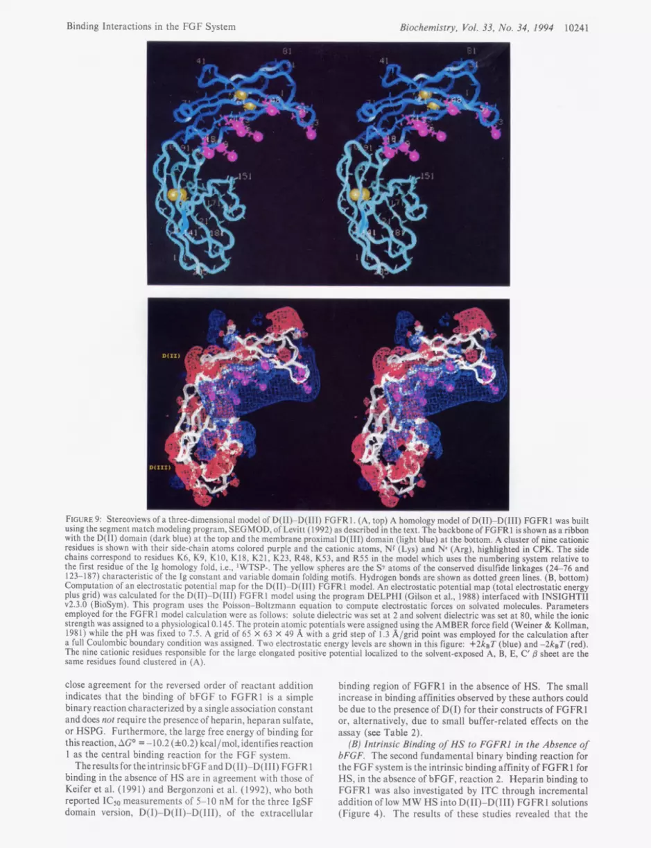
Binding Interactions in the FGF System Biochemistry, Vol. 33, No. 34, 1994 10241
I I
I I L
FIGURE 9: Stereoviews of a three-dimensional model of D(I1)-D(II1) FGFRl. (A, top) A homology model of D(I1)-D(II1) FGFRl was built using the segment match modeling program, SEGMOD, of Levitt (1992) as described in the text. The backbone of FGFRl is shown as a ribbon with the D(I1) domain (dark blue) at the top and the membrane proximal D(II1) domain (light blue) at the bottom. A cluster of nine cationic residues is shown with their side-chain atoms colored purple and the cationic atoms, NC (Lys) and Nc (Arg), highlighted in CPK. The side chains correspond to residues K6, K9, K10, K18, K21, K23, R48, K53, and R55 in the model which uses the numbering system relative to the first residue of the Ig homology fold, Le., ‘WTSP-. The yellow spheres are the Sr atoms of the conserved disulfide linkages (24-76 and 123-187) characteristic of the Ig constant and variable domain folding motifs. Hydrogen bonds are shown as dotted green lines. (B, bottom) Computation of an electrostatic potential map for the D(I1)-D(II1) FGFRl model. An electrostatic potential map (total electrostatic energy plus grid) was calculated for the D(I1)-D(II1) FGFRl model using the program DELPHI (Gilson et al., 1988) interfaced with INSIGHT11 v2.3 .O (BioSym). This program uses the Poisson-Boltzmann equation to compute electrostatic forces on solvated molecules. Parameters employed for the FGFRl model calculation were as follows: solute dielectric was set at 2 and solvent dielectric was set at 80, while the ionic strength was assigned to a physiological 0.145. The protein atomic potentials were assigned using the AMBER force field (Weiner & Kollman, 1981) while the pH was fixed to 7.5. A grid of 65 X 63 X 49 A with a grid step of 1.3 A/grid point was employed for the calculation after a full Coulombic boundary condition was assigned. Two electrostatic energy levels are shown in this figure: + 2 k ~ T (blue) and - 2 k ~ T (red). The nine cationic residues responsible for the large elongated positive potential localized to the solvent-exposed A, B, E, C’ /3 sheet are the same residues found clustered in (A).
close agreement for the reversed order of reactant addition indicates that the binding of bFGF to FGFRl is a simple binary reaction characterized by a single association constant and does not require the presence of heparin, heparan sulfate, or HSPG. Furthermore, the large free energy of binding far this reaction, AGO = -10.2 (f0.2) kcal/mol, identifies reaction 1 as the central binding reaction for the FGF system.
The results for the intrinsic bFGF and D(I1)-D(II1) FGFRl binding in the absence of HS are in agreement with those of Keifer et al. (1 99 1) and Bergonzoni et al. (1 992), who both reported ICs0 measurements of 5-10 nM for the three IgSF domain version, D(1)-D(I1)-D(III), of the extracellular
binding region of FGFRl in the absence of HS. The small increase in binding affinities observed by these authors could be due to the presence of D(1) for their constructs of FGFRl or, alternatively, due to small buffer-related effects on the assay (see Ta& 2 ) .
(B) Intrinsic Binding of HS to FGFRl in the Absence of bFGF. The second fundamental binary binding reaction for the FGF system is the intrinsic binding affinity of FGFRl for HS, in the absence of bFGF, reaction 2. Heparin binding to FGFR 1 was also investigated by ITC through incremental addition of low MW HS into D(I1)-D(II1) FGFRl solutions (Figure 4). The results of these studies revealed that the

10242
stoichiometry of this reaction is 1.39 (k0.06) HS to 1 .O D(I1)- D(II1) FGFRl (using MW = 3000 for low MW HS) with an observed dissociation constant of KRH = 104 (f17) pM. Similar results were obtained for RD Heparin 5000 (Figure
Although the binding affinity of reaction 2 was found to be relatively weak in comparison to other more familiar HS binding sites such as thrombin (Olson et al., 1991), anti- thrombin I11 (Olson & Bjork, 1991), and bFGF (see below), the HS binding site of D(I1)-D(II1) FGFRl is believed to be biologically relevant because of the high expression levels of HSPG on the surface of cells that coexpress FGFR1. Yanagishita and Hascall (1992) have found that typical expression of HSPG is in the range of 105-106 copies per cell. As pointed out by Brandts and Jacobson (1983) in their thermodynamic treatment of membrane receptor clustering mechanisms for transmembrane signal transduction, the mass law dependence for reactions involving membrane receptors is more conveniently treated using two-dimensional leaflet mole fractions, XR = NR/(O.~NL + NR), as the concentration units, where NR = number of embedded receptor molecules and NL = number of lipid molecules (0.5 is due to the bilayer nature of biological membranes) since the receptors can be assumed to have no significant degrees of freedom in the 2-direction perpendicular to the plane of the bilayer. For lo6 copies of a membrane receptor on the surface of an erythrocyte- sized cell, the bulk concentration of receptor in the membrane expressed in terms of two-dimensional leaflet mole fractions was estimated to be X R = 5 X (Brandts & Jacobson, 1983). Therefore, a KRH = 85-104 pM binding affinity measured in vitro in three-dimensional concentration units for HS binding to D(I1)-D(II1) FGFRl is believed to be relevant for cases where HSPG-like surface molecules are expressed to such a high level that theeffective two-dimensional concentrations can be estimated to be 5-50-fold above the observed Kd.’ Similar calculations for the estimated 2000 copies of the FGFRl expressed on the surface of the same cell lines yield an effective two-dimensional leaflet mole fraction of X R = 1 X 1 0-5 for FGFRl .
(C) Pentosan Polysulfate Binding to D(II)-D(III) FGFRl . The binding of PPS to D(I1)-D(II1) FGFRl (Figure 6) is intriguing since this sulfated oligosaccharide is a well-known inhibitor of angiogenesis and is currently being investigated for the treatment of breast cancer (Lippman, 1993; Harris et al., 1992) and AIDS-related Kaposi’s skin cancer (Wellstein etal., 1991;Nakamuraet al., 1992). Themechanismofaction of PPS was presumed to be through the competitive inhibition of HSPG binding to FGF (reaction 3) and subsequent interference with the previously held view that HSPG/FGF complex formation is required for binding to FGFRI, a view that now appears to be untenable when the results in Table 1 are considered. The observations described above for reaction 1, together with the observation that PPS binds to FGFRl with a IO-fold higher affinity than HS [Kd = 10.9 (f3.5) pM], however, may provide an opportunity to reexamine the mechanism of action of this sulfated oligosac- charide and other related angiogenesis inhibitors such as the sulfated polysaccharide peptidoglycan SP-PG (Nakamura et al., 1992) and the sulfated naphthalene, suramin (Harris et al., 1992). In this regard, it has been reported that PPS inhibits growth of both AIDS-Kaposi sarcoma cells and normal human
Biochemistry, Vol. 33, NO. 34, 1994
5 ) .
Pantoliano et al.
This calculation assumes one FGFRl binding site per HSPG, which is most likely an underestimate depending on how many sites can be accommodated per HS chain with a typical MW of 30 000. The evidence provided in Figures 4 and 5 is that there is one FGFRl site per -3-5 kDa of HS.
endothelial cells from umbilical vein (H-UVE) with IC50(s of 2 and 8 pM, respectively (Nakamura et al., 1992). These growth inhibition data are in close agreement with the observed binding affinity of PPS to FGFRl shown in Figure 6.
(0) Intrinsic Binding of HS to bFGF in the Absence of FGFRl , The third fundamental binary binding reaction, reaction 3, is the intrinsic binding affinity of bFGF for HS, in the absence of FGFRl. A detailed characterization of this reaction through the use of molecular modeling of docked heparin/bFGF interactions and site-directed mutagenesis of bFGF, in addition to isothermal titrating calorimetry, has been reported (Thompson et al., 1994). Briefly, the wild- type bFGF was found to bind to low MW heparin (- 3 kDa) to yield a KFH = 470 (f20) nM when titrated under conditions identical to those in Tables 1 and 2, Le.: 50 mM Hepes- NaOH, pH 7.5,O.lO M NaCl, 1.0 mM DTT, and 26 OC. The value of the equilibrium constant was also found to be independent of the order of addition of the two reactants.
Conditional Reactions of bFGFand FGFRl in the Presence of Low M W Heparin. The conditional reactions of bFGF and FGFRl in the presence of heparin were examined under circumstances that saturate only the bFGF heparin site (1.5 equiv of HS/bFGF) or saturate the HS binding sites of both bFGF and FGFRl (1.0 mM HS). Both 3- and 5-kDa low MW heparin were found to give - IO-fold increase in affinity for FGFRl binding to bFGF (K1 = 5 nM) when [HS] = 1.0 mM, relative to the reaction with no heparin (Figure 7 , Tables 3 and 4). The presence of HS, at a minimum of 1 S equiv/ bFGF, was found to induce a second FGFRl molecule to bind to a lower affinity secondary site on FGFRl/bFGF/HS (K2 = 1.9 f 0.7 pM) in an entropy-dominated reaction to yield a final quaternary complex of (FGFR1)2/bFGF/HS. Equi- librium sedimentation experiments were found to be consistent with this result when conducted at [FGFRl],/[bFGF], = 2.0 in the presence of 1 .O mM low MW heparin (- 3 kDa) (Table 6). The titrations of bFGF and FGFRl in both directions in the presence of saturating levels of heparin are in close agreement (Tables 4 and 5) and strongly suggest that heparin binding events on both of these proteins induce bFGF-mediated receptor dimerization.
The endothermic nature of the second receptor binding event observed for both 1 .O mM heparin and the minimum 1.5 equiv/ bFGF can be explained in terms of the burial of a large nonpolar surface area upon binding to form the quater- nary complex (FGFRl)z/bFGF/HS or quinary complex (FGFRI)z/bFGF/(HS), as might be expected for protein/ protein interactions that involved the IgSF (see below).
Allosteric Modulation of the Multivalent Binding Equi- libria of FGFRl, bFGF, and Heparin. The dissection of the binding reactions of the three primary reactants into the three fundamental binary reactions (1-3) reveals that each reactant is multivalent: FGFRl binds growth factor and HS, bFGF binds receptor and HS, and HS binds FGFRl and bFGF. The multivalent nature of each primary reactant suggests that many unanswered questions for this growth factor system that relate to the role of heparin in high-affinity binding and the mechanism of mitogenic signal transduction can be understood in terms of well-studied classical multiple ligand binding mechanisms (Wyman, 1964; Weber, 1975). In the presence of heparin, at a minimum of 1.5 equiv/bFGF, there is an - 10-fold potentiation of reaction 1 and the induction of a fourth reaction, which is the binding of a second molecule of FGFRl to yield a quaternary complex:
FGFRl/bFGF/HS + FGFRl Q
a 8 r K ~ ~ = 1.9 rM
(FGFR l),/bFGF/HS (4)

Binding Interactions in the FGF System Biochemistry, Vol. 53, No. 34, 1994 10243
=\It*- I
.
e = eV-(AGe/KO = Ke, where AGs = fne: energy of coupling for H and R2 binding ta Rls
FIGURE 10: Allosteric modulation of the multivalent binding equilibria of FGFR1, bFGF, and heparin. This schematic illustrates how the multiple ligand binding equilibria of the three reactants of the FGF system can be understood in terms of an allosteric mechanism for the dimerization of the FGF receptor, where R = FGFRl (R1 = R2; the subscripts are used to denote the first and second receptor to bind to the growth factor), F = bFGF, and H = low MW heparin (-3 or 5 kDa). The three binary equilibrium dissociation constants, KRF, Km, and KFH, characterize the binding of R to F, R to H, and F to H, respectively, as discussed in the text. The reactions are color coded so that all R/F binding reactions are in yellow and lie in the X-direction, while all heparin binding reactions, either F/H or R/H, are in purple and lie in the 2-direction. A fourth binary binding reaction is postulated, KRR, which is the dimerization of R to yield RI-RZ, in the absence of all ligands. All the receptor dimerization reactions are in blue and lie in the Y-direction. The KRR reaction is assumed to be a very weak association and undetectable in the absence of other ligands but was observed in the presence of both the ligands, F and H. This can be understood if the binding of each of the ligands modifies the affinity of the other ligands by a certain factor, and the principle of microscopic reversibility suggests five such interaction factors: a, 0, y, 6, and t. For example, it was observed that H potentiates the binding of F to its receptor R, by - 10-fold, so that the reaction of R and FH is characterized by CVKRF = 4.9 f 2.0 nM, where a = exp[-(AG,/RT)] = K, = - 10 and AGa = --1.4 kcal/mol = free energy of coupling for bFGF and H binding to R1 (Weber, 1975). Similarly, the binding of FH to R1 (1.5 equiv of H/F) is believed to potentiate the binding of R1 to a second receptor, R2, so that the resultant receptor dimerization is characterized by (Y&KRR = 1.9 (f0.7) pM, where /3 = exp[-(AGp/RT)] = Kp and AGp = free energy of coupling for bFGF and R2 binding to R1 and where y = exp[-(AG,/RT)] = K, and AG, = free energy of coupling for H (bound to bFGF) and R1F binding to R2. Additional interaction factors that may play a role for the mechanism of receptor dimerization when the [HS] = 1 .O mM are 6 = exp[-(AGa/RT)] = Ks, where AGs = free energy of coupling for H and bFGF/H binding to R1, and t = exp[-(AG,/RT)] = K,, where AGe = free energy of coupling for H and R2 binding to R1.
An allosteric mechanism that attempts to explain how FGF and HS together induce the dimerization of FGFRl, reaction 4, is shown in Figure 10. The foundation of this mechanism lies with the three binary equilibrium dissociation constants, KRF, KRH, and KFH, as discussed above. A fourth binary binding reaction is postulated, KRR, which is the dimerization of FGFRl to yield R1-R2, in the absence of all ligands (R1 = Rz; the subscripts are used to indicate the first and second receptor to bind to the ligand, FGF/HS). This reaction is assumed to be a very weak association and undetectable in the absence of other ligands at [FGFRl] = 30 p M but was observed in the presence of both the ligands, F and H. The reason for this shift in equilibrium comes from the evidence that the binding of each of the ligands modifies the affinity of the other ligands by a certain factor, and the principle of microscopic reversibility suggests five such interaction fac- tors: cy, p, y, 6, and e. This is a consequence of the trivalent binding preferences of F, the trivalent binding characteristics of R, and the bivalent binding properties of H.
For example, if one begins with growth factor, F, in Figure 10, this reactant has a choice: it can bind to H, shown as the purple reaction characterized by KFH = 0.48 pM, leading to
the formation of F-H; or it can bind to R1 shown as the yellow reaction characterized by KRF = 41 nM, leading to the binary complex, R1-F. The binary complexes, F-H and Rl-F, can then react with R1, or H, respectively, to yield thesame ternary complex R1-F-H. It was observed that H increases the binding affinity of F to its receptor R, by - 1 0-fold, so that the reaction of R and FH is characterized by ~ X K R F = 4.9 f 2.0 nM, where cy = exp[-(AGa/R7')] = K, = - 10 and AGa = --1.4 kcal/ mol = free energy of coupling for bFGF and H binding to R1 (Weber, 1975). The principle of microscopic reversibility, implicit in the linkage of multivalent binding reactions, suggests that R should increase the binding affinity of F to H by the same interaction factor, a, to yield ~ K F H . It is also possible to arrive at the ternary complex RI-F-H in Figure 10 by starting at R1 and reacting with FH, shown as the yellow reaction also characterized by (XKRF = 4.9 f 2.0 nM.
Similarly, the binding of FH to R1 (1.5 equiv H/F) is believed to increase the binding affinity of R1 to a second receptor, R2, so that the resultant receptor dimerization is characterized by &KRR = 1.9 f 0.7 pM, where p = exp[-(AGb/R7')] = Kb and AGb = free energy of coupling for bFGF and R2 binding to R1 and where y = exp[-( AG,/RT)]
I
obtained after energy minimization usin ation in the Y-direction will place it in
D(II1) FGFRl model.
the interaction
is required to account fo quaternary complex, (F presence of 1.5 equiv of the most important of observation that C @ ~ K R R =
uced receptor dimerization sence of bFGF, just as bFGF-in
was not observed in the absence of HS here, but may occur at high [
reactions 1 and is not unlike those mechanisms invo s of one ligand binding on another in o multivalent protein systems
similar to that recently ivation of N-methyl+-
intracellular tyrosine kinase domains a manner that leads to cross-phospho
9. This enabled a corre binding reactions with
s case a 13 monosaccharide approximate a low
een that of heparin 3-( 6-OS03)-a( 1-4)-
of the 1,4 linkages and also to optimize the

Binding Interactions in the FGF System
repulsion of the 4 0 3 - and -COO- groups after computer synthesis. This helical structure is consistent with the known single-stranded helical nature for related glycosaminoglycans, such as dermatan sulfate, studied by X-ray diffraction (Arnott & Mitra, 1984; Mitra et al., 1983).
The juxtaposition of this 13-monosaccharide model of low MW HS near the clustered cationic groups at the A, B, E, C’ p sheet reveals how a translation of -5 A in the +Y- direction would dock the HS ligand against the A, B, E, C’ 0 surface of D(I1) in such a way that would complement the positive electrostatic potential map in Figure 9B. In this binding configuration, six or seven of the H S monosaccharides would interact with the D(I1) binding cleft, but the remaining six or seven would be sterically precluded for binding another FGFRl molecule. This observation, based on the poised structural models of Figure 11, is consistent with the 1:l binding stoichiometry observed for the 10 monosaccharide sized heparin 3000, the 15 monosaccharide sized, RD Heparin 5000, and the 12 monosaccharide sized pentosan polysulfate (MW -4500) reported for the binding experiments of Figures
An elongated clustering of positively charged residues that focus positive electrostatic potential at protein surfaces has the capability to interact with opposite sides of the carbo- hydrate in a two-sided binding mode. This heparin binding structural motif has also been observed for the HS binding proteins thrombin (Bode et al., 1992), antithrombin I11 (Grootenhuis &van Boeckel, 1991), and bFGF (Zhang et al., 1991; Thompson et al., 1994) and strongly suggests that these nine FGFR 1 cationic residues, discussed above, constitute the greater part of the HS binding domain of FGFR1. Identification of sulfated proteoglycan binding regions in other proteins for which no X-ray or NMR information is available has also been successfully employed by Sali et al. (1993) through a similar strategy of homology modeling and electrostatic potential calculations.
During the course of these investigations the existence of a heparin binding site for FGFRl was identified in the D(I1) domain using peptide mapping and site-directed mutagenesis of FGFRl (Kan et al., 1993). The binding data and modeling exercises described here are consistent with the results of these authors. When comparing the location of the clustered nine cationic residues in Figure 9 with the peptide sequence K18K, NH2-KMEKKLHAVPAAKTVKFK-COOH, identified by Kan et al. (1993) as comprising at least part of the heparin binding site of FGFR1, we found that six of the lysines in the model, residues K6, K9, K10, K18, K21, and K23 located in the A and B strands of domain D(II), corresponded to the six lysine residues in the K18K peptide. The close agreement between the results of Kan et al. (1993) and the electrostatic potential calculations for the three-dimensional structural model of FGFRl further supports theassertion that the heparin binding site of FGFRl is fairly accurately represented in Figures 9 and 11.
The feasibility of side-by-side adjacent binding of bFGF and FGFR 1 to a heparin polymer of sufficient length to bridge the growth factor and receptor was explored through docking simulations that aligned the HS binding domains of these two proteins. The HS binding domain of bFGF was recently defined by a combination of molecular modeling, site-directed mutagenesis, and isothermal titrating calorimetry (Thompson et al., 1994). Approximately 95% of the binding free energy for the bFGF/HS interaction, KFH, was accounted for through individual substitutions of eleven residues, K26, N27, R8 1, K119, R120, T121,Q123, K125, K129,Q134, and K135, of bFGF to alanine residues. The locations of many of these
4-6.
Biochemistry, Vol. 33, No. 34, I994 10245
residues were consistent with those adjacent to bound sulfate anions that cocrystallized with bFGF (Zhang et al., 1991; Eriksson et al., 1991) and also the site of sucrose octasulfate binding (Zhu et al., 1993). Docking simulations of FGFRl and bFGF revealed that the heparin binding domains of each of these proteins could be aligned to yield a continuous stretch of heparin binding residues of -60 A that transverse both proteins (Figure 12). The 13 monosaccharide sized heparin model that mimics low MW heparin is also shown poised to interact with this continuous reach of heparin binding residues without any need to perturb the modeled HS helical confor- mation. This simulation reveals that side-by-side adjacent binding of growth factor and receptor is quite reasonable for H S of N 10-1 5 monosaccharide length.
Concomitant with the heparin binding domain alignment of FGF and FGFR1, it was also possible to simultaneously juxtapose the putative receptor binding loop of bFGF, 110KYTSW114 (Bairdet al., 1988), against theexteriorD(II)/ D(II1) domain elbow junction of FGFRl (Figure 12), which is analogous to the protein A binding site of the homology- matched IgFc. This locus (white surface of Figure 12) is the most likely site for FGF binding, given the sequence com- parisons between D(I1) and Ig cH2 and the X-ray structural evidence for the involvement of the exterior elbow junction for the HGH/HGH receptor interaction (De Voset al., 1992). The alignment of the heparin binding residues for FGF and FGFR1, together with the docking of the Il°KYTSW114 loop of bFGF (Bairdet al., 1988) against the proposed FGF binding site of FGFR 1, thereby provides the means for a binary docking process that results in a two-point landing of the two proteins and eliminates many alternative orientations (Stoddard & Koshland, 1992). This outcome is instructive, since it suggests an explanation for the manner in which two adjacent molecular recognition sites of bFGF, the heparin binding domain and the putative receptor binding loop (Baird et al., 1988), can collaborate with their counterparts on FGFRl to produce a bFGF/FGFRl /HS complex and thereby provide a structural explanation for the experimental observation that the HS (1 3 monosaccharide sized) binding events on FGFRl and bFGF, reactions 2 and 3, are energetically coupled to growth factor/ receptor binding events, reactions 1 and 4. The apparent greater dependence on the presence of HS for reaction 4 over reaction 1 may suggest that the 110KYTSW114/D(II)-D(III) FGFRl interface is involved with the second receptor binding event (Figure 10). Characterization of the binding interface, 1*oKYTSW114/D(II)-D(III) FGFR1, by site-directed mu- tagenesis (Springer et al., unpublished results) further supports the belief that this represents the binding interface that is the product of the second receptor binding event, reaction 4, characterized by ~ & K R R = 1.9 pM, in the receptor dimer- ization mechanism (Figure 10).
Asecond D(I1)-D(II1) FGFRl moiety added to the ternary complex of Figure 12 revealed how the three reactants of the FGF system were assembled to satisfy all of the multivalent binding results but still remain within the framework of the identified structural binding components of the X-ray structure of bFGF and the homology model of D(I1)-D(II1) FGFRl (Figure 13). This simulation was also a binary docking process in that it employed a two-point landing of the proposed FGF binding site of FGFRl (white surface) onto a second receptor binding site of bFGF (red surface - 180’ from ll°KYTSW114 on left also identified by site-directed mutagenesis mapping; Springer et al., unpublished results), and a concomitant D(III)/ D(II1) dimerization (green surfaces) to be consistent with the results for the HGH receptor (De Vos et al., 1992). The quaternary complex of FGFRl, bFGF, and HS that results

10246 Biochemistry, Vol. 33, No. 34, 1994 Pantoliano et al.
I -
a Y
FIGURE 12: Stereoview of a model for the alignment of the heparin binding domains of D(I1)-D(II1) FGFRl and bFGF. The heparin binding domain of bFGF was found by Thompson et al. (1994) to be comprised of 11 residues, K26, N27, R81, K119, R120, T121,Q123, K125, K129, 4134, and K135, whose side-chain atoms are colored purple and the cationic atoms, Nf (Lys) and NE (Arg), highlighted in CPK. A yellow ribbon represents the remainder of the bFGF structure (Zhang et al., 1991). These residues are shown as they were found to interact with a docked pentasaccharide heparin model as described by Thompson et al. (1994). This heparin binding site for bFGF was then aligned with the heparin binding site for D(I1)-D(II1) FGFRl as described in Figures 9 and 11. This resulted in the docking of bFGF to FGFRl in such a way that yielded a continuous stretch of heparin binding residues (purple side chains) of -60 A that transverse both proteins. Also shown is the 13-monosaccharide model of low MW HS, as described in Figure 11, again positioned -5 A below the span of cationic residues for both proteins. This alignment of HS sites in the two proteins concomitantly juxtaposed the putative receptor binding p turn of bFGF, 110KYTSW"4 (Baird et al., 1988), represented by the red solvent-accessible surface area on the right, against the white solvent-accessible surface of FGFR1, essentially the protein A binding site of the homologous Fc fragment (Deisenhofer, 1981). It is composed of three loop regions that connect (1) A and B p strands in the D(I1) domain, (2) E and F /3 strands in the D(I1) domain, and (3) F and G p strands in the D(II1) domain. The second solvent accessible surface area on the left (red) for bFGF was identified as another FGFRl binding site by site-directed mutagenesis mapping (Springer et al., unpublished results), consistent with the binding stoichiometry of bFGF observed in the presence of HS (Figure 7).
91 r
FIGURE 13: Structural model for the quaternary complex of FGFRl , bFGF, and HS. This three-dimensional structural model adds a second D(I1)-D(II1) FGFRl moiety to the ternary complex of Figure 12 to reveal how the three reactants of the FGF system can be assembled to satisfy all of the multivalent binding results but still remain within the framework of the identified structural components of the X-ray structure of bFGF and the homology model of D(I1)-D(II1) FGFR1, described here and elsewhere. In accordance with the ITC results, the FGF is shown to be trivalent, binding two FGFRls at the red solvent-accessible surfaces and one HS at the purple site (Thompson et al., 1994). FGFRl is also shown to be trivalent, binding one FGF at the proposed growth factor binding site (white surface), binding one HS at the positive electrostatic locus identified in Figure 9, and binding one FGFRl via a D(II1) dimer interface (green surface). Similarly, low MW HS is shown to be bivalent, binding FGF and FGFRl . The stronger HS dependence for reaction 4 over reaction 1 suggests that the putative receptor binding loop 110KYTSW114 (red surface of bFGF on the right) is the secondary receptor binding locus, site 2, characterized by &KRR = 1.9 (f0.7) pM when HS is present at 1.5 equiv/bFGF. This supposition is supported by site-directed mutagenesis changes in the 110KYTSW114 0 turn of bFGF (Springer et al., unpublished results). When HS is present at 1.0 mM, it is probable that a second low MW HS binds to the other FGFRl heparin binding site forming a quinary complex, as shown in Figure 10. is consistent with all the experimental and structural results reported here: (1) the stoichiometry observed for the ITC experiments with a minimum of 1.5 equiv HS/bFGF; (2) equilibrium sedimentation data for [ FGFRl],/ [bFGF], = 2.0 in the presence of 1.0 mM HS; (3) the alignment of the continuous span of heparin binding residues for FGFRl and bFGF; (4) the alignment of the putative receptor binding loop, IloKYTSW1l4, with the proposed FGF binding site of FGFR1; and ( 5 ) site-directed mutagenesis mapping of FGFRl binding sites on bFGF to be reported elsewhere (Springer et
al., unpublished results). Figure 13, therefore, shows a possible structural consequence of a trivalent bFGF, a trivalent D(I1)- D(II1) FGFR1, and a bivalent HS assembled in a data-driven manner.
The surfaces involved in bFGF/FGFR 1 interactions are shown in Figure 13 as solvent-accessible surface areas calculated with a 1.4-A water probe (Richards, 1977). Each of the receptor binding sites on bFGF (red surfaces) was found to be -400 A2, indicating that the proposed growth factor binding site of FGFRl (white surface) is also -400 A2. The
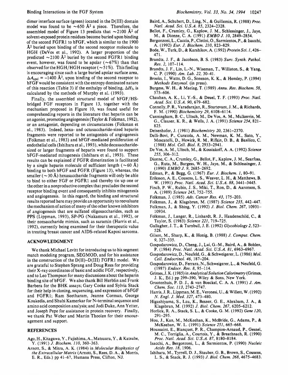
Binding Interactions in the FGF System
dimer interface surface (green) located in the D(II1) domain model was found to be -650 A2 a piece. Therefore, the assembled model of Figure 13 predicts that -2100 A2 of solvent-exposed protein residues become buried upon binding of the second FGFRl to bFGF, which is similar to the 1900 A2 buried upon binding of the second receptor molecule to HGH (DeVos et al., 1992). A larger proportion of the predicted -2100 A2 buried by the second FGFRl binding event, however, was found to be apolar (-67%) than that observed for the HGH/HGH receptor (- 5 1%). This finding is encouraging since such a large buried apolar surface area, Map, = -1400 A2, upon binding of the second receptor to bFGF would be consistent with the entropy dominated nature of this reaction (Table 3) if the enthalpy of binding, AH2, is calculated by the methods of Murphy et al. (1993).
Finally, the assembled structural model of bFGF/HS- bridged FGF receptors in Figure 13, together with the mechanism proposed in Figure 10, was found useful for comprehending reports in the literature that heparin can be an agonist, promoting angiogenesis (Taylor & Folkman, 1982), or an antagonist, depending on circumstances (Folkman et al., 1983). Indeed, hexa- and octasaccharide-sized heparin fragments were reported to be antagonists of angiogenesis (Folkman et al., 1983) and bFGF-mediated proliferation of endothelial cells (Ishihara et al., 1993), while decasaccharide- sized or larger fragments of heparin were found to support bFGF-mediated mitogenesis (Ishihara et al., 1993). These results can be explained if FGFR dimerization is facilitated by a single heparin molecule of sufficient length (-60 A) binding to both bFGF and FGFR (Figure 13), whereas, the smaller ( - 30 A) hexasaccharide fragments will only be able to bind to either FGF or FGFRl and thereby tie up one or the other in a nonproductive complex that precludes the second receptor binding event and consequently inhibits mitogenesis and angiogenesis. In this regard, the binding and structural results reported here may provide an opportunity to reevaluate themechanism of actionof many of theother known inhibitors of angiogenesis that are sulfated oligosaccharides, such as PPS (Lippman, 1993), SP-PG (Nakamura et al., 1992), or their nonsaccharide mimics, such as suramin (Harris et al., 1992), currently being examined for their therapeutic value in treating breast cancer and AIDS-related Kaposi sarcoma.
ACKNOWLEDGMENT
We thank Michael Levitt for introducing us to his segment match modeling program, SEGMOD, and for his assistance in the construction of the D(I1)-D(II1) FGFRl model. We are grateful to Stephen Sprang and Doug Rees for providing their X-ray coordinates of basic and acidic FGF, respectively, and to Leo Thompson for many discussions about the heparin binding site of bFGF. We also thank Bill Herblin and Frank Barbera for the BHK assays; Gary Cooke and Sylvia Stack for their help in cloning, sequencing, and expression of bFGF and FGFRl ; Ram Seetharam, Jeanne Corman, George Koukedis, and Shubi Kamerkar for N-terminal sequence and amino acid composition analysis; and Jodi Duke, Ann Yetter, and Joseph Pepe for assistance in protein recovery. Finally, we thank Pat Weber and Martin Thoolen for their encour- agement and support.
REFERENCES
Ago, H., Kitagawa, Y., Fujishima,A., Matsuura, Y., & Katsube, Y. (1991) J . Biochem. 110, 360-363.
Arnott, S., & Mitra, A. K. (1984) in Molecular Biophysics of the Extracellular Matrix (Arnott, S . , Rees, D. A., & Morris, E. R., Eds.) pp 41-67, Humana Press, Clifton, NJ.
Biochemistry, Vol. 33, No. 34, 1994 10247
Baird, A., Schubert, D., Ling, N., & Guillemin, R. (1988) Proc.
Bellot, F., Crumley, G., Kaplow, J. M., Schlessinger, J., Jaye,
Bergonzoni, L., Caccia, P., Cletini, O., Sarmientos, P., & Isacchi,
Bode, W., Turk, D., & Karshikov, A. (1992) Protein Sci. 1,426-
Brandts, J. F., & Jacobson, B. S. (1983) Surv. Synth. Pathol.
Brandts, J. F., Lin, L.-N., Wiseman, T., Williston, S., & Yang,
Brooks, I., Watts, D. G., Soneson, K. K., & Hensley, P. (1994)
Burgess, W. H., & Maciag, T. (1989) Annu. Rev. Biochem. 58,
Chauhan, A. K., Li, Y.-S., & Deuel, T. F. (1993) Proc. Natl. Acad. Sci. U.S.A. 90, 679-682.
Connelly, P. R., Varadarajan, R., Sturtevant, J. M., & Richards, F. M. (1990) Biochemistry 29, 6108-61 14.
Cunningham, B. C., Ultsch, M., De Vos, A. M., Mulkerrin, M. G., Clauser, K. R., & Wells, J. A. (1991) Science 254, 821- 825.
Natl. Acad. Sci. U.S.A. 85, 2324-2328.
M., & Dionne, C. A. (1991) EMBO J. 10, 2849-2854.
A. (1992) Eur. J . Biochem. 210, 823-829.
471.
Res. 2, 107-114.
C. P. (1990) Am. Lab. 22, 30-41.
Methods Enzymol. (in press).
575-606.
Deisenhofer, J. (1981) Biochemistry 20, 2361-2370. Delli-Bovi, P., Curatola, A. M., Newman, K. M., Sato, Y.,
Moscatelli, D., Hewick, R. M., Rifkin, D. B., & Basilico, C. (1988) Mol. Cell. Biol. 8, 2933-2941.
De Vos, A. M., Ultsch, M., & Kossiakoff, A. A. (1992) Science
Dionne, C. A., Crumley, G., Bellot, F., Kaplow, J. M., Searfoss, G., Ruta, M., Burgess, W. H., Jaye, M., & Schlessinger, J.
255, 306-3 12.
(1990) EMBO J . 9, 2685-2692. Edman, P., & Begg, G. (1967) Eur. J . Biochem. I , 80-91. Eriksson, A. E., Cousens, L. S., Weaver, L. H., & Matthews, B.
W. (1991) Proc. Natl. Acad. Sci. U.S.A. 88, 3441-3445. Finch, P. W., Rubin, J. S., Miki, T., Ron, D., & Aaronson, S.
A. (1989) Science 245, 752-755. Folkman, J. (1985) Adv. Cancer Res. 43, 175-203. Folkman, J., & Klagsbrun, M. (1987) Science 235, 442-447. Folkman, J., & Shing, Y. (1992) J . Biol. Chem. 267, 10931-
Folkman, J., Langer, R., Linhardt, R. J., Haudenschild, C., &
Gallagher, J. T., & Turnbull, J. E. (1992) Glycobiology 2, 523-
Gilson, M., Sharp, K., & Honig, B. (1988) J . Comput. Chem.
Gospodarowicz, D., Cheng, J., Lui, G.-M., Baird, A., & Bohlen, P. (1984) Proc. Natl. Acad. Sci. U.S.A. 81, 6963-6967.
Gospodarowicz, D., Neufeld, G., & Schweigerer, L. (1986) Mol. Cell. Endocrinol. 46, 187-204.
Gospodarowicz, D., Ferrara, N., Schweigerer, L., & Neufeld, G. (1987) Endocr. Rev. 8, 95-114.
Grimes, J. K. (1985) in AnalyticalSolution Calorimetry (Grimes, J. K., Ed.) pp 299-390, Wiley & Sons, New York.
Grootenhuis, P. D. J., & van Boeckel, C. A. A. (1991) J . Am. Chem. SOC. 113, 2743-2747.
Harris, J. R., Lippman, M. E., Veronesi, U., & Willett, W. (1992) N . Engl. J . Med. 327, 473-480.
Higashiyama, S., Lau, K., Besner, G. E., Abraham, J. A., & Klagsbrun, M. (1992) J. Biol. Chem. 267, 6205-6212.
Horlick, R. A., Stack, S. L., & Cooke, G. M. (1 992) Gene 120,
Hou, J., Kan, M., McKeehan, K., McBride, G., Adams, P., & McKeehan, W. L. (1991) Science 251, 665-668.
Houssaint, E., Blanquet, P. R., Champion-Arnaud, P., Gesnel, M. C., Torriglia, A,, Courtois, Y., & Breathnach, R. (1990) Proc. Natl. Acad. Sci. U.S.A. 87, 8180-8184.
Isacchi, A,, Bergonzoni, L., & Sarmientos, P. (1990) Nucleic Acids Res. 18, 1906.
Ishihara, M., Tyrrell, D. J., Stauber, G. B., Brown, S., Cousens, L. S., & Stack, R. J. (1993) J. Biol. Chem. 268,4675-4683.
10934.
Taylor, S. (1983) Science 221, 719-725.
528.
9, 327-335.
29 1-295.

10248 Biochemistry, Vol. 33, No. 34, 1994 Pantoliano et al.
Jaye, M., Schlessinger, J., & Dionne, C. A. (1992) Biochim. Biophys. Acta 11 35, 185-1 99.
Johnson, D. E., Lee, P. L., Lu, J., & Williams, L. T. (1990) Mol. Cell. Biol. 10, 4728-4736.
Johnson, D. E., Lu, J., Chen, H., Werner, S., & Williams, L. T. (1991) Mol. Cell. Biol. 11, 4627-4634.
Kan, M., Wang, F., Xu, J., Crabb, J. W., Hou, J., & McKeehan, W. L. (1993) Science 259, 1918-1921.
Kiefer, M. C., Baird, A,, Nguyen, T., George-Nascimento, C., Mason, 0. B., Boley, L. J., Valenzuela, P., & Barr, P. J. (1 99 1) Growth Factors 5 , 115-127.
Klagsbrun, M., & Baird, A. (1991) Cell 67, 229-231. Kuntz, I. D. (1992) Science 257, 1078-1082. Laemmli, U. K. (1970) Nature 227, 680-695. Leahy, D. J., Hendrickson, W. A,, Aukhil, I., & Erickson, H. P.
Lee,P. L., Johnson, D. E.,Cousens, L. S., Fried, V. A., & Williams,
Levitt, M. (1983) J. Mol. Biol. 168, 595-620. Levitt, M. (1992) J . Mol. Biol. 226, 507-533. Lin, L.-N., Mason, A. B., Woodworth, R. C., & Brandts, J. F.
Lippman, M. E. (1993) Science 259, 631-632. Lobb, R. R., & Fett, J. W. (1984) Biochemistry 23,6295-6299. Luyten, F. P., Cunningham, N. S. , Ma, S., Muthukumaran, N.,
Hammonds, R. G., Nevins, W. B., Wood, W. I., & Reddi, A. H. (1989) J . Biol. Chem. 264, 13377-13380.
Marics, I., Adelaide, J., Raybaud, F., Mattei, M. G., Coulier, F., Planche, J., de Lapeyriere, O., & Birnbaum, D. (1989) Oncogene 4, 335-340.
MarvizQ, J.-C., & Baudry, M. (1993) Anal. Biochem. 21 3,3-11. Melnyk, V. O., Shipley, G. D., Sternfeld, M. D., Sherman, L.,
& Rosenbaum, J.T. (1990) Arthritis Rheum. 33, 493-500. Miki, T., Bottaro, D. P., Fleming T. P., Smith, C. L., Burgess,
W. H., Chan, A. M.-L., & Aaronson, S. A. (1992) Proc. Natl. Acad. Sci. U.S.A. 89, 246-250.
Mitra, A. K., Arnott, S., Atkins, E. D. T., & Isaac, D. H. (1983) J. Mol. Biol. 169, 873-901.
(1992) Science 258, 987-991.
L. T. (1989) Science 245, 57-60.
(1991) Biochemistry 30, 11660-1 1669.
Sali, A., Matsumoto, R., McNeil, H. P., Karplus, M., & Stevens, R. L. (1993) J. Biol. Chem. 268, 9023-9034.
Sambrook, J., Fritsch, E. F., & Maniatis, T. (1989) Molecular Cloning: A Laboratory Manual, 2nd ed., Cold Spring Harbor Laboratory Press, Cold Spring Harbor, NY.
Sharpe, R. J., Arndt, K. A,, Bauer, S. I . , & Maione, T. E. (1989) Arch. Dermatol. 125, 1359-1362.
Sheriff, S., Silverton, E. W., Padlan, E. A,, Cohen, G. H., Smith- Gill, s. J., Finzel, B. C., & Davies, D. R. (1987) Proc. Natl. Acad. Sci. U.S.A. 84, 8075-8079.
Shing, Y., Folkman, J., Sullivan, R., Butterfield, C., Murray, J., & Klagsbrun, M. (1984) Science 223, 1296-1299.
Sivalingam, A., Kenney, J., Brown, G. C., Benson, W. E., & Donoso, L. (1990) Arch. Ophthalmol. 108, 869-872.
Squires, C. H., Childs, J., Eisenberg, S. P., Polverini, P. J., & Sommer, A. (1988) J . Biol. Chem. 263, 16297-16302.
Stoddard, B. L., & Koshland, D. E. (1992) Nature 358, 774- 716.
Summers, M. D., & Smith, G. E. (1987) A Manual of Methods for Baculovirus Vectors and Insect Cell Culture Procedures, Texas Agricultural Experimental Station Bulletin 1555, College Station, TX.
Taira, M., Yoshida, T., Miyagawa, K., Sakamoto, H., Terada, M., & Sugimura, T. (1987) Proc. Natl. Acad. Sci. U.S.A. 84, 2980-2984.
Tarentino, A. L., Gbmez, C. M., & Plummer, T. H., Jr. (1985) Biochemistry 24, 4665-467 1.
Taylor, S., & Folkman, J., (1982) Nature 297, 307. Thompson, L. D., Pantoliano, M. W., & Springer, B. A. (1994)
Biochemistry 33, 383 1-3840. Tyrrell, D. J., Ishihara, M., Rao, N., Horne, A., Kiefer, M. C.,
Stauber, G. B., Lam, L. H., & Stack, R. J. (1993) J . Biol. Chem. 268, 4684-4689.
Ueno, H., Gum, M., Dell, K., Tseng, A., & Williams, L. (1992) J . Biol. Chem. 267, 1470-1476.
Moore, R., Casey, G,, Brookes, S., Dixon, M., Peters, G., &
Moscatelli, D. (1987) J . Cell Physiol. 131, 123-130.
Murphy, K. p., Xi% D.9 Garcia, K. c.9 Amzel, M.3 & Freire, E. (1993) Proteins: Struct., Funct., Genet. 15, 113-120.
Nakamura, S., Sakurada, S., Salahuddin, S. Z., Osada, Y., Tanaka, N. G., Sakamoto, N., Sekiguchi, M., & Gallo, R. C. (1992) Science 255, 1437-1440.
Neufeld. G.. & GosDodarowicz. D. (1985) J . Biol. Chem. 260.
Ullrich, A., & Schlessinger, J. (1990) Cell 61, 203-212. Vialard, J., Lalumiere, M., Vernet, T., Briedis, D., Alkhatib, G.,
Henning, D., Levin, D., & Richardson, C. (1990) J. Virol. 64, Dickson, C. (1986) EMBO J . 5, 919-924.
Moses, M. A., & Langer, R. (1991) Bio/Technology 9,630-634. 37-50. Weber, G. (1975) Ado. Protein Chem. 29, 1-83. Weber, P. C., Wendoloski, J. J., Pantoliano, M. W., & Salemme,
Weber, P. C., Pantoliano, M. W., Simons, D. M., & Salemme, F. R. (1992) J . Am. Chem. SOC. 114, 3197-3200.
F. R. (1994) J. Am. Chem. SOC. 116, 2717-2724. I . ,
1386613868. A
8883. Nugent, M. A., & Edelman, E. R. (1992) Biochemistry 31,8876-
Olson, S . T., & Bjork, I. (1991) J. Biol. Chem. 266,6353-6364. Olson, S . T., Halvorson, H. R., & Bjork, I. (1991) J . Biol. Chem.
Ornitz, D. M., Yayon, A., Flanagan, J. G., Svahn, C. M., Levi, E., & Leder, P. (1992) Mol. Cell. Biol. 12, 240-247.
Pantoliano, M. W., Bird, R. E., Johnson, S., Asel, E. D., Dodd, S. W., Wood, J. F., & Hardman, K. D. (1991) Biochemistry
Partanen, J., Makela, T. P., Eerola, E., Korhonen, J., Hirvonen, H., Claesson-Welsh, L., & Alitalo, K. (1991) EMBO J . 10,
Pasquale, E. B. (1990) Proc. Natl. Acad. Sci U.S.A. 87, 5812-
Pasquale, E. B., & Singer, S. J. (1989) Proc. Natl. Acad. Sci.
Petitou, M., Lormeau, J.-C., & Choay, J. (1991) Nature 350
Rapraeger, A. C., Krufka, A., & Olwin, B. B. (1991) Science
Richards, F. M. (1977) Annu. Rev. Biophys. Bioeng. 6,151-176. Roberts, R., Gallagher, J., Spooncer, E., Allen, T. D., Bloomfield,
266, 6342-6352.
30, 101 17-10125.
1347-1 354.
5816.
U.S.A. 86, 5449-5453.
(Suppl.), 30-33.
252, 1705-1708.
F., & Dexter, T. M. (1988) Nature 332, 376-378.
Weiner, P. K., & Kollman, P. A. (1981) J. Comput. Chem. 2,
Wellstein, A., Zugmaier, G., Califano, J. A., 111, Kern, F., Paik, S., & Lippman, M. E. (1991) J . Natl. Cancer Znst. 83, 716- 720.
Wiseman, T., Williston, S . , Brandts, J. F., & Lin, L.-N. (1989) Anal. Biochem. 179, 131-137.
Wozney, J. M., Rosen, V., Celeste, A. J., Mitsock,L. M., Whitters, M. J., Kriz, R. W., Hewick, R. M., & Wang, E. A. (1988) Science 242, 1528-1 534.
287-303.
Wyman, J., Jr. (1964) Adv. Protein Chem. 19, 223-285. Yanagishita, M., & Hascall, V. C. (1992) J. Biol. Chem. 267,
Yayon, A., Klagsbrun, M., Esko, J. D., Leder, P., & Ornitz, D. M. (1991) Cell 64, 841-848.
Zhan, X., Bates, B., Hu, X., & Goldfarb, M. (1988) Mol. Cell. Biol. 8, 3487-3495.
Zhang, J., Cousens, L. S., Barr, P. J., & Sprang, S. R. (1991) Proc. Natl. Acad. Sci. U.S.A. 88, 3446-3450.
Zhu, X., Komiya, H., Chirino, A., Faham, S., Fox, G. M., Arakawa, T., Hsu, B.T., & Rees, D. C. (1991) Science 251, 90-93.
Zhu, X., Hsu, B. T., & Rees, D. C. (1993) Structure 1, 27-34.
9451-9454.