Importance of basal processes in simulations of a surging ...
Upload
historiauexCategory
view
5download
0
Dioxin Receptor Expression Inhibits Basal and TransformingGrowth Factor �-induced Epithelial-to-mesenchymalTransition*□S
Received for publication, October 3, 2012, and in revised form, January 28, 2013 Published, JBC Papers in Press, February 4, 2013, DOI 10.1074/jbc.M112.425009
Eva M. Rico-Leo‡1, Alberto Alvarez-Barrientos§, and Pedro M. Fernandez-Salguero‡2
From the ‡Departamento de Bioquímica y Biología Molecular, Facultad de Ciencias and the §Servicio de Técnicas Aplicadas a lasBiociencias, Universidad de Extremadura, 06071 Badajoz, Spain
Background: The dioxin receptor (AhR) regulates cell migration and has a role in TGF� activation.Results: AhR expression inhibits basal and TGF�-induced epithelial-to-mesenchymal transition (EMT).Conclusion: AhR has an intrinsic role in EMT and cross talks with TGF�.Significance: The involvement of AhR in EMT can help explain its functions in organ homeostasis and tumor progression.
Recent studies have emphasized the role of the dioxin recep-tor (AhR) inmaintaining cellmorphology, adhesion, andmigra-tion. These novel AhR functions depend on the cell phenotype,and although AhR expression maintains mesenchymal fibro-blasts migration, it inhibits keratinocytesmotility. These obser-vations prompted us to investigate whether AhR modulates theepithelial-to-mesenchymal transition (EMT). For this, we haveused primary AhR�/� and AhR�/� keratinocytes and NMuMGcells engineered to knock down AhR levels (sh-AhR) or toexpress a constitutively active receptor (CA-AhR). BothAhR�/�
keratinocytes and sh-AhR NMuMG cells had increased migra-tion, reduced levels of epithelial markers E-cadherin and�-catenin, and increased expression of mesenchymal markersSnail, Slug/Snai2, vimentin, fibronectin, and �-smooth muscleactin. Consistently, AhR�/� and CA-AhR NMuMG cells hadreducedmigration and enhanced expression of epithelial mark-ers. AhR activation by the agonist FICZ (6-formylindolo[3,2-b]carbazole) inhibited NMuMG migration, whereas the an-tagonist �-naphthoflavone induced migration as did AhRknockdown. Exogenous TGF� exacerbated the promigratorymesenchymal phenotype in both AhR-expressing and AhR-depleted cells, although the effects on the latter were morepronounced. Rescuing AhR expression in sh-AhR cellsreduced Snail and Slug/Snai2 levels and cell migration andrestored E-cadherin levels. Interference of AhR in humanHaCaT cells further supported its role in EMT. Interestingly, co-immunoprecipitation and immunofluorescence assays showedthat AhR associates in common protein complexes with E-cad-herin and�-catenin, suggesting the implication of AhR in cell-celladhesion. Thus, basal or TGF�-induced AhR down-modulationcould be relevant in the acquisition of amotile EMT phenotype inboth normal and transformed epithelial cells.
The aryl hydrocarbon receptor (AhR)3 is a basic-helix-loop-helix (bHLH) transcription factor (1)well known for its relevantrole in xenobiotic-induced toxicity and carcinogenesis and,more recently, for its implication in different cellular processesincluding proliferation, differentiation, cell adhesion, andmigration and organ homeostasis (2–6). Cell morphology,adhesion, and migration are essential cell properties requiringendogenousAhR activity. AhR exerts a differential effect on cellmotility depending on the mesenchymal, epithelial, or endo-thelial phenotype of the target cell. Thus, immortalized andembryonic primarymurine fibroblasts lackingAhRhave spreadmorphology and impaired adhesion and migration (7–9). Con-trary to mesenchymal fibroblasts, AhR knockout in epidermalkeratinocytes increases their motility and migration both invitro and in vivo (10). In additional cell types such as primarymouse endothelial cells (11) and CD4�CD8� thymocytes (4,12), AhR activation promoted cell migration to newly formedblood vessels and to the spleen, respectively. The fact that AhRdepletion increased primary keratinocytes migration andimproved wound healing in vivo led us to suggest that AhRcould be involved in the epithelial-to-mesenchymal transition(EMT).EMT is a phenotypic switch that permanently or transiently
converts epithelial cells into motile mesenchymal-like cells.During this process, epithelial cells suffer a spectrumof changesthat affect their adhesion to neighboring cells and to the sub-stratum, their migration, and their normal functioning (13).EMT is essential during embryonic development and in tissuerepair, although a large body of evidence indicates that it alsocontributes to pathology (13–15). Because EMT enables epi-thelial cells with migration and invasion capabilities, it is gen-erally accepted that it contributes to the early stages of tumormetastasis (15, 16). Among the EMT features that are con-served in most epithelial cell types are the repression of the
* This work was funded by grants from the Spanish Ministry of Science andInnovation (SAF2008-00462 and BFU2011-22678), the Red Temática deInvestigación Cooperativa en Cáncer, Fondo de Investigaciones Sanitarias,Carlos III Institute, Spanish Ministry of Health (RD06/0020/1016 and RD12/0036/0032), and the Junta de Extremadura (GRU10008). All Spanish fund-ing was co-sponsored by the European Union FEDER (Fondo Europeo deDesarrollo Regional) program.
□S This article contains supplemental Table S1 and Fig. S1.1 Supported by the Red Temática de Investigación Cooperativa en Cáncer.2 To whom correspondence should be addressed. Tel.: 34924289422; Fax:
34924289419; E-mail: [email protected].
3 The abbreviations used are: AhR, dioxin receptor; EMT, epithelial-to-mesen-chymal transition; �-naph, �-naphthoflavone; CA-AhR, constitutivelyactive AhR receptor; E-Cad and N-Cad, E-cadherin and N-cadherin, respec-tively; �-Cat, �-catenin; FICZ, 6-formylindolo[3,2-b]carbazole; sh-AhR,small-hairpin for AhR; TER, trans-epithelial resistance; qRT, quantitativereal-time; EV, empty vector.
THE JOURNAL OF BIOLOGICAL CHEMISTRY VOL. 288, NO. 11, pp. 7841–7856, March 15, 2013© 2013 by The American Society for Biochemistry and Molecular Biology, Inc. Published in the U.S.A.
MARCH 15, 2013 • VOLUME 288 • NUMBER 11 JOURNAL OF BIOLOGICAL CHEMISTRY 7841
at UN
IV D
E E
XT
RE
MA
DU
RA
, on March 15, 2013
ww
w.jbc.org
Dow
nloaded from
http://www.jbc.org/content/suppl/2013/02/04/M112.425009.DC1.html Supplemental Material can be found at:
adherents junctions protein E-cadherin (E-Cad), the up-regu-lation of mesenchymal markers vimentin, fibronectin, andN-cadherin (N-Cad), and the change toward a mesenchymal-like morphology (13, 17, 18). Several transcription factors pro-mote EMT through the down-regulation of E-Cad (13, 15), anda central role has been given to members of the Snail family ofproteins (Snail and Slug/Snai2), which repress E-Cad by bind-ing to an E-box located in its promoter (19–21). Interestingly,Snail and Slug/Snai2modulate common as well as specific generegulatory pathways that likely differentiate their contribu-tion to cancer progression and dissemination (22). An addi-tional inducer of EMT is the extracellular cytokine trans-forming growth factor � (TGF�), which can be produced andsecreted by tumor cells or by the stroma. TGF� induces EMTand cancer metastasis (23–25) possibly by promoting theearly dissolution of the tight junctions that interconnect epi-thelial cells (26, 27).AhR is functionally related to TGF� in different cell types
and in vivo. Several studies have shown that AhR-null fibro-blasts have increased expression and activation of TGF� thatpresumably contribute to their lower proliferation ability(28–31). In addition, TGF� secreted by the stroma was ableto increase the migration efficiency of primary AhR�/� kera-tinocytes in vitro and in vivo (10). Interestingly, TGF� exertscell type-specific effects on AhR by inhibiting receptorexpression and activation in A549 lung cancer cells whileenhancing receptor function in HepG2 hepatoma cells (32,33). Thus, it is likely that AhR and TGF� could cross-talkduring EMT.In this study we have investigated the role of AhR in EMT
under both basal andTGF�-induced conditionswith the aim todetermine whether or not AhR expression restrains the acqui-sition of a migratory EMT phenotype in epithelial cells. Thus,we have used primary keratinocytes fromAhR�/� andAhR�/�
mice and NMuMG cells engineered to have either depleted orenhanced AhR activity to investigate the role of AhR in EMT.We have found that AhR knockdown by itself triggers an EMT-like phenotype characterized by changes in epithelial and mes-enchymal markers and by increased cell migration. Moreover,AhR deficiency seems to cooperate with TGF� to enhance theEMT process. These results support a mechanism by whichAhR is a negative regulator of EMT and suggest that metastasisof TGF�-responsive tumors could be exacerbated if theyundergo AhR down-modulation.
EXPERIMENTAL PROCEDURES
Primary Keratinocytes and Cell Lines—Primary keratino-cytes were obtained from newborn AhR�/� and AhR�/� miceat 2–3 days of age. Pups were sequentially washed in povidonesolution, sterile water, and 70% ethanol in PBS. Legs and tailwere removed, and the complete skin was dissected using for-ceps. The skins were floated dermis side down in 0.25% trypsinfor 16–18 h at 4 °C. Next, the epidermis was separated from thedermis and minced in 2–3 ml/mouse of plating medium (min-imum essential medium Eagle containing gentamycin and 4%FBS pretreated with Chelex and 0.2 mM Ca2�). Tissues weredigested by gentle incubation for 45 min at 4 °C and filteredthrough a 140-�mmesh to remove aggregates and undigested
material. The resulting cell suspension was seeded at 2 � 106cells in 60-mm culture plates pretreated with 5 �g/ml colla-gen or 15 �g/ml fibronectin. After 24 h, keratinocytes werewashed with PBS and grown in CnT-0.7 culture medium(CELLnTEC) to promote proliferation and to inhibit differ-entiation. NMuMG mouse epithelial cells were grown inDMEM containing 10% FBS, 100 units/ml penicillin, 100�g/ml streptomycin, 2 mM L-glutamine, and 10 �g/ml insu-lin at 37 °C in a 5% CO2 atmosphere. Human HaCaT andPhoenix packing cells were cultured in the above mediumwithout added insulin at 37 °C and 5% CO2 atmosphere. Celllines were trypsinized and passaged at 1:3 dilution when theyreached 70–80% confluence.Antibodies, Reagents, Expression Vectors, and Treatments—
Antibodies against the following proteins were used: �-catenin(BDBiosciences); E-cadherin (Calbiochem);AhR (Immunostepand Biomol); TGF� (R&D Systems); p-Smad2 (Cell Signaling);Slug/Snai2 (Santa Cruz); N-cadherin (Invitrogen); fibronectin(Chemicon), vimentin, �-smooth muscle actin, and �-actin(Sigma). The AhR agonist 6-formylindolo[3,2-b]carbazole(FICZ) was from Enzo, and the AhR antagonist �-naphthofla-vone (�-naph) was from Sigma. The pharmacological inhibitorof the TGF� pathway SB431542 was from Selleckchem. Rhod-amine-phalloidin was from Invitrogen. Matrigel solution wasfrom BD Biosciences. TaqDNA polymerase was from Ecogen.iScript reverse transcription supermix and SYBRGreenmastermix were obtained from Bio-Rad. Small hairpin RNA was fromSigma. Small interfering RNA for AhR and scrambled siRNAwere synthesized by Dharmacon. The constitutively activeform of the AhR (CA-AhR) was produced from the wild typemouse receptor by deleting the minimal PAS-B motif (aminoacids 288–421) without altering the N-terminal half of thebinding domain (PAS-A). This constitutively active receptorheterodimerizes with ARNT and has intrinsic transcriptionalactivity in a ligand-independent manner (34). Recombinanthuman TGF� (Sigma) was added to the cultures at 10 ng/ml(primary keratinocytes and HaCaT cells) or 5 ng/ml (NMuMGcells). Control cultures were treated with the same volume ofsolvent (PBS).Retroviral Transduction—NMuMG cells were stably trans-
duced with expression vectors containing a small hairpin RNAfor AhR (sh-AhR) or a constitutively active form of the protein(CA-AhR) as described (Stanford University Medical Center).In brief, constructs LMP-sh-AhR, pBABE-CA-AhR, or theempty vectors pBABE�LMP were transfected by calciumphosphate precipitation in Phoenix cells, and virus productionwas allowed for 48 h. NMuMG cells were exposed overnight tothe viral supernatants, and 48 h later selection was started with1 �g/ml puromycin for 14 days. Individual clones survivingselection were isolated by cell sorting and then analyzed forAhR expression by immunoblotting or for the Cyp1a1 AhRtarget gene by qRT-PCR.Transient Transfection and RNA Interference—NMuMG
cells were transiently transfected using the Turbofect reagent(Fermentas). Briefly, the DNA was incubated for 15 min atroom temperature with Turbofect, and the mix was added tothe cells in complete medium. After 24 h-48 h of transfection,cultures were processed and analyzed. HaCaT cells were trans-
AhR Expression Blocks EMT
7842 JOURNAL OF BIOLOGICAL CHEMISTRY VOLUME 288 • NUMBER 11 • MARCH 15, 2013
at UN
IV D
E E
XT
RE
MA
DU
RA
, on March 15, 2013
ww
w.jbc.org
Dow
nloaded from
fected with small interfering RNAs for AhR (si-AhR) or scram-bled siRNA by nucleoporation using a MicroPorator MP-100(Digital-Bio). In brief, cells were trypsinized, washed, and resus-pended in transfection solution. si-AhR or scrambled siRNAwere added at 50 nM to the cells, and nucleofection was per-formed with a single pulse of 1400 V for 30 ms. After 24 h, cellswere transferred to fresh complete medium, and culture wascontinued for an additional 48 h.Cellular Morphology—Cell area and circularity of AhR�/�
andAhR�/� keratinocytes andNMuMGcells were determinedby blinded analysis in different random fields for each culture asindicated (7). Cellular contour and axis were analyzed using theImageJ software (Version 1.45S). Significance of the data wasanalyzed as indicated below (“Statistical Analyses”).Trans-epithelial Resistance (TER)—Cells seeded on transwell
filter inserts (BD Biosciences) of 0.4-�mpore size were allowedto reach confluence. A volume of 2 ml of mediumwas added tothe inner insert chamber and 4 ml to the outer chamber, withdaily replacement of fresh medium. TER was measured everyday using the Millicell Electrical Resistance System accordingto the manufacturer’s instructions.Clonogenic Assays—Clonogenic assays in two dimensions
(2-D) cultures were done by platting 5 � 102 or 103 sh-AhR orCA-AhR NMuMG cells in plain tissue culture dishes. Emptyvector-transduced (EV) NMuMG cells were used as controls.After 5 days of culture, medium was removed, and clones werewashed in PBS, stained for 10minwith 0.5% (w/v) crystal violet,and counted. Clone formation in three dimensions (3-D) wasanalyzed by growing each cell line onMatrigel as described (11).Briefly, 5 � 103 sh-AhR, CA-AhR, or EV NMuMG cells wereseeded on Matrigel plugs for 3 days in complete medium.Clones formed were counted and analyzed for cell spreadingusing the ImageJ software (Version 1.45S).Cell Migration—Cell migration was analyzed using wound
healing experiments as previously reported (9, 10).Immunofluorescence and Rhodamine-phalloidin—Fluores-
cence analysis was done using a Fluoview 1000 confocal micro-scope (Olympus) on cultures fixed for 20 min at room temper-ature in 4% paraformaldehyde (Polysciences, Inc). Secondaryantibodies labeled with Alexa 488 for E-cadherin (rat monoclo-nal ECCd2), �-catenin (mouse monoclonal), Snail (mousemonoclonal), and Slug/Snai2 (goat polyclonal) or Alexa 633 forAhR (rabbit polyclonal) were used. DAPI staining served tolabel cell nuclei. Cells stained with only secondary antibodywere used as negative controls. To label actin stress fibers, cellswere also incubated with rhodamine-phalloidin as indicated(9). Fluorescence distribution analysis was performed using theFV1000 software (Olympus).Immunoprecipitation and Immunoblotting—Immunopre-
cipitation from NMuMG cells was performed essentially asdescribed (35, 36). Briefly, cells were lysed, and 1 mg of totalcellular protein was precleared by incubation with protein A/Gplus-agarose beads. Extracts were then mixed with 2 �g of thecorresponding specific antibody and fresh protein A/G-Sep-harose beads. After overnight incubation at 4 °C, beads werewashed twice with buffer A (20 mM Tris-HCl pH 7.4, 50 mM
NaCl, 1% Nonidet P-40, 10 mM EDTA, 1 mM sodiumorthovanadate, 50 mM NaF, 0.5 mM PMSF, and 4 mg/ml Com-
plete protease inhibitormixture) and buffer B (25mMTris-HCl,pH 7.5, 150 mM NaCl, and 1 mM EDTA). Washed beads werethen used for immunoblotting. SDS-PAGE and immunoblot-ting were performed as described (9, 31).Reverse Transcription and Real-time PCR—Total RNA was
isolated from primary keratinocytes, NMuMG, and HaCaTcells using the RNeasy kit (Qiagen). Reverse transcription wasperformed using random hexamers and iScript reverse tran-scription super mix (Bio-Rad). Real-time PCR was done toquantify the levels of human and mouse Snail and Slug/Snai2and of mouse Cyp1a1mRNAs.GapdhmRNAwas used to nor-malize individual gene expression (�Ct). Reactions were doneusing SYBR Green I/QTaq DNA polymerase mix on an iCyclerequipment (Bio-Rad) as described (37). 2���Ct was used to cal-culate Gapdh-normalized gene expression with respect to thecontrol or untreated conditions. Primer sequences used in thisstudy are indicated in supplemental Table 1.Statistical Analyses—Data are shown as themean� S.D. Sta-
tistical comparison between experimental conditions was doneusingGraphPad Prism 4.0 software (GraphPad). Student’s t testor analysis of variance were applied. Experiments were done induplicate or triplicate in two or three cultures of each cell line.
RESULTS
AhR Knockdown Induces EMT under Basal Cell Conditions—To analyze the role of AhR in EMT under normal cellular con-ditions, we have used primary keratinocytes from AhR�/� andAhR�/� mouse pups and NMuMG epithelial cells engineeredby retroviral transduction to encode a small hairpin RNA forAhR (sh-AhR) or a constitutively active AhR receptor (CA-AhR). NMuMG cells we also transduced with an empty retro-virus to account for the basal AhR expression. After antibioticselection, clones carrying each of those constructs were iso-lated. Immunoblotting analysis with an AhR-specific antibodyrevealed a marked knockdown of the endogenous AhR expres-sion in the sh-AhR cell line (supplemental Fig. S1A). Constitu-tively active AhR was ectopically expressed in CA-AhR cells,and it was functional as determined by the increase in mRNAexpression observed for its canonical target gene Cyp1a1 (sup-plemental Fig. S1B).EMT is generally followed by a change inmorphology toward
a mesenchymal-like phenotype in parallel with a switch in thepattern of expression of cell adhesion molecules and their reg-ulators. AhR depletion significantly reduced the circularity ofNMuMG cells, inducing a more elongated morphology,whereas AhR over-activation further stressed their circularform (Fig. 1A). AhR has a role in that phenotype as primarykeratinocytes from AhR-null mice were significantly moreelongated than theirAhR�/� counterparts (Fig. 1B).Mesenchy-mal cells usually have more relaxed cell-cell interactions thanepithelial cells when grown at high confluence, thus producingless compact monolayers. The TER of sh-AhR confluent cul-tures was significantly lower than that of wild type (EV) or CA-AhR NMuMG cultures (Fig. 1C), supporting that reduction ofAhR levels in epithelial cells favors a more mesenchymalphenotype.The acquisition of an EMT phenotype implies a reduction in
the expression of epithelial markers such as E-Cad and a gain in
AhR Expression Blocks EMT
MARCH 15, 2013 • VOLUME 288 • NUMBER 11 JOURNAL OF BIOLOGICAL CHEMISTRY 7843
at UN
IV D
E E
XT
RE
MA
DU
RA
, on March 15, 2013
ww
w.jbc.org
Dow
nloaded from

mesenchymal molecules like fibronectin, N-cadherin, andvimentin. sh-AhR NMuMG cells had a significant reduction inE-Cad and �-Cat protein levels and an increase in fibronectin(Fig. 2A). CA-AhR cells, on the contrary, increased their E-Cadprotein content by about 3-fold and reduced vimentin andfibronectin expression (Fig. 2A). Immunofluorescence experi-ments showed that AhR depletion reduced the cellular contentof E-Cad and �-Cat and altered their location at the plasmamembrane while increasing the levels of F-actin fibers, suggest-ing that low AhR expression could destabilize cell-cell interac-tions in epithelial cells (Fig. 2C). Oppositely, CA-AhR cells con-centrated E-Cad and �-Cat at the plasma membrane and hadmuch fewer prominent F-actin fibers (Fig. 2C). PrimaryAhR�/� keratinocytes had marked reductions in E-Cad and�-Cat and higher contents of vimentin and �-smooth muscleactin as compared with AhR�/� cells (Fig. 2B), confirming thatAhR down-modulation triggers phenotypical changes indica-tive of EMT. TGF� is considered a potent EMT inducer indifferent cell types (24, 25), and we have previously shown thatAhR negatively regulates TGF� activation (29–31). Proteinexpression analysis revealed that AhR depletion increasedTGF� content in sh-AhRNMuMGcells by close to 2-foldwith-out a significant effect in AhR�/� keratinocytes (Fig. 2, A andB). Yet both cell types had a significant increase in the activationof the TGF� pathway based on the phosphorylation of pSmad2(Fig. 2, A and B), indicating that TGF�-dependent signalingcould mediate the pro-EMT effects induced by reduced AhRexpression. To further analyze this possibility, we treated sh-
AhR NMuMG cells with the TGF�-RI inhibitor SB431542.SB431542 was effective in blocking TGF�-dependent signalingin this cell line, as determined by a significant reduction inSmad2 phosphorylation (Fig. 2D). SB431542 increased E-Cadexpression (Fig. 2D), suggesting that enhanced TGF� signalingcould contribute to the EMT-like features observed underabsent or reduced AhR expression.Because E-Cad is regulated by transcription factors Snail and
Slug/Snai2 (20, 21), we next investigated if their expression wasAhR-dependent. As shown in Fig. 3A, AhR knockdown signif-icantly increased Slug/Snai2 mRNA expression in NMuMGcells (upper panel) and in primary keratinocytes (lower panel);Snail expression was largely increased in primary cells (lowerpanel) but only marginally in NMuMG cells (upper panel).Consistently, constitutive AhR activation diminished Slug/Snai2 and Snail mRNA levels in NMuMG cells (Fig. 3A).Immunocytochemical analysis showed that Slug/Snai2 andSnail were barely detectable inwild type andCA-AhRNMuMGcells but significantly expressed in sh-AhR cells (Fig. 3B). Thesedata support that AhR represses Slug/Snai2 and Snail andmaintains the expression and localization of E-Cad at theplasma membrane.We then explored if the mesenchymal-like status of sh-AhR
NMuMG cells affected their clonogenic and migratory poten-tials. Experiments in two dimensions (2-D) cultures revealedthat AhR knockdown produced a larger number of clones thatwere significantly more spread and that contained mesenchy-mal-like cells readily moving out from the periphery (Fig. 4A).
FIGURE 1. AhR knockdown alters the morphology of NMuMG cells and of primary mouse keratinocytes. A, wild type (EV), sh-AhR, and CA-AhR cell lineswere grown in plain tissue culture dishes, and their morphology was determined by measuring the circularity factor (4� � area/perimeter2). n.s., not significant.B, cell morphology of wild type (AhR�/�) and AhR-null (AhR�/�) primary keratinocytes was also determined as indicated above. C, TER of each NMuMG cell linewas measured in confluent cultures grown in transwell filter inserts as indicated under “Experimental Procedures.” Determinations were done in triplicate in atleast three independent cultures of each cell line. Data are shown as mean � S.D. Bar, 200 �m.
AhR Expression Blocks EMT
7844 JOURNAL OF BIOLOGICAL CHEMISTRY VOLUME 288 • NUMBER 11 • MARCH 15, 2013
at UN
IV D
E E
XT
RE
MA
DU
RA
, on March 15, 2013
ww
w.jbc.org
Dow
nloaded from

sh-AhR NMuMG cells grown in three dimensions (3-D) cul-tures also formed more and larger clones than wild type (EV)and CA-AhR cells (Fig. 4B). The EMT generally induces apro-migratory phenotype (15) that can be associated to theclonogenic potential of the cell. Wound healing experimentsindicated that sh-AhR NMuMG cells had a moderateincrease in migration with respect to wild type (EV) cells,unlike CA-AhR cells, which had a significantly reducedmotility as compared with both EV and sh-AhR cells (Fig.5A). Likewise, primary AhR�/� keratinocytes had increasedmigration rates with respect to AhR�/� cells (Fig. 5B). Thus,the EMT process induced by AhR depletion concurs withenhanced migration and increased clonogenic potential.Pharmacological Modulation of AhR Affects EMT Markers
and Epithelial Cell Migration—These results suggested thatnon-toxic (e.g. potentially endogenous) molecules known toregulate AhR could be potential EMT modulators. To addressthis issue,wehave investigated the effects of theAhRantagonist�-naphthoflavone (38) and of the potential AhR endogenous
ligand derived from tryptophan FICZ (39, 40) on relevant EMTparameters (Fig. 6). FICZ was an efficient AhR agonist inNMuMG cells markedly inducing Cyp1a1 mRNA expression(Fig. 6A). Although its effects on fibronectin protein levels wereonly slight, FICZ significantly increased E-Cad levels (Fig. 6B)and inhibited cell migration in wound healing assays (Fig. 6C).Treatment ofNMuMGcells with the antagonist�-naph, on thecontrary, reduced AhR levels and increased fibronectin expres-sion while diminishing E-Cad amounts (Fig. 6D). Regarding itseffects on migration, �-naph increased the ability of NMuMGcells to close wounds and to spread out at the margin of thewounds (Fig. 6E and insets). These results support our hypoth-esis that increasingAhRactivity by an endogenous ligand inhib-its EMT-like processes, whereas blockade of AhR enhancesEMT properties in epithelial cells.AhR Knockdown Has Synergistic Effects with TGF� in EMT—
Considering the results above, we decided to address howknockdown or constitutive activation of AhR affected cellresponse to exogenous TGF�. In wild type NMuMG cells,
FIGURE 2. AhR depletion alters the pattern of EMT molecular markers in NMuMG cell lines and in primary keratinocytes. A, total cell extracts from wildtype (EV), sh-AhR, and CA-AhR NMuMG cells were analyzed for the expression of relevant EMT markers by immunoblotting. B, total cell extracts from AhR�/�
and AhR�/� primary keratinocytes were also analyzed for EMT markers as above. The expression of �-actin was used to normalize protein levels. C, theexpression patterns of E-cad, �-cat, and F-actin were also studied by confocal microscopy in NMuMG cell lines. Cell nuclei were stained with DAPI. D, sh-AhRNMuMG cells were treated with 10 �M TGF�-RI inhibitor SB431542, and the expression of E-Cad and pSmad2 was analyzed by immunoblotting. �-Actin wasused to normalize protein levels. Experiments were done in duplicate in at least three independent cultures. Numbers under the figures stand for theexpression levels relative to the wild type condition (EV or AhR�/�). Bar, 50 �m. Statistical significance: *, p � 0.05; **, p � 0.001; ***, p � 0.0001.
AhR Expression Blocks EMT
MARCH 15, 2013 • VOLUME 288 • NUMBER 11 JOURNAL OF BIOLOGICAL CHEMISTRY 7845
at UN
IV D
E E
XT
RE
MA
DU
RA
, on March 15, 2013
ww
w.jbc.org
Dow
nloaded from

TGF� induced a mesenchymal-like morphology that was exac-erbated in sh-AhR but not in CA-AhR cells (Fig. 7A). Immuno-blot analysis of EMT markers confirmed the lower basal levelsof E-Cad and the higher amounts of fibronectin in sh-AhR cells(Fig. 7, B–D). TGF� treatment reduced E-Cad to almost unde-tectable levels in sh-AhR cells, although a significant reductionwas also observed inCA-AhR cells (Fig. 7,B andD). Fibronectincontent was enhanced by 48 and 72 h of TGF� treatment in allcell lines (Fig. 7, B and C). Interestingly, TGF� reduced AhR
protein levels in all three cell lines, particularly in EV and CA-AhR cells (Fig. 7E). Primary keratinocytes from AhR-null micetreated with TGF� grew as more elongated cells with loosencell-cell interactions (Fig. 7F). E-Cad protein levels wereadversely affected by TGF� in both primary cell lines, reachingvery low levels in AhR�/� cells (Fig. 7, G and I). �-Smoothmuscle actin was overexpressed in AhR�/� cells and remainedunchanged after TGF� treatment (Fig. 7G); fibronectin expres-sion, on the other hand, increased uponTGF� addition inAhR-
FIGURE 3. AhR modulates the expression of EMT regulators Snail and Slug/Snai2. A, Snail and Slug/Snai2 mRNA expression was determined by qRT-PCR intotal RNA isolated from wild type (EV), sh-AhR, and CA-AhR NMuMG cell lines (upper) or primary AhR�/� and AhR�/� keratinocytes (lower). mRNA levels werenormalized by the expression of Gapdh and referred to the expression of control cells (EV or AhR�/�). B, the expression patterns of Snail and Slug/Snai2 proteinswere analyzed by confocal microscopy in NMuMG cell lines. Cell nuclei were stained with DAPI. The experiment was done in triplicate in at least three culturesof each genotype. Data are shown as the mean � S.D. Bar, 100 �m.
FIGURE 4. AhR down-modulation in NMuMG cells increases their clonogenicity. A, wild type (EV), sh-AhR, and CA-AhR NMuMG cell lines were grown in plaintissue culture dishes at low density (2-D), and the clones formed were counted and analyzed for their morphology and spreading. B, the clonogenicity andinvasive potential of NMuMG cell lines were also analyzed in Matrigel-treated culture dishes (3-D). Clone number was determined using the ImageJ software(Version 1.45S). The experiments were done in duplicate in three cultures of each genotype. Data are shown as the mean � S.D. Bar, 200 �m.
AhR Expression Blocks EMT
7846 JOURNAL OF BIOLOGICAL CHEMISTRY VOLUME 288 • NUMBER 11 • MARCH 15, 2013
at UN
IV D
E E
XT
RE
MA
DU
RA
, on March 15, 2013
ww
w.jbc.org
Dow
nloaded from
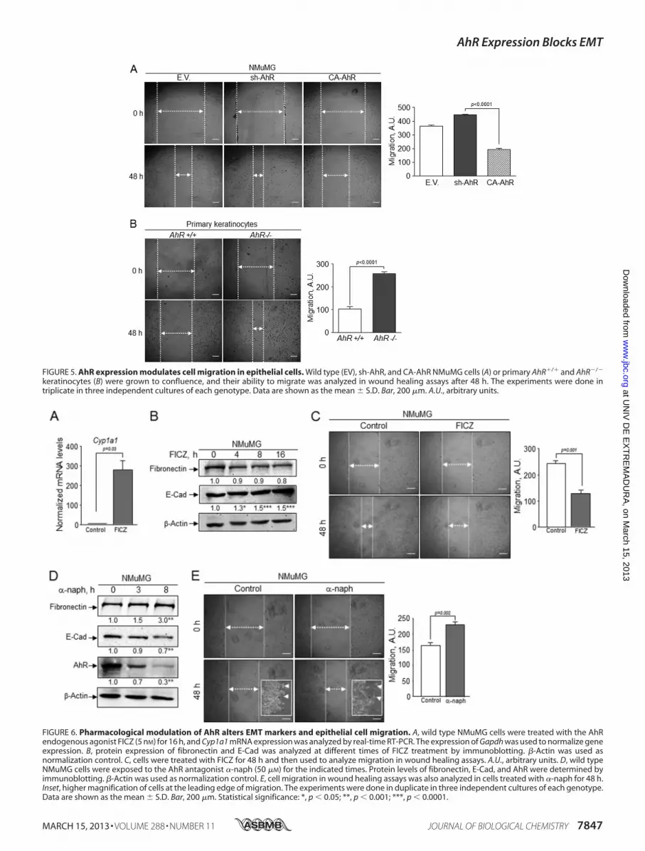
FIGURE 5. AhR expression modulates cell migration in epithelial cells. Wild type (EV), sh-AhR, and CA-AhR NMuMG cells (A) or primary AhR�/� and AhR�/�
keratinocytes (B) were grown to confluence, and their ability to migrate was analyzed in wound healing assays after 48 h. The experiments were done intriplicate in three independent cultures of each genotype. Data are shown as the mean � S.D. Bar, 200 �m. A.U., arbitrary units.
FIGURE 6. Pharmacological modulation of AhR alters EMT markers and epithelial cell migration. A, wild type NMuMG cells were treated with the AhRendogenous agonist FICZ (5 nM) for 16 h, and Cyp1a1 mRNA expression was analyzed by real-time RT-PCR. The expression of Gapdh was used to normalize geneexpression. B, protein expression of fibronectin and E-Cad was analyzed at different times of FICZ treatment by immunoblotting. �-Actin was used asnormalization control. C, cells were treated with FICZ for 48 h and then used to analyze migration in wound healing assays. A.U., arbitrary units. D, wild typeNMuMG cells were exposed to the AhR antagonist �-naph (50 �M) for the indicated times. Protein levels of fibronectin, E-Cad, and AhR were determined byimmunoblotting. �-Actin was used as normalization control. E, cell migration in wound healing assays was also analyzed in cells treated with �-naph for 48 h.Inset, higher magnification of cells at the leading edge of migration. The experiments were done in duplicate in three independent cultures of each genotype.Data are shown as the mean � S.D. Bar, 200 �m. Statistical significance: *, p � 0.05; **, p � 0.001; ***, p � 0.0001.
AhR Expression Blocks EMT
MARCH 15, 2013 • VOLUME 288 • NUMBER 11 JOURNAL OF BIOLOGICAL CHEMISTRY 7847
at UN
IV D
E E
XT
RE
MA
DU
RA
, on March 15, 2013
ww
w.jbc.org
Dow
nloaded from

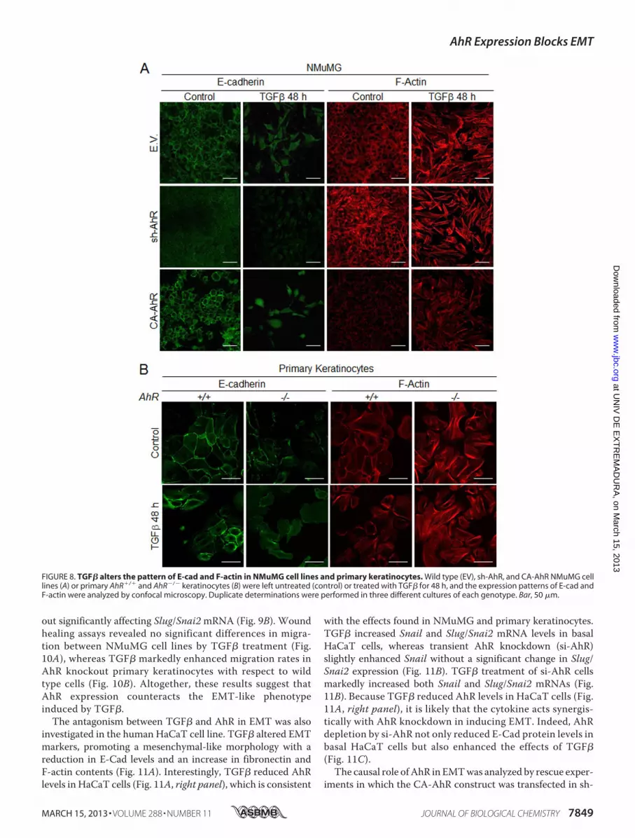
null keratinocytes (Fig. 7,G andH). Similarly to NMuMG cells,TGF� also reduced AhR protein amounts in AhR�/� cells (Fig.7,G and J). Confocal microscopy was used to analyze by immu-nocytochemistry the pattern of EMT regulators in NMuMGcells and in primary keratinocytes exposed to TGF� (Fig. 8).E-Cad staining was reduced and delocalized from the plasmamembrane in sh-AhR NMuMG cells, and such a pattern wasstressed by 48 h treatment with TGF� (Fig. 8A). The welldefinedmembrane location of E-Cadwas disturbed by the cyto-kine in wild type (EV) and CA-AhR cells, although to a lesserdegree than in AhR-depleted cells. F-actin was enhanced byTGF� to a similar extent in each cell line despite its higher basallevels in sh-AhR cells (Fig. 8A). Primary keratinocytes
responded alike to TGF�, delocalizing E-Cad from the plasmamembrane and increasing F-actin fibers content, although asfor NMuMG cells, both effects were more apparent in AhR�/�
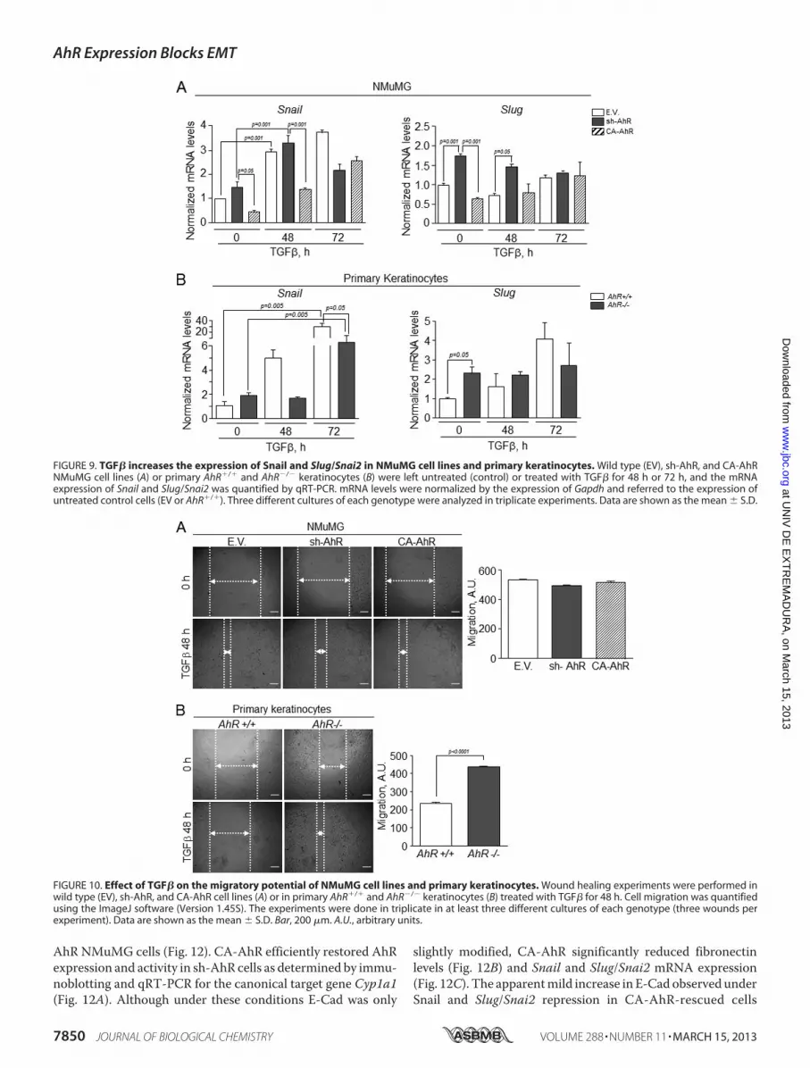
than inAhR�/� cells (Fig. 8B).We subsequently examined howAhR expression modulates the effects of TGF� on Snail andSlug/Snai2. In agreement with the effects seen on E-Cad, TGF�increased Snail mRNA levels in sh-AhR, wild type (EV) andCA-AhR NMuMG cells (Fig. 9A, left panel). Despite its higherbasal levels in sh-AhR cells, Slug/Snai2 was not significantlyinduced by TGF� treatment in either cell line (Fig. 9A, rightpanel). Similar results were also found for primary keratino-cytes, inwhichTGF� increased their SnailmRNA levels (with adelayed kinetics inAhR�/� with respect toAhR�/� cells) with-
FIGURE 7. NMuMG cell lines and primary keratinocytes respond to TGF�-induced EMT. Wild type (EV), sh-AhR, and CA-AhR NMuMG cell lines (A) or primaryAhR�/� and AhR�/� keratinocytes (F) were left untreated (control) or treated with TGF� for 48 h or 72 h, and their morphology was analyzed. Protein expressionfor molecular EMT markers was analyzed in NMuMG cell lines (B) or primary keratinocyte cultures (G) exposed to TGF�. Quantification of E-Cad (D and I),fibronectin (C and H) and AhR (E and J) protein expression in NMuMG cells and primary keratinocytes is shown, respectively. The expression of �-actin was usedto normalize protein levels. Determinations were done in duplicate of two different cultures. Data are shown as the mean � S.D. Bar, 100 �m.
AhR Expression Blocks EMT
7848 JOURNAL OF BIOLOGICAL CHEMISTRY VOLUME 288 • NUMBER 11 • MARCH 15, 2013
at UN
IV D
E E
XT
RE
MA
DU
RA
, on March 15, 2013
ww
w.jbc.org
Dow
nloaded from
out significantly affecting Slug/Snai2mRNA (Fig. 9B). Woundhealing assays revealed no significant differences in migra-tion between NMuMG cell lines by TGF� treatment (Fig.10A), whereas TGF� markedly enhanced migration rates inAhR knockout primary keratinocytes with respect to wildtype cells (Fig. 10B). Altogether, these results suggest thatAhR expression counteracts the EMT-like phenotypeinduced by TGF�.
The antagonism between TGF� and AhR in EMT was alsoinvestigated in the human HaCaT cell line. TGF� altered EMTmarkers, promoting a mesenchymal-like morphology with areduction in E-Cad levels and an increase in fibronectin andF-actin contents (Fig. 11A). Interestingly, TGF� reduced AhRlevels in HaCaT cells (Fig. 11A, right panel), which is consistent
with the effects found in NMuMG and primary keratinocytes.TGF� increased Snail and Slug/Snai2 mRNA levels in basalHaCaT cells, whereas transient AhR knockdown (si-AhR)slightly enhanced Snail without a significant change in Slug/Snai2 expression (Fig. 11B). TGF� treatment of si-AhR cellsmarkedly increased both Snail and Slug/Snai2 mRNAs (Fig.11B). Because TGF� reduced AhR levels in HaCaT cells (Fig.11A, right panel), it is likely that the cytokine acts synergis-tically with AhR knockdown in inducing EMT. Indeed, AhRdepletion by si-AhR not only reduced E-Cad protein levels inbasal HaCaT cells but also enhanced the effects of TGF�(Fig. 11C).The causal role ofAhR in EMTwas analyzed by rescue exper-
iments in which the CA-AhR construct was transfected in sh-
FIGURE 8. TGF� alters the pattern of E-cad and F-actin in NMuMG cell lines and primary keratinocytes. Wild type (EV), sh-AhR, and CA-AhR NMuMG celllines (A) or primary AhR�/� and AhR�/� keratinocytes (B) were left untreated (control) or treated with TGF� for 48 h, and the expression patterns of E-cad andF-actin were analyzed by confocal microscopy. Duplicate determinations were performed in three different cultures of each genotype. Bar, 50 �m.
AhR Expression Blocks EMT
MARCH 15, 2013 • VOLUME 288 • NUMBER 11 JOURNAL OF BIOLOGICAL CHEMISTRY 7849
at UN
IV D
E E
XT
RE
MA
DU
RA
, on March 15, 2013
ww
w.jbc.org
Dow
nloaded from
AhR NMuMG cells (Fig. 12). CA-AhR efficiently restored AhRexpression and activity in sh-AhR cells as determined by immu-noblotting and qRT-PCR for the canonical target gene Cyp1a1(Fig. 12A). Although under these conditions E-Cad was only
slightly modified, CA-AhR significantly reduced fibronectinlevels (Fig. 12B) and Snail and Slug/Snai2 mRNA expression(Fig. 12C). The apparentmild increase in E-Cad observed underSnail and Slug/Snai2 repression in CA-AhR-rescued cells
FIGURE 9. TGF� increases the expression of Snail and Slug/Snai2 in NMuMG cell lines and primary keratinocytes. Wild type (EV), sh-AhR, and CA-AhRNMuMG cell lines (A) or primary AhR�/� and AhR�/� keratinocytes (B) were left untreated (control) or treated with TGF� for 48 h or 72 h, and the mRNAexpression of Snail and Slug/Snai2 was quantified by qRT-PCR. mRNA levels were normalized by the expression of Gapdh and referred to the expression ofuntreated control cells (EV or AhR�/�). Three different cultures of each genotype were analyzed in triplicate experiments. Data are shown as the mean � S.D.
FIGURE 10. Effect of TGF� on the migratory potential of NMuMG cell lines and primary keratinocytes. Wound healing experiments were performed inwild type (EV), sh-AhR, and CA-AhR cell lines (A) or in primary AhR�/� and AhR�/� keratinocytes (B) treated with TGF� for 48 h. Cell migration was quantifiedusing the ImageJ software (Version 1.45S). The experiments were done in triplicate in at least three different cultures of each genotype (three wounds perexperiment). Data are shown as the mean � S.D. Bar, 200 �m. A.U., arbitrary units.
AhR Expression Blocks EMT
7850 JOURNAL OF BIOLOGICAL CHEMISTRY VOLUME 288 • NUMBER 11 • MARCH 15, 2013
at UN
IV D
E E
XT
RE
MA
DU
RA
, on March 15, 2013
ww
w.jbc.org
Dow
nloaded from

could derive from slow accumulation kinetics not fullyaddressed in the 48-h time frame of the experiment. Yet invitrowound healing showed that CA-AhR was able to reducethe migration potential of sh-AhR cells (Fig. 12D). There-fore, AhR re-expression can partially reestablish EMTmark-ers and impair the migration of cells undergoing EMT byAhR down-modulation.
AhR Associates to E-Cad and �-Cat in NMuMG EpithelialCells—Themechanisms bywhichAhR participates in signalingpathways controlling EMT are practically unknown. One pos-sibility is that AhR interacts with the function of moleculesmaintaining cell-cell adhesions such as E-Cad and �-Cat. Co-immunoprecipitation experiments for AhR showed that it wasassociated in a common protein complex with both E-Cad and
FIGURE 11. TGF� induces EMT in HaCaT cells and alters AhR expression. A, human HaCaT cells were left untreated or treated with TGF� for 48 h or 72 h, andthe expression of E-cad and F-actin was determined by confocal microscopy (left panel). Protein expression of E-cad, fibronectin, and AhR was also determinedby immunoblotting using specific antibodies (right panel). B, the effects of TGF�, a siRNA against AhR (si-AhR), or both on Snail and Slug/Snai2 mRNA expressionwere analyzed by qRT-PCR. mRNA levels were normalized by the expression of Gapdh and referred to the expression of control cells (EV). C, the effects of TGF�on the expression of EMT markers in the absence or presence of a si-AhR were also analyzed by immunoblotting. The expression of �-actin was used tonormalize protein levels. Three experiments were performed in at least three independent HaCaT cultures. Data are shown as mean � S.D. Bar, 50 �m.Statistical significance: * p � 0.05, **p � 0.001, ***p � 0.0001.
AhR Expression Blocks EMT
MARCH 15, 2013 • VOLUME 288 • NUMBER 11 JOURNAL OF BIOLOGICAL CHEMISTRY 7851
at UN
IV D
E E
XT
RE
MA
DU
RA
, on March 15, 2013
ww
w.jbc.org
Dow
nloaded from

�-Cat in wild type (EV) and CA-AhR NMuMG cells (Fig. 13A).Consistently, E-Cad was also able to co-immunoprecipitatewith AhR in both cell lines (Fig. 13B). For �-Cat, co-immuno-precipitation with AhR was more evident in CA-AhR than inwild type (EV) cells (Fig. 13C), although the presence of AhR inthe immunoprecipitates was reproducibly observed in the lat-ter. The plausible association of AhR in a common proteincomplex with E-Cad and �-Cat gained additional support byconfocal microscopy analysis. As shown in Fig. 13D, AhRco-localized with both E-Cad and �-Cat at the periphery ofNMuMG cells (yellow color inmerge panels). Thus, one likelymechanism for AhR tomodulate EMT is through interactionwith proteins preserving epithelial integrity and cell-cellinteractions. In addition, the fact that the constitutively
active AhR did not show an increased association to E-Cadsuggests that AhR could modulate additional signaling path-ways unrelated to its intrinsic transcriptional activity.
DISCUSSION
The interest in understanding the physiological functions ofAhR has significantly increased as recent studies have antici-pated an important and perhaps causal role of this receptor inreproduction and organ homeostasis (6). In addition to the con-trol of cell cycle and cell proliferation (28, 41, 42), cell adhesionand migration are recognized as important cellular functionsrequiring AhR activity (43). This assumption is partially basedon in vitro studies using MCF-7 breast tumor cells (44–46),thymocytes (4, 12, 47), and hematopoietic stem cells (48)
FIGURE 12. Rescue of AhR expression in AhR knockdown NMuMG cells reduces the EMT markers and inhibits cell migration. sh-AhR NMuMG cells weretransiently transfected with the CA-AhR construct (A), and the expression of E-cad and fibronectin was analyzed by immunoblotting (B). The expression of�-actin was used to normalize protein levels. C, the effects of CA-AhR on the mRNA levels of Snail and Slug/Snai2 in sh-AhR NMuMG cells were analyzed byqRT-PCR. mRNA levels were normalized by the expression of Gapdh and referred to the expression of control cells (sh-AhR). D, sh-AhR NMuMG cells transfectedwith CA-AhR were treated with TGF� and studied for cell migration using would healing assays. The experiments were done in duplicate in at least threeindependent transfections. Data are shown as the mean � S.D. Bar, 200 �m. Statistical significance: **, p � 0.001. A.U., arbitrary units.
AhR Expression Blocks EMT
7852 JOURNAL OF BIOLOGICAL CHEMISTRY VOLUME 288 • NUMBER 11 • MARCH 15, 2013
at UN
IV D
E E
XT
RE
MA
DU
RA
, on March 15, 2013
ww
w.jbc.org
Dow
nloaded from

treated with the potent exogenous AhR ligand dioxin. AhR isalso involved in cell adhesion and migration under physiologi-cal conditions through cell type-specific mechanisms (6, 49).An important observation is that although the lack of AhRalters the morphology and compromises the adhesion andmigration of mesenchymal fibroblasts (7, 9), its deficiencyenhances keratinocyte motility and epithelial wound healing invivo (10). From these studies it can be proposed that the variationsin AhR levels, which presumably take place during developmentand disease, might contribute to altered cell migration. Closely
related to such a scenario is the epithelial-to-mesenchymal transi-tion that provides epithelial cells with some, but generally not all,mesenchymal characteristics of a migratory cell (13, 15, 50).In this study we have decided to investigate if the cellular
levels of AhR are enough to block EMT and if this receptorantagonizes the effects of TGF�. The latter hypothesis is basedon previous studies revealing a cross-regulation between AhRand TGF� in epithelial cells (10, 32) and mesenchymal fibro-blasts (28, 30, 31).Cell morphology is closely related and, in a large part
dependent, on cell-cell and cell-substratum interactions (51).As a result, changes in cell attachment largely influence cellmigration and can be relevant in processes such as EMT (52).AhR expression affects epithelial cell morphology as receptordepletion in NMuMG cells induced a mesenchymal-like phe-notype thatwasAhR-dependent because it was also observed inprimaryAhR�/� keratinocytes but not inNMuMGcells havinga constitutively active receptor. The mesenchymal-like mor-phology of AhR knockout/knockdown cells apparently reducedthe strength of their cell-cell interactions as revealed by theirlower trans-epithelial resistance when grown at confluence.Based on these results, we considered the possibility that AhRdown-modulation could alter the expression of proteins main-taining epithelial identity. InAhR-interferedNMuMGcells andprimary AhR-null keratinocytes, epithelial markers E-Cad and�-Cat were significantly reduced, whereasmesenchymalmark-ers fibronectin, vimentin, �-smooth muscle actin, and F-actinwere increased, suggesting that AhR expression could impairEMT under normal cellular conditions and in the absence ofexogenous stimuli. Additional sets of data strengthen this con-clusion. First, a constitutively active CA-AhRwas able to rescueepithelialmarkers and to inhibit cellmigration inAhR-depletedcells, showing thatAhRhas a causal role in the EMTphenotype.Second, AhR appears to be associated to and to co-localize withE-Cad and �-Cat in the periphery of NMuMG cells, indicatingthatAhRdeficiency could enhance EMT-dependent cellmigra-tion through altered cell-cell adhesion. In agreement with theseresults, early studies showed that AhR becomes induced in10T1/2 fibroblasts after de-assembling of cell-cell contacts (53).Finally, the increasedmigration ofAhR knockdown cells is con-sistent with the more efficient wound healing found in AhR-null mice due to increased keratinocyte migration (10). Impor-tantly, the fact that AhR inhibition by the antagonist �-naphswitched E-Cad, fibronectin, andmigration to a pattern similarto that of bothAhR�/� and sh-NMuMG cells, strongly supportthat AhR depletion favors an EMT-like phenotype. Moreover,the effects induced by the potential endogenous AhR ligandFICZ not only indicate that AhR expression could impair EMTbut also offer the possibility to block EMT by pharmacologicalmodulation of AhR activity.Interestingly, although AhR knockout/knockdown homoge-
neously affectsmost EMTmarkers, constitutiveAhR activationproduces a more variable phenotype that significantly alterssuchmarkers as E-Cad and fibronectin but that does not signif-icantly influence cell morphology, TER, or �-Cat levels.Althoughmore extensive studies need to be done, it is probablethat AhR functionally interacts with different signaling path-ways requiring increased transcriptional activity of the recep-
FIGURE 13. AhR associates in a common protein complex with E-cad and�-cat in NMuMG cells. A, AhR was immunoprecipitated (IP) in wild type (EV),sh-AhR, and CA-AhR NMuMG cell lines, and the amounts of associated E-cadand �-cat were determined by immunoblotting using specific antibodies.AhR association to E-cad (B) and �-cat (C) was also determined in E-cad and�-cat immunoprecipitates, respectively. Total cell lysates are shown on theright side of panels A, B, and C. D, the pattern of association of AhR to E-cad and�-cat was analyzed by confocal microscopy. Merge for each pair of proteins isshown on the right panel. Determinations were done in triplicate in threeindependent cultures of each genotype. Bar, 30 �m.
AhR Expression Blocks EMT
MARCH 15, 2013 • VOLUME 288 • NUMBER 11 JOURNAL OF BIOLOGICAL CHEMISTRY 7853
at UN
IV D
E E
XT
RE
MA
DU
RA
, on March 15, 2013
ww
w.jbc.org
Dow
nloaded from

tor. Alternatively, wild typeAhR activity could suffice to represscertain EMT markers, thus attenuating the potential effects ofconstitutive AhR activation.It is generally accepted that the initial steps in EMT require
the induction of the E-Cad transcriptional repressors Snailand/or Slug/Snai2 (19–21, 54). The reduced levels of E-Cad inAhR-depleted cells was accompanied by a significant increasein Snail and Slug/Snai2 expression, further supporting that alack of AhR induces an EMT-like phenotype. Indeed, AhRdeficiency increased clonogenicity, spreading and migration ofepithelial cells, demonstrating that AhR is functionally relevantin EMT.The link betweenAhR and Slug/Snai2 appears likely aswe have found that both proteins cooperate to repress theexpression of genes harboring a novel murine SINE B1-X35Sretrotransposon in their promoters (55). In addition, treatmentof rat mammary cells with the tumor promoter DMBA (7,12-dimethylbenz(a)anthracene) induced EMT by activating c-Rel/CK2-dependent signaling and the downstream target proteinsAhR and Slug/Snai2 (56). Although this study suggests thatSlug/Snai2 can be a putative AhR target gene, our data clearlyindicate that steady AhR knockdown significantly increasesSlug/Snai2 expression, represses E-Cad, and promotes cellmigration, in agreement with an EMT process. Interestingly,Slug/Snai2 was similarly induced in sh-AhR NMuMG and pri-maryAhR�/� keratinocytes, whereas Snail induction wasmoresignificant in the latter, suggesting that the two proteins can bedifferentially affected by cell type-specific AhR expression. Insupport of such a possibility, we have found that the AhR-de-pendent B1-X35S retrotransposon has insulator activity whenbound to Slug/Snai2 but not to Snail (37), and an additionalstudy in MDCK epithelial cells has shown that Snail and Slug/Snai2 induce common as well as specific genetic programs dur-ing EMT (22).TGF� addition enhanced EMT features in AhR knockdown
cells (sh-AhR NMuMG and primary keratinocytes) withrespect to either wild type (EV and AhR�/� keratinocytes) orconstitutively active CA-AhR. This includes the repression ofE-Cad and the induction of Snail, Slug/Snai2, fibronectin, andF-actin. Accordingly, TGF� increased themigration of primaryAhR�/� keratinocytes but not of sh-AhR NMuMG cells, per-haps because the addition of exogenous TGF� to this cell lineoverrides the stimulatory effect induced by AhR depletion. Thecross-talk betweenAhR andTGF� in EMTwas also observed inHaCaT cells in which AhR down-modulation potentiated theeffects of TGF� on Snail and Slug/Snai2 expression and onE-Cad repression. It has been shown (57) that short term (e.g.1 h) treatment with the AhR ligand 3-methylcholanthrene ofsi-AhR-interfered HaCaT cells blocked Slug/Snai2 mRNAexpression, a result differing from ours most probably becauseof the use of an exogenous ligand (versus none) and a muchshorter time of treatment (1 versus 48 h). In fact, the increasedresponse to exogenous TGF� exhibited by AhR-depleted cellscould help explain the effects seen under basal cell conditions.Immunoblotting experiments revealed that sh-AhR NMuMGcells and primary AhR�/� keratinocytes had slightly higherTGF� protein content and markedly increased levels of acti-vated pSmad2, suggesting that their basal EMT-like phenotypeis at least partially due to enhancedTGF�-dependent signaling.
This possibility is in agreementwith previous reports indicatingthat AhR negatively regulates TGF� activation under basal cellconditions inmouse fibroblasts (8, 28, 30, 58), hepatocytes (59),and dermal fibroblasts (10). Therefore, TGF�, by reducingAhRlevels, could have synergistic activity in inducingmolecular andphenotypical features of EMT in epithelial cells.In summary, we propose that AhR deficiency in epithelial
cells is enough to trigger morphological and phenotypicalchanges indicative of EMT, likely because of increased TGF�signaling. ExogenousTGF� further intensifies the EMTpheno-type induced by AhR deficiency, probably because of its inhib-itory role on AhR expression. This cross-talk could be relevantto the study of tumors in which AhR activity presumably variesduring metastatic progression of the disease. In this context,several studies suggest that AhR has tumor suppressor activityand that certain human tumors tend to reduce their AHRexpression. For instance, AHR expression was lowered by pro-moter hypermethylation in close to 35% of acute lymphoblasticleukemia patients (60). We have also found using tissuemicroarrays that AhR levels are significantly reduced in a largefraction of human glioblastomas andmelanomas but not in lowgrade astrocytomas or benign nevi,4 again supporting an inhib-itory role for this receptor in certain types of cancers. A recentstudy analyzing a panel of 947 human tumor cell lines identifiedAHR as a predictor of drug sensitivity associated with the effi-cacy of MEK inhibitors in NRAS-mutant lines (61).
Acknowledgments—We are very grateful to Drs. AlbertoMuñoz, XoseR. Bustelo, and Jesús M. Paramio for technical assistance with TERmeasurements, retroviral transduction, and primary keratinocytescultures, respectively. We also thank Drs. Antonio Garcia de Herrerosfor the NMuMG cells and the anti-Snail antibody, Yoshiaki Fujii-Kuriyama for the CA-AhR construct, and Miguel Quintanilla for theHaCaT cell line. The technical help of Eva Barrasa is greatly appre-ciated. Support of the Servicio de Técnicas Aplicadas a las Biocienciasof the Universidad de Extremadura is greatly acknowledged.
REFERENCES1. Massari,M. E., andMurre, C. (2000)Helix-loop-helix proteins. Regulators
of transcription in eucaryotic organisms.Mol. Cell. Biol. 20, 429–4402. Abbott, B. D., Birnbaum, L. S., and Perdew, G. H. (1995) Developmental
expression of two members of a new class of transcription factors. I. Ex-pression of aryl hydrocarbon receptor in the C57BL/6N mouse embryo.Dev. Dyn. 204, 133–143
3. Barouki, R., Coumoul, X., and Fernandez-Salguero, P. M. (2007) The arylhydrocarbon receptor, more than a xenobiotic-interacting protein. FEBSLett. 581, 3608–3615
4. Esser, C., Rannug, A., and Stockinger, B. (2009) The aryl hydrocarbonreceptor in immunity. Trends Immunol. 30, 447–454
5. Furness, S. G., Lees, M. J., and Whitelaw, M. L. (2007) The dioxin (arylhydrocarbon) receptor as a model for adaptive responses of bHLH/PAStranscription factors. FEBS Lett. 581, 3616–3625
6. Pohjanvirta, R. (2012) The AH Receptor in Biology and Toxicology, JohnWiley & Sons, New York
7. Carvajal-Gonzalez, J. M., Mulero-Navarro, S., Roman, A. C., Sauzeau, V.,Merino, J. M., Bustelo, X. R., and Fernandez-Salguero, P. M. (2009) Thedioxin receptor regulates the constitutive expression of the vav3 proto-
4 E. M. Rico-Leo, M. Contador-Troca, I. Catalina-Fernandez, A. Gomez-Duran, J. Saenz-Santamarıa, and P. M. Fernandez-Salguero, unpub-lished observations.
AhR Expression Blocks EMT
7854 JOURNAL OF BIOLOGICAL CHEMISTRY VOLUME 288 • NUMBER 11 • MARCH 15, 2013
at UN
IV D
E E
XT
RE
MA
DU
RA
, on March 15, 2013
ww
w.jbc.org
Dow
nloaded from

oncogene and modulates cell shape and adhesion. Mol. Biol. Cell 20,1715–1727
8. Gomez-Duran, A., Carvajal-Gonzalez, J. M., Mulero-Navarro, S., Santia-go-Josefat, B., Puga, A., and Fernandez-Salguero, P. M. (2009) Fitting axenobiotic receptor into cell homeostasis. How the dioxin receptor inter-acts with TGF� signaling. Biochem. Pharmacol. 77, 700–712
9. Mulero-Navarro, S., Pozo-Guisado, E., Pérez-Mancera, P. A., Alvarez-Barrientos, A., Catalina-Fernández, I., Hernández-Nieto, E., Sáenz-Sant-amaria, J., Martínez, N., Rojas, J. M., Sánchez-García, I., and Fernández-Salguero, P. M. (2005) Immortalized mouse mammary fibroblasts lackingdioxin receptor have impaired tumorigenicity in a subcutaneous mousexenograft model. J. Biol. Chem. 280, 28731–28741
10. Carvajal-Gonzalez, J. M., Roman, A. C., Cerezo-Guisado, M. I., Rico-Leo,E. M., Martin-Partido, G., and Fernandez-Salguero, P. M. (2009) Loss ofdioxin-receptor expression accelerates wound healing in vivo by a mech-anism involving TGF�. J. Cell Sci. 122, 1823–1833
11. Roman, A. C., Carvajal-Gonzalez, J. M., Rico-Leo, E. M., and Fernandez-Salguero, P. M. (2009) Dioxin receptor deficiency impairs angiogenesis bya mechanism involving VEGF-A depletion in the endothelium and trans-forming growth factor-� overexpression in the stroma. J. Biol. Chem. 284,25135–25148
12. Temchura, V. V., Frericks,M.,Nacken,W., and Esser, C. (2005) Role of thearyl hydrocarbon receptor in thymocyte emigration in vivo. Eur. J. Immu-nol. 35, 2738–2747
13. Thiery, J. P., and Sleeman, J. P. (2006) Complex networks orchestrateepithelial-mesenchymal transitions. Nat. Rev. Mol. Cell Biol. 7, 131–142
14. Radisky, D. C. (2005) Epithelial-mesenchymal transition. J. Cell Sci. 118,4325–4326
15. Thiery, J. P., Acloque, H., Huang, R. Y., and Nieto, M. A. (2009) Epithelial-mesenchymal transitions in development and disease. Cell 139, 871–890
16. Prall, F. (2007) Tumour budding in colorectal carcinoma. Histopathology50, 151–162
17. Hajra, K. M., and Fearon, E. R. (2002) Cadherin and catenin alterations inhuman cancer. Genes Chromosomes Cancer 34, 255–268
18. Huber, M. A., Kraut, N., and Beug, H. (2005) Molecular requirements forepithelial-mesenchymal transition during tumor progression.Curr. Opin.Cell Biol. 17, 548–558
19. Batlle, E., Sancho, E., Francí, C., Domínguez, D., Monfar, M., Baulida, J.,and García De Herreros, A. (2000) The transcription factor snail is a re-pressor of E-cadherin gene expression in epithelial tumour cells.Nat. CellBiol. 2, 84–89
20. Cano, A., Pérez-Moreno,M. A., Rodrigo, I., Locascio, A., Blanco,M. J., delBarrio,M. G., Portillo, F., andNieto,M. A. (2000) The transcription factorsnail controls epithelial-mesenchymal transitions by repressing E-cad-herin expression. Nat. Cell Biol. 2, 76–83
21. Bolós, V., Peinado, H., Pérez-Moreno,M. A., Fraga,M. F., Esteller,M., andCano, A. (2003) The transcription factor Slug represses E-cadherin ex-pression and induces epithelial to mesenchymal transitions. A compari-son with Snail and E47 repressors. J. Cell Sci. 116, 499–511
22. Moreno-Bueno, G., Cubillo, E., Sarrió, D., Peinado, H., Rodríguez-Pinilla,S. M., Villa, S., Bolós, V., Jordá, M., Fabra, A., Portillo, F., Palacios, J., andCano, A. (2006) Genetic profiling of epithelial cells expressing E-cadherinrepressors reveals a distinct role for Snail, Slug, and E47 factors in epithe-lial-mesenchymal transition. Cancer Res. 66, 9543–9556
23. Bhowmick, N. A., and Moses, H. L. (2005) Tumor-stroma interactions.Curr. Opin. Genet. Dev. 15, 97–101
24. Fischer, A. N., Herrera, B., Mikula, M., Proell, V., Fuchs, E., Gotzmann, J.,Schulte-Hermann, R., Beug, H., andMikulits,W. (2005) Integration of Rassubeffector signaling in TGF-� mediated late stage hepatocarcinogenesis.Carcinogenesis 26, 931–942
25. Janda, E., Lehmann, K., Killisch, I., Jechlinger, M., Herzig, M., Downward,J., Beug, H., and Grünert, S. (2002) Ras and TGF� cooperatively regulateepithelial cell plasticity and metastasis. Dissection of Ras signaling path-ways. J. Cell Biol. 156, 299–313
26. Barrios-Rodiles,M., Brown, K. R., Ozdamar, B., Bose, R., Liu, Z., Donovan,R. S., Shinjo, F., Liu, Y., Dembowy, J., Taylor, I. W., Luga, V., Przulj, N.,Robinson, M., Suzuki, H., Hayashizaki, Y., Jurisica, I., and Wrana, J. L.(2005) High-throughput mapping of a dynamic signaling network in
mammalian cells. Science 307, 1621–162527. Ozdamar, B., Bose, R., Barrios-Rodiles, M., Wang, H. R., Zhang, Y., and
Wrana, J. L. (2005) Regulation of the polarity protein Par6 by TGF� re-ceptors controls epithelial cell plasticity. Science 307, 1603–1609
28. Chang, X., Fan, Y., Karyala, S., Schwemberger, S., Tomlinson, C. R., Sartor,M.A., and Puga, A. (2007) Ligand-independent regulation of transforminggrowth factor �1 expression and cell cycle progression by the aryl hydro-carbon receptor.Mol. Cell. Biol. 27, 6127–6139
29. Gomez-Duran, A., Ballestar, E., Carvajal-Gonzalez, J. M., Marlowe, J. L.,Puga, A., Esteller, M., and Fernandez-Salguero, P. M. (2008) Recruitmentof CREB1 and histone deacetylase 2 (HDAC2) to the mouse Ltbp-1 pro-moter regulates its constitutive expression in a dioxin receptor-dependentmanner. J. Mol. Biol. 380, 1–16
30. Gomez-Duran, A., Mulero-Navarro, S., Chang, X., and Fernandez-Sal-guero, P. M. (2006) LTBP-1 blockade in dioxin receptor-null mouse em-bryo fibroblasts decreases TGF-� activity. Role of extracellular proteasesplasmin and elastase. J. Cell. Biochem. 97, 380–392
31. Santiago-Josefat, B., Mulero-Navarro, S., Dallas, S. L., and Fernandez-Sal-guero, P. M. (2004) Overexpression of latent transforming growth factor-�-binding protein 1 (LTBP-1) in dioxin receptor-null mouse embryo fi-broblasts. J. Cell Sci. 117, 849–859
32. Döhr, O., Sinning, R., Vogel, C., Münzel, P., and Abel, J. (1997) Effect oftransforming growth factor-� 1 on expression of aryl hydrocarbon recep-tor and genes of Ah gene battery. Clues for independent down-regulationin A549 cells.Mol. Pharmacol. 51, 703–710
33. Wolff, S.,Harper, P.A.,Wong, J.M.,Mostert,V.,Wang,Y., andAbel, J. (2001)Cell-specific regulation of human aryl hydrocarbon receptor expression bytransforming growth factor-� (1).Mol. Pharmacol. 59, 716–724
34. McGuire, J., Okamoto, K., Whitelaw, M. L., Tanaka, H., and Poellinger, L.(2001) Definition of a dioxin receptor mutant that is a constitutive activa-tor of transcription. Delineation of overlapping repression and ligandbinding functions within the PAS domain. J. Biol. Chem. 276,41841–41849
35. Pozo-Guisado, E., Lorenzo-Benayas, M. J., and Fernández-Salguero, P. M.(2004) Resveratrol modulates the phosphoinositide 3-kinase pathwaythrough an estrogen receptor �-dependent mechanism. Relevance in cellproliferation. Int. J. Cancer 109, 167–173
36. Santiago-Josefat, B., and Fernandez-Salguero, P. M. (2003) Proteasomeinhibition induces nuclear translocation of the dioxin receptor through anSp1 and protein kinase C-dependent pathway. J. Mol. Biol. 333, 249–260
37. Román, A. C., González-Rico, F. J., Moltó, E., Hernando, H., Neto, A.,Vicente-Garcia, C., Ballestar, E., Gómez-Skarmeta, J. L., Vavrova-Ander-son, J., White, R. J., Montoliu, L., and Fernández-Salguero, P. M. (2011)Dioxin receptor and SLUG transcription factors regulate the insulatoractivity of B1 SINE retrotransposons via an RNA polymerase switch. Ge-nome Res. 21, 422–432
38. Merchant, M., Krishnan, V., and Safe, S. (1993) Mechanism of action of�-naphthoflavone as an Ah receptor antagonist in MCF-7 human breastcancer cells. Toxicol. Appl. Pharmacol. 120, 179–185
39. Bergander, L., Wahlström, N., Alsberg, T., Bergman, J., Rannug, A., andRannug, U. (2003) Characterization of in vitro metabolites of the arylhydrocarbon receptor ligand 6-formylindolo[3,2-b]carbazole by liquidchromatography-mass spectrometry and NMR. Drug Metab. Dispos. 31,233–241
40. Wincent, E., Amini, N., Luecke, S., Glatt, H., Bergman, J., Crescenzi, C.,Rannug, A., and Rannug, U. (2009) The suggested physiologic aryl hydro-carbon receptor activator and cytochrome P4501 substrate 6-formylin-dolo[3,2-b]carbazole is present in humans. J. Biol. Chem. 284, 2690–2696
41. Marlowe, J. L., and Puga, A. (2005) Aryl hydrocarbon receptor, cell cycleregulation, toxicity, and tumorigenesis. J. Cell. Biochem. 96, 1174–1184
42. Puga, A., Marlowe, J., Barnes, S., Chang, C. Y., Maier, A., Tan, Z., Kerzee,J. K., Chang, X., Strobeck, M., and Knudsen, E. S. (2002) Role of the arylhydrocarbon receptor in cell cycle regulation. Toxicology 181, 171–177
43. Fernandez-Salguero, P. M. (2010) A remarkable new target gene for thedioxin receptor. THE Vav3 proto-oncogene links AhR to adhesion andmigration. Cell Adh. Migr. 4, 172–175
44. Bui, L. C., Tomkiewicz, C., Chevallier, A., Pierre, S., Bats, A. S., Mota, S.,Raingeaud, J., Pierre, J., Diry, M., Transy, C., Garlatti, M., Barouki, R., and
AhR Expression Blocks EMT
MARCH 15, 2013 • VOLUME 288 • NUMBER 11 JOURNAL OF BIOLOGICAL CHEMISTRY 7855
at UN
IV D
E E
XT
RE
MA
DU
RA
, on March 15, 2013
ww
w.jbc.org
Dow
nloaded from

Coumoul, X. (2009) Nedd9/Hef1/Cas-L mediates the effects of environ-mental pollutants on cell migration and plasticity. Oncogene 28,3642–3651
45. Diry, M., Tomkiewicz, C., Koehle, C., Coumoul, X., Bock, K. W., Barouki,R., andTransy, C. (2006) Activation of the dioxin/aryl hydrocarbon recep-tor (AhR)modulates cell plasticity through a JNK-dependentmechanism.Oncogene 25, 5570–5574
46. Seifert, A., Rau, S., Küllertz, G., Fischer, B., and Santos, A. N. (2009) TCDDinduces cell migration via NFATc1/ATX-signaling in MCF-7 cells. Toxi-col. Lett. 184, 26–32
47. Frericks, M., Burgoon, L. D., Zacharewski, T. R., and Esser, C. (2008)Promoter analysis of TCDD-inducible genes in a thymic epithelial cell lineindicates the potential for cell-specific transcription factor crosstalk in theAhR response. Toxicol. Appl. Pharmacol. 232, 268–279
48. Casado, F. L., Singh, K. P., and Gasiewicz, T. A. (2011) Aryl hydrocarbonreceptor activation in hematopoietic stem/progenitor cells alters cellfunction and pathway-specific gene modulation reflecting changes in cel-lular trafficking and migration.Mol. Pharmacol. 80, 673–682
49. Roman, A. C., Carvajal-Gonzalez, J. M., Mulero-Navarro, S., Gomez-Du-ran, A., Rico-Leo, E., Merino, J. M., and Fernandez-Salguero, P. M. (2012)in The AH receptor in Biology and Toxicology (Pohjanvirta, R., ed.) pp.485–497, John Wiley & Sons, New York
50. Peinado,H.,Olmeda,D., andCano,A. (2007) Snail, Zeb, and bHLH factorsin tumour progression. An alliance against the epithelial phenotype?Nat.Rev. Cancer 7, 415–428
51. Mitra, S. K., Hanson, D. A., and Schlaepfer, D. D. (2005) Focal adhesionkinase. In command and control of cellmotility.Nat. Rev.Mol. Cell Biol. 6,56–68
52. Li, S., Guan, J. L., and Chien, S. (2005) Biochemistry and biomechanics ofcell motility. Annu. Rev. Biomed. Eng. 7, 105–150
53. Cho, Y. C., Zheng, W., and Jefcoate, C. R. (2004) Disruption of cell-cellcontact maximally but transiently activates AhR-mediated transcriptionin 10T1/2 fibroblasts. Toxicol. Appl. Pharmacol. 199, 220–238
54. Barrallo-Gimeno, A., and Nieto, M. A. (2005) The Snail genes as inducersof cell movement and survival. Implications in development and cancer.Development 132, 3151–3161
55. Roman, A. C., Benitez, D. A., Carvajal-Gonzalez, J. M., and Fernandez-
Salguero, P. M. (2008) Genome-wide B1 retrotransposon binds the tran-scription factors dioxin receptor and Slug and regulates gene expression invivo. Proc. Natl. Acad. Sci. U.S.A. 105, 1632–1637
56. Belguise, K., Guo, S., Yang, S., Rogers, A. E., Seldin, D. C., Sherr, D. H., andSonenshein, G. E. (2007) Green tea polyphenols reverse cooperation be-tween c-Rel andCK2 that induces the aryl hydrocarbon receptor, slug, andan invasive phenotype. Cancer Res. 67, 11742–11750
57. Ikuta, T., and Kawajiri, K. (2006) Zinc finger transcription factor Slug is anovel target gene of aryl hydrocarbon receptor. Exp. Cell Res. 312,3585–3594
58. Elizondo, G., Fernandez-Salguero, P., Sheikh, M. S., Kim, G. Y., Fornace,A. J., Lee, K. S., and Gonzalez, F. J. (2000) Altered cell cycle control at theG2/M phases in aryl hydrocarbon receptor-null embryo fibroblast. Mol.Pharmacol. 57, 1056–1063
59. Zaher, H., Fernandez-Salguero, P. M., Letterio, J., Sheikh, M. S., Fornace,A. J., Jr., Roberts, A. B., and Gonzalez, F. J. (1998) The involvement of arylhydrocarbon receptor in the activation of transforming growth factor-�and apoptosis.Mol. Pharmacol. 54, 313–321
60. Mulero-Navarro, S., Carvajal-Gonzalez, J. M., Herranz, M., Ballestar, E.,Fraga,M. F., Ropero, S., Esteller,M., and Fernandez-Salguero, P.M. (2006)The dioxin receptor is silenced by promoter hypermethylation in humanacute lymphoblastic leukemia through inhibition of Sp1 binding.Carcino-genesis 27, 1099–1104
61. Barretina, J., Caponigro, G., Stransky, N., Venkatesan, K., Margolin, A. A.,Kim, S., Wilson, C. J., Lehár, J., Kryukov, G. V., Sonkin, D., Reddy, A., Liu,M., Murray, L., Berger, M. F., Monahan, J. E., Morais, P., Meltzer, J., Ko-rejwa, A., Jané-Valbuena, J., Mapa, F. A., Thibault, J., Bric-Furlong, E.,Raman, P., Shipway, A., Engels, I. H., Cheng, J., Yu, G. K., Yu, J., Aspesi, P.,Jr., de Silva, M., Jagtap, K., Jones, M. D., Wang, L., Hatton, C., Palescan-dolo, E., Gupta, S., Mahan, S., Sougnez, C., Onofrio, R. C., Liefeld, T.,MacConaill, L., Winckler, W., Reich, M., Li, N., Mesirov, J. P., Gabriel,S. B., Getz, G., Ardlie, K., Chan, V., Myer, V. E., Weber, B. L., Porter, J.,Warmuth, M., Finan, P., Harris, J. L., Meyerson, M., Golub, T. R., Morris-sey, M. P., Sellers, W. R., Schlegel, R., and Garraway, L. A. (2012) TheCancer Cell Line Encyclopedia enables predictive modelling of anticancerdrug sensitivity. Nature 483, 603–607
AhR Expression Blocks EMT
7856 JOURNAL OF BIOLOGICAL CHEMISTRY VOLUME 288 • NUMBER 11 • MARCH 15, 2013
at UN
IV D
E E
XT
RE
MA
DU
RA
, on March 15, 2013
ww
w.jbc.org
Dow
nloaded from
Copyright © 2022 FDOKUMEN