Basal sauropodomorphs from the Ischigualasto Formation
20
This article was downloaded by: [Cecilia Apaldetti] On: 10 October 2013, At: 07:24 Publisher: Taylor & Francis Informa Ltd Registered in England and Wales Registered Number: 1072954 Registered office: Mortimer House, 37-41 Mortimer Street, London W1T 3JH, UK Journal of Vertebrate Paleontology Publication details, including instructions for authors and subscription information: http://www.tandfonline.com/loi/ujvp20 Basal sauropodomorphs from the Ischigualasto Formation Ricardo N. MartÍnez a , Cecilia Apaldetti a b & Diego Abelin a a Instituto y Museo de Ciencias Naturales , Universidad Nacional de San Juan , 5400 , San Juan , Argentina b Consejo Nacional de Investigaciones Científicas y Técnicas , Buenos Aires , Argentina Published online: 08 Oct 2012. To cite this article: Ricardo N. MartÍnez , Cecilia Apaldetti & Diego Abelin (2012) Basal sauropodomorphs from the Ischigualasto Formation, Journal of Vertebrate Paleontology, 32:sup1, 51-69, DOI: 10.1080/02724634.2013.819361 To link to this article: http://dx.doi.org/10.1080/02724634.2013.819361 PLEASE SCROLL DOWN FOR ARTICLE Taylor & Francis makes every effort to ensure the accuracy of all the information (the “Content”) contained in the publications on our platform. However, Taylor & Francis, our agents, and our licensors make no representations or warranties whatsoever as to the accuracy, completeness, or suitability for any purpose of the Content. Any opinions and views expressed in this publication are the opinions and views of the authors, and are not the views of or endorsed by Taylor & Francis. The accuracy of the Content should not be relied upon and should be independently verified with primary sources of information. Taylor and Francis shall not be liable for any losses, actions, claims, proceedings, demands, costs, expenses, damages, and other liabilities whatsoever or howsoever caused arising directly or indirectly in connection with, in relation to or arising out of the use of the Content. This article may be used for research, teaching, and private study purposes. Any substantial or systematic reproduction, redistribution, reselling, loan, sub-licensing, systematic supply, or distribution in any form to anyone is expressly forbidden. Terms & Conditions of access and use can be found at http:// www.tandfonline.com/page/terms-and-conditions
Transcript of Basal sauropodomorphs from the Ischigualasto Formation
This article was downloaded by: [Cecilia Apaldetti]On: 10 October 2013, At: 07:24Publisher: Taylor & FrancisInforma Ltd Registered in England and Wales Registered Number: 1072954 Registered office: Mortimer House,37-41 Mortimer Street, London W1T 3JH, UK
Journal of Vertebrate PaleontologyPublication details, including instructions for authors and subscription information:http://www.tandfonline.com/loi/ujvp20
Basal sauropodomorphs from the IschigualastoFormationRicardo N. MartÍnez a , Cecilia Apaldetti a b & Diego Abelin aa Instituto y Museo de Ciencias Naturales , Universidad Nacional de San Juan , 5400 , SanJuan , Argentinab Consejo Nacional de Investigaciones Científicas y Técnicas , Buenos Aires , ArgentinaPublished online: 08 Oct 2012.
To cite this article: Ricardo N. MartÍnez , Cecilia Apaldetti & Diego Abelin (2012) Basal sauropodomorphs from theIschigualasto Formation, Journal of Vertebrate Paleontology, 32:sup1, 51-69, DOI: 10.1080/02724634.2013.819361
To link to this article: http://dx.doi.org/10.1080/02724634.2013.819361
PLEASE SCROLL DOWN FOR ARTICLE
Taylor & Francis makes every effort to ensure the accuracy of all the information (the “Content”) containedin the publications on our platform. However, Taylor & Francis, our agents, and our licensors make norepresentations or warranties whatsoever as to the accuracy, completeness, or suitability for any purpose of theContent. Any opinions and views expressed in this publication are the opinions and views of the authors, andare not the views of or endorsed by Taylor & Francis. The accuracy of the Content should not be relied upon andshould be independently verified with primary sources of information. Taylor and Francis shall not be liable forany losses, actions, claims, proceedings, demands, costs, expenses, damages, and other liabilities whatsoeveror howsoever caused arising directly or indirectly in connection with, in relation to or arising out of the use ofthe Content.
This article may be used for research, teaching, and private study purposes. Any substantial or systematicreproduction, redistribution, reselling, loan, sub-licensing, systematic supply, or distribution in anyform to anyone is expressly forbidden. Terms & Conditions of access and use can be found at http://www.tandfonline.com/page/terms-and-conditions
Society of Vertebrate Paleontology Memoir 12Journal of Vertebrate PaleontologyVolume 32, Supplement to Number 6: 51–69© 2013 by the Society of Vertebrate Paleontology
BASAL SAUROPODOMORPHS FROM THE ISCHIGUALASTO FORMATION
RICARDO N. MARTINEZ,*,1 CECILIA APALDETTI,1,2 and DIEGO ABELIN1
1Instituto y Museo de Ciencias Naturales, Universidad Nacional de San Juan, 5400 San Juan, Argentina, [email protected];[email protected];
2Consejo Nacional de Investigaciones Cientıficas y Tecnicas, Buenos Aires, Argentina, [email protected]
ABSTRACT—Basal sauropodomorphs from the Ischigualasto Formation include Eoraptor lunensis, Panphagia protos, andChromogisaurus novasi. Few comparisons have been made between these taxa, because Eoraptor was only recently reassessedas a basal sauropodomorph and because Panphagia and Chromogisaurus were described nearly simultaneously. We describe indetail the fully prepared bones of the holotype of Chromogisaurus novasi, examine the evidence for its taxonomic distinction,and analyze the phylogenetic relationships among basal sauropodomorphs. Our results support Chromogisaurus novasi as avalid genus and species and provide weak phylogenetic evidence favoring a series of stem taxa at the base of Sauropodomorpha.The analysis positions Panphagia as the basal-most sauropodomorph, followed by Eoraptor, Pampadromaeus, and a clade thatincludes Chromogisaurus and Saturnalia.
RESUMEN—Los sauropodomorfos basales de la Formacion Ischigualasto incluyen Eoraptor lunensis, Panphagia protos yChromogisaurus novasi. Pocas comparaciones se han hecho entre estos taxones, porque Eoraptor fue reevaluado reciente-mente como sauropodomorfo basal y porque Panphagia y Chromogisaurus se describieron de forma casi simultanea. De-scribimos en detalle los huesos totalmente preparados del holotipo de Chromogisaurus novasi, examinamos la evidencia desu distincion taxonomica, y analizamos la relacion filogenetica entre sauropodomorfos basales. Nuestros resultados apoyan aChromogisaurus novasi como un genero y especie valido y proporcionan debil soporte filogenetico favoreciendo un arregloparafiletico en la base de Sauropodomorpha. El analisis posiciona a Panphagia como el sauropodomorfo mas basal, seguido deEoraptor, Pampadromaeus y un clado que incluye a Chromogisaurus y Saturnalia.
SUPPLEMENTAL DATA—Supplemental materials are available for this article for free at www.tandfonline.com/UJVP
INTRODUCTION
Basal sauropodomorphs are represented in two of the oldestdinosaur-bearing geologic units, namely the Ischigualasto Forma-tion of Argentina (Ezcurra et al., 2008; Ezcurra, 2010; Martınezand Alcober, 2009; Martınez et al., 2011) and the Santa MariaFormation of Brazil (Langer et al., 1999; Cabreira et al., 2011).For many years dinosaurs were thought to comprise a relativelysmall proportion of the archosaurs in the Ischigualasto fauna, in-cluding only the ornithischian Pisanosaurus (Casamiquela, 1967)and the theropods Herrerasaurus (Reig, 1963; Sereno and Novas,1992) and Eoraptor (Sereno et al., 1993). Sauropodomorphs hadyet to be recorded.
In recent years, however, four new dinosaurs have been de-scribed, including the basal sauropodomorphs Panphagia pro-tos (Martınez and Alcober, 2009) and Chromogisaurus novasi(Ezcurra, 2010), the herrerasaurid theropod Sanjuansaurus gordil-loi (Alcober and Martınez, 2010), and the basal theropod Eo-dromaeus murphi (Martınez et al., 2011). All of these dinosaurswere recovered from the Scaphonyx-Exaeretodon-Herrerasaurusbiozone, which is dated to approximately 231 Ma (Rogers et al.,1993; Martınez et al., 2011).
The similar morphology and near-synchronous, independentdescription of the two aforementioned sauropodomorphs, Pan-phagia (Martınez and Alcober, 2009) and Chromogisaurus(Ezcurra, 2010), leave their validity an open question. Priorto a detailed comparison, we completely prepared the holo-type and only known specimen of Chromogisaurus. The recentreinterpretation of Eoraptor as a basal sauropodomorph closelyrelated to Panphagia (Martınez et al., 2011) underscored theneed to effectively compare these three contemporaneous basalsauropodomorphs. Although missing data are a problem for all
*Corresponding author.
taxa considered except Eoraptor and Saturnalia, we analyzed thephylogenetic relationships among known basal sauropodomorphs.
METHODS
Comparative Specimens
Comparisons were made using the holotypic specimens ofChromogisaurus (PVSJ 845), Panphagia (PVSJ 874), and San-juansaurus (PVSJ 605), holotypic and referred specimens of Eo-dromaeus (PVSJ 534, 561, 562), Eoraptor (PVSJ 512, 559), Sat-urnalia (MCP 3844-PV, 3845-PV, 3846-PV), and Guaibasaurus(MCN PV 2355, 2356, UFRGS PV 0725T), and referred speci-mens of Herrerasaurus (PVSJ 373, 380, 407). Other comparisonswere based on published figures.
Photography
Some bone surfaces of Chromogisaurus (PVSJ 845) werepainted with neutral gray acrylic paint to remove color distractionsprior photography.
Anatomical and Taxonomic Terminology
We employ traditional, or ‘Romerian,’ anatomical and direc-tional terms over veterinarian alternatives (Wilson, 2006). ‘An-terior’ and ‘posterior,’ for example, are used as directional termsrather than the veterinarian alternatives ‘rostral’ or ‘cranial’ and‘caudal.’ We also follow recent recommendations regarding theidentification of vertebral laminae (Wilson, 1999).
We used the phylogenetic definitions for basal taxawithin Dinosauria proposed by Sereno (2005a, 2005b, 2007).Sauropodomorpha, for example, has a stem-based definition inopposition to Theropoda and does not require the monophyly ofSaurischia or Prosauropoda, as do other definitions (e.g., Galtonand Upchurch, 2004). In this way, Sauropodomorpha is defined as
51
Dow
nloa
ded
by [
Cec
ilia
Apa
ldet
ti] a
t 07:
24 1
0 O
ctob
er 2
013
52 SOCIETY OF VERTEBRATE PALEONTOLOGY, MEMOIR 12
the most inclusive clade containing Saltasaurus loricatus but notPasser domesticus or Triceratops horridus.
Institutional Abbreviations—MCN, Museu de Ciencias Natu-rais, Rio Grande do Sul, Brazil; MCP, Museo de Ciencias e Tec-nologıa, Porto Alegre, Brazil; PVSJ, Instituto y Museo de Cien-cias Naturales, Universidad Nacional de San Juan, San Juan,Argentina; UFRGS, Universidade Federal de Rio Grande do Sul,Porto Alegre, Brazil.
SYSTEMATIC PALEONTOLOGY
DINOSAURIA Owen, 1842SAURISCHIA Seeley, 1887
SAUROPODOMORPHA Huene, 1932CHROMOGISAURUS Ezcurra, 2010
CHROMOGISAURUS NOVASI Ezcurra, 2010(Figs. 4–8, 9A–E, 10–13, 14A–E, 15)
Type Specimen—PVSJ 845, partial skeleton comprising one an-terior and two mid-caudal vertebrae, anterior chevron, glenoid re-gion of the left scapulocoracoid, right ilium lacking preacetabu-lar process, acetabular portion of the left ilium, shaft of the rightfemur and proximal and distal ends of the left femur, completeright tibia and proximal end of the left tibia, right fibula lack-ing proximal and distal ends and proximal end of the left fibula,right metatarsal 2, right digit III phalanges, and unidentified bonefragments.
Type Locality—Cancha de Bochas, Hollada de Ischigualasto,Ischigualasto Provincial Park, San Juan Province, Argentina.
Age and Distribution—The horizon is located at the southernoutcrops of the Ischigualasto Formation, 40 m above the base ofthe unit in the Cancha de Bochas Member (sensu Currie et al.,2009). The holotypic location belongs to the lower Scaphonyx-Exaeretodon-Herrerasaurus biozone (Martınez et al., 2011). Themid-Carnian age is based on a radioisotopic date near the base ofthe formation (Rogers et al., 1993) that was recently recalibratedto 231.4 Ma (Martınez et al., 2011).
Revised Diagnosis—Basal sauropodomorph diagnosed by thefollowing combination of features (asterisk indicates an autapo-morphy): marked posterior projection of the iliac postacetabu-lar process; incipient perforation of the iliac portion of the ac-etabulum; low lateral projection of the iliac supraacetabular crest;strongly concave acetabular surface of the supraacetabular crest;femoral lateral condyle smaller than the medial condyle; medialsurface of the proximal end of the fibula with an elongate rugos-ity adjacent to the anterior margin of the shaft∗; metatarsal 2 withstrongly dorsoventrally asymmetric distal condyles∗.
DESCRIPTION
We offer alternative identifications for several elements de-scribed by Ezcurra (2010), based on comparisons with other basalsaurischians from the Ischigualasto Formation, namely, Eoraptor(Sereno et al., 1993), Panphagia (Martınez and Alcober, 2009),and Eodromaeus (Martınez et al., 2011).
Reidentification of Ulna, Ischium, and Metatarsal
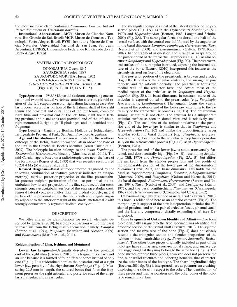
Lower Jaw Fragment—Originally described as the proximalend of the right ulna (Ezcurra, 2010), this fragment is clearly notan ulna because it is formed of four different bones instead of onlyone (Fig. 1). It is reidentified here as the posterior end of a rightlower jaw, possibly of the rhynchosaur Scaphonyx (Fig. 1). Mea-suring 29.5 mm in length, the sutured bones that form the frag-ment preserves the right articular and posterior ends of the angu-lar, surangular, and prearticular.
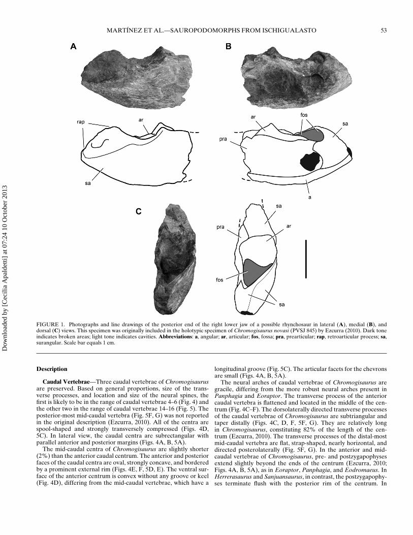
The surangular comprises most of the lateral surface of the pre-served piece (Fig. 1A), as in the rhynchosaurs Scaphonyx (Sill,1970) and Hyperodapedon (Benton, 1983; Langer and Schultz,2000) (Fig. 2A). The surangular forms the dorsal one-half of thelateral surface, with the ventral one-half formed by the angular, asin the basal dinosaurs Eoraptor, Panphagia, Herrerasaurus, Tawa(Nesbitt et al., 2009), and Lesothosaurus (Galton, 1978; Knoll,2002). In the fragment in question, the surangular wraps aroundthe posterior end of the retroarticular process (Fig. 1C), as also oc-curs in Scaphonyx and Hyperodapedon (Fig. 2C). The posteroven-tral surface of the surangular is eroded, exposing the internal tex-ture of the bone. Ezcurra (2010) interpreted this feature as thestrongly striated surface of the olecranon.
The posterior portion of the prearticular is broken and eroded(Fig. 1B). It contacts the angular ventrally, the surangular pos-teriorly, and the articular dorsally. The prearticular forms themedial wall of the adductor fossa and covers most of themedial aspect of the articular, as in Scaphonyx and Hypero-dapedon (Fig. 2B). In basal dinosaurs, in contrast, more of thearticular is exposed dorsal to the prearticular (e.g., Panphagia,Herrerasaurus, Lesothosaurus). The angular forms the ventralmargin of the posterior end of the lower jaw, extending to the ex-tremity of the retroarticular process (Fig. 1B). The prearticular-surangular suture is not clear. The articular has a subquadratearticular surface as seen in dorsal view and is relatively small(Fig. 1C). The small size of the articular relative to the poste-rior end of the lower jaw is similar to that in Scaphonyx andHyperodapedon (Fig. 2C) and unlike the proportionately largerarticular socket in basal dinosaurs (e.g., Panphagia, Eoraptor,Herrerasaurus). Posterior to the articular, a deep fossa opens dor-sally on the retroarticular process (Fig. 1C), as in Hyperodapedon(Benton, 1983).
The posterior end of the lower jaw is stout, transversely flat-tened, and dorsoventrally high (Fig. 1A, B), resembling Scapho-nyx (Sill, 1970) and Hyperodapedon (Fig. 2A, B), but differ-ing markedly from the slender proportions and low profile ofthe comparable portion of the lower jaw of the dinosauriformSilesaurus (Dzik, 2003) and basal dinosaurs. This holds for thebasal sauropodomorphs Panphagia, Eoraptor, Adeopapposaurus(Martınez, 2009), and Pantydraco (Galton and Kermack, 2011),the basal theropods Eodromaeus, Herrerasaurus (Sereno and No-vas, 1994), Tawa (Nesbitt et al., 2009), and Coelophysis (Raath,1977), and the basal ornithischians Pisanosaurus (Casamiquela,1967) and Heterodontosaurus (Crompton and Charig, 1962).
Chevron—Originally described as metatarsal 5 (Ezcurra, 2010),this bone is reidentified here as an anterior chevron (Fig. 6). Themorphology in support of the new interpretation includes the ‘Y’-shaped proximal end with a pair of articular facets, a hemal canal,and the laterally compressed, distally expanding shaft (see De-scription).
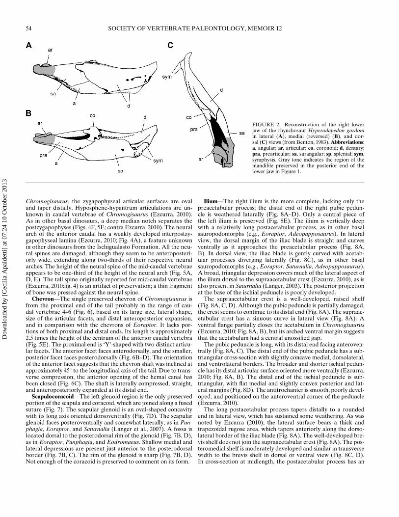
Bone Fragments of Unknown Identity and Affinity—One bonepiece originally assigned to the type specimen was identified as aprobable section of the ischial shaft (Ezcurra, 2010). The squaredsection and massive size of the bone (Fig. 3) does not closelyresemble the triangular section and slender proportions of theischium in basal saurischians (e.g., Eoraptor, Saturnalia, Eodro-maeus). Two other bone pieces originally included as part of theholotype have similar size, cross-sectional shape, and surface de-tail, suggesting that they may belong to the same bone (Fig. 3). Thebone surface of these three pieces, however, does not preserve thefine, subparallel fractures and adhering hematite that character-ize the other bones of the holotype. The sharp longitudinal ridge(Ezcurra, 2010:fig. 7B) is interpreted here as postmortem crushing,displacing one side with respect to the other. The identification ofthese pieces and their association with the other bones of the holo-type remain uncertain.
Dow
nloa
ded
by [
Cec
ilia
Apa
ldet
ti] a
t 07:
24 1
0 O
ctob
er 2
013
MARTINEZ ET AL.—SAUROPODOMORPHS FROM ISCHIGUALASTO 53
FIGURE 1. Photographs and line drawings of the posterior end of the right lower jaw of a possible rhynchosaur in lateral (A), medial (B), anddorsal (C) views. This specimen was originally included in the holotypic specimen of Chromogisaurus novasi (PVSJ 845) by Ezcurra (2010). Dark toneindicates broken areas; light tone indicates cavities. Abbreviations: a, angular; ar, articular; fos, fossa; pra, prearticular; rap, retroarticular process; sa,surangular. Scale bar equals 1 cm.
Description
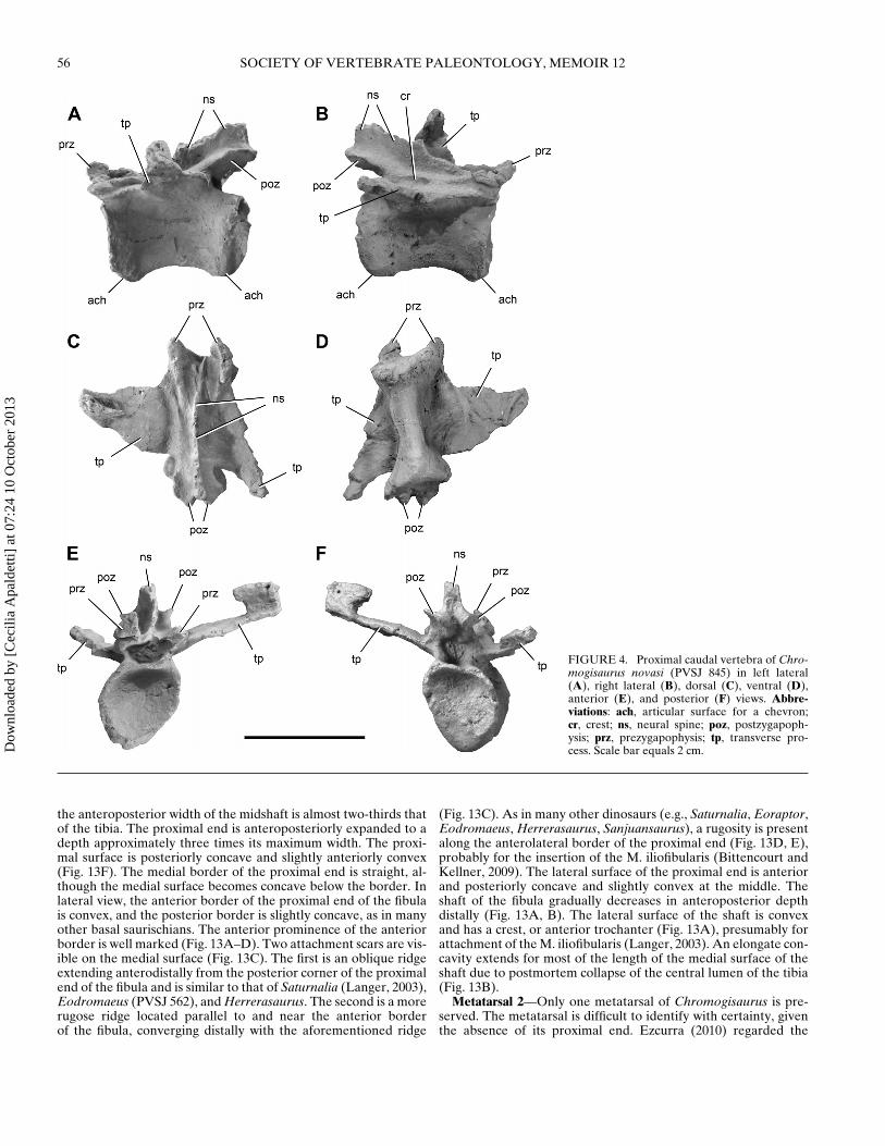
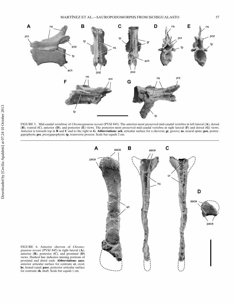
Caudal Vertebrae—Three caudal vertebrae of Chromogisaurusare preserved. Based on general proportions, size of the trans-verse processes, and location and size of the neural spines, thefirst is likely to be in the range of caudal vertebrae 4–6 (Fig. 4) andthe other two in the range of caudal vertebrae 14–16 (Fig. 5). Theposterior-most mid-caudal vertebra (Fig. 5F, G) was not reportedin the original description (Ezcurra, 2010). All of the centra arespool-shaped and strongly transversely compressed (Figs. 4D,5C). In lateral view, the caudal centra are subrectangular withparallel anterior and posterior margins (Figs. 4A, B, 5A).
The mid-caudal centra of Chromogisaurus are slightly shorter(2%) than the anterior caudal centrum. The anterior and posteriorfaces of the caudal centra are oval, strongly concave, and borderedby a prominent external rim (Figs. 4E, F, 5D, E). The ventral sur-face of the anterior centrum is convex without any groove or keel(Fig. 4D), differing from the mid-caudal vertebrae, which have a
longitudinal groove (Fig. 5C). The articular facets for the chevronsare small (Figs. 4A, B, 5A).
The neural arches of caudal vertebrae of Chromogisaurus aregracile, differing from the more robust neural arches present inPanphagia and Eoraptor. The transverse process of the anteriorcaudal vertebra is flattened and located in the middle of the cen-trum (Fig. 4C–F). The dorsolaterally directed transverse processesof the caudal vertebrae of Chromogisaurus are subtriangular andtaper distally (Figs. 4C, D, F, 5F, G). They are relatively longin Chromogisaurus, constituting 82% of the length of the cen-trum (Ezcurra, 2010). The transverse processes of the distal-mostmid-caudal vertebra are flat, strap-shaped, nearly horizontal, anddirected posterolaterally (Fig. 5F, G). In the anterior and mid-caudal vertebrae of Chromogisaurus, pre- and postzygapophysesextend slightly beyond the ends of the centrum (Ezcurra, 2010;Figs. 4A, B, 5A), as in Eoraptor, Panphagia, and Eodromaeus. InHerrerasaurus and Sanjuansaurus, in contrast, the postzygapophy-ses terminate flush with the posterior rim of the centrum. In
Dow
nloa
ded
by [
Cec
ilia
Apa
ldet
ti] a
t 07:
24 1
0 O
ctob
er 2
013
54 SOCIETY OF VERTEBRATE PALEONTOLOGY, MEMOIR 12
FIGURE 2. Reconstruction of the right lowerjaw of the rhynchosaur Hyperodapedon gordoniin lateral (A), medial (reversed) (B), and dor-sal (C) views (from Benton, 1983). Abbreviations:a, angular; ar, articular; co, coronoid; d, dentary;pra, prearticular; sa, surangular; sp, splenial; sym,symphysis. Gray tone indicates the region of themandible preserved in the posterior end of thelower jaw in Figure 1.
Chromogisaurus, the zygapophyseal articular surfaces are ovaland taper distally. Hyposphene-hypantrum articulations are un-known in caudal vertebrae of Chromogisaurus (Ezcurra, 2010).As in other basal dinosaurs, a deep median notch separates thepostzygapophyses (Figs. 4F, 5E; contra Ezcurra, 2010). The neuralarch of the anterior caudal has a weakly developed interpostzy-gapophyseal lamina (Ezcurra, 2010; Fig. 4A), a feature unknownin other dinosaurs from the Ischigualasto Formation. All the neu-ral spines are damaged, although they seem to be anteroposteri-orly wide, extending along two-thirds of their respective neuralarches. The height of the neural spine of the mid-caudal vertebraeappears to be one-third of the height of the neural arch (Fig. 5A,D, E). The tall spine originally reported for mid-caudal vertebrae(Ezcurra, 2010:fig. 4) is an artifact of preservation; a thin fragmentof bone was pressed against the neural spine.
Chevron—The single preserved chevron of Chromogisaurus isfrom the proximal end of the tail probably in the range of cau-dal vertebrae 4–6 (Fig. 6), based on its large size, lateral shape,size of the articular facets, and distal anteroposterior expansion,and in comparison with the chevrons of Eoraptor. It lacks por-tions of both proximal and distal ends. Its length is approximately2.5 times the height of the centrum of the anterior caudal vertebra(Fig. 5E). The proximal end is ‘Y’-shaped with two distinct articu-lar facets. The anterior facet faces anterodorsally, and the smaller,posterior facet faces posterodorsally (Fig. 6B–D). The orientationof the anterior facet suggests that the chevron shaft was inclined atapproximately 45◦ to the longitudinal axis of the tail. Due to trans-verse compression, the anterior opening of the hemal canal hasbeen closed (Fig. 6C). The shaft is laterally compressed, straight,and anteroposteriorly expanded at its distal end.
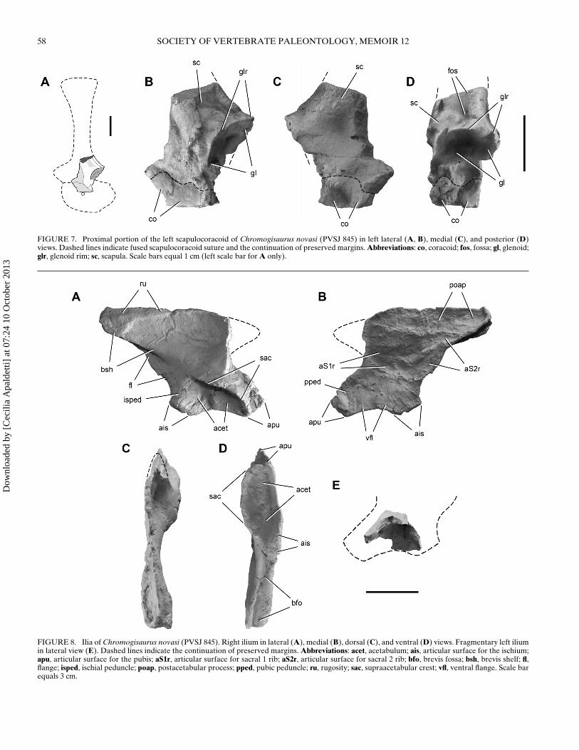
Scapulocoracoid—The left glenoid region is the only preservedportion of the scapula and coracoid, which are joined along a fusedsuture (Fig. 7). The scapular glenoid is an oval-shaped concavitywith its long axis oriented dorsoventrally (Fig. 7D). The scapularglenoid faces posteroventrally and somewhat laterally, as in Pan-phagia, Eoraptor, and Saturnalia (Langer et al., 2007). A fossa islocated dorsal to the posterodorsal rim of the glenoid (Fig. 7B, D),as in Eoraptor, Panphagia, and Eodromaeus. Shallow medial andlateral depressions are present just anterior to the posterodorsalborder (Fig. 7B, C). The rim of the glenoid is sharp (Fig. 7B, D).Not enough of the coracoid is preserved to comment on its form.
Ilium—The right ilium is the more complete, lacking only thepreacetabular process; the distal end of the right pubic pedun-cle is weathered laterally (Fig. 8A–D). Only a central piece ofthe left ilium is preserved (Fig. 8E). The ilium is vertically deepwith a relatively long postacetabular process, as in other basalsauropodomorphs (e.g., Eoraptor, Adeopapposaurus). In lateralview, the dorsal margin of the iliac blade is straight and curvesventrally as it approaches the preacetabular process (Fig. 8A,B). In dorsal view, the iliac blade is gently curved with acetab-ular processes diverging laterally (Fig. 8C), as in other basalsauropodomorphs (e.g., Eoraptor, Saturnalia, Adeopapposaurus).A broad, triangular depression covers much of the lateral aspect ofthe ilium dorsal to the supraacetabular crest (Ezcurra, 2010), as isalso present in Saturnalia (Langer, 2003). The posterior projectionat the base of the ischial peduncle is poorly developed.
The supraacetabular crest is a well-developed, raised shelf(Fig. 8A, C, D). Although the pubic peduncle is partially damaged,the crest seems to continue to its distal end (Fig. 8A). The supraac-etabular crest has a sinuous curve in lateral view (Fig. 8A). Aventral flange partially closes the acetabulum in Chromogisaurus(Ezcurra, 2010; Fig. 8A, B), but its arched ventral margin suggeststhat the acetabulum had a central unossified gap.
The pubic peduncle is long, with its distal end facing anteroven-trally (Fig. 8A, C). The distal end of the pubic peduncle has a sub-triangular cross-section with slightly concave medial, dorsolateral,and ventrolateral borders. The broader and shorter ischial pedun-cle has its distal articular surface oriented more ventrally (Ezcurra,2010; Fig. 8A, B). The distal end of the ischial peduncle is sub-triangular, with flat medial and slightly convex posterior and lat-eral margins (Fig. 8D). The antitrochanter is smooth, poorly devel-oped, and positioned on the anteroventral corner of the peduncle(Ezcurra, 2010).
The long postacetabular process tapers distally to a roundedend in lateral view, which has sustained some weathering. As wasnoted by Ezcurra (2010), the lateral surface bears a thick andtrapezoidal rugose area, which tapers anteriorly along the dorso-lateral border of the iliac blade (Fig. 8A). The well-developed bre-vis shelf does not join the supraacetabular crest (Fig. 8A). The pos-teromedial shelf is moderately developed and similar in transversewidth to the brevis shelf in dorsal or ventral view (Fig. 8C, D).In cross-section at midlength, the postacetabular process has an
Dow
nloa
ded
by [
Cec
ilia
Apa
ldet
ti] a
t 07:
24 1
0 O
ctob
er 2
013
MARTINEZ ET AL.—SAUROPODOMORPHS FROM ISCHIGUALASTO 55
FIGURE 3. Bone fragments of unknown affinity and identity in dor-sal view. These fragments were originally identified as a possible portionof the ischial shaft and included in the Chromogisaurus novasi holotype(PVSJ 845) by Excurra (2010). Abbreviations: ri, ridge. Scale bar equals1 cm.
inverted ‘V’-shape. The posteromedial shelf extends anteroven-trally and dissipates as a rounded ridge on the medial surface ofthe iliac blade dorsal to the supraacetabular crest. Only the baseof the preacetabular process is preserved (Fig. 8A, B). The ventralmargin is thicker than the dorsal margin, so that the broken cross-section of the process is subtriangular. The ventral margin of theprocess is everted dorsal to the level of the supraacetabular crest.
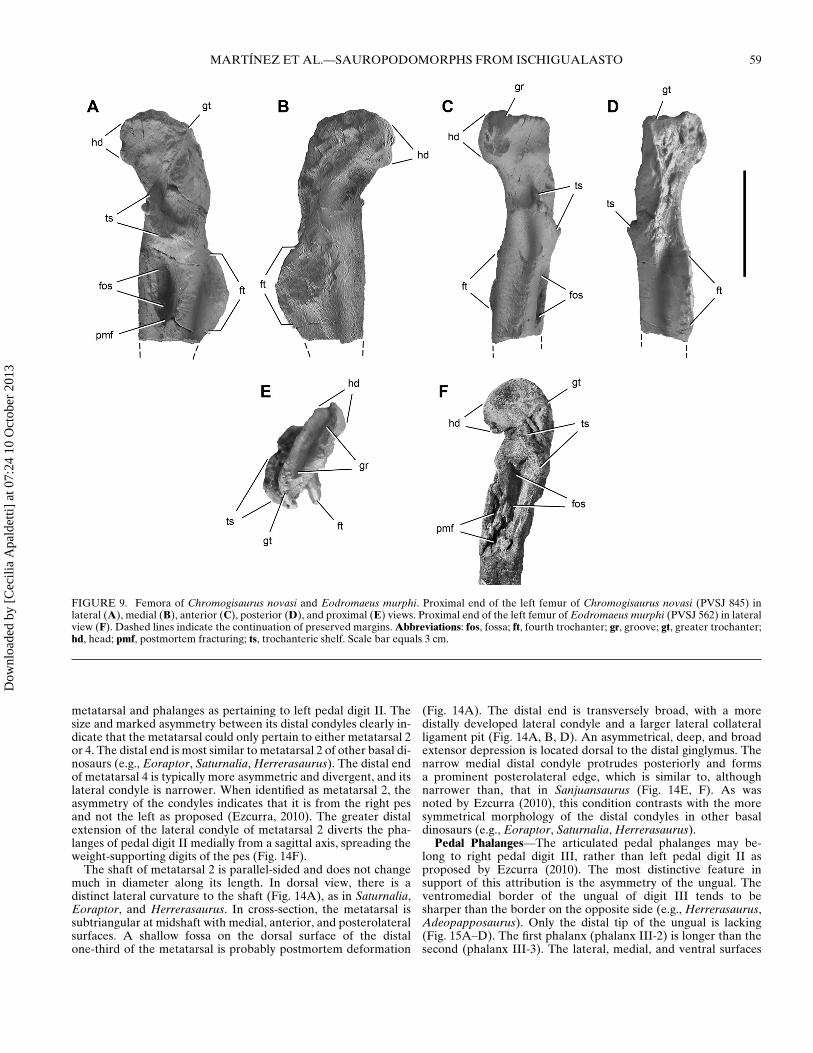
Femur—Although both femora are incomplete (Figs. 9A–E, 10,11), they overlap to allow an estimate of the form and length(ca. 170 mm) of the bone. The femur is gracile—its minimumcircumference-to-length ratio equals 0.28—and slightly shorterthan the tibia (Fig. 9F). In lateral view, the shaft of the femurhas a sigmoid curve, as in other basal dinosaurs. In anterior view,it is straight with the femoral head rotated anteromedially. Theshaft of the right femur of Chromogisaurus is transversely crushed,which may have flattened the medial projection of the head (com-
pare Fig. 9C and F). The proximal surface of the femoral head iskidney-shaped, as in most other basal dinosaurs, but it faces dor-sally and is almost straight in posteromedial view (Fig. 9C, D).The proximal surface bounded by sharp edges, between which liesa groove extending anteromedially from the greater trochanter(Fig. 9E), as in basal dinosaurs such as Eoraptor (PVSJ 855), Sat-urnalia, Herrerasaurus, and Staurikosaurus (Galton, 1977). Thegreater trochanter is developed as a prominent ridge (Fig. 9A, D,E). The anterior surface of the femoral neck bears a sharp ridgethat extends from the anteromedial end of the head and tapersdistally (Fig. 9B, C). The trochanteric shelf extends along the lat-eral aspect of the proximal end, dissipating on its posterior aspectdorsal to the fourth trochanter (Ezcurra, 2010; Fig. 9).
A deep, elliptical fossa is present ventral the trochanteric shelfon both femora (Figs. 9A, 10A), which was considered an au-tapomorphy by Ezcurra (2010). Careful observation of the better-preserved left femur reveals compressional fractures surroundingeach fossa (Fig. 9A). A similar fossa is present in some specimensof Eodromaeus (PVSJ 534, PVSJ 562), which also shows evidenceof postmortem transverse compression (Fig. 9F). We regard thisstructure as an artifact of crushing and collapse of the internal lu-men of the femoral shaft.
A prominent, crescentic fourth trochanter projects from theproximal half of the femoral shaft (Fig. 9A, B, D, E). The dis-tal portion of the fourth trochanter is damaged on both sides.Whether the trochanter extends distally to end with an asym-metric profile, as in many basal saurischians (e.g., Eoraptor, Sat-urnalia, Herrerasaurus, Staurikosaurus, Eodromaeus), cannot bedetermined. A marked rugose scar for insertion of the M. caud-ofemoralis longus (Langer, 2003) is present on the medial aspectof the fourth trochanter and extends distally as a shallow striatedfossa (Fig. 9B).
The distal end of the left femur, although partially broken, hasa nearly subcircular shape as preserved (Fig. 11E). The medialborder is flat, and the medial (tibial) condyle is oval and antero-posteriorly longer than the lateral condyle. The posterior surfaceis marked by a deep popliteal fossa that separates the condyles(Figs. 9F, 11D). At the distal end of the femur, a large rugose at-tachment scar is present facing anteriorly (Fig. 11C).
Tibia—The right tibia and proximal end of the left tibia are pre-served, both of which have been flattened transversely (Fig. 12).The tibia is slender and measures 175 mm long. The tibiofemoralratio is 1.03, similar to the values of Eoraptor (Sereno et al., 2013)and Eodromaeus (Martınez et al., 2011). The proximal articu-lar surface is subtriangular and deeply concave, which has beenexaggerated by transverse compression (Fig. 12E). As noted byEzcurra (2010), the lateral condyle is located slightly anterior tothe medial condyle (Fig. 12E). A median notch separates the prox-imal condyles (Fig. 12D, I). A low and symmetrical cnemial crestforms the anterior margin of the proximal end (Fig. 12A–C, E,H). A very pronounced longitudinal tuberosity is present on thelateral surface posterior to the concavity formed adjacent to thecnemial crest (Ezcurra, 2010; Fig. 12A, G). The shaft of the tibia isstraight (Fig. 12A–D). The distal end has been transversely com-pressed postmortem, so that its transverse width is only about 40%of its anteroposterior depth (Fig. 12F). The medial tip of the pos-terolateral process projects distally (Fig. 12A–C). A groove on thelateral aspect of the distal end separates the posterolateral flangefrom the shaft of the tibia (Fig. 12A). As in other basal dinosaurs,the distal articular surface of the tibia is split between a broad as-tragalar articular surface and a convex ‘L’-shaped surface formedby the posterolateral flange (Fig. 12F).
Fibula—Portions of both fibulae are preserved. On the rightside, both the proximal and distal ends are broken away(Fig. 13A, B). On the left side, the proximal end is well preserved(Fig. 13C–F). The fibula is narrower transversely than the tibia;
Dow
nloa
ded
by [
Cec
ilia
Apa
ldet
ti] a
t 07:
24 1
0 O
ctob
er 2
013
56 SOCIETY OF VERTEBRATE PALEONTOLOGY, MEMOIR 12
FIGURE 4. Proximal caudal vertebra of Chro-mogisaurus novasi (PVSJ 845) in left lateral(A), right lateral (B), dorsal (C), ventral (D),anterior (E), and posterior (F) views. Abbre-viations: ach, articular surface for a chevron;cr, crest; ns, neural spine; poz, postzygapoph-ysis; prz, prezygapophysis; tp, transverse pro-cess. Scale bar equals 2 cm.
the anteroposterior width of the midshaft is almost two-thirds thatof the tibia. The proximal end is anteroposteriorly expanded to adepth approximately three times its maximum width. The proxi-mal surface is posteriorly concave and slightly anteriorly convex(Fig. 13F). The medial border of the proximal end is straight, al-though the medial surface becomes concave below the border. Inlateral view, the anterior border of the proximal end of the fibulais convex, and the posterior border is slightly concave, as in manyother basal saurischians. The anterior prominence of the anteriorborder is well marked (Fig. 13A–D). Two attachment scars are vis-ible on the medial surface (Fig. 13C). The first is an oblique ridgeextending anterodistally from the posterior corner of the proximalend of the fibula and is similar to that of Saturnalia (Langer, 2003),Eodromaeus (PVSJ 562), and Herrerasaurus. The second is a morerugose ridge located parallel to and near the anterior borderof the fibula, converging distally with the aforementioned ridge
(Fig. 13C). As in many other dinosaurs (e.g., Saturnalia, Eoraptor,Eodromaeus, Herrerasaurus, Sanjuansaurus), a rugosity is presentalong the anterolateral border of the proximal end (Fig. 13D, E),probably for the insertion of the M. iliofibularis (Bittencourt andKellner, 2009). The lateral surface of the proximal end is anteriorand posteriorly concave and slightly convex at the middle. Theshaft of the fibula gradually decreases in anteroposterior depthdistally (Fig. 13A, B). The lateral surface of the shaft is convexand has a crest, or anterior trochanter (Fig. 13A), presumably forattachment of the M. iliofibularis (Langer, 2003). An elongate con-cavity extends for most of the length of the medial surface of theshaft due to postmortem collapse of the central lumen of the tibia(Fig. 13B).
Metatarsal 2—Only one metatarsal of Chromogisaurus is pre-served. The metatarsal is difficult to identify with certainty, giventhe absence of its proximal end. Ezcurra (2010) regarded the
Dow
nloa
ded
by [
Cec
ilia
Apa
ldet
ti] a
t 07:
24 1
0 O
ctob
er 2
013
MARTINEZ ET AL.—SAUROPODOMORPHS FROM ISCHIGUALASTO 57
FIGURE 5. Mid-caudal vertebrae of Chromogisaurus novasi (PVSJ 845). The anterior-most preserved mid-caudal vertebra in left lateral (A), dorsal(B), ventral (C), anterior (D), and posterior (E) views. The posterior-most preserved mid-caudal vertebra in right lateral (F) and dorsal (G) views.Anterior is towards top in B and C and to the right in G. Abbreviations: ach, articular surface for a chevron; gr, groove; ns, neural spine; poz, postzy-gapophysis; prz, prezygapophysis; tp, transverse process. Scale bar equals 2 cm.
FIGURE 6. Anterior chevron of Chromo-gisaurus novasi (PVSJ 845) in right lateral (A),anterior (B), posterior (C), and proximal (D)views. Dashed line indicates missing portions ofproximal and distal ends. Abbreviations: aace,anterior articular surface for centrum; cr, crest;hc, hemal canal; pace, posterior articular surfacefor centrum; sh, shaft. Scale bar equals 1 cm.
Dow
nloa
ded
by [
Cec
ilia
Apa
ldet
ti] a
t 07:
24 1
0 O
ctob
er 2
013
58 SOCIETY OF VERTEBRATE PALEONTOLOGY, MEMOIR 12
FIGURE 7. Proximal portion of the left scapulocoracoid of Chromogisaurus novasi (PVSJ 845) in left lateral (A, B), medial (C), and posterior (D)views. Dashed lines indicate fused scapulocoracoid suture and the continuation of preserved margins. Abbreviations: co, coracoid; fos, fossa; gl, glenoid;glr, glenoid rim; sc, scapula. Scale bars equal 1 cm (left scale bar for A only).
FIGURE 8. Ilia of Chromogisaurus novasi (PVSJ 845). Right ilium in lateral (A), medial (B), dorsal (C), and ventral (D) views. Fragmentary left iliumin lateral view (E). Dashed lines indicate the continuation of preserved margins. Abbreviations: acet, acetabulum; ais, articular surface for the ischium;apu, articular surface for the pubis; aS1r, articular surface for sacral 1 rib; aS2r, articular surface for sacral 2 rib; bfo, brevis fossa; bsh, brevis shelf; fl,flange; isped, ischial peduncle; poap, postacetabular process; pped, pubic peduncle; ru, rugosity; sac, supraacetabular crest; vfl, ventral flange. Scale barequals 3 cm.
Dow
nloa
ded
by [
Cec
ilia
Apa
ldet
ti] a
t 07:
24 1
0 O
ctob
er 2
013
MARTINEZ ET AL.—SAUROPODOMORPHS FROM ISCHIGUALASTO 59
FIGURE 9. Femora of Chromogisaurus novasi and Eodromaeus murphi. Proximal end of the left femur of Chromogisaurus novasi (PVSJ 845) inlateral (A), medial (B), anterior (C), posterior (D), and proximal (E) views. Proximal end of the left femur of Eodromaeus murphi (PVSJ 562) in lateralview (F). Dashed lines indicate the continuation of preserved margins. Abbreviations: fos, fossa; ft, fourth trochanter; gr, groove; gt, greater trochanter;hd, head; pmf, postmortem fracturing; ts, trochanteric shelf. Scale bar equals 3 cm.
metatarsal and phalanges as pertaining to left pedal digit II. Thesize and marked asymmetry between its distal condyles clearly in-dicate that the metatarsal could only pertain to either metatarsal 2or 4. The distal end is most similar to metatarsal 2 of other basal di-nosaurs (e.g., Eoraptor, Saturnalia, Herrerasaurus). The distal endof metatarsal 4 is typically more asymmetric and divergent, and itslateral condyle is narrower. When identified as metatarsal 2, theasymmetry of the condyles indicates that it is from the right pesand not the left as proposed (Ezcurra, 2010). The greater distalextension of the lateral condyle of metatarsal 2 diverts the pha-langes of pedal digit II medially from a sagittal axis, spreading theweight-supporting digits of the pes (Fig. 14F).
The shaft of metatarsal 2 is parallel-sided and does not changemuch in diameter along its length. In dorsal view, there is adistinct lateral curvature to the shaft (Fig. 14A), as in Saturnalia,Eoraptor, and Herrerasaurus. In cross-section, the metatarsal issubtriangular at midshaft with medial, anterior, and posterolateralsurfaces. A shallow fossa on the dorsal surface of the distalone-third of the metatarsal is probably postmortem deformation
(Fig. 14A). The distal end is transversely broad, with a moredistally developed lateral condyle and a larger lateral collateralligament pit (Fig. 14A, B, D). An asymmetrical, deep, and broadextensor depression is located dorsal to the distal ginglymus. Thenarrow medial distal condyle protrudes posteriorly and formsa prominent posterolateral edge, which is similar to, althoughnarrower than, that in Sanjuansaurus (Fig. 14E, F). As wasnoted by Ezcurra (2010), this condition contrasts with the moresymmetrical morphology of the distal condyles in other basaldinosaurs (e.g., Eoraptor, Saturnalia, Herrerasaurus).
Pedal Phalanges—The articulated pedal phalanges may be-long to right pedal digit III, rather than left pedal digit II asproposed by Ezcurra (2010). The most distinctive feature insupport of this attribution is the asymmetry of the ungual. Theventromedial border of the ungual of digit III tends to besharper than the border on the opposite side (e.g., Herrerasaurus,Adeopapposaurus). Only the distal tip of the ungual is lacking(Fig. 15A–D). The first phalanx (phalanx III-2) is longer than thesecond (phalanx III-3). The lateral, medial, and ventral surfaces
Dow
nloa
ded
by [
Cec
ilia
Apa
ldet
ti] a
t 07:
24 1
0 O
ctob
er 2
013
60 SOCIETY OF VERTEBRATE PALEONTOLOGY, MEMOIR 12
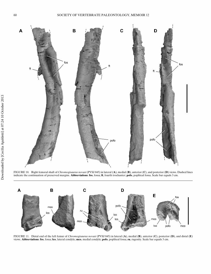
FIGURE 10. Right femoral shaft of Chromogisaurus novasi (PVSJ 845) in lateral (A), medial (B), anterior (C), and posterior (D) views. Dashed linesindicate the continuation of preserved margins. Abbreviations: fos, fossa; ft, fourth trochanter; pofo, popliteal fossa. Scale bar equals 3 cm.
FIGURE 11. Distal end of the left femur of Chromogisaurus novasi (PVSJ 845) in lateral (A), medial (B), anterior (C), posterior (D), and distal (E)views. Abbreviations: fos, fossa; lco, lateral condyle; mco, medial condyle; pofo, popliteal fossa; ru, rugosity. Scale bar equals 3 cm.
Dow
nloa
ded
by [
Cec
ilia
Apa
ldet
ti] a
t 07:
24 1
0 O
ctob
er 2
013
MARTINEZ ET AL.—SAUROPODOMORPHS FROM ISCHIGUALASTO 61
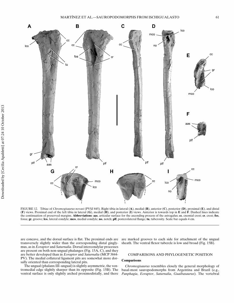
FIGURE 12. Tibiae of Chromogisaurus novasi (PVSJ 845). Right tibia in lateral (A), medial (B), anterior (C), posterior (D), proximal (E), and distal(F) views. Proximal end of the left tibia in lateral (G), medial (H), and posterior (I) views. Anterior is towards top in E and F. Dashed lines indicatethe continuation of preserved margins. Abbreviations: aas, articular surface for the ascending process of the astragalus; cc, cnemial crest; cr, crest; fos,fossa; gr, groove; lco, lateral condyle; mco, medial condyle; no, notch; plf, posterolateral flange; tu, tuberosity. Scale bar equals 4 cm.
are concave, and the dorsal surface is flat. The proximal ends aretransversely slightly wider than the corresponding distal gingly-mus, as in Eoraptor and Saturnalia. Dorsal intercondylar processesare present on both non-ungual phalanges (Fig. 15A, C), and theyare better developed than in Eoraptor and Saturnalia (MCP 3844-PV). The medial collateral ligament pits are somewhat more dor-sally oriented than corresponding lateral pits.
The ungual (phalanx III–ungual) is slightly asymmetric, the ven-tromedial edge slightly sharper than its opposite (Fig. 15B). Theventral surface is only slightly arched proximodistally, and there
are marked grooves to each side for attachment of the ungualsheath. The ventral flexor tubercle is low and broad (Fig. 15B).
COMPARISONS AND PHYLOGENETIC POSITION
Comparisons
Chromogisaurus resembles closely the general morphology ofbasal-most sauropodomorphs from Argentina and Brazil (e.g.,Panphagia, Eoraptor, Saturnalia, Guaibasaurus). The vertebral
Dow
nloa
ded
by [
Cec
ilia
Apa
ldet
ti] a
t 07:
24 1
0 O
ctob
er 2
013
62 SOCIETY OF VERTEBRATE PALEONTOLOGY, MEMOIR 12
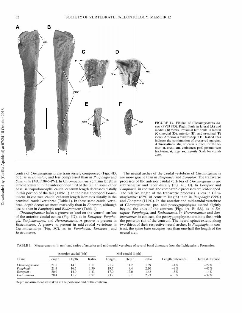
FIGURE 13. Fibulae of Chromogisaurus no-vasi (PVSJ 845). Right fibula in lateral (A) andmedial (B) views. Proximal left fibula in lateral(C), medial (D), anterior (E), and proximal (F)views. Anterior is towards top in F. Dashed linesindicate the continuation of preserved margins.Abbreviations: afe, articular surface for the fe-mur; cr, crest; em, eminence; pmf, postmortemfracturing; ri, ridge; ru, rugosity. Scale bar equals2 cm.
centra of Chromogisaurus are transversely compressed (Figs. 4D,5C), as in Eoraptor, and less compressed than in Panphagia andSaturnalia (MCP 3846-PV). In Chromogisaurus, centrum length isalmost constant in the anterior one-third of the tail. In some otherbasal sauropodomorphs, caudal centrum length decreases distallyin this portion of the tail (Table 1). In the basal theropod Eodro-maeus, in contrast, caudal centrum length increases distally in theproximal caudal vertebrae (Table 1). In these same caudal verte-brae, depth decreases more markedly than in Eoraptor, althoughless so than in Panphagia and Eodromaeus (Table 1).
Chromogisaurus lacks a groove or keel on the ventral surfaceof the anterior caudal centra (Fig. 4D), as in Eoraptor, Panpha-gia, Sanjuansaurus, and Herrerasaurus. A groove is present inEodromaeus. A groove is present in mid-caudal vertebrae inChromogisaurus (Fig. 5C), as in Panphagia, Eoraptor, andEodromaeus.
The neural arches of the caudal vertebrae of Chromogisaurusare more gracile than in Panphagia and Eoraptor. The transverseprocesses of the anterior caudal vertebra of Chromogisaurus aresubtriangular and taper distally (Fig. 4C, D). In Eoraptor andPanphagia, in contrast, the comparable processes are leaf-shaped.The relative length of the transverse processes is less in Chro-mogisaurus (82% of centrum length) than in Panphagia (99%)and Eoraptor (111%). In the anterior and mid-caudal vertebraeof Chromogisaurus, pre- and postzygapophyses extend slightlybeyond the ends of the centrum (Figs. 4A, B, 5A), as in Eo-raptor, Panphagia, and Eodromaeus. In Herrerasaurus and San-juansaurus, in contrast, the postzygapophyses terminate flush withthe posterior rim of the centrum. The neural spines extend alongtwo-thirds of their respective neural arches. In Panphagia, in con-trast, the spine base occupies less than one-half the length of theneural arch.
TABLE 1. Measurements (in mm) and ratios of anterior and mid-caudal vertebrae of several basal dinosaurs from the Ischigualasto Formation.
Anterior caudal (4th) Mid-caudal (14th)
Taxon Length Depth Ratio Length Depth Ratio Length difference Depth difference
Chromogisaurus 21.6 14.3 1.51 21.2 11.2 1.89 −1% −22%Panphagia 21.4 16.5 1.30 19.7 9.4 2.10 −8% −43%Eoraptor 20.0 14.0 1.43 17.0 12.0 1.42 −15% −14%Eodromaeus 20.4 11.9 1.71 23.7 8.1 2.93 +13% −32%
Depth measurement was taken at the posterior end of the centrum.
Dow
nloa
ded
by [
Cec
ilia
Apa
ldet
ti] a
t 07:
24 1
0 O
ctob
er 2
013
MARTINEZ ET AL.—SAUROPODOMORPHS FROM ISCHIGUALASTO 63
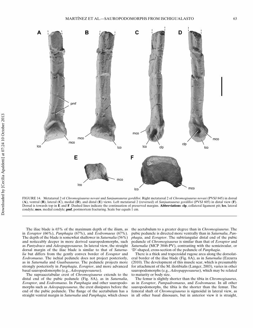
FIGURE 14. Metatarsal 2 of Chromogisaurus novasi and Sanjuansaurus gordilloi. Right metatarsal 2 of Chromogisaurus novasi (PVSJ 845) in dorsal(A), ventral (B), lateral (C), medial (D), and distal (E) views. Left metatarsal 2 (reversed) of Sanjuansaurus gordilloi (PVSJ 605) in distal view (F).Dorsal is towards top in E and F. Dashed lines indicate the continuation of preserved margins. Abbreviations: clp, collateral ligament pit; lco, lateralcondyle; mco, medial condyle; pmf, postmortem fracturing. Scale bar equals 1 cm.
The iliac blade is 65% of the maximum depth of the ilium, asin Eoraptor (66%), Panphagia (67%), and Eodromaeus (67%).The depth of the blade is somewhat shallower in Saturnalia (56%)and noticeably deeper in more derived sauropodomorphs, suchas Pantydraco and Adeopapposaurus. In lateral view, the straightdorsal margin of the iliac blade is similar to that of Saturna-lia but differs from the gently convex border of Eoraptor andEodromaeus. The ischial peduncle does not project posteriorly,as in Saturnalia and Guaibasaurus. The peduncle projects morestrongly posteriorly in Panphagia, Eoraptor, and more advancedbasal sauropodomorphs (e.g., Adeopapposaurus).
The supraacetabular crest of Chromogisaurus extends to thedistal end of the pubic peduncle (Fig. 8A), as in Saturnalia,Eoraptor, and Eodromaeus. In Panphagia and other sauropodo-morphs such as Adeopapposaurus, the crest dissipates before theend of the pubic peduncle. The flange of the acetabulum has astraight ventral margin in Saturnalia and Panphagia, which closes
the acetabulum to a greater degree than in Chromogisaurus. Thepubic peduncle is directed more ventrally than in Saturnalia, Pan-phagia, and Eoraptor. The subtriangular distal end of the pubicpeduncle of Chromogisaurus is similar than that of Eoraptor andSaturnalia (MCP 3846-PV), contrasting with the semicircular, or‘D’-shaped, cross-section of the peduncle of Panphagia.
There is a thick and trapezoidal rugose area along the dorsolat-eral border of the iliac blade (Fig. 8A), as in Saturnalia (Ezcurra(2010). The development of this muscle scar, which is presumablyfor attachment of the M. iliotibialis (Langer, 2003), varies in othersauropodomorphs (e.g., Adeopapposaurus), which may be relatedto maturity or body size.
The femur is slightly shorter than the tibia in Chromogisaurus,as in Eoraptor, Pampadromaeus, and Eodromaeus. In all othersauropodomorphs, the tibia is the shorter than the femur. Thefemoral shaft of Chromogisaurus is sigmoidal in lateral view, asin all other basal dinosaurs, but in anterior view it is straight,
Dow
nloa
ded
by [
Cec
ilia
Apa
ldet
ti] a
t 07:
24 1
0 O
ctob
er 2
013
64 SOCIETY OF VERTEBRATE PALEONTOLOGY, MEMOIR 12
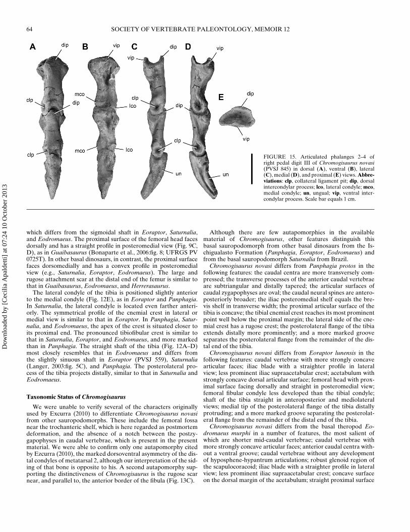
FIGURE 15. Articulated phalanges 2–4 ofright pedal digit III of Chromogisaurus novasi(PVSJ 845) in dorsal (A), ventral (B), lateral(C), medial (D), and proximal (E) views. Abbre-viations: clp, collateral ligament pit; dip, dorsalintercondylar process; lco, lateral condyle; mco,medial condyle; un, ungual; vip, ventral inter-condylar process. Scale bar equals 1 cm.
which differs from the sigmoidal shaft in Eoraptor, Saturnalia,and Eodromaeus. The proximal surface of the femoral head facesdorsally and has a straight profile in posteromedial view (Fig. 9C,D), as in Guaibasaurus (Bonaparte et al., 2006:fig. 8; UFRGS PV0725T). In other basal dinosaurs, in contrast, the proximal surfacefaces dorsomedially and has a convex profile in posteromedialview (e.g., Saturnalia, Eoraptor, Eodromaeus). The large andrugose attachment scar at the distal end of the femur is similar tothat in Guaibasaurus, Eodromaeus, and Herrerasaurus.
The lateral condyle of the tibia is positioned slightly anteriorto the medial condyle (Fig. 12E), as in Eoraptor and Panphagia.In Saturnalia, the lateral condyle is located even farther anteri-orly. The symmetrical profile of the cnemial crest in lateral ormedial view is similar to that in Eoraptor. In Panphagia, Satur-nalia, and Eodromaeus, the apex of the crest is situated closer toits proximal end. The pronounced tibiofibular crest is similar tothat in Saturnalia, Eoraptor, and Eodromaeus, and more markedthan in Panphagia. The straight shaft of the tibia (Fig. 12A–D)most closely resembles that in Eodromaeus and differs fromthe slightly sinuous shaft in Eoraptor (PVSJ 559), Saturnalia(Langer, 2003:fig. 5C), and Panphagia. The posterolateral pro-cess of the tibia projects distally, similar to that in Saturnalia andEodromaeus.
Taxonomic Status of Chromogisaurus
We were unable to verify several of the characters originallyused by Excurra (2010) to differentiate Chromogisaurus novasifrom other sauropodomorphs. These include the femoral fossanear the trochanteric shelf, which is here regarded as postmortemdeformation, and the absence of a notch between the postzy-gapophyses in caudal vertebrae, which is present in the presentmaterial. We were able to confirm only one autapomorphy citedby Ezcurra (2010), the marked dorsoventral asymmetry of the dis-tal condyles of metatarsal 2, although our interpretation of the sid-ing of that bone is opposite to his. A second autapomorphy sup-porting the distinctiveness of Chromogisaurus is the rugose scarnear, and parallel to, the anterior border of the fibula (Fig. 13C).
Although there are few autapomorphies in the availablematerial of Chromogisaurus, other features distinguish thisbasal sauropodomorph from other basal dinosaurs from the Is-chigualasto Formation (Panphagia, Eoraptor, Eodromaeus) andfrom the basal sauropodomorph Saturnalia from Brazil.
Chromogisaurus novasi differs from Panphagia protos in thefollowing features: the caudal centra are more transversely com-pressed; the transverse processes of the anterior caudal vertebraeare subtriangular and distally tapered; the articular surfaces ofcaudal zygapophyses are oval; the caudal neural spines are antero-posteriorly broader; the iliac posteromedial shelf equals the bre-vis shelf in transverse width; the proximal articular surface of thetibia is concave; the tibial cnemial crest reaches its most prominentpoint well below the proximal margin; the lateral side of the cne-mial crest has a rugose crest; the posterolateral flange of the tibiaextends distally more prominently; and a more marked grooveseparates the posterolateral flange from the remainder of the dis-tal end of the tibia.
Chromogisaurus novasi differs from Eoraptor lunensis in thefollowing features: caudal vertebrae with more strongly concavearticular faces; iliac blade with a straighter profile in lateralview; less prominent iliac supraacetabular crest; acetabulum withstrongly concave dorsal articular surface; femoral head with prox-imal surface facing dorsally and straight in posteromedial view;femoral fibular condyle less developed than the tibial condyle;shaft of the tibia straight in anteroposterior and mediolateralviews; medial tip of the posterolateral flange of the tibia distallyprotruding; and a more marked groove separating the posterolat-eral flange from the remainder of the distal end of the tibia.
Chromogisaurus novasi differs from the basal theropod Eo-dromaeus murphi in a number of features, the most salient ofwhich are shorter mid-caudal vertebrae; caudal vertebrae withmore strongly concave articular faces; anterior caudal centra with-out a ventral groove; caudal vertebrae without any developmentof hyposphene-hypantrum articulations; robust glenoid region ofthe scapulocoracoid; iliac blade with a straighter profile in lateralview; less prominent iliac supraacetabular crest; concave surfaceon the dorsal margin of the acetabulum; straight proximal surface
Dow
nloa
ded
by [
Cec
ilia
Apa
ldet
ti] a
t 07:
24 1
0 O
ctob
er 2
013
MARTINEZ ET AL.—SAUROPODOMORPHS FROM ISCHIGUALASTO 65
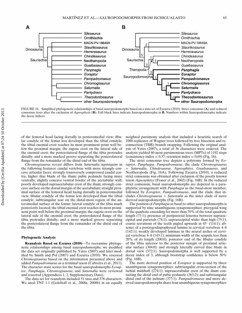
FIGURE 16. Simplified phylogenetic relationships of basal sauropodomorphs based on a data set of Ezcurra (2010). Strict consensus (A) and reducedconsensus trees after the exclusion of Agnosphytis (B). Full black lines indicate Sauropodomorpha in B. Numbers within Sauropodomorpha indicatethe decay indices.
of the femoral head facing dorsally in posteromedial view; fibu-lar condyle of the femur less developed than the tibial condyle;the tibial cnemial crest reaches its most prominent point well be-low the proximal margin; the rugose crest on the lateral side ofthe cnemial crest; the posterolateral flange of the tibia protrudesdistally; and a more marked groove separating the posterolateralflange from the remainder of the distal end of the tibia.
Chromogisaurus novasi differs from Saturnalia tupiniquim inthe following features: caudal vertebrae with more strongly con-cave articular faces; strongly transversely compressed caudal cen-tra; higher iliac blade of the ilium; pubic peduncle facing moreventrally; slightly concave ventral border of the acetabular wall;poorly developed supraacetabular crest of the ilium; strongly con-cave surface on the dorsal margin of the acetabulum; straight prox-imal surface of the femoral head facing dorsally in posteromedialview; fibular condyle of the femur less developed than the tibialcondyle; subtriangular scar on the distal-most region of the an-teromedial surface of the femur; lateral condyle of the tibia muchposteriorly located; the tibial cnemial crest reaches its most promi-nent point well below the proximal margin; the rugose crest on thelateral side of the cnemial crest; the posterolateral flange of thetibia protrudes distally; and a more marked groove separatingthe posterolateral flange from the remainder of the distal end ofthe tibia.
Phylogenetic Analysis
Reanalysis Based on Ezcurra (2010)—To reexamine phyloge-netic relationships among basal sauropodomorphs, we modifiedthe data set originally published by Yates (2007) and later mod-ified by Smith and Pol (2007) and Ezcurra (2010). We rescoredChromogisaurus based on the information presented above andadded Pampadromaeus as a terminal taxon (Cabreira et al., 2011).The character-state scores for the basal sauropodomorphs Eorap-tor, Panphagia, Chromogisaurus, and Saturnalia were reviewedand rescored (Appendices 1, 2, Supplementary Data).
The data set for reanalysis included 51 taxa and 378 characters.We used TNT 1.1 (Goloboff et al., 2008a, 2008b) in an equally
weighted parsimony analysis that included a heuristic search of1000 replicates of Wagner trees followed by tree bisection and re-connection (TBR) branch swapping. Following the original anal-ysis of Yates (2007), a total of 36 characters were ordered. Theanalysis yielded 60 most parsimonious trees (MPTs) of 1192 steps(consistency index = 0.37; retention index = 0.69) (Fig. 16).
The strict consensus tree depicts a polytomy formed by Eo-raptor, Panphagia, Pampadromaeus, the clade Chromogisaurus+ Saturnalia, Chindesaurus, Agnosphitys, Guaibasaurus, andNeotheropoda (Fig. 16A). Following Ezcurra (2010), a reducedstrict consensus was obtained after exclusion of the poorly knowntaxon Agnosphitys (Fraser et al., 2002) (Fig. 16B). In the reducedstrict consensus, basal sauropodomorphs are depicted in a para-phyletic arrangement with Panphagia as the basal-most member,followed by Eoraptor, Pampadromaeus, and the clade that in-cludes Chromogisaurus + Saturnalia as the sister clade of morederived sauropodomorphs (Fig. 16B).
The position of Panphagia as basal to other sauropodomorphs issupported by nine unambiguous synapomorphies: pterygoid wingof the quadrate extending for more than 70% of the total quadratelength (71:1); presence of postparietal fenestra between supraoc-cipital and parietals (74:1); supraoccipital wider than high (75:1);coarse serrations of the teeth angled upwards at 45◦ (114:1); ab-sence of a postzygodiapophyseal lamina in cervical vertebrae 4–8(142:1); weakly developed laminae in the neural arches of cervi-cal vertebrae 4–8 (143:1); minimum width of the scapula less than20% of its length (200:0); posterior end of the fibular condyleof the tibia anterior to the posterior margin of proximal artic-ular surface (304:0); and strongly laterally curved iliac blade indorsal view (372:1). Sauropodomorpha is well supported by adecay index of 3, although bootstrap confidence is below 50%(Fig. 16B).
The more derived position of Eoraptor is supported by threeunambiguous synapomorphies: subtriangular cross-section of theischial midshaft (274:1); supraacetabular crest of the ilium con-tacting the distal end of pubic peduncle (363:2); and subtriangulardistal end of the ischium (377:1). Pampadromaeus and more de-rived sauropodomorphs share four unambiguous synapomorphies:
Dow
nloa
ded
by [
Cec
ilia
Apa
ldet
ti] a
t 07:
24 1
0 O
ctob
er 2
013
66 SOCIETY OF VERTEBRATE PALEONTOLOGY, MEMOIR 12
squamosal bordering the laterotemporal fenestra for more than50% of its depth (62:0); length of the base of proximal cau-dal neural spines greater than half the length of the neural arch(184:1); transverse width of the distal humerus greater than 33%of its length (211:1); and length of the pubic peduncle of the il-ium greater than twice the anteroposterior width of its distal end(252:1).
The Chromogisaurus + Saturnalia clade is supported by threeunambiguous synapomorphies: presence of a caudosacral vertebra(178:1); strong trapezoidal rugosity for the origin of the M. flexortibialis and iliotibialis on the postacetabular process of the ilium(362:1); and concave posterolateral corner of the distal end of thetibia (375:1). This clade is particularly weak, with a decay index of1 and bootstrap frequency below 50% (Fig. 16B).
Comparisons with Recent Analyses—In the analysis of Ezcurra(2010), Panphagia, Guaibasaurus, and Chromogisaurus + Satur-nalia joined a polytomy within the clade Guaibasauridae (Bona-parte et al., 1999; Ezcurra and Novas, 2009). In the present anal-ysis, Guaibasaurus is recovered as a theropod, the sister taxon toNeotheropoda (Fig. 16B), a very tentative result for this poorlyknown taxon that mirrors previous analyses (e.g., Smith and Pol,2007; Yates, 2007; Langer et al., 2011).
Eoraptor was originally viewed as a theropod (Sereno et al.,1993; Novas, 1996; Sereno, 1999; Rauhut, 2003; Yates, 2007;Ezcurra, 2010) or basal saurischian (Langer and Benton, 2006;Brusatte et al., 2010, 2011). Here Eoraptor is retrieved as a basalsauropodomorph (Fig. 16B), as in Martınez et al. (2011) andas discussed in detail in Sereno and Martınez (in review). Thesauropodomorph affinity of Eoraptor is generated by alteration ofseveral character states for this taxon (Appendix 2).
Ezcurra (2010) coined the subfamily Saturnaliinae to includeSaturnalia plus Chromogisaurus. He based this clade on threesynapomorphies: ulna with an extremely enlarged olecranonprocess; iliac postacetabular process with pointed posteroventralcorner and rounded posterodorsal margin; and strong trapezoidalrugosity for the origin of the M. flexor tibialis and iliotibialis. Al-though we show that the ulna of Chromogisaurus is not preserved,our analysis also generates this clade (Fig. 16B), based on onlyone of the original synapomorphies: strong trapezoidal rugosityfor the origin of the M. flexor tibialis and iliotibialis (362:1).
CONCLUSIONS
Most of the small-bodied (<15 kg) primary consumersfrom the Ischigualasto Formation appear to have been sauro-podomorphs. This trophic niche was shared with at least onesilesaurid (Martınez et al., 2013) and possibly the ornithischianPisanosaurus, which is thus far known only from the upper levelsof the formation. The three sauropodomorphs, Panphagia, Eorap-tor, and Chromogisaurus, come from the Scaphonyx-Exaeretodon-Herrerasaurus biozone (Martınez et al., 2011) and thus were likelycontemporaries sharing the same ecosystem. The presence inBrazil of the similar-age basal sauropodomorphs Saturnalia andPampadromaeus suggests that these herbivores had achieved alevel of diversity in southern Pangaea during the Carnian. Asimilar diversity of sauropodomorphs (three) is present in theoverlying Los Colorados Formation, which was deposited about15 million years later in the mid-Norian (Santi Malnis et al.,2011). The main difference between these sauropodomorphs isthat in the Los Colorados Formation, they are the most abundantvertebrates and also attained large body size. The fossil recordof sauropodomorphs across these two formations suggests thatsauropodomorphs have been an important component from theemergence of dinosaurs, but that their size and abundance in-creased markedly in the Norian toward the end of the Triassic.
ACKNOWLEDGMENTS
We thank C. Abraczinskas for assistance in preparing the finalfigures. We also thank the Earthwatch Institute for support of fieldwork and for the many Earthwatch field volunteers and the Insti-tuto y Museo de Ciencias Naturales of the Universidad Nacionalde San Juan and CICITCA for their support of this research. Wethank P. Mannion, A. Yates, and M. Langer for their critical re-views, which improved the quality of the manuscript.
LITERATURE CITED
Alcober, O. A., and R. N. Martınez. 2010. A new herrerasaurid (Di-nosauria, Saurischia) from the Upper Triassic Ischigualasto Forma-tion of northwestern Argentina. ZooKeys 63:55–81.
Benton, M. J. 1983. The Triassic reptile Hyperodapedon from Elgin: func-tional morphology and relationships. Philosophical Transactions ofthe Royal Society of London, Series B 302:605–717.
Bittencourt, J. S., and A. W. A. Kellner. 2009. The anatomy and phyloge-netic position of the Triassic dinosaur Staurikosaurus pricei Colbert,1970. Zootaxa 2079:1–56.
Bonaparte, J. F., J. Ferigolo, and A. M. Ribeiro. 1999. A new early LateTriassic saurischian dinosaur from Rio Grande do Sul State, Brazil;pp. 89–109 in Y. Tomida, T. H. Rich, and P. Vickers-Rich (eds.), Pro-ceedings of the Second Gondwanan Dinosaur Symposium, BuenosAires 26–30 July, 1999. National Science Museum Monographs 15.
Bonaparte, J. F., G. Brea, C. L. Schultz, and A. G. Martinelli. 2006. Anew specimen of Guaibasaurus candelariensis (basal Saurischia) fromthe Late Triassic Caturrita Formation of southern Brazil. HistoricalBiology 19:73–82.
Brusatte, S. L., M. J. Benton, G. T. Lloyd, M. Ruta, and S. C. Wang.2011. Macroevolutionary patterns in the evolutionary radiation of ar-chosaurs (Tetrapoda: Diapsida). Earth and Environmental ScienceTransactions of the Royal Society of Edinburgh 101:367–382.
Brusatte, S. L., S. J. Nesbitt, R. B Irmis, R. J. Butler, M. J. Benton, and M.A. Norell. 2010. The origin and early radiation of dinosaurs. Earth-Science Reviews 101:68–100.
Cabreira, S. F., C. L. Schultz, J. S. Bittencourt, M. B. Soares, D. C. Fortier,L. R. Silva, and M. C. Langer. 2011. New stem-sauropodomorph (Di-nosauria, Saurischia) from the Triassic of Brazil. Naturwissenschaften938:1035–1040.
Casamiquela, R. M. 1967. Un nuevo dinosaurio ornitisquio Triasico(Pisanosaurus mertii; Ornithopoda) de la Formacion Ischigualasto,Argentina. Ameghiniana 4:47–64.
Crompton, A. W., and A. J. Charig. 1962. A new ornithischian from theUpper Triassic of South Africa. Nature 196:1074–1077.
Currie, B. S., C. E. Colombi, N. J. Tabor, T. C. Shipman, and I. P.Montanez. 2009. Stratigraphy and architecture of the Upper Trias-sic Ischigualasto Formation, Ischigualasto Provincial Park, San Juan,Argentina. Journal of South American Earth Sciences 27:74–87.
Dzik, J. 2003. A beaked herbivorous archosaur with dinosaur affinitiesfrom the early Late Triassic of Poland. Journal of Vertebrate Pale-ontology 23:556–574.
Ezcurra, M. D., A. Lecuona, and R. Irmis. 2008. A new early dinosaur fromthe Carnian Ischigualasto Formation (NW Argentina) and the originof dinosaurs; p. 88 in J. O. Calvo, R. J. Valieri, J. D. Porfiri, and D. dosSantos (eds.), Actas de Resumenes, III Congreso Latinoamericanode Paleontologıa de Vertebrados, Neuquen 21–24 September 2008.Universidad Nacional del Comahue.
Ezcurra, M. D. 2010. A new early dinosaur (Saurischia: Sauropodomor-pha) from the Late Triassic of Argentina: a reassessment of di-nosaur origin and phylogeny. Journal of Systematic Palaeontology8:371–425.
Ezcurra, M. D., and F. E. Novas. 2009. Guaibasauridae, a new clade of Tri-assic basal sauropodomorphs. Journal of Vertebrate Paleontology,Program and Abstracts 2009:92A.
Fraser, N. C., K. Padian, G. M. Walkden, and A. L. M. Davis. 2002.Basal dinosauriform remains from Britain and the diagnosis of theDinosauria. Palaeontology 45:79–95.
Galton, P. M. 1977. On Staurikosaurus pricei, an early saurischian dinosaurfrom the Triassic of Brazil, with notes on the Herrerasauridae andPoposauridae. Palaontologische Zeitschrift 51:234–245.
Dow
nloa
ded
by [
Cec
ilia
Apa
ldet
ti] a
t 07:
24 1
0 O
ctob
er 2
013
MARTINEZ ET AL.—SAUROPODOMORPHS FROM ISCHIGUALASTO 67
Galton, P. M. 1978. Fabrosauridae, the basal family of ornithischiandinosaurs (Reptilia: Ornithopoda). Palaontologische Zeitschrift52:138–159.
Galton, P. M., and D. Kermack. 2011. The anatomy of Pantydraco caducus,a very basal sauropodomorph from the Rhaetian (Upper Triassic) ofSouth Wales, UK. Revue de Paleobiology 29:341–404.
Galton, P. M., and P. Upchurch. 2004. Prosauropoda; pp. 232–258 in D.B. Weishampel, P. Dodson, and H. Osmolska (eds.), The Dinosauria,second edition. University of California Press, Berkeley, California.
Goloboff, P. A., J. S. Farris, and K. Nixon. 2008a. TNT: Tree Anal-ysis Using New Technology, version 1.1 (Willi Hennig SocietyEdition). Available at http://www.zmuc.dk/public/phylogeny/tnt. Ac-cessed November 30, 2008.
Goloboff, P. A., J. S. Farris, and K. Nixon. 2008b. TNT, a free program forphylogenetic analysis. Cladistics 24:774–786.
Huene, F. von. 1932. Die fossile Reptil-Ordnung Saurischia, ihre Entwick-lung und Geschichte. Monographien zur Geologie und Palaontologie4:1–361.
Knoll, F. 2002. Nearly complete skull of Lesothosaurus (Dinosauria: Or-nithischia) from the Upper Elliot Formation (Lower Jurassic: Het-tangian) of Lesotho. Journal of Vertebrate Paleontology 22:238–243.
Langer, M. C. 2003. The pelvic and hind limb anatomy of the stem-sauropodomorph Saturnalia tupiniquim (Late Triassic, Brazil). Pale-oBios 23:1–30.
Langer, M. C., and M. J. Benton. 2006. Early dinosaurs: a phylogeneticstudy. Journal of Systematic Palaeontology 4:309–358.
Langer, M. C., and C. L. Schultz. 2000. A new species of the Late Trias-sic rhynchosaur Hyperodapedon from the Santa Marıa Formation ofSouth Brazil. Palaeontology 43:633–652.
Langer, M. C., J. S. Bittencourt, and C. L. Schultz. 2011. A reassessmentof the basal dinosaur Guaibasaurus candelariensis, from the Late Tri-assic Caturrita Formation of South Brazil. Earth and EnvironmentalScience Transactions of the Royal Society of Edinburgh 101:1–32.
Langer, M. C., M. A. G. Franca, and S. Gabriel. 2007. The pectoral gir-dle and forelimb anatomy of the stem sauropodomorph Saturnaliatupiniquim (Late Triassic, Brazil). Special Papers in Palaeontology77:113–137.
Langer, M. C., F. Abdala, M. Richter, and M. J. Benton. 1999. Asauropodomorph dinosaur from the Upper Triassic (Carnian) ofsouthern Brazil. Comptes Rendus de l’Academie des Sciences, Sci-ences de la Terre et des Planetes 329:511–517.
Martınez, R. N. 2009. Adeopapposaurus mognai gen. et sp. nov. (Di-nosauria: Sauropodomorpha) with comments on adaptations of basalSauropodomorpha. Journal of Vertebrate Paleontology 29:142–164.
Martınez, R. N., and O. A. Alcober. 2009. A basal sauropodomorph(Dinosauria: Saurischia) from the Ischigualasto Formation (Triassic,Carnian) and the early evolution of Sauropodomorpha. PLoS ONE4:e4397. doi: 4310.1371/journal.pone.0004397.
Martınez, R. N., P. C. Sereno, O. A. Alcober, C. E. Colombi, P. R.Renne, I. P. Montanez, and B. S. Currie. 2011. A basal dinosaurfrom the dawn of the dinosaur era in southwestern Pangaea. Science331:201–210.
Martınez R. N., C. Apaldetti, O. A. Alcober, C. Colombi, P. C. Sereno,E. Fernandez, P. Santi Malnis, G. Correa, and D. Abelın. 2013. Ver-tebrate succession in the Ischigualasto Formation; pp. 10–30 in P. C.Sereno (ed.), Basal sauropodomorphs and the vertebrate fossil recordof the Ischigualasto Formation (Late Triassic: Carnian–Norian) ofArgentina. Society of Vertebrate Paleontology Memoir 12.
Nesbitt, S. J., N. D. Smith, R. B. Irmis, A. H. Turner, A. Downs, and M. A.Norell. 2009. A complete skeleton of a Late Triassic saurischian andthe early evolution of dinosaurs. Science 326:1530–1533.
Novas, F. E. 1996. Dinosaur monophyly. Journal of Vertebrate Paleontol-ogy 16:723–741.
Owen, R. 1842. Report on British Fossil Reptiles. Part II. Reports of theBritish Association for the Advancement of Science 11:60–204.
Raath, M. A. 1977. The anatomy of the Triassic theropod Syntarsus rhode-siensis (Saurischia: Podokesauridae) and a consideration of its bi-ology. Ph.D. dissertation, Rhodes University, Grahamstown, SouthAfrica, 233 pp.
Rauhut, O. W. M. 2003. The interrelationships and evolution of basaltheropod dinosaurs. Special Papers in Palaeontology 69:1–213.
Reig, O. A. 1963. La presencia de dinosaurios saurisquios en los “Es-tratos de Ischigualasto” (MesoTriasico superior) de las provincias
de la San Juan y La Rioja (Republica Argentina). Ameghiniana 3:3–20.
Rogers, R. R., C. C. Swisher, P. C. Sereno, A. M. Monetta, C. A. Forster,and R. N. Martınez. 1993. The Ischigualasto tetrapod assemblage(Late Triassic, Argentina) and 40Ar/39Ar dating of dinosaur origins.Science 260:794–797.
Santi Malnis, P., D. V. Kent, C. E. Colombi, and S. E. Geuna. 2011. Que-brada de la Sal magnetostratigraphic section, Los Colorados Forma-tion, Upper Triassic Ischigualasto Villa Union Basin, Argentina. Lat-inmag Letters Proceedings 1:1–7.
Seeley, H. G. 1887. On the classification of the fossil animals commonlynamed Dinosauria. Proceedings of the Royal Society of London43:165–171.
Sereno, P. C. 1999. The evolution of dinosaurs. Science 284:2137–2147.Sereno, P. C. 2005a. The logical basis for phylogenetic taxonomy. System-
atic Biology 54:595–619.Sereno, P. C. 2005b. TaxonSearch: a relational database for suprageneric
taxa and phylogenetic definitions. PhyloInformatics 8:1–21.Sereno, P. C. 2007. The phylogenetic relationships of early dinosaurs: a
comparative report. Historical Biology 19:145–155.Sereno, P. C., and F. E. Novas. 1992. The complete skull and skeleton of
an early dinosaur. Science 258:1137–1140.Sereno, P. C., and F. E. Novas. 1994. The skull and neck of the basal thero-
pod Herrerasaurus ischigualastensis. Journal of Vertebrate Paleontol-ogy 13:451–476.
Sereno, P. C., C. A. Forster, R. R. Rogers, and A. M. Monetta. 1993. Prim-itive dinosaur skeleton from Argentina and the early evolution of theDinosauria. Nature 361:64–66.
Sereno, P. C., R. N. Martınez, and O. A. Alcober. 2013. Osteology ofEoraptor lunensis (Dinosauria: Sauropodomorpha); pp. 83–179 inP. C. Sereno (ed.), Basal sauropodomorphs and the verte-brate fossil record of the Ischigualasto Formation (Late Triassic:Carnian–Norian) of Argentina. Society of Vertebrate PaleontologyMemoir 12.
Sill, W. D. 1970. Scaphonyx sanjuanensis, nuevo rincosaurio (Reptilia) dela formacion Ischigualasto, Triasico de San Juan, Argentina. Amegh-iniana 7:341–354.
Smith, N. D., and D. Pol. 2007. Anatomy of a basal sauropodomorphdinosaur from the Early Jurassic Hanson Formation of Antarctica.Acta Palaeontologia Polonica 52:657–674.
Wilson, J. A. 1999. A nomenclature for vertebral laminae in sauropodsand other saurischian dinosaurs. Journal of Vertebrate Paleontology19:639–653.
Wilson, J. A. 2006. Anatomical nomenclature of fossil vertebrates: stan-dardized terms or ‘lingua franca.’ Journal of Vertebrate Paleontology26:511–518.
Yates, A. M. 2007. The first complete skull of the Triassic dinosaurMelanorosaurus Haughton (Sauropodomorpha: Anchisauria); pp.9–55 in P. M. Barrett and D. J. Batten (eds.), Evolution and Palaeo-biology of Early Sauropodomorph Dinosaurs. Special Papers inPalaeontology 77.
Submitted December 27, 2012; revisions received June 2, 2013; acceptedJune 21, 2013.Handling editor: Jeffrey Wilson.
APPENDIX 1. Character-state scores modified from the datamatrix of Ezcurra (2010).
Chromogisaurus novasi
Character 215: state (?) instead of (2). Chromogisaurus wasoriginally scored as having greatly enlarged olecranon pro-cess on the proximal ulna, with a separate ossification form-ing a strongly striated portion (2). As we demonstrate, theulna is unknown in Chromogisaurus, and the element identi-fied by Ezcurra (2010) as an ulna is actually part of a rhyn-chosaur mandible.
Character 374: state (1) instead of (?). The femoral head wasscored as unknown in the original description (Ezcurra,2010), but it can be scored. The head is strongly inturned,
Dow
nloa
ded
by [
Cec
ilia
Apa
ldet
ti] a
t 07:
24 1
0 O
ctob
er 2
013
68 SOCIETY OF VERTEBRATE PALEONTOLOGY, MEMOIR 12
oriented at less than 120◦ from the main axis of the femoralhead, and distinctively separated from the shaft by a well-developed femoral neck.
Character 376: state (?) instead of (2). Although pedal digit IIwas originally described for Chromogisaurus (Ezcurra, 2010),we interpret it to be pedal digit III. Accordingly, we scorepedal digit II as unknown in Chromogisaurus.
Eoraptor lunensis (see Sereno et al., 2013)
Character 24: state (1) instead of (0). The posterolateral processof the nasal overlaps the lacrimal in Eoraptor.
Character 71: state (1) instead of (?). The pterygoid wing occu-pies more than 70% of the length of the quadrate in Eorap-tor.
Character 74: state (1) instead of (0). The postparietal fen-estra between supraoccipital and parietals is present inEoraptor.
Character 75: state (1) instead of (0). The supraoccipital of Eo-raptor is wider than high.
Character 100: state (1) instead of (?). The first dentary toothof Eoraptor is inset one tooth’s width from the symphysis(Martınez et al., 2011).
Character 128: state (1) instead of (0). The lengths of middle toposterior cervical centra (cervicals 6–8) are greater than thelength of the axial centrum in Eoraptor.
Character 142: state (1) instead of (?). The postzygodiapophy-seal lamina is absent in cervical neural arches 4–8 ofEoraptor.
Character 143: state (1) instead of (?). The laminae of the cer-vical neural arches 4–8 are weakly developed low ridges inEoraptor.
Character 200: state (0) instead of (1). The minimum width ofthe scapula is lesser than 20% of its length in Eoraptor.
Character 205: state (1) instead of (0). The length of thehumerus of Eoraptor is 56% of the length of the femur.
Character 215: state (1) instead of (0). The olecranon processon the proximal ulna is absent in Eoraptor.
Character 234: state (1) instead of (?). The transverse axis of thedistal end of the first phalanx of manual digit I is ventrolater-ally twisted relative to its proximal end in an angle of 35◦ inEoraptor (Martinez et al., 2011).
Character 238: state (0) instead of (1). The length of the penul-timate phalanx of manual digit II is less than the length of thesecond metacarpal in Eoraptor.
Character 242: state (1) instead of (0). The length of the ungualof manual digit II is equal to the length of the ungual of man-ual digit I in Eoraptor.
Character 251: state (1) instead of (2). Although in the holotypicspecimen of Eoraptor the acetabulum is hidden by the femur,the referred specimen PVSJ 754 has the acetabulum partiallyopen.
Character 255: state (2) instead of (0). Length of the postac-etabular process of the ilium. This character was originallyscored by Yates (2007) as state (1)—less than 40% of thedistance between the pubic and ischial peduncles. Ezcurra(2010) modified this character to state (0)—between 40%and 100%. This character is partially obscured in the holo-typic specimen, but the referred specimen PVSJ 754 showsthat its length is more than 100% of the distance between thepeduncles (state 2).
Character 257: state (0) instead of (1). The anterior end of thebrevis shelf is not connected to the supracetabular crest.
Character 274: state (1) instead of (0). The shape of the trans-verse section of the ischial shaft is triangular in Eoraptor.
Character 308: state (0) instead of (1). The posterolateral pro-cess of the distal end of the tibia does not make significantcontact with the fibula.
Character 364: state (0) instead of (1). The subnarial gap (i.e.,posterior part of premaxillary alveolar margin edentulous,resulting in an interruption of the upper tooth row) is absentin Eoraptor.
Character 365: state (0) instead of (1). The alveolar margin ofthe anterior-most maxilla is slightly convex in Eoraptor.
Character 369: state (1) instead of (0). The medial distal condyleof metacarpal I of Eoraptor is dorsoventrally smaller than thelateral distal condyle.
Character 371: state (1) instead of (2). The supraacetabularcrest of the ilium in Eoraptor is a well-developed raised shelf,but does not flare lateroventrally to form a hood-like struc-ture that hides the anterodorsal half of acetabulum in lateralview.
Character 372: state (1) instead of (0). The iliac blade in dorsalview curves strongly laterally with a deeply concave lateralborder.
Character 373: state (1) instead of (0). The pubic shaft is nearlystraight in Eoraptor.
Character 377: state (1) instead of (0). The distal outline of is-chium in Eoraptor is subtriangular.
Character 378: state (1) instead of (0). The pubic shaft of Eorap-tor is oriented anteroventrally, almost perpendicular to thelongitudinal axis of the ilium.
Pampadromaeus barberenai
Character 13: state (1) instead of (0). The subnarial foramen inPampadromaeus is present. It is small and positioned outsideof narial fossa (Cabreira et al., 2011:fig. S3).
Panphagia protos
Character 47: state (1) instead of (0). Panphagia has a small andslender anterior process of the frontal intruding between theprefrontal and the nasal (see Martınez et al., 2013).
Character 74: state (1) instead of (0). The postparietal fenestrais present in Panphagia (see Martınez et al., 2013).
Character 131: state (0) instead of (1). The length of anteriorcervical centrum (c4?) of Panphagia is less than 2.5 times theheight of its anterior face (Martınez and Alcober, 2009).
Character 201: state (0) instead of (1). The posterior margin ofthe acromion process of the scapula of Panphagia rises fromthe blade at angle of 55◦ from the long axis of the scapula atits steepest point.
Character 252: state (0) instead of (1). The length of the pubicpeduncle of the ilium in Panphagia is less than twice the an-teroposterior width of its distal end.
Character 306: state (1) instead of (0). The transverse width ofthe distal tibia is greater than its anteroposterior length.
Character 363: state (1) instead of (2). The supraacetabularcrest of the ilium extends along the pubic peduncle as a faintridge, but does not contact its distal end.
Saturnalia tupiniquim
Character 200: state (0) instead of (1). The minimum widthof the scapula of Saturnalia is less than 13% of its length
Dow
nloa
ded
by [
Cec
ilia
Apa
ldet
ti] a
t 07:
24 1
0 O
ctob
er 2
013
MARTINEZ ET AL.—SAUROPODOMORPHS FROM ISCHIGUALASTO 69
(Langer et al., 2007:table 1), but it was originally scored asbeing greater than 20%.
APPENDIX 2. Scorings of the basal dinosaurs Chromogisaurus,Eoraptor, Pampadromaeus, Panphagia, and Saturnalia for 378characters from the data matrix of Ezcurra (2010). Scores inparentheses indicate that both states are present.
Chromogisaurus
?????????????????????????????????????????????????????????????????????????????????????????????????????????????????????????????????????????????????????????????????????????????????1000??1100??1???????????????????????0??????????????????????????????0????111002202??????????????????????0??????1???10110??1000000010100010????????????????????????0??0???000???10????????12???????11?11???
Eoraptor
0001?001?01010?0011000111010000010000110100000?10110010100?011000010011??11?1????????????????00000010???0?0?00?1010000001?????110000000000?1011??000??00?00???00?000??00000011??10???100??001?00??000?0000??1?10??00101??????20?00011?00010?0000010110100110002201??0??00?0???10?1011?10??0?0110000101100000?1?0011010??1?0??0?????01????0?0???00?0010000000??000?0????1?02001111011110011
Pampadromaeus
00???00211001?1?011??0???000?1000000?110?0?0?0??0?10?1??0???10????111?1??????????????????0???00000000??0010?100101000000???????????????????1??????0?0?01??001?110?01000?000000?0??0000?11?0??01????00?1010??1?????1?0?1?????????????????????????????00100111010202?????????????????????0000111110011011100?00?000??????010??????????????????????????????????????0??0???0??210011??11?1????
Panphagia
?0????????1??????????0?1???????????????????0??1????????1?1?0???????10?1?111??????????????????1?0?0?1010000?10?0101000000????????00000100011?01101?000?01???01?10000?0?0000000??0???00??????????00??0??000???????????????????????????????????????????0??0?110102201?00??000????1??0??10?????????????????????001000110100???010?1010?????????????0??????????????????0??????01???????111?1?0?
Saturnalia
10????????????????????????1??1????00?100?0??????????????????10?????1??00???0?0??0?0000??????????01000??????0000100000000????????0010010010??01101?00000100001100100000000000000001000101??0??1000??0??10101111100010102?????????????????????????????00000111(01)0220210000000100110010010000001001(01)00010110001000000010100010010010100000001000000001011000?000?00100000000012?000???11111?11
Dow
nloa
ded
by [
Cec
ilia
Apa
ldet
ti] a
t 07:
24 1
0 O
ctob
er 2
013