Update of the European guidelines for basal cell carcinoma management
18
doi:10.1684/ejd.2014.2271 EJD 2014 (epub ahead of print) 1 To cite this article: Trakatelli M, Morton C, Nagore E, Ulrich C, Del Marmol V, Peris K, Basset-Seguin N. Update of the European guidelines for basal cell carcinoma management. Eur J Dermatol 2014 (epub ahead of print) doi:10.1684/ejd.2014.2271 European Dermatology Forum Eur J Dermatol 2014 (epub ahead of print) Myrto TRAKATELLI 1 Colin MORTON 2 Eduardo NAGORE 3 Claas ULRICH 4 Veronique DEL MARMOL 5 Ketty PERIS 6 Nicole BASSET-SEGUIN 7 1 Second Department of Venereology and Dermatology, Papageorgiou Hospital, Thessaloniki, Greece 2 Department of Dermatology, Stirling Community Hospital, Stirling, FK8 2AU, UK 3 Department of Dermatology, Instituto Valenciano de Oncología, Valencia, Spain 4 Skin Cancer Centre, Department of Dermatology, Charité Universitätsmedizin, Berlin, Germany 5 Université Libre de Bruxelles, Department of Dermatology, Hôpital Erasme, Bruxelles, Belgium 6 Department of Dermatology, University of L’Aquila, L’Aquila, Italy 7 Département de Dermatologie, Hôpital Saint-Louis, 1, avenue Claude Vellefaux, 75017 Paris, France Reprints: N. Basset-Seguin <[email protected]> Article accepted on 1/11/2014 Update of the European guidelines for basal cell carcinoma management * Developed by the Guideline Subcommittee of the European Dermatology Forum Background European guidelines for the management of basal cell carcinoma (BCC) prepared by the former BCC subcommittee of the Guidelines Committee of the European Dermatology Forum (EDF) were published in 2006. Objectives To present updated guidelines that include consensual expert definitions on various BCC types, prognosis and risk factors for BCC as well as review recommendations for diagno- sis and treatment reflecting current published evidence. Methods These guidelines (S1 type) were prepared by the new BCC subgroup of the European Dermatology Forum (EDF)’s Guidelines Committee through extensive literature review (up to 2012) and expert experience; they were extensively discussed within the EDF subcommittee and approved by peer reviewers of the EDF. Results BCC is a common tumour with an incidence rising worldwide. Three major clinical types of BCC are recog- nized: nodular, superficial and morpheaform. Four histological subtypes are defined: superficial, nodular, infiltrative and morpheaform. On the basis of the risk of relapse, three prognosis groups have been identified: high, intermediate and low risk. According to these classifications and evidence-based evaluation of the therapeutic strategies available, a deci- sion tree is proposed for the management of BCCs. Conclusions. The guidelines offer a useful tool that will help dermatologists to select the most appropriate treatment for individual patients. B asal Cell Carcinoma (BCC) is the most com- mon malignancy in the fair skinned population. It accounts for around 80% of all non-melanoma skin cancers (NMSC) [4]. It is a slow growing tumour, which rarely metastasizes but can cause substantial mor- bidity due to its location on the face, its tendency to relapse, its multiplicity and the potential to invade and destroy local tissues. BCCs are a heterogeneous group of tumours rang- * Disclaimer This update of the BCC EDF guidelines is based on the initial EDF guidelines published in 2006 [1], the French guidelines and the British Association of Dermatologists’ guidelines published in 2006 [2] and 2008 [3]. It presents consensual expert definitions on various BCC types, prog- nosis and risk factors for BCC and treatment options reflecting current published evidence. These guidelines (S1 type), were prepared by the BCC subgroup of the European Dermatology Forum (EDF)’s guidelines committee. The members of the BCC subgroup who co-authored this update were chosen among expert leaders in oncodermatology throughout Europe. Literature analysis was based on Pubmed searches and papers were graded on the basis of supporting evidence according to Telfer NR [3]. ing from superficial to deeply-invasive tumours than can be life-threatening. The present guidelines aim at updating the current definition and classification of BCC and selection of the most appropriate treatment for individual patients. Incidence/prevalence BCC incidence is difficult to estimate as NMSC are usually not included in cancer registries [5]. Additionally, there are marked geographical variations in the incidence of NMSC [5]. In France, in the Haut-Rhin area, the cancer registry standardised incidence was estimated at 75.4/100,000 inhabitants in men and 60.5/100,000 inhabitants in women [6]. In South Wales (UK), the corresponding numbers are 128/105 male/female/100,000 inhabitants. In Girona, Spain, a recent study reported an age-adjusted incidence of 44.6 per 100,000 inhabitants [7]. In the US, age standardized yearly rates have been estimated at up to 407/100,000 inhabitants in men and 212/100,000 inhabi- tants in women [8]. In Australia an incidence of as high as 2% per year has been reported in some regions [4].
-
Upload
independent -
Category
Documents
-
view
2 -
download
0
Transcript of Update of the European guidelines for basal cell carcinoma management

Journal Identification = EJD Article Identification = 2271 Date: April 3, 2014 Time: 1:31 pm
doi:1
0.16
84/e
jd.2
014.
2271
EJD 2014 (epub ahead of print) 1To cite this article: Trakatelli M, Morton C, Nagore E, Ulrich C, Del Marmol V, Peris K, Basset-Seguin N. Update of the European guidelines for basal cell carcinomamanagement. Eur J Dermatol 2014 (epub ahead of print) doi:10.1684/ejd.2014.2271
EuropeanDermatologyForum
Eur J Dermatol 2014 (epub ahead of print)
Myrto TRAKATELLI1
Colin MORTON2
Eduardo NAGORE3
Claas ULRICH4
Veronique DEL MARMOL5
Ketty PERIS6
Nicole BASSET-SEGUIN7
1 Second Department of Venereology andDermatology,Papageorgiou Hospital,Thessaloniki, Greece2 Department of Dermatology,Stirling Community Hospital,Stirling, FK8 2AU, UK3 Department of Dermatology,Instituto Valenciano de Oncología,Valencia, Spain4 Skin Cancer Centre,Department of Dermatology,Charité Universitätsmedizin,Berlin, Germany5 Université Libre de Bruxelles,Department of Dermatology,Hôpital Erasme,Bruxelles, Belgium6 Department of Dermatology,University of L’Aquila,L’Aquila, Italy7 Département de Dermatologie,Hôpital Saint-Louis,1, avenue Claude Vellefaux,75017 Paris, France
Reprints: N. Basset-Seguin<[email protected]>
Article accepted on 1/11/2014
Update of the European guidelines for basal cellcarcinoma management*
Developed by the Guideline Subcommittee of theEuropean Dermatology Forum
Background European guidelines for the management of basal cellcarcinoma (BCC) prepared by the former BCC subcommittee of theGuidelines Committee of the European Dermatology Forum (EDF)were published in 2006. Objectives To present updated guidelines thatinclude consensual expert definitions on various BCC types, prognosisand risk factors for BCC as well as review recommendations for diagno-sis and treatment reflecting current published evidence. Methods Theseguidelines (S1 type) were prepared by the new BCC subgroup of theEuropean Dermatology Forum (EDF)’s Guidelines Committee throughextensive literature review (up to 2012) and expert experience; they wereextensively discussed within the EDF subcommittee and approved bypeer reviewers of the EDF. Results BCC is a common tumour with anincidence rising worldwide. Three major clinical types of BCC are recog-nized: nodular, superficial and morpheaform. Four histological subtypesare defined: superficial, nodular, infiltrative and morpheaform. On thebasis of the risk of relapse, three prognosis groups have been identified:high, intermediate and low risk. According to these classifications andevidence-based evaluation of the therapeutic strategies available, a deci-sion tree is proposed for the management of BCCs. Conclusions. Theguidelines offer a useful tool that will help dermatologists to select themost appropriate treatment for individual patients.
B asal Cell Carcinoma (BCC) is the most com-mon malignancy in the fair skinned population.It accounts for around 80% of all non-melanoma
skin cancers (NMSC) [4]. It is a slow growing tumour,which rarely metastasizes but can cause substantial mor-bidity due to its location on the face, its tendency to relapse,its multiplicity and the potential to invade and destroy localtissues. BCCs are a heterogeneous group of tumours rang-
! DisclaimerThis update of the BCC EDF guidelines is based on the initial EDFguidelines published in 2006 [1], the French guidelines and the BritishAssociation of Dermatologists’ guidelines published in 2006 [2] and 2008[3]. It presents consensual expert definitions on various BCC types, prog-nosis and risk factors for BCC and treatment options reflecting currentpublished evidence. These guidelines (S1 type), were prepared by theBCC subgroup of the European Dermatology Forum (EDF)’s guidelinescommittee. The members of the BCC subgroup who co-authored thisupdate were chosen among expert leaders in oncodermatology throughoutEurope. Literature analysis was based on Pubmed searches and paperswere graded on the basis of supporting evidence according to Telfer NR[3].
ing from superficial to deeply-invasive tumours than can belife-threatening. The present guidelines aim at updating thecurrent definition and classification of BCC and selectionof the most appropriate treatment for individual patients.
Incidence/prevalence
BCC incidence is difficult to estimate as NMSC are usuallynot included in cancer registries [5]. Additionally, there aremarked geographical variations in the incidence of NMSC[5]. In France, in the Haut-Rhin area, the cancer registrystandardised incidence was estimated at 75.4/100,000inhabitants in men and 60.5/100,000 inhabitants in women[6]. In South Wales (UK), the corresponding numbersare 128/105 male/female/100,000 inhabitants. In Girona,Spain, a recent study reported an age-adjusted incidenceof 44.6 per 100,000 inhabitants [7]. In the US, agestandardized yearly rates have been estimated at up to407/100,000 inhabitants in men and 212/100,000 inhabi-tants in women [8]. In Australia an incidence of as high as2% per year has been reported in some regions [4].

Journal Identification = EJD Article Identification = 2271 Date: April 3, 2014 Time: 1:31 pm
2 EJD 2014 (epub ahead of print)
The incidence of BCC continues to increase worldwide.A recent paper from Denmark reported an increase in age-adjusted incidence of BCC from 27.1 to 96.6 cases/100,000inhabitants in women and from 34.2 to 91.2 cases/100,000inhabitants for men between 1978 and 2007 [9]. Addition-ally, age incidence rates in the Netherlands were shown toincrease approximately three fold (from 40 to 148/100,000in males and from 34 to 141/100,000 in females between1973 and 2008) [10]. In a study from Spain, for both sexes,age-adjusted incidence increased from 48.5 (1994-1995) to60.5 (2004-05) [7].A study of population-based incidence of first and multi-ple BCC in 4 European regions (Finland, Malta, SoutheastNetherlands and Scotland) reported that age incidence offirst BCC was estimated to vary between 77 and 158 per100,000 person years [11]. This work showed that consid-ering only the number of first BCC underestimates the totalnumber of BCC in a given year. This study suggested thatthe incidence of the first BCC should be multiplied by 1.3for an estimate of the total numbers of patients diagnosedwith a BCC in a given year.
Risk factors
The most significant aetiologic factor for skin carcinomais exposure to sunlight (UV). While squamous cell carci-nomas appear strongly related to cumulative sun exposure,the link between sun-exposure and risk of BCC is morecomplex. Sun exposure in childhood and recreational sunexposure seem to be critical in the development of BCC inadult life [12-14]. In 1996, Rosso et al. [15] reported thatthe risk of developing a BCC exhibited a 2-fold increaseof risk for lower exposure (8,000-10,000 cumulated hoursin a lifetime) but with a plateau and a slight decrease ofrisk for the highest exposures (100,000 cumulated hoursor more). However, a recent case control study suggestedthat sun exposure is associated with both BCC and SCCrisk regardless of the pattern in which the exposure wasreceived (i.e. intermittent vs continuous) [16].Furthermore, in a systematic review and meta-analysisBauer et al. [17] recently reported that outdoor workers areat a significantly increased risk for BCC and this risk shouldbe taken into account for effective prevention strategies.Phenotypical factors including fair skin, red or blondhair, light eye colour that influence sensitivity to UVare also independent risk factors [4]. Additionally, radia-tion, arsenic, psoralen and UVA exposure can participatein BCC development [4]. Immunosuppression, such asthat observed in organ transplant recipients (OTR), alsoincreases the risk of NMSC. Although the risk is muchhigher for squamous-cell carcinoma (SCC), with a 1:4BCC/SCC ratio, the risk of development of BCC in OTR isalso estimated to be increased by 10 [18-21].Genetic factors also predispose to BCC. This is highlightedby the development of multiple BCC in Gorlin’s/naevoidbasal cell carcinoma syndrome (NBCCS) patients, whohave a germline mutation in the PATCH1 gene, whichencodes for the patched protein involved in the patch sonichedgehog pathway controlling embryonic development andis downregulated in most normal adult tissues. However, thepatch sonic hedgehog pathway seems to play a limited rolein adult homeostasis [22, 23]. Loss of the second allele of
PATCH in BCC tumours in Gorlin’s patients is thought tooccur according to the two-hit hypothesis of Knudson [24].However, some other mechanisms of inactivation, includ-ing haplo-insufficiency or dominant negative effect, havealso been reported [25]. Almost all sporadic tumours arethought to be driven by activation of the sonic hedgehogpathway, through inactivating mutation of PATCH1 or acti-vating mutations of smo or HH [26, 27]. Other geneticdiseases can predispose to the formation of BCC [28].Among them the most well-known is xeroderma pigmen-tosum, which is due to germline mutation in DNA repairgenes. These patients develop multiple tumours, includingBCC but also melanoma and SCC, and often at an earlyage. Other more common genetic traits may predispose toNMSC including gene polymorphisms in the DNA repairgene, Melanocortin 1 receptor (MC1R) gene, or even thepatch gene, among others [29-35].
Socioeconomic status and BCC
A recent paper from Denmark has suggested that highsocioeconomic status, measured by both education and dis-posable income, was strongly associated with a higher riskof BCC, which was not the case for SCC [36]. This findingmost probably reflects different patterns of sun-exposurerelated to the socio-economic status.
Cell of origin and molecular pathwayof transformation
The cell of origin for BCC is still not clearly known.Whereas it was long thought to arise from the hair folli-cle bulge stem cell [37], a recent paper claimed that BCCstem cells were located in the interfollicular epidermis andin the infundibulum, and not in the hair bulge [38]. It canbe hypothesized that different stem cell compartments canbe targeted, according to the carcinogenic agent involved.
Diagnosis
French guidelines are the only ones that have defined differ-ent clinical and histological subtypes of BCC. Accordingto the French working group, BCCs should be dividedinto three clinical and four histological subtypes. Clini-cal subtypes include nodular, superficial and morpheaform.Nodular BCC presents as a papule or a nodule with over-lying telangiectasias. The superficial type presents as aflat, scaly, erythematous, well-demarcated patch or plaque.The morpheaform type appears as an indurated, scar-likewhitish plaque with indistinct borders. Pigmentation orulceration can be observed in all these forms. The fibroep-ithelial tumour of Pinkus is considered by some authorsto be a rare anatomic and clinical form of BCC [2]. Thefour histological variants that are recognized are: nodular,superficial, infiltrating and morpheaform. Two additionalhistological forms have also been identified:

Journal Identification = EJD Article Identification = 2271 Date: April 3, 2014 Time: 1:31 pm
EJD 2014 (epub ahead of print) 3
– Metatypical BCC: This is defined as a BCC that includessquamous carcinomatous differentiation. Classifying thislesion as a histological subtype of BCC or as a transitionalform with SCC remains controversial.– Mixed or composite carcinoma: This is defined as a com-bination of a BCC with a SCC, each component beinghistologically clearly distinguishable.
Aggressive histological subtypes include infiltrating (ormicronodular), morpheaform and the rarer metatypicalbasosquamous forms. Perineural invasion also seems to bea histological sign of aggressiveness [39].The diagnosis of BCC is suspected clinically but is usuallyconfirmed by histology (except for small typical lesionswhere an excisional biopsy can be performed). The biopsyconfirms the diagnosis and can help to define the patho-logical subtype. However, appreciation of the histologicalsubtype is more reliably made upon examination of thewhole tumour. A combination of histological subtypes maybe present, in which case the subtype of the least favourablecomponent is the one to be adopted. In a review of 1039 con-secutive cases of BCC, Sexton et al. [40] found that 38.6%are mixed, 21% are nodular, 17.4% superficial and 14.5 %micronodular.Variations exist in histological subtypes by body site[41]. A large cohort study (n: 13,457) in which onlythree different histological subtypes (superficial, nodularand morpheaform) were considered, found that superficiallesions are more frequent in men on the trunk, whereasnodular and morpheaform lesions are more frequent on theface and in women.
Dermoscopy
Dermoscopy may be useful for the clinical diagnosis bothof pigmented and non-pigmented BCC. A retrospectivestudy of 609 BCC demonstrated that these lesions show alarge spectrum of global and local dermoscopic features[42]. Expert observers provided an accurate (sensitiv-ity: 97%) and reliable (K: 87%) dermoscopic diagnosisof BCC, although significant differences in specificity(p: 0.0002) and positive predictive value (p: 0.0004)were found. Classic BCC patterns include arborizingtelangiectasias, blue/grey ovoid nests, ulceration, multi-ple blue/grey globules, leaf-like areas and spoke-wheelareas. Non-classic BCC patterns include fine superficialtelangiectasias, multiple small erosions, concentric struc-tures and multiple in-focus blue/grey dots. Arborizingtelangiectasias, leaf-like areas and large blue/grey ovoidnests represent the most reliable and robust diagnostic der-moscopy parameters.
Emerging techniques in digital imagingdiagnostics
Over the past decade, novel non-invasive diagnostic tech-niques, including in-vivo reflectance confocal microscopy(RCM), multiphoton microscopy (MPT) and optical coher-ence tomography (OCT), have become available for the
in-vivo diagnosis of skin tumours at near-histologicalresolution. Of these techniques, RCM has shown high diag-nostic accuracy for the diagnosis of BCC, with a sensitivityof 100% and a specificity of 88.5% in a large multicentrestudy [43]. Although MPT and OCT also show good histo-morphological correlation of BCC features, the diagnosticaccuracy of these techniques still needs to be determined inlarger studies [44, 45].
Evolution
Most primary BCC can easily be treated by surgery or,for superficial subtypes, by non-surgical methods. Recur-rent BCC need to be treated with wider margins, up to15 mm. The risk of recurrence increases with tumour size,poorly-defined margins, aggressive histological subtypeand previous recurrences. Additionally, some tumours candestroy adjacent structures (muscle, bone, cartilage etc).This local destruction is often due to lack of treatment ofthe tumour for many years, but in rare cases, some tumourscan also be rapidly destructive. These BCCs are calledlocally advanced BCC. Imaging (RMN or scanner) may benecessary for evaluation of advanced tumours. When mul-tiple local recurrences make surgery and/or radiotherapynot feasible, or when invasion of extra-cutaneous structuresoccurs, an interdisciplinary approach is recommended tomanage these patients. Metastasis very rarely occurs, withan incidence ranging from 0.0028 to 0.55% of cases. Metas-tasis is most often observed in the regional lymph nodes,followed by the lungs and liver. The prognosis of metastaticBCC is very poor, with a mean survival ranging from 8months to 3.6 years [46].
Definition of prognostic groups
The prognostic groups of BCC are defined according to thelikelihood of cure, which depends on several factors. Theseprognostic groups help to select the treatment options.
Prognostic factors
– Tumour size (increasing size confers higher risk of recur-rence).– Tumour location (high-risk zones include the nose, peri-orificial areas of the head and neck, intermediate-risk zonesare the forehead, cheek, chin, scalp and neck, and low-riskzones are the trunk and limbs).– Definition of clinical margins (poorly-defined lesionsare at higher risk).– Histological subtype (aggressive forms: morpheaform,infiltrating and metatypical form) or histological featuresof aggression: perineural invasion.– Failure of previous treatment (recurrent lesions are athigher risk).– The role of immunosuppression as a prognosis factor isnot clear.
According to these prognostic factors, guidelines have pro-posed the concept of low- and high-risk tumours [1-3].

Journal Identification = EJD Article Identification = 2271 Date: April 3, 2014 Time: 1:31 pm
4 EJD 2014 (epub ahead of print)
Table 1. Prognostic groups of BCC (according to Dandurand et al. –[2]).
Classification of BCC according to risk for recurrence
Low risk Intermediate risk High risk
Superficial primary BCC Superficial recurrent BCC Clinical forms:Morpheaform or ill-defined
Nodular primary BCC when :<1 cm in intermediate risk area<2 cm in low risk area
Nodular primary BCC when :<1 cm in high risk area>1 cm in intermediate risk area>2 cm in low risk area
Nodular primary BCC when:>1 cm in high risk area
Pinkus tumor BCC
Histological forms:Aggressive
Recurrent forms(apart from superficial BCC)
This classification is based on data from large retrospectivestudies that have defined prognostic factors involved in therisk of recurrence. However this classification has not beenvalidated in prospective studies. High-risk BCC are tumoursharbouring or ‘that present with’ one or more poor prognos-tic factors. Low-risk tumours are superficial BCC (sBCC),Pinkus tumour and small nodular BCC on intermediate-or low-risk zones. French guidelines have defined a thirdgroup: intermediate prognosis group, to separate recurrentsBCC from other recurrent BCC, and some nodular BCCaccording to size and location, whose risk of recurrenceseems lower [2] (table 1).
Treatment options
Surgical excisionSurgical removal of the tumour with a variable margin ofclinically-uninvolved surrounding skin is the standard treat-ment of BCC to which other techniques should be compared[47]. This procedure allows the histologic assessment of thewhole tumour and of the surgical margins.The width of surgical margins is variable; it depends onsome tumour characteristics and the local anatomy, whichinfluence the degree of subclinical extension of the tumour[48-51]. The tumour size is crucial and a BCC with a diam-eter less than 2 cm would need a minimum margin of 4 mmto totally eradicate the tumour in more than 95% of cases[52]. However, the margins are also different for the differ-ent types of BCC and also depend on whether the tumouris primary or recurrent or incompletely excised, and on thepresence or absence of perineural invasion [53, 54]. Forexample, a high-risk primary BCC of 2 cm would need asafety margin of at least 13 mm for relative certainty ofremoval of the tumour in 95% of cases [55]. In all cases,particularly for lesions on the head, the deep margins shouldreach the fascia, perichondrium or the periosteum, whereverappropriate. Particularly in nodular and sBCC, the use ofcurettage prior to excision of primary BCC may increasethe cure rate by defining more precisely the true limits ofthe lesion [56]. Examination of excision margins can beperformed using different techniques. The most commontechnique is by using postoperative vertical (bread-loaf)sections obtained from formalin-fixed, paraffin-embeddedtissue [52]. The main limitation of this technique is that less
than 1% of the tissue margins are examined and thus nocertainty about completeness of excision can be obtainedin cases where no tumour cells are found on the sectionmargins [57]. This is especially important in those tumourtypes displaying an irregular lateral and deep infiltrationgrowth pattern, i.e. infiltrative or morpheaform BCC. It isadvisable to mark the excised tumour with a suture or tis-sue dyes for subsequent orientation. Before closure of thedefect, particularly in cases with complex reconstruction,information about completeness of excision is mandatory.A recent meta-analysis searched for the best surgical mar-gins for BCC through reading 89 articles referring to 16,066lesions. Recurrence rates for 5, 4, 3 and 2 mm surgicalmargins were 0.39, 1.62, 2.56, and 3.96%, respectively.The authors concluded that for lesions of 2 cm or smaller,non-morpheaform, a 3 mm surgical margin was sufficientto obtain 95% cure rates. In addition, they showed that apositive margin had an average recurrence rate of 27% [58].Surgical excision is very effective for primary BCC treat-ment. Recurrence rates vary from less than 2% to 8% at 5years after surgery [59-61]. Remarkably, one-third of therecurrences appear in the first year, 50% of the recurrencesoccur between the second and the fifth year of follow-up; up to 18% of recurrent BCC may present even later[61, 62]. Cure rates for recurrent BCC are inferior to thoseof primary lesions, with figures of 11.6 to 17.4% for re-recurrence at 5 years [61, 63, 64]. Very few randomisedtrials are available for surgery. Neuman et al. [64] com-pared a group of patients treated by surgical excision witha group of patients treated by Mohs surgery. Their resultsshowed a significantly lower recurrence rate at 5 years forrecurrent BCC treated with Mohs surgery (2.4%) comparedto surgical excision (12.1%); whereas the difference is notsignificant for primary BCC (2.1 and 4.1% respectively).
Evidence level (3)
Surgical excision is a good treatment for primary BCC(Strength of recommendation: A, quality of evidence I)
Incompletely excised BCCIncomplete excision, where one or more surgical marginsare involved with tumour, has been reported in 4.7 to 24%

Journal Identification = EJD Article Identification = 2271 Date: April 3, 2014 Time: 1:31 pm
EJD 2014 (epub ahead of print) 5
of excisions, influenced by surgical experience, anatomi-cal site, histological subtype of tumour and the excision ofmultiple lesions during one procedure [65, 66]. Besides,these percentages might be underestimated because of thehistopathological analysis procedure itself. This reflects theextent of subclinical tumour spread that is not completelypredictable by the above-discussed features. Recurrenceafter surgery of incompletely-excised BCC is not as highas might be expected; it ranges from 26 to 41% after 2to 5 years of follow-up, and the maximum number oftumour recurrences has been detected in series with a pre-dominance of morpheaform BCC [67-69]. An absence ofresidual tumour has been observed in the surgical spec-imens in half of BCCs after re-excision due to positivesurgical margins [70, 71]. However, the risk of further recur-rences among tumours that have recurred once is over 50%,especially when both lateral and deep margins are involved[70, 72]; besides, the treatment of lesions in some areas,e.g. the face, can be difficult and regrettably there is nosingle characteristic that defines which cases will have noremaining tumour cells and thus be candidates for clinicalsurveillance [73]. Some incompletely-excised lesions maypresent with a more aggressive histological subtype whenthe lesion recurs [74]. Therefore, data support re-treatmentof the tumour, particularly when it involves the midface orother risk sites. Special attention should be paid to lesionswith surgical defects repaired with skin flaps or grafts,those with a deep surgical margin involved and those ofaggressive histological subtypes [75]. Mohs micrographicsurgery should be considered in the latter situations [76];however, clinical follow-up could also be considered fornon-aggressive, small lesions on the trunk.Lesions with surgical margins that are extremely close tothe tumour should be managed as incompletely excised.
Evidence level (3)
Tumours which have been incompletely excised, espe-cially high-risk BCC and lesions incompletely excisedat the deep margin, are at high risk of recurrence andshould be re-excised (Strength of recommendation A,quality of evidence II-i)
Micrographic surgeryMohs micrographic surgery, most commonly known asMohs surgery (MS), is a specialized surgical procedure thatexamines the margins using intraoperative frozen sections.With MS, serial sections are excised with precise mappingof the operation field so that the whole undersurface andouter edges of the tumour can be examined microscopi-cally. This technique allows the surgeon to take additionalstages only from those areas with persistent foci of tumourand thus it spares as much uninvolved skin as possible [77].The procedure begins with a precise drawing of the tumour,followed by careful assessment and marking of the clinicalborders. The tumour is then often debulked with a curetteor scalpel. Then the curetted wound, including a small mar-gin of epidermal layer, is excised at an angle of 45". Thespecimen is cut into small parts and the cutting edges are
coloured to allow correct orientation of the removed tis-sue. After careful flattening by pressure, horizontal sectionsare obtained, including the whole resection margin (boththe deeper and epidermal layers). This surgical techniqueresults in extremely high cure rates, including in high-risklesions, with maximal preservation of uninvolved tissues[78]. As disadvantages, MS is time-consuming and needsspecial laboratory processing and microscopic examina-tion.According to several retrospective studies, the overall curerates for BCC treated with MS range from 97-99% forprimary tumours and 93-98% for recurrences, after 3 to5 years of follow-up [62, 63, 79-83]. Some studies basedon large series with BCC on specific locations (such as theear or the eyelid) that have been treated with MS surgery,have shown similar cure rates [84, 85]. Two prospectivestudies from Australia reported a 5-year cure rate of 100%and 92.2% for primary and recurrent tumours, respectively,on the periocular region [86] and 98.6% for primary and96% for recurrent BCC on the head and neck [87].MS has been prospectively compared with surgical exci-sion for the treatment of BCC of the face in a series of408 primary and 204 recurrent BCCs [64]. The authorsconcluded that MS might be considered cost-effective forrecurrent, but not for primary BCCs, since the difference inrecurrence rates was not statistically significant for primarytumours. However, due to the design of the study and thefact that some patients moved from one arm to the other,a clear selection bias was present and there were muchmore aggressive tumours in the group of patients treatedwith MS than in the group treated with surgical excision.According to some authors, MS is cost-effective comparedto surgical excision [88]. In addition, other authors havealso shown that MS does not generate significantly highercosts than conventional surgery at least in selected patientswith high-risk facial BCCs [89].
Evidence level (3)
Mohs micrographic surgery is a good treatment for high-risk BCC and for high-risk recurrent BCC (Strength ofrecommendation : A, quality of evidence I)
Curettage and electrodesiccation/cauteryThis technique consists of the curettage of the tumour usingcurettes of several sizes in order to minimize removal of sur-rounding tissue. The curettage is applied firmly and usedin multiple directions over the tumour and immediate adja-cent skin. The wound is desiccated (coagulated), with theelectrode making direct contact with the tissue. The entireprocess may be repeated one or two more times dependingon the lesion characteristics. However, there is no consensuson the best protocol.This technique is particularly useful in friable tumours thatare not embedded in a fibrous stroma [90]. Therefore, itmight be considered in nodular or superficial BCC but notin the aggressive subtypes of BCC, such as morpheaform,infiltrating, micronodular and recurrent tumours, which areusually not friable.

Journal Identification = EJD Article Identification = 2271 Date: April 3, 2014 Time: 1:31 pm
6 EJD 2014 (epub ahead of print)
Residual tumour can be found if wounds created after curet-tage and electrodesiccation are immediately re-excised, andthey are much more frequently found on head and neck(47%) than the trunk or limbs (8.3%) [91].Overall 5-year recurrence rates for primary tumours treatedwith this technique vary from 3.3% in low-risk sites to18.8% in high-risk ones [62, 92]. The rates are higher forrecurrent BCCs, with figures of 60% [63]. However, thesehigh rates might be due to the size and characteristics ofthe BCCs treated during the period evaluated in the studies;much lower rates are expected in carefully-selected tumours[93, 94].
Evidence level (3)
Curettage and cautery is a good treatment for low-riskBCC (Strength of recommendation: A, Quality of evi-dence II-iii)
CryosurgeryThe basic concept of cryosurgery is based on the induc-tion of selective necrosis by using cryogenic materials.Each freeze/thaw cycle leads to changes in tissue tex-ture or even to destruction. Prior to the freezing cycles,the tumour can be curetted carefully to diminish its mass.Liquid nitrogen is applied to the clinically apparent lesion.It uses the effects of extreme cold (tissue temperatures of -50 to -60 "C) to achieve deep destruction of the tumour andsurrounding tissues. No single standard technique exists.Both open and closed spray techniques, with either singleor multiple freeze/thaw cycles) have been proposed. Themain disadvantage is the lack of histological control for thecompleteness of treatment.Double freeze/thaw cycles are generally recommended forthe treatment of facial BCC, although superficial lesionson the trunk may require only a single treatment cycle.Wounds usually heal with good cosmetic results, althoughtwo cycles of 20 seconds freeze and 60 seconds thaw areassociated with significantly worse cosmetic outcome thanstandard surgical excision for superficial and nodular BCCsof the head and neck [95]. Recurrence rates vary greatly (8-40%), but in selected lesions and in expert hands, they maybe as low as 1% [96-99].
Evidence level (3)
Cryosurgery is a good treatment for low-risk BCC(Strength of recommendation: A, Quality of evidenceII-ii)
LaserCarbon dioxide (CO2) laser ablation is an infrequently usedtreatment for BCC. This procedure provides a bloodlessfield, minimal postoperative pain and good postoperativeappearance without scar formation. Therefore, it may beconsidered when a bleeding diathesis is present, as bleed-
ing is unusual when this laser is used. However, the maindisadvantage of this technique is the great variability inreported recurrence rates [100].
Evidence level (3)
Carbon dioxide laser ablation may be effective in thetreatment for low-risk BCC (Strength of recommenda-tion: C, Quality of evidence III)
Medical treatmentsMedical treatments can be indicated for low-risk BCC.Their main advantages are good cosmetic outcomes,preservation of surrounding tissue and potential for homeapplication.
5-fluorouracilAlthough 5-fluorouracil (5-FU) has been widely used onactinic keratosis and in situ SCC, there are only a fewstudies investigating its use in BCC. In the first one [101],the cream was applied twice daily for 11 weeks with90% clearance observed 3 weeks after treatment but noclinical follow-up was provided. However, a recent, single-blinded, non-inferiority, randomised, controlled trial fromthe Netherlands compared the use of 5-FU (twice daily for4 weeks) with imiquimod cream (once daily, five times aweek for 6 weeks) and photodynamic therapy (PDT) (twosessions with an interval of 1 week) also in sBCC [102].This study demonstrated that topical 5-FU is non-inferiorto PDT in a direct head-to-head comparison and thereforeit can be considered as a therapeutic option for this type ofBCC.
Evidence level (3)
5-Fluorouracil appears effective for the treatment ofsuperficial BCC (Strength of Recommendation A, Qual-ity of Evidence I)
ImiquimodThe major biological effects of imiquimod or (1-2methylpropyl)-1 H-imidazo (4,5c) quinolin-4amine) aremediated through agonistic activity towards toll-like recep-tors (TLR) 7 and 8 and consecutively, activation of NuclearFactor kappa B (NFkB). The result of this activity is theinduction of pro-inflammatory cytokines, chemokines andother mediators, leading to activation of antigen-presentingcells and other components of innate immunity and, finally,the mounting of a profound T helper (Th1)-weightedantitumoural cellular immune response. Moreover, inde-pendently of TLR-7 and TLR-8, imiquimod appears tointerfere with adenosine receptor signalling pathways andalso induces apoptosis of tumour cells at higher concentra-tion [103]. Imiquimod may also exert tumour suppressionfunction via induction of Notch signalling [104].The side-effects of imiquimod are mainly local site reac-tions, including erosion, ulceration and induration, as well

Journal Identification = EJD Article Identification = 2271 Date: April 3, 2014 Time: 1:31 pm
EJD 2014 (epub ahead of print) 7
as itching, burning or pain, affecting from 58 to 92%trial participants [105]. A positive correlation was shownbetween the severity of local site reaction and clinicalresponse rate [106]. In the 2007 Cochrane review [107], allstudies (except one, [109]) were judged to be of mediumquality. It was also reported that, in a pooled analysis of 5studies, testing higher and lower dosing regimens for BCC(not only sBCC), there was a 50% reduction in the riskof early treatment failure with the more frequent dosingregimen than the less frequent. Many different treatmentregimens were used but clinical utility as a topical treat-ment for treating sBCC has been established when used 5or 7 times per week for six weeks [108, 109]. 5 times perweek for 6 to 12 weeks is now currently approved in the EUand the USA for the treatment of sBCC <2 cm in diameteron the neck, the trunk and the extremities (excluding thehands and feet) in immunocompetent adults. The followingtext mostly refers to this treatment regimen.Concerning sBCC, pooled results collecting prospective,retrospective and case studies using SORT recommendationtaxonomy showed that, in class A studies, within a group of515 patients treated at least daily and for six to 12 weeks,81% of patients were histologically tumour-free at six or 12weeks [110]. These studies did not include tumours in high-risk locations (within 1 cm of the hairline, eyes, nose, mouthor ear, or tumours in the anogenital, hand, foot regions) ortumours >2 cm2 [111].Studies including 5-year follow-up showed similar results:5-year follow-up results were available in one study thatincluded 182 patients and showed that the estimated prob-ability of overall treatment success was 77.9% after oncedaily application five days per week for six weeks. Whenmost patients had completed the 12-week visit with a his-tological evaluation, the probability of overall treatmentsuccess reached 80.9% [103]. Most recurrences occurredearly, indicating that careful follow up is warranted duringthe first year of treatment. Another 5-year follow-up studyshowed an 80.9% overall estimate of treatment success at60 months, but recurrent tumours were observed during thefirst 24 months of follow-up [112].Concerning nodular BCC, the larger study included 167patients treated with multiple regimens. Tumours within1 cm of the hairline, eyes, nose, mouth and ear were alsoexcluded and tumour size ranged from 0.5 to 1.5 cm2 totalarea. This study reported 76% histological clearance at sixweeks when applying imiquimod daily for 12 weeks and42% histological clearance at 8 weeks when applying thedrug twice daily three days per week for 10 weeks.One study, also including infiltrative BCC treated withimiquimod, showed 5-year clearance rates of 63 and 56%,depending on the regimen used [113, 114].The main conclusion from these initial studies was thatimiquimod can be a first-line treatment of superficial (but ofneither nodular nor infiltrative) BCC not located in high-risklocations.The more recent literature also proposes the use ofimiquimod for specific body sites (the face and specifi-cally the eyelids) in combination with other non-surgicaltreatments such as photodynamic therapy and cryosurgery,or for locally recurring lesions, even for larger lesions incombination with other therapies or even MS, and finallyin specific clinical settings, such as in immunosuppressedpatients.
Interestingly, a comparative cost-effectiveness study ofsurgery and imiquimod 5% cream showed that the latteris a cost-effective alternative to excision surgery in patientswith sBCC [115].
Evidence level (3)
Topical Imiquimod appears effective in the treatmentof primary small superficial BCC (Strength of recom-mendation A, Quality of evidence I); it may have a rolein the treatment of primary nodular BCC (Strength ofrecommendation C, Quality of evidence I)
Photodynamic therapyPhotodynamic therapy (PDT) is licensed for the treatmentof some BCC in many European countries. Many studiesused 5-aminolaevulinic acid (ALA) as the prodrug, appliedunder occlusion for 4-6 hours, but more recent studies useits lipophilic methylester, methyl-aminolaevulinate (MAL),with a licensed protocol for 3 hour incubation betweenapplication and illumination by red light (75 J/cm2 570-670 nm or equivalent dose of narrowband red light) andrepeat treatment after 7 days. Various light sources can beused, although practitioners now typically use narrowbandred LED sources, to maximize the depth of action by target-ing the 630/635nm peak of protoporphyrin IX and hence topromote the photodynamic reaction.MAL-PDT cleared 92-97% of sBCC in two pivotal mul-ticentre randomized comparison studies, with recurrencerates of 9% in each study at one year [116, 117]. PDT wasas effective as cryotherapy with equivalent 5-year recur-rence rates of 22% and 20% respectively, despite a possiblesuboptimal PDT protocol with a single initial treatmentfollowed by two further sessions at 3 months. Cosmetic out-come was superior following PDT. In the 1-year comparisonstudy of PDT (two treatments seven days apart, repeated atthree months if required) with surgery, no lesions recurredwith surgery but cosmetic outcome was again superior withPDT [117]. A weighted initial clearance rate of 87% wasreported for sBCC treated by ALA-PDT in a review of12 studies [118]. No statistically-significant difference inresponse was observed when ALA-PDT was compared withcryotherapy for both superficial and nodular BCC, althoughhealing times were shorter and cosmesis superior with PDT[119]. Clearance at three months of 91% of primary nodu-lar BCC following MAL-PDT using the currently approvedprotocol has an estimated 5-year sustained lesion clear-ance response rate of 76% [120, 121]. PDT was inferiorto surgery when recurrence rates were compared (91% vs98% initial clearance, 14% and 4% 5-year recurrence).Histologically-confirmed response rates were observed intwo randomized studies of MAL-PDT for nodular BCC,using the standard protocol. Treatment site excisions (atsix months for responders) revealed an overall clearancerate of 73%, most effectively for facial lesions, where 89%achieved complete histological response [122]. In a follow-up study of 53 BCCs <3.5 mm thick treated by ALA-PDTusing the penetration enhancer dimethylsulfoxide, 81% ofsites remained tumour-free at 72 months [123].

Journal Identification = EJD Article Identification = 2271 Date: April 3, 2014 Time: 1:31 pm
8 EJD 2014 (epub ahead of print)
Nodular subtype and location on the limbs were predictorsof failure in a large multicentre series of BCC treated byMAL-PDT, with an 82% clearance rate for sBCC but only33% of nodular lesions clearing following standard protocol[124].Gentle removal of overlying crusts/scales is commonly per-formed for sBCC. Some have observed reduced efficacy iflesions are not debrided prior to PDT. Lesion preparation isprobably more important when treating nodular BCC withrecommended practice to gently remove overlying crustwith a curette/scalpel in a manner insufficient to cause painand thus not requiring local anaesthesia. A small compar-ative study found no difference in efficacy between ALAand MAL-PDT. Residual tumour was more often observedin nodular BCC that had not been debulked [125].Discontinuous illumination using two light fractions of 20J/cm2, then 80 J/cm2 four and six hours after application,has improved the responsiveness of sBCC to ALA-PDTcompared with single illumination (97% vs 89% clearancerate 12 months after therapy), but is dependent on proto-col, with a low initial dose important [126]. In a furtherstudy with an average follow-up of two years, the same doseschedule achieved complete lesion clearance of 97% sBCCand 80% of nodular BCC [127]. An alternative fractiona-tion protocol of two doses of 75 J/cm2 at four and five hourswas associated with an initial 94% clearance rate for nodu-lar BCC, but with a cumulative failure rate of 30% by threeyears [128]. This response difference with fractionated lighthas yet to be replicated with MAL-PDT.PDT has been used to treat patients with Gorlin syn-drome/NBCCS, with a large cohort of 33 patients treatedby topical or systemic PDT, depending on whether lesionswere less or greater than 2 mm in thickness as assessedby ultrasonography [129]. A recent short report found thatMAL-PDT for NBCCS improves patient satisfaction andreduces the need for surgical procedures [130].Topical PDT has been used to treat BCC in immuno-suppressed patients with ALA-PDT clearing 30/32 facialtumours (including 21 BCC) in 5 OTR after one to threetreatments [131]. PDT has also been assessed for its abilityto prevent/delay new cancer development in OTR. A sin-gle treatment of MAL-PDT delayed (9.6 vs 6.8 months forcontrol sites) the development of new lesions (BCC, actinickeratosis, keratoacanthoma, SCC or warts) in an open intra-patient randomised study of 27 renal OTR with 2-10 skinlesions in two contralateral 5 cm areas [132]. By 12 months,62% of treated areas were free from new lesions comparedto only 35% in control areas, with no new BCC or SCCobserved during this follow-up time.Pain/burning sensation is often experienced during PDT; itusually develops within minutes of commencing light expo-sure and is more likely when large lesions and fields aretreated, with treatments to the face and scalp more likely tobe painful [133]. Pain may be less in BCC compared withactinic keratosis, although this may be due to the area oftreatment. Greater pain has been observed with increasinglesion size [133, 134]. Most patients tolerate PDT with-out anaesthesia but a variety of methods of pain relief canbe provided, including lesional injected anaesthesia andnerve blockade. Topical anaesthetics have shown no bene-fit but a simple cold-air fan can reduce discomfort. Using adevice to blow air at a temperature of -35 "C reduced painduration and severity in a study of ALA-PDT for Bowen’sdisease and BCC [135]. Modifying the method of delivery
of PDT can reduce pain, with low intensity ambulatory lightless painful than delivering PDT using conventional lightsources [136]. PDT is otherwise well tolerated, althoughlocalised erythema and oedema are common, with erosion,crust formation and healing over 2–6 weeks, and treatmentsites can remain light-sensitive for up to 48 hours. The costof topical PDT depends on several variables but a detailedanalysis of cost per full responder calculated that MAL-PDT was better value for money in BCC compared withexcision over five years (to allow time for recurrences)[137]. In a real-life practice study, total cost of care perpatient was 318D for nodular BCC and 298D for sBCC,consistent with the predicted cost-effectiveness in the abovemodel [138].Topical PDT is most appropriate for primary superficialand thin nodular BCC in patients with large or multiplelesions and those in sites of high cosmetic importance,although responsiveness is influenced by tumour thickness[139].
Evidence level (3)
PDT appears effective for the treatment of superficialBCC (Strength of Recommendation A, Quality of Evi-dence I) and for the treatment of nodular BCC (Strengthof Recommendation B, Quality of Evidence I)
RadiotherapyRadiotherapy (RT) is an efficient treatment modality interms of local control of many clinicopathological formsof BCC. It requires prior histological confirmation of thediagnosis. It uses low-energy X-ray (particularly suitablefor treating BCC), brachytherapy (for curved surfaces)or high-energy radiotherapy (photons or electrons) whichpenetrates deeper tissues, depending on the clinical presen-tation. However, given the superiority of surgery to controlBCC and the fact that surgery is always more complicatedon irradiated tissues, a multidisciplinary approach is rec-ommended before starting RT to treat BCC.Careful patient selection can result in very high cure rates;in a series of 412 BCCs treated with RT, 5-year cure ratesof 90.3% were achieved [140]. In a prospective trial, where93 patients with BCC were randomized to receive eithercryosurgery or radiation therapy, the 2-year cure rate forthe RT group was 96% [141]. Two reviews of all studiespublished since 1945 and 1947 showed overall 5-yearcure rates of 90.2% and 91.3% respectively, following RTfor primary BCC [142, 143]. RT can be used to treat evenBCC overlying bone and cartilage, although it is probablyless suitable for the treatment of large tumours in criticalsites, as very large BCC are often both resistant and requireradiation doses that closely approach tissue tolerance. How-ever, the only study comparing surgery and RT showed thatsurgery should always be preferred for BCC of the face mea-suring <4 cm in diameter, as long-term follow-up showsrecurrence rates of 0.7% for surgery and 7.25 % for RT[144]. RT is also not indicated for BCCs on areas subjectto repeated trauma, such as the extremities or trunk, andfor young patients, as the late-onset changes of cutaneousatrophy and telangiectasias may result in a cosmetic result

Journal Identification = EJD Article Identification = 2271 Date: April 3, 2014 Time: 1:31 pm
EJD 2014 (epub ahead of print) 9
inferior to that achieved with surgery [145, 146]. It can alsobe difficult to use RT to re-treat BCCs that have recurredfollowing RT. Modern fractionated dose therapy has manyadvantages but requires multiple visits. Late-onset fibrosismay cause problems such as epiphora and ectropion fol-lowing treatment of lower eyelid and inner canthal lesions,where cataract formation is also a recognized risk, althoughthis can be minimized by the use of protective contact lenses[147]. In the elderly, infirm patient, single-fraction regimensare still used, as the long-term cosmetic result of treatmentis less of a concern. There is some evidence suggesting thatBCCs recurring following RT may behave in a particularlyaggressive fashion, although this may simply reflect thefact that these lesions were of an aggressive, high-risk typefrom the very beginning [148, 149]. A recent retrospectivestudy on 148 patients (64 women/84 men; mean age, 69years) with 175 BCC of different subtypes (103 nodular,25 superficial and 47 sclerosing) treated with RT, found anoverall 5-year recurrence rate of 15.8% (8.2% for nodu-lar, 26.1% for superficial and 27.7% for sclerosing BCC).86.4% of all recurrences occurred within three years follow-ing treatment. The authors concluded that sclerosing BCCwas a risk factor for recurrence after RT. In contrast, excel-lent results were achieved for patients with predominantlynodular BCC [150]. A recent long-term analysis of theefficacy of a hypofractionated schedule for electron-beamtherapy has shown for BCC (n: 332) an actuarial 3-year localrecurrence-free rate of 97.6% for tumours treated with 54Gy and 96.9% for those treated with 44 Gy. In view of thesimilar efficacy and patient’s convenience of the hypofrac-tionated schedule, the authors suggested that 44 Gy in 10fractions could be regarded as the radiation schedule ofchoice [151]. RT has short-, medium- and long-term sideeffects (tissue necrosis, radiodermatitis, pigmentation) thatcan progress over time. Additionally, surgery is difficultfor the treatment of BCC recurring after RT; besides, RThas long-term carcinogenic properties that can favour thedevelopment of secondary carcinomas. Accordingly, RT iscontra-indicated in genetic syndromes predisposing to skincancers such as the Nevoid Basal-cell carcinoma syndromeand Xeroderma Pigmentosum, and is not recommended: a)as first-line treatment if excision surgery is possible, b) inpatients aged under 60 years, c) for morpheaform BCC, andd) on the ears, hands, feet, legs and genitalia.RT (with minimum safety margins of 5-10 mm appliedto the irradiated volume depending on tumour prognosis)should be reserved for cases where surgery is not possi-ble because of contra-indications or patient’s refusal). Inthese circumstances, the best indications are: a) BCC withincomplete excision, b) recurrent BCC, c) nodular BCC ofthe head and neck < 2 cm, and d) BCC invading bones orcartilage.In BCC with perineural invasion, surgery and adjuvantradiotherapy (median dose 55 Gy) has provided a high localcontrol rate (97%) [152].
Evidence level (3)
Radiotherapy is a good treatment for some primary BCCand for recurrent BCC (with the exception of recurrencefollowing previous RT) (Strength of recommendation A,Quality of evidence I)
ChemotherapyChemotherapy has been used both for the management ofuncontrolled local disease and for patients with metastaticBCC. Metastatic BCC is an extremely rare but rapidly fatalcondition with a median survival time of only 8 months[153, 154]. No standard therapy for metastatic BCC or evenfor cases of locally advanced tumours exists. Due to theabsence of randomized trials or even large case series, treat-ment is guided by anecdotal evidence or the availability ofclinical trials. Published data suggest that platinum-basedtherapy can induce responses in metastatic BCC and shouldbe considered first for such patients if treatment is warranted[155-157]. However, there are issues to be considered whendeciding to begin therapy in these patients. Patients withBCC are often elderly and present significant comorbidities.Treatment with cisplatin requires adequate kidney func-tion and is associated with important bone-marrow toxicity[157]. The duration of responses after platinum-based ther-apy varies; in the absence of randomized trials, the survivalbenefit and effect on quality-of-life of this treatment regi-men is unclear, so before chemotherapy initiation all datashould be taken into account.
Evidence level (3)
Presently no level of evidence supports the use ofchemotherapy in the treatment of advanced BCC(Strength of recommendation: C, Quality of evidenceIV)
Targeted therapiesIn recent years, novel tumor-specific and pathogenesis-based molecules have been developed and are currentlyunder investigation for the treatment of BCC [158]. Theyinclude several compounds that can be categorized intothree groups: natural products (e.g. cyclopamine and itsderivatives), synthetic Hh-signaling antagonists (e.g. GDC-0449 or vismodegib) and Hh-signaling modulators (e.g.vitamin D3 and tazarotene).The Hedgehog (Hh) signaling pathway, which has a crucialrole during morphogenesis and organogenesis, is a driverelement in the pathogenesis of BCC. Indeed, inactivationof PTCH releases the inhibition of SMO allowing a cascadeof downstream events such as transcription of Gli proteinsand Hh target gene expression. Mutations of the PTCH1gene are common in BCC of patients with Nevoid Basal-Cell Carcinoma syndrome (NBCCS) and in sporadic BCCs.Activation of the sonic hedgehog pathway is a driver ele-ment in the development of BCC. More rarely, activatingmutations of SMO have been detected in sporadic BCC.The first SMO antagonist discovered is cyclopamine, anaturally-occurring steroid alkaloid derived from the plantVeratrum californicum, California corn lily. It was ini-tially observed that sheep eating lily plants containingcyclopamine during pregnancy gave birth to offspring withsevere developmental defects such as holoprosencephalyand cyclopia.In phase I clinical trials, patients affected by locally-advanced BCC (laBCC) and metastatic BCCs (mBCC)were treated with 150-270 mg/day of a synthetic SMO

Journal Identification = EJD Article Identification = 2271 Date: April 3, 2014 Time: 1:31 pm
10 EJD 2014 (epub ahead of print)
inhibitor (GDC-0449 or vismodegib) for a median of 10months. The overall response rate was 60% in laBCC and50% in mBCC [159, 160]. In a subsequent phase II trialthat included 104 patients treated with vismodegib 150 mgonce daily for a median of 7.6 months, the response ratewas 42.9% in laBCC and 30.3% in mBCC [161]. In bothphase I and II trials, the mean duration of clinical responsewas eight months. Notably, a significant decrease of thesize of existing BCCs and reduction of newly developedBCCs were described in a double blind phase II trialon 41 patients with NBCCS treated with vismodegib forat least eight months [162]. Regression of palmoplantarpits and jaw odontogenic keratocysts was also observedin patients with NBCCS [162, 163]. The most commonside effects were muscle spasms, dysgeusia, hair loss andfatigue.Long-term use of vismodegib is limited by these sideeffects; indeed, almost half of NBCCS patients discon-tinue the drug because of them. Clinical studies are beingdeveloped to check if intermittent doses (on and off treat-ment protocols) can improve tolerance without reducingefficacy. The initial phase I study tested several dosesand the lowest one (150mg/day) was retained for furtherstudies. As far as we know, lower doses have not yet beentested. The mechanism of recurrence of BCC after treat-ment discontinuation as well as drug resistance is beingstudied. Vismodegib is currently licensed in the USA forthe treatment of advanced BCC in adult patients and hasreceived from European Medicines Agency a conditionalmarketing authorization for the treatment of symptomaticmBCC or laBCC inappropriate for surgery or RT.Additional inhibitors of the Hh pathway that are beinginvestigated in phase I/II clinical trials include systemicBMS-833923 (XL139) and topical LED225 in patientswith NBCCS and in laBCC and mBCC (NCI clinical trialdatabase).
Evidence level (3)
Anti-smo agents are effective against locally advancedor metastatic BCC (Strength of recommendation A,Quality of evidence II-i)
Future therapies
Ingenol mebutateIngenol mebutate (PEP005) is a diterpene ester extractedand purified from the plant Euphorbia peplus. It has beensuccessfully used as a topical treatment for actinic kerato-sis [164]. The results of a phase I/II study showed thatingenol mebutate gel 0.05% applied to nodular and sBCConce daily for three consecutive days achieved 82% com-plete clinical response rate at one month and histologicalclearance in 57% of cases [160]. In another recent phaseIIa trial, complete histological clearance was achieved in38% and 63% of patients with sBCC treated with ingenolmebutate gel 0.05% for two consecutive days or at days 1and 8, respectively [163]. Side-effects consisted of mild-to-moderate erythema, that may extend beyond the applicationsite and may persist for some months, flaking/scaling, painon treatment site, and headache [165, 166].
Evidence level (3)
Presently no recommendation can be made for ingenolmebutate gel 0.05% for the treatment of BCC.
Topical retinoidsSystemic retinoids have been used as chemopreventiveagents in patients with BCC with rather controversial resultsand high recurrence rates observed after treatment discon-tinuation. One phase II study assessing tazarotene 0.1% gel,a topical receptor-selective retinoid, applied once daily for12-24 months to BCCs located on the chest and back, iscurrently ongoing [http://clinicaltrials.gov].
Evidence level (3)
Presently no recommendation can be made for topicalretinoids for the treatment of BCC.
Follow-upThere is no official consensus on either the frequency ortotal duration of follow-up of patients that have developed aprimary BCC. However, long-term surveillance of patientshaving presented a BCC is advisable, especially for patientswith high-risk and recurrent BCC, as is patient educationregarding sun-protection measures and self-examination.It has become clearer that such a practice is important as apatient who has been treated for a BCC is both at risk for theappearance of new primary lesions as well as for treatmentfailure and the appearance of local recurrence.Concerning the appearance of new lesions, the NCCN 2011guidelines state that 30-50% of NMSC patients will developanother NMSC within 5 years [167], that these patients arealso at an increased risk of developing cutaneous melanoma[168], and suggest complete skin examination every 6-12months for life.The possibility of developing additional BCC after theappearance of a first tumour has been studied by severalauthors. McLoone et al. [169] found that patients diag-nosed with BCC had an 11.6% risk of developing a newBCC in the first year and a 6.3% risk in the second year fol-lowing treatment. Kiiski et al. [167] have recently shownthat the 3-year cumulative risk of a subsequent BCC after afirst tumour was around 44%. A review and meta-analysisof seven studies [171] assessing the risk of developing asecond BCC reported that the 3-year cumulative riskranged from 33% to 70% (mean 44%), representing anapproximately 10-fold increase over the rate expected in acontrol population. The highest rates (60-70%) came fromstudies including large populations of patients with at leasttwo (sometimes more) previous BCCs, suggesting that asthe number of BCCs increases, so does the risk of devel-oping more tumours. By contrast, patients with only theirindex BCC who remain disease-free for three years appearto have a decreased on-going risk of further BCC. There wasno general agreement on particular factors that might con-fer a higher risk of subsequent BCC. Several other authors

Journal Identification = EJD Article Identification = 2271 Date: April 3, 2014 Time: 1:31 pm
EJD 2014 (epub ahead of print) 11
have tried to identify specific factors associated with anincreased risk of developing further BCC. Van Iersel et al.[172] identified a possible higher risk in older patients, thosewith multiple BCC at first presentation and those with anindex tumour >1 cm in size. Others reported that the riskof subsequent BCC is greater if the age is above 60 yearsat presentation, the initial occurrence is on the trunk, is ofsuperficial subtype and with male sex [173].The risk of local recurrence of a treated BCC is an indi-vidual risk, based upon the tumour characteristics and thetreatment used. Recurrence rates are higher in lesions thathave already recurred in the past. As BCCs are slowly-growing tumours, recurrent disease may take up to 5 yearsto present clinically, with up to 18% of recurrent BCC pre-senting even later, making a long-term follow-up appearnecessary for high-risk tumours [174]. This need is alsoconfirmed by a review study showing that for primary (pre-viously untreated) BCCs treated by a variety of modalities,less than one-third of all recurrences occurred in the firstyear following treatment, 50% appeared within 2 years, and66% within 3 years [175].
It seems reasonable to have at least one follow-up visitfor all BCC patients, to counsel them for sun-protectionmeasures, to explain the risk of developing new lesionsand to stress the importance of self-monitoring.Ideally all patients presenting with BCC should beoffered a lifelong follow-up yearly. However, as sucha scenario is unfeasible for some public health systems,follow-up every 6-12 months for 3-5 years (if not life-long) should be proposed to:- patients who are at high-risk for recurrences- those who have already been treated for recurrent BCC(increased risk of further recurrence following all typesof treatment)- those with a history of multiple BCC (significantlyincreased risk of further BCC)In case of metastatic BCC, follow-up should be prac-tised by a multidisciplinary team at a frequency dictatedby each individual case.
PreventionThe use of sunscreens to prevent the development of BCCis still a matter of debate, as controversial data have beenreported so far [176, 177]. A recent systematic review [170]showed that, although regular sunscreen use may preventSCC, it is unclear whether it can prevent BCC. Indeed, somestudies showed no effect of sunscreen use on BCC preven-tion. In a case-control Italian study [178], the frequent use ofsunscreens entailed a non-significant protective effect (OR0.6, 95% CI 0.3-1.4) and a recent Brazilian case-controlstudy carried out in subjects aged 18-80 years found noeffect of sunscreen or protective clothing use on BCC risk[179]. Finally, two cohort studies did not show a decreasein SCC or BCC risk with sunscreen use after adjusting forskin phenotype and sun-exposure [180, 181]. In contrast, aprotective effect of sunscreen use on BCC prevention has
been suggested by several case-control and cohort studies,and in clinical trials.Recent clinical trials [182-184] showed that individualsrandomly-assigned to regular sunscreen use had a decreasedrisk for SCC after eight years of follow-up (RR 0.65, CI,0.45–0.94) but no statistically-significant decreased riskfor BCC. Notably, at eight years, a substantial proportionof participants had only passive follow-up with pathologyrecords. Two additional case-control studies suggested aprotective effect of sunscreen for BCC, although both usedcrude measures of sunscreen use and neither study adjustedfor sun-exposure [185, 186].A trend toward a lower risk of subsequent BCC was foundin sunscreen users enrolled in an Australian randomizedtrial [187]. Gordon et al. [188] demonstrated that the useof sunscreens in Australia was a good strategy to preventskin cancer and to lower costs associated with skin cancermanagement; moreover, it has been reported that patientswith a history of BCC had fewer subsequent BCCs if theyprotected themselves from UV exposure [189].A recent study on potential risk factors for sporadic BCCin a subset of young adults (19 to 40 years) showed thatsunscreen use had a protective effect. The influence ofsun-protective measures taken by parents during patients’childhood on BCC development was also evaluated and aprotective effect was found, supporting that sun-protectionduring childhood prevents skin carcinogenesis [190]. Theregular use of sunscreens may prevent the development offurther BCCs in OTR [191]. Finally, sunburn avoidancehas been shown to decrease the incidence of sporadic BCC[192].
Evidence level (3)
The use of sunscreens may protect from the developmentof subsequent BCC, but currently insufficient evidencesupports the use of sunscreens in BCC prevention.
Conclusion
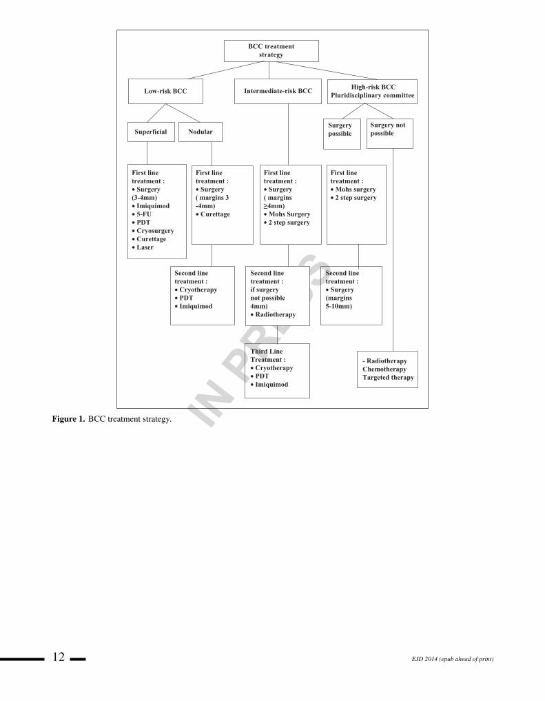
This review aims to present updated guidelines that includeconsensual expert definitions on various BCC types, prog-nosis and risk factors for BCC as well as recommendationsfor treatment reflecting current published evidence. Theprognostic groups of BCC first presented by the FrenchNational Guidelines were defined according to the likeli-hood of cure and are based on data from large retrospectivestudies that have revealed prognostic factors involved inthe risk of recurrence. Though this classification has notbeen validated in prospective studies, the experts partic-ipating in the BCC EDF guidelines subcommittee, afterextensive discussions and review of the current literature,agreed unanimously to present them as such in order to helpguide the choice of treatment. Figure 1 shows a treatmentalgorithm with options according to the risk of recurrenceof the tumour and table 2 summarizes the treatment recom-mendations for primary, recurrent or incompletely-excisedBCC, as well as for locally advanced and metastatic BCCnaccording to the existing evidence. We hope these tools
Journal Identification = EJD Article Identification = 2271 Date: April 3, 2014 Time: 1:31 pm
12 EJD 2014 (epub ahead of print)
BCC treatmentstrategy
Low-risk BCC Intermediate-risk BCC High-risk BCCPluridisciplinary committee
Superficial Nodular
First linetreatment :• Surgery(3-4mm)• Imiquimod• 5-FU• PDT• Cryosurgery• Curettage• Laser
First linetreatment :• Surgery( margins 3-4mm)• Curettage
Second linetreatment :• Cryotherapy• PDT• Imiquimod
First linetreatment :• Surgery( margins!4mm)• Mohs Surgery• 2 step surgery
Second linetreatment :if surgerynot possible4mm)• Radiotherapy
Third LineTreatment :• Cryotherapy• PDT• Imiquimod
Surgerypossible
Surgery notpossible
First linetreatment :• Mohs surgery• 2 step surgery
- RadiotherapyChemotherapyTargeted therapy
Second linetreatment :• Surgery(margins5-10mm)
Figure 1. BCC treatment strategy.

Journal Identification = EJD Article Identification = 2271 Date: April 3, 2014 Time: 1:31 pm
EJD 2014 (epub ahead of print) 13
Table 2. Therapeutic recommendations according to existing evidence
SUMMARY OF THERAPEUTIC RECOMMENDATIONS FOR BCC
A. PRIMARY BCC:1. Surgery
- Surgical excision is a good treatment for primary BCC (Strength of recommendation: A, Quality of evidence I)- Mohs micrographic surgery is a good treatment for high-risk BCC (Strength of recommendation: A, Quality of evidence I)
2. Ablative TreatmentsI. Curettage and cautery (C/C)
- C/C is a good treatment for low-risk BCC (Strength of recommendation: A, quality of evidence II-iii)II. Cryosurgery
- Cryosurgery is a good treatment for low-risk BCC (Strength of recommendation: A, Quality of evidence II-ii)III. Carbon dioxide (CO2) laser ablation
- CO2 laser ablation may be effective in the treatment of low-risk BCC (Strength of recommendation: C, Quality of evidence III)
3. Topiccal Medical TreatmentsI. 5-FU:
- 5-Fluorouracil appears effective for the treatment of superficial BCC (Strength of Recommendation A, Quality of evidence I)II. Imiquimod:
- Topical Imiquimod appears effective in the treatment of primary small superficial BCC (Strength of recommendation A, Quality ofevidence I)
- Topical imiquimod may have a role in the treatment of primary nodular BCC (Strength of recommendation C, Quality of evidence I)III. Ingenol Mebutate:
- Presently no recommendation can be made for ingenol mebutate gel 0.05% for the treatment of BCCIV. Topical Retinoids:
- Presently no recommendation can be made for topical retinoids for the treatment of BCC
4. Photodynamic Therapy (PDT)- PDT appears effective for the treatment of superficial BCC (Strength of Recommendation A, Quality of evidence I)- PDT appears effective for the treatment of nodular BCC (Strength of Recommendation B, Quality of evidence I)
5. Radiotherapy- Radiotherapy is a good treatment for certain primary BCC (Strength of recommendation A, Quality of evidence I)
B. INCOMPLETELY EXCISED OR RECURRENT BCC:1. Surgery
- Tumours which have been incompletely excised, especially high-risk BCC and lesions incompletely excised at the deep margin areat high risk of recurrence and should be re-excised (Strength of recommendation A, Quality of evidence II-i)
- Mohs micrographic surgery is a good treatment for high-risk recurrent BCC (Strength of recommendation: A, Quality of evidence I)
2. Radiotherapy- Radiotherapy is a good treatment for recurrent BCC except if recurrence has followed previous RT (Strength of recommendation
A, Quality of evidence I)
C. LOCALLY ADVANCED OR METASTATIC BCC:1. Chemotherapy
- Presently no level of evidence supports the use of chemotherapy in the treatment of advanced BCC (Strength of recommendation:C, Quality of evidence IV)
2. Targeted Therapy- Anti-smo agents are effective against locally-advanced or metastatic BCC (Strength of recommendation A, Quality of evidence II-i)
will help clinicians with decision taking for BCC man-agement, pending large prospective studies that will shedmore light into BCC prognosis and response to differenttreatments. !
Disclosure. Financial support: none.Conflict of interest: N. Basset-Seguin is a board memberfor Roche, Meda and Leo companies, a consultant forRoche, Meda and Leo, member of the speaker’s bureau ofRoche and Leo and has received travelling/accommodationgrants by Roche, BMS and Galderma. C. Morton is aboard member for Leo and Almirall companies, memberof the speaker’s bureau of Leo and Galderma and hasreceived travelling/accommodation grants by Leo and Gal-derma. E. Nagorre is a member of the speaker’s bureau ofMeda and has received traveling/ accommodation grantsby Roche, Galderma and Meda. C. Ulrich has receivedresearch grants and was/is member of the speaker’s bureauof Almirall, MEDA, Novartis, Galderma, Pfizer and Spirig.
K Perris is a board member of Roche, Meda, Galdermaand Leo, a consultant for Roche, Meda and Leo a memberof the speaker’s bureau of Roche and has received trav-elling/accommodation grants by Roche and Leo. V. DelMarmol is a board member for Roche, Abbott and Leo anda member of the speaker’s bureau of Roche. M Trakatelliis a member of the speaker’s bureau of Meda and hasreceived travelling/accommodation grants from Janssen-Cilag, Meda, Leo, and Uriage.
References
1. Sterry W. European Dermatology Forum Guideline Committee.Guidelines: the management of basal cell carcinoma. Eur J Dermatol2006; 16: 467-75.

Journal Identification = EJD Article Identification = 2271 Date: April 3, 2014 Time: 1:31 pm
14 EJD 2014 (epub ahead of print)
2. Dandurand M, Petit T, Martel P, Guillot B. Management of basalcell carcinoma in adults: Clinical practice guidelines. Eur J Dermatol2006; 16: 394-401.3. Telfer NR, Colver GB, Morton CA. Guidelines for the managementof basal cell carcinoma. Br J Dermatol 2008; 159: 35-48.4. Rubin AI, Chen EH, Ratner D. Basal-cell carcinoma. N Engl J Med2005; 353: 2262-9.5. Trakatelli M, Ulrich C, del Marmol V, Euvrard S, Stockfleth E, AbeniD. Epidemiology of nonmelanoma skin cancer (NMSC) in Europe:accurate and comparable data are needed for effective public healthmonitoring and interventions. Br J Dermatol 2007; 156(Suppl 3):1-7.6. Halna JM, Grandadam M, Bueni A. Epidemiologic study of skincancers from french population (1988-1996). Report of cancer regis-tration of “Haut Rhin” area. Nouv Dermatol 2000; 19: 48-55.7. Vilar-Coromina N, Miró-Queralt J, Cano-Bautista A, Vilardell-Gil L,Torres Babié P, Marcos-Gragera R. Non-melanoma skin cancer: inci-dence time trends analysis in Girona, Spain, 1994-2007. Med Clin(Barc) 2011; 137: 145-51.8. Miller DL, Weinstock MA. Nonmelanoma skin cancer in the UnitedStates: incidence. J Am Acad Dermatol 1994; 30(5 Pt 1): 774-8.9. Birch-Johansen F, Jensen A, Mortensen L, Olesen AB, Kjær SK.Trends in the incidence of non melanoma skin cancer in Denmark1978-2007: Rapid incidence increase among young Danish women.Int J Cancer 2010; 127: 2190-8.10. Flohil SC, de Vries E, Neumann HA, Coebergh JW, Nijsten T. Inci-dence, prevalence and future trends of primary basal cell carcinomain the Netherlands. Acta Derm Venereol 2011; 91: 24-30.11. de Vries E, Micallef R, Brewster DH, et al. Population-basedestimates of the occurrence of multiple vs first primary basal cellcarcinomas in 4 European regions. Arch Dermatol 2012; 148:347-54.12. Gallagher RP, Hill GB, Bajdik CD, et al. Sunlight exposure, pig-mentary factors, and risk of nonmelanocytic skin cancer. I. Basal cellcarcinoma. Arch Dermatol 1995; 131: 157-63.13. Armstrong BK, Kricker A. The epidemiology of UV induced skincancer. J Photochem Photobiol 2001; 63: 8-18.14. Kricker A, Armstrong BK, English DR, Heenan PJ. Does intermit-tent sun exposure cause basal cell carcinoma? A case-control study inWestern Australia. Int J Cancer 1995; 60: 489-94.15. Rosso S, Zanetti R, Martinez C, et al. The multicentre south Euro-pean study ‘Helios’. II: Different sun exposure patterns in the aetiologyof basal cell and squamous cell carcinomas of the skin. Br J Cancer1996; 73: 1447-54.16. Iannacone MR, Wang W, Stockwell HG, et al. Patterns and timingof sunlight exposure and risk of basal cell and squamous cell carcino-mas of the skin–a case-control study. BMC Cancer 2012; 12: 417.17. Bauer A, Diepgen TL, Schmitt J. Is occupational solar ultravioletirradiation a relevant risk factor for basal cell carcinoma? A system-atic review and meta-analysis of the epidemiological literature. Br JDermatol 2011; 165: 612-25.18. Berg D, Otley CC. Skin cancer in organ transplant recipients:epidemiology, pathogenesis and management. J Am Acad Dermatol2002; 47: 1-17.19. Jensen P, Hansen S, Møller B, et al. Skin cancer in kidney andheart transplant recipients and different long-term immunosuppressivetherapy regimens. J Am Acad Dermatol 1999; 40(2 Pt 1): 177-86.20. Wisgerhof HC, Edelbroek JR, de Fijter JW, et al. Subsequentsquamous- and basal-cell carcinomas in kidney-transplant recipientsafter the first skin cancer: cumulative incidence and risk factors. Trans-plantation 2010; 89: 1231-8.21. Euvrard S, Kanitakis J, Decullier E, et al. Subsequent skin cancersin kidney and heart transplant recipients after the first squamous cellcarcinoma. Transplantation 2006; 81: 1093-100.22. Johnson RL, Rothman AL, Xie J, et al. Human homolog ofpatched, a candidate gene for the basal cell nevus syndrome. Science1996; 272: 1668-71.23. Teperino R, Amann S, Bayer M, et al. Hedgehog Partial Ago-nism Drives Warburg-like Metabolism in Muscle and Brown Fat. Cell2012; 151: 414-26.24. Knudson AG Jr. Mutation and cancer: statistical study ofretinoblastoma. Proc Natl Acad Sci USA 1971; 68: 820-3.
25. Pan S, Dong Q, Sun LS, Li TJ. Mechanisms of inactivation of PTCH1gene in nevoid basal cell carcinoma syndrome: modification of thetwo-hit hypothesis. Clin Cancer Res 2010; 16: 442-50.26. Boukamp P. Non-melanoma skin cancer: what drives tumor devel-opment and progression? Carcinogenesis 2005; 26: 1657-67.27. Epstein EH. Basal cell carcinomas: attack of the hedgehog. NatRev Cancer 2008; 8: 743-54.28. Castori M, Morrone A, Kanitakis J, Grammatico P. Geneticskin diseases predisposing to basal cell carcinoma. Eur J Dermatol2012; 22: 299-309.29. Liboutet M, Portela M, Delestaing G, et al. MC1R and PTCH genepolymorphism in French patients with basal cell carcinomas. J InvestDermatol 2006; 126: 1510-7.30. Stacey SN, Sulem P, Masson G, et al. New common vari-ants affecting susceptibility to basal cell carcinoma. Nat Genet2009; 41: 909-14.31. Rizzato C, Scherer D, Rudnai P, et al. POMC and TP53 geneticvariability and risk of basal cell carcinoma of skin: interaction betweenhost and genetic factors. J Dermatol Sci 2011; 63: 47-54.32. Rizzato C, Canzian F, Rudnai P, et al. Interaction between func-tional polymorphic variants in cytokine genes, established risk factorsand susceptibility to basal cell carcinoma of skin. Carcinogenesis2011; 32: 1849-54.33. Stacey SN, Gudbjartsson DF, Sulem P, et al. Common variantson 1p36 and 1q42 are associated with cutaneous basal cell car-cinoma but not with melanoma or pigmentation traits. Nat Genet2008; 40: 1313-8.34. Gudbjartsson DF, Sulem P, Stacey SN, et al. ASIP and TYR pig-mentation variants associate with cutaneous melanoma and basal cellcarcinoma. Nat Genet 2008; 40: 886-91.35. Scherer D, Bermejo JL, Rudnai P, et al. MC1R variants associ-ated susceptibility to basal cell carcinoma of skin: interaction withhost factors and XRCC3 polymorphism. Int J Cancer 2008; 122:1787-93.36. Steding-Jessen M, Birch-Johansen F, Jensen A, Schüz J, Kjær SK,Dalton SO. Socioeconomic status and non-melanoma skin cancer: anationwide cohort study of incidence and survival in Denmark. CancerEpidemiol 2010; 34: 689-95.37. Wang GY, Wang J, Mancianti ML, Epstein EH Jr. Basal cell car-cinomas arise from hair follicle stem cells in Ptch1(+/-) mice. CancerCell 2011; 19: 114-24.38. Youssef KK, Van Keymeulen A, Lapouge G, et al. Identificationof the cell lineage at the origin of basal cell carcinoma. Nat Cell Biol2010; 12: 299-305.39. Ratner D, Lowe L, Johnson TM, Fader DJ. Perineural spread ofbasal cell carcinomas treated with Mohs micrographic surgery. Cancer2000; 88: 1605-13.40. Sexton M, Jones DB, Maloney ME. Histologic pattern analysis ofbasal cell carcinoma. Study of a series of 1039 consecutive neoplasms.J Am Acad Dermatol 1990; 23(6 Pt 1): 1118-26.41. Scrivener Y, Grosshans E, Cribier B. Variations of basal cell car-cinomas according to gender, age, location and histopathologicalsubtype. Br J Dermatol 2002; 147: 41-7.42. Altamura D, Menzies SW, Argenziano G, et al. Dermoscopyof basal cell carcinoma: Morphologic variability of global andlocal features and accuracy of diagnosis. J Am Acad Dermatol2010; 62: 67-75.43. Guitera P, Menzies SW, Longo C, Cesinaro AM, Scolyer RA,Pellacani G. In vivo confocal microscopy for diagnosis of melanomaand basal cell carcinoma using a two-step method: analysisof 710 consecutive clinically equivocal cases. J Invest Dermatol2012; 132: 2386-94.44. Seidenari S, Arginelli F, Bassoli S, et al. Diagnosis of BCC bymultiphoton laser tomography. Skin Res Technol 2013; 19: e297-304.45. Mogensen M, Joergensen TM, Nürnberg BM, et al. Assess-ment of optical coherence tomography imaging in the diagnosis ofnon-melanoma skin cancer and benign lesions versus normal skin:observer-blinded evaluation by dermatologists and pathologists. Der-matol Surg 2009; 35: 965-72.46. Walling HW, Fosko SW, Geraminejad PA, Whitaker DC, ArpeyCJ. Aggressive basal cell carcinoma: presentation, pathogenesis, andmanagement. Cancer Metastasis Rev 2004; 23: 389-402.

Journal Identification = EJD Article Identification = 2271 Date: April 3, 2014 Time: 1:31 pm
EJD 2014 (epub ahead of print) 15
47. Bath-Hextall F, Bong J, Perkins W, Williams H. Interven-tions for basal cell carcinoma of the skin: systematic review. BMJ2004; 329: 705.48. Breuninger H, Dietz K. Prediction of subclinical tumor infiltrationin basal cell carcinoma. J Dermatol Surg Oncol 1991; 17: 574-8.49. Batra RS, Kelley LC. Predictors of extensive subclinical spread innonmelanoma skin cancer treated with Mohs micrographic surgery.Arch Dermatol 2002; 138: 1043-51.50. Ratner D, Bagiella E. The efficacy of curettage in delineatingmargins of basal cell carcinoma before Mohs micrographic surgery.Dermatol Surg 2003; 29: 899-903.51. Ro KW, Seo SH, Son SW, Kim IH. Subclinical infiltration of basalcell carcinoma in asian patients: assessment after mohs micrographicsurgery. Ann Dermatol 2011; 23: 276-81.52. Wolf DJ, Zitelli JA. Surgical margins for basal cell carcinoma. ArchDermatol 1987; 123: 340-4.53. Leibovitch I, Huilgol SC, Selva D, Richards S, Paver R. Basalcell carcinoma treated with Mohs surgery in Australia III. Perineuralinvasion. J Am Acad Dermatol 2005; 53: 458-63.54. Feasel AM, Brown TJ, Bogle MA, Tschen JA, Nelson BR. Per-ineural invasion of cutaneous malignancies. Dermatol Surg 2001; 27:531-42.55. Kuijpers DIM, Thissen MRTM, Neumann MHA. Basal cell carci-noma: treatment options and prognosis, a scientific approach to acommon malignancy. Am J Clin Dermatol 2002; 3: 247-59.56. Chiller K, Passaro D, McCalmont T, Vin-Christian K. Efficacy ofcurettage before excision in clearing surgical margins of nonmelanomaskin cancer. Arch Dermatol 2000; 136: 1327-32.57. Abide JM, Nahai F, Bennett RG. The meaning of surgical margins.Plast Reconstr Surg 1984; 73: 492-7.58. Gulleth Y, Goldberg N, Silverman RP, Gastman BR. What is thebest surgical margin for a Basal cell carcinoma: a meta-analysis of theliterature. Plast Reconstr Surg 2010; 126: 1222-31.59. Walker P, Hill D. Surgical treatment of basal cell carcinomas usingstandard postoperative histological assessment. Australas J Dermatol2006; 47: 1-12.60. Griffiths RW, Suvarna SK, Stone J. Do basal cell carcinomasrecur after complete conventional surgical excision? Br J Plast Surg2005; 58: 795-805.61. Silverman MK, Kopf AW, Bart RS, Grin CM, Levenstein MS. Recur-rence rates of treated basal cell carcinomas. Part 3: Surgical excision.J Dermatol Surg Oncol 1992; 18: 471-6.62. Rowe DE, Carroll RJ, Day CL Jr. Long-term recurrence rates inpreviously untreated (primary) basal cell carcinoma: implications forpatient follow-up. J Dermatol Surg Oncol 1989; 15: 315-28.63. Rowe DE, Carroll RJ, Day CL Jr. Mohs surgery is the treatmentof choice for recurrent (previously treated) basal cell carcinoma. JDermatol Surg Oncol 1989; 15: 424-31.64. Mosterd K, Krekels GAM, Nieman FH, et al. Surgical excisionversus Mohs’ micrographic surgery for primary and recurrent basal-cell carcinoma of the face: a prospective randomised controlled trialwith 5-years’ follow-up. Lancet Oncol 2008; 9: 1149-56.65. Kumar P, Orton CI, McWilliam LJ, Watson S. Incidence of incom-plete excision in surgically treated basal cell carcinoma: a retrospectiveclinical audit. Br J Plast Surg 2000; 53: 563-6.66. Dieu T, Macleod AM. Incomplete excision of basal cell carcino-mas: a retrospective audit. ANZ J Surg 2002; 72: 219-21.67. Nagore E, Grau C, Molinero J, Fortea JM. Positive marginsin basal cell carcinoma: relationship to clinical features and recur-rence risk. A retrospective study of 248 patients. J Eur Acad DermatolVenereol 2003; 17: 167-70.68. De Silva SP, Dellon AL. Recurrence rate of positive margin basalcell carcinoma: results of a five-year prospective study. J Surg Oncol1985; 28: 72-4.69. Park AJ, Strick M, Watson JD. Basal cell carcinomas: do they needto be followed up? J R Coll Surg Edinb 1994; 39: 109-11.70. Sei JF. [Excision limits and reoperation in cutaneous carcinoma].Ann Dermatol Venereol 1997; 124: 421-6.71. Wilson AW, Howsam G, Santhanam V, et al. Surgical manage-ment of incompletely excised basal cell carcinomas of the head andneck. Br J Oral Maxillofac Surg 2004; 42: 311-4.
72. Menn H, Robins P, Kopf AW, Bart RS. The recurrent basal cellepithelioma. A study of 100 cases of recurrent, re-treated basal cellepitheliomas. Arch Dermatol 1971; 103: 628-31.73. Angulo J, Serra-Guillén C, Traves V, et al. [Mohs micrographicsurgery for repeat excision of basal cell carcinomas on the head withpositive margins]. Actas Dermosifiliogr 2011; 102: 797-804.74. Boulinguez S, Grison-Tabone C, Lamant L, et al. Histologicalevolution of recurrent basal cell carcinoma and therapeutic impli-cations for incompletely excised lesions. Br J Dermatol 2004; 151:623-6.75. Robinson JK, Fisher SG. Recurrent basal cell carcinoma afterincomplete resection. Arch Dermatol 2000; 136: 1318-24.76. Bieley HC, Kirsner RS, Reyes BA, Garland LD. The use of Mohsmicrographic surgery for determination of residual tumor in incom-pletely excised basal cell carcinoma. J Am Acad Dermatol 1992; 26(5Pt 1): 754-6.77. Swanson NA. Mohs surgery. Technique, indications, applications,and the future. Arch Dermatol 1983; 119: 761-73.78. Williford PM, Feldman SR. Surgery for basal-cell carcinoma of theface. Lancet 2004; 364: 1732-3.79. Emmett AJ. Surgical analysis and biological behaviour of 2277basal cell carcinomas. ANZ J Surg 1990; 60: 855-63.80. Miller PK, Roenigk RK, Brodland DG, Randle HW. Cuta-neous micrographic surgery: Mohs procedure. Mayo Clin Proc1992; 67: 971-80.81. Swanson NA. Mohs surgery. Technique, indications, applica-tions, and the future. Arch Dermatol 1983; 119: 761-73.82. Smeets NWJ, Kuijpers DIM, Nelemans P, et al. Mohs’ micro-graphic surgery for treatment of basal cell carcinoma of the face–resultsof a retrospective study and review of the literature. Br J Dermatol2004; 151: 141-7.83. Paoli J, Daryoni S, Wennberg AM, et al. 5-year recurrence ratesof Mohs micrographic surgery for aggressive and recurrent facial basalcell carcinoma. Acta Derm Venereol 2011; 91: 689-93.84. Mohs F, Larson P, Iriondo M. Micrographic surgery for the micro-scopically controlled excision of carcinoma of the external ear. J AmAcad Dermatol 1988; 19: 729-37.85. Mohs FE. Micrographic surgery for the microscopically con-trolled excision of eyelid cancers. Arch Ophthalmol 1986; 104:901-9.86. Malhotra R, Huilgol SC, Huynh NT, Selva D. The Australian Mohsdatabase, part II: periocular basal cell carcinoma outcome at 5-yearfollow-up. Ophthalmology 2004; 111: 631-6.87. Leibovitch I, Huilgol SC, Selva D, Richards S, Paver R. Basal cellcarcinoma treated with Mohs surgery in Australia II. Outcome at 5-yearfollow-up. J Am Acad Dermatol 2005; 53: 452-7.88. Cook J, Zitelli JA. Mohs micrographic surgery: a cost analysis. JAm Acad Dermatol 1998; 39(5 Pt 1): 698-703.89. Blázquez-Sánchez N, de Troya-Martín M, Frieyro-Elicegui M,Fúnez-Liébana R, Martín-Márquez L, Rivas-Ruiz F. [Cost analysis ofMohs micrographic surgery in high-risk facial basal cell carcinoma].Actas Dermosifiliogr 2010; 101: 622-8.90. Spiller WF, Spiller RF. Treatment of basal cell epithelioma bycurettage and electrodesiccation. J Am Acad Dermatol 1984; 11(5 Pt1): 808-14.91. Suhge d’Aubermont PC, Bennett RG. Failure of curettage andelectrodesiccation for removal of basal cell carcinoma. Arch Dermatol1984; 120: 1456-60.92. Kopf AW, Bart RS, Schrager D, Lazar M, Popkin GL. Curettage-electrodesiccation treatment of basal cell carcinomas. Arch Dermatol1977; 113: 439-43.93. Chren M-M, Torres JS, Stuart SE, Bertenthal D, Labrador RJ,Boscardin WJ. Recurrence after treatment of nonmelanoma skin can-cer: a prospective cohort study. Arch Dermatol 2011; 147: 540-6.94. Barlow JO, Zalla MJ, Kyle A, DiCaudo DJ, Lim KK, Yiannias JA.Treatment of basal cell carcinoma with curettage alone. J Am AcadDermatol 2006; 54: 1039-45.95. Thissen MR, Nieman FH, Ideler AH, Berretty PJ, Neumann HA.Cosmetic results of cryosurgery versus surgical excision for primaryuncomplicated basal cell carcinomas of the head and neck. DermatolSurg 2000; 26: 759-64.

Journal Identification = EJD Article Identification = 2271 Date: April 3, 2014 Time: 1:31 pm
16 EJD 2014 (epub ahead of print)
96. Kuflik EG. Cryosurgery for skin cancer: 30-year experience andcure rates. Dermatol Surg 2004; 30(2 Pt 2): 297-300.97. Hall VL, Leppard BJ, McGill J, Kesseler ME, White JE, GoodwinP. Treatment of basal-cell carcinoma: comparison of radiotherapy andcryotherapy. Clin Radiol 1986; 37: 33-4.98. Jaramillo-Ayerbe F. Cryosurgery in difficult to treat basal cell car-cinoma. Int J Dermatol 2000; 39: 223-9.99. Tuppurainen K. Cryotherapy for eyelid and periocular basal cellcarcinomas: outcome in 166 cases over an 8-year period. GraefesArch Clin Exp Ophthalmol 1995; 233: 205-8.100. Nouri K, Chang A, Trent JT, Jiménez GP. Ultrapulse CO2 usedfor the successful treatment of basal cell carcinomas found in patientswith basal cell nevus syndrome. Dermatol Surg 2002; 28: 287-90.101. Gross K, Kircik L, Kricorian G. 5% fuorouacile cream for thetreatment of small superficial basal cell carcinoma: efficacy, cosmeticoutcome, and patient satisfaction Dermatol Surg 2007; 33: 433-40.102. Arits AH, Mosterd K, Essers BA, et al. Photodynamic therapyversus topical imiquimod versus topical fluorouracil for treatment ofsuperficial basal-cell carcinoma: a single blind, non-inferiority, ran-domised controlled trial. Lancet Oncol 2013; 7: 647-54.103. Schon MP, Schon M. Imiquimod : mode of action. Br J Dermatol2007; 157 (suppl 2): 8-13.104. Wuest M, Dummer R, Urosevic M. Induction of the membersof Notch pathway in superfical basal cell carcinomas treated withimiquimod. Arch Dermatol Res 2007; 299: 493-8.105. Schulze HJ, Cribier B, Requena L, et al. Imiquimod 5% creamfor the treatment of superficail basal cell carcinoma: results from arandomized vehicle-controlled study. Br J Dermatol 2005; 152: 939-47.106. Gollnick H, Barona CG, Frank RG, et al. Recurrence rate ofsuperficial basal cell carcinoma following successful treatment withimiquimod 5% cream: conclusion of a 5 year long term follow up studyin Europe. Eur J Dermatol 2008; 18: 677-82.107. Bath-Hextall FJ, Perkins W, Bong J,Williams HC. Interventionsfor basal cell carcinoma of the skin. Cochrane Data Syst Rev 2007;CD003412.108. Geisse J, Caro I, Lindholm J, Golitz I, Stampone P, Owens M.Imiquimod 5% cream for the treatment of superficial basal cell carci-noma: results from two phase III randomized vehicle controlled studies.J Am Acad Dermatol 2004; 50: 722-33.109. Sterry W, Ruzicka T, Herrera E, et al. Imiquimod cream 5%cream for the treatment of superficial and nodular basal cell carcinoma:randomized studies comparing low frequency dosing with and withoutocclusion. Br J Dermatol 2002; 147: 1227-36.110. Ebell MH, Siwek J, Weiss BD. Strength of recommendation tax-onomy (SORT): A patient-centered approach to grading evidence inmedical litterature. Am Fam Physician 2004; 69: 548-56.111. Love E, Bernhard J, Bordeaux J. Topical imiquimod or fluouraciltherapy for basal and squamous cell carcinoma-systematic review.Arch Dermatol 2009; 145: 1431-8.112. Quikck C, Gebauer K, De Ambrosis B, Slacke H, Meng T.Sustained clearance of superficial basal cell carcinomas treated withimiquimod cream 5%: results of a prospective 5-year Study. Cutis2010; 85: 318-24.113. Vidal D, Matias-Gulu X, Alomar A. An open study of the efficacyand mechanism of action of topical imiquimod in basal carcinoma. ClinExp Dermatol 2004; 29: 518-25.114. Vidal D, Matias-Gulu X, Alomar A. Forty basal cell carcinomatreated with topical imiquimod: outcome 5 years follow up. Arch Der-matol 2007; 143: 266-8.115. Vanalocha F, Dauden E, Badia X, et al. Cost-effectiveness oftreatment of superficial basal cell carcinoma: surgical excision vs.imiquimod 5% cream. Br J Dermatol 2007; 156: 769-71.116. Basset-Seguin N, Ibbotson SH, Emtestam L, et al. Topicalmethyl-aminolaevulinate photodynamic therapy versus cryotherapy forsuperficial basal cell carcinoma: a 5 year randomized trial. Eur J Der-matol 2008; 18: 547-53.117. Szeimies RM, Ibbotson S, Murrell DF, et al. A clinical study com-paring methyl aminolevulinate photodynamic therapy and surgery insmall superficial basal cell carcinoma (8-20 mm), with a 12-monthfollow-up. J Eur Acad Dermatol Venereol 2008; 22: 1302-11.
118. Peng Q, Warloe T, Berg K, et al. 5-Aminolevulinic acid-basedphotodynamic therapy. Clinical research and future challenges. Can-cer 1997; 79: 2282-308.119. Wang I, Bendsoe N, Klinteberg CA, et al. Photodynamic therapyvs. cryosurgery of basal cell carcinomas: results of a phase III clinicaltrial. Br J Dermatol 2001; 144: 832-40.120. Rhodes LE, de Rie M, Enstrom Y, et al. Photodynamic therapyusing topical methyl aminolevulinate vs surgery for nodular basal cellcarcinoma: results of a multicenter randomized prospective trial. ArchDermatol 2004; 140: 17-23.121. Rhodes LE, de Rie MA, Leifsdottir R, et al. Five-year follow-upof a randomized prospective trial of topical methyl aminolevulinate-photodynamic therapy versus surgery for nodular basal cell carcinoma.Arch Dermatol 2007; 143: 1131-6.122. Foley P, Freeman M, Menter A, et al. Photodynamic therapywith methyl aminolevulinate for primary nodular basal cell carcinoma:results of two randomized studies. Int J Dermatol 2009; 48: 1236-45.123. Christensen E, Skogvoll E, Viset T, Warloe T, Sundstrøm S. Pho-todynamic therapy with 5-aminolaevulinic acid, dimethylsulfoxide andcurettage in basal cell carcinoma: a 6-year clinical and histologicalfollow-up. J Eur Acad Dermatol Venereol 2009; 23: 58-66.124. Fantini F, Greco A, Del Giovane C, et al. Photodynamic ther-apy for basal cell carcinoma: clinical and pathological determinantsof response. J Eur Acad Dermatol Venereol 2011; 25: 896-901.125. Kuijpers D, Thissen MR, Thissen CA, Neumann MH. Similareffectiveness of methyl aminolevulinate and 5-aminolevulinate in topi-cal photodynamic therapy for nodular basal cell carcinoma. J DrugsDermatol 2006; 5: 642-5.126. de Haas ER, Kruijt B, Sterenborg HJ, et al. Fractionated illu-mination significantly improves the response of superficial basal cellcarcinoma to aminolevulinic acid photodynamic therapy. J Invest Der-matol 2006; 126: 2679-86.127. de Haas ER, de Vijlder HC, Sterenborg HJ, et al. Fraction-ated aminolevulinic acid-photodynamic therapy provides additionalevidence for the use of PDT for non-melanoma skin cancer. J Eur AcadDermatol Venereol 2008; 22: 426-30.128. Mosterd K, Thissen M, Nelemans P, et al. Fractionated 5-aminolaevulinic acid-photodynamic therapy vs. surgical excision in thetreatment of nodular basal cell carcinoma: results of a randomizedcontrolled trial. Br J Dermatol 2008; 159: 864-70.129. Loncaster J, Swindell R, Slevin F, et al. Efficacy of photodynamictherapy as a treatment for Gorlin Syndrome-related basal cell carcino-mas. Clin Oncol 2009; 21: 502-8.130. Pauwels C, Mazereeuw-Hautier J, Basset-Seguin N, et al. Topicalmethyl aminolevulinate photodynamic therapy for management ofbasal cell carcinomas in patients with basal cell nevus syndromeimproves patient’s satisfaction and reduces the need for surgical pro-cedures. J Eur Acad Dermatol Venereol 2011; 25: 861-4.131. Schleier P, Hyckel P, Berndt A, et al. Photodynamic therapyof virus-associated epithelial tumours of the face in organ transplantrecipients. J Cancer Res Clin Oncol 2004; 130: 279-84.132. Wulf HC, Pavel S, Stender I, Bakker-Wensveen C. Topical photo-dynamic therapy for prevention of new skin lesions in renal transplantrecipients. Acta Derm Venereol 2006; 86: 25-8.133. Ibbotson SI. Adverse effects of topical photodynamic therapy.Photoderm Photoimmunol Photomedicine 2011; 27: 116-30.134. Grapengiesser S, Ericson M, Gudmundsson F, et al. Paincaused by photodynamic therapy of skin cancer. Clin Exp Dermatol2002; 27: 493-7.135. Pagliaro J, Elliott T, Bulsara M, et al. Cold air analgesia inphotodynamic therapy of basal cell carcinomas and Bowen’s dis-ease: an effective addition to treatment: a pilot study. Dermatol Surg2004; 30: 63-6.136. Attili SK, Lesar A, McNeill A, et al. An open pilot study of ambu-latory photodynamic therapy using a wearable low-irradiance organiclight-emitting diode light source in the treatment of nonmelanoma skincancer. Br J Dermatol 2009; 161: 170-3.137. Caekelbergh K, Annemans L, Lambert J, Roelands R. Economicevaluation of methyl aminolevulinate photodynamic therapy in the man-agement of actinic keratoses and basal cell carcinoma. Br J Dermatol2006; 155: 784-90.

Journal Identification = EJD Article Identification = 2271 Date: April 3, 2014 Time: 1:31 pm
EJD 2014 (epub ahead of print) 17
138. Annemans L, Roelandts R, Boonen H, et al. Real-life practicestudy of the clinical outcome and cost-effectiveness of photodynamictherapy using methyl aminolevulinate (MAL-PDT) in the manage-ment of actinic keratosis and basal cell carcinoma. Eur J Dermatol2008; 18: 539-46.139. Morton CA, MacKie RM, Whitehurst C, et al. Photodynamictherapy for basal cell carcinoma: effect of tumour thickness andduration of photosensitizer application on response. Arch Dermatol1998; 134: 248-9.140. Dubin N, Kopf A. Multivariate risk score for recurrence of cuta-neous basal cell carcinomas. Arch Dermatol 1983; 119: 373-7.141. Hall VL, Leppard BJ, McGill J, Kesseler ME, White JE, GoodwinP. Treatment of basal-cell carcinoma: comparison of radiotherapy andcryotherapy. Clin Radiol 1986; 37: 33-4.142. Rowe DE, Carroll RJ, Day CL Jr. Long-term recurrence rates inpreviously untreated (primary) basal cell carcinoma: implications forpatient follow-up. J Dermatol Surg Oncol 1989; 15: 315-28.143. Rowe DE, Carroll RJ, Day CL Jr. Mohs surgery is the treatmentof choice for recurrent (previously treated) basal cell carcinoma. J Der-matol Surg Oncol 1989; 15: 424-31.144. Avril MF, Auperin A, Margulis A, et al. Basal cell carcinoma ofthe face: surgery or radiotherapy? Results of a randomized study. Br JCancer 1997; 76: 100-6.145. Silverman MK, Kopf AW, Gladstein AH, Bart RS, Grin CM,Levenstein MJ. Recurrence rates of treated basal cell carcinomas. Part4: X-ray therapy. J Dermatol Surg Oncol 1992; 18: 549-54.146. Cooper JS. Radiation therapy for cancers of the skin. DermatolClin 1991; 9: 683-7.147. Orton CI. The treatment of basal cell carcinoma by radiotherapy.Clin Oncol 1978; 4: 317-22.148. Smith SP, Foley EH, Grande DJ. Use of Mohs micrographicsurgery to establish quantitative proof of heightened tumor spread inbasal cell carcinoma recurrent following radiotherapy. J Dermatol SurgOncol 1990; 16: 1012-6.149. Smith SP, Grande DJ. Basal cell carcinoma recurring after radio-therapy: a unique, difficult treatment subclass of recurrent basal cellcarcinoma. J Dermatol Surg Oncol 1991; 17: 26-30.150. Zagrodnik B, Kempf W, Seifert B, et al. Superficial radiotherapyfor patients with basal cell carcinoma: recurrence rates, histologic sub-types, and expression of p53 and Bcl-2. Cancer 2003; 98: 2708-14.151. van Hezewijk M, Creutzberg CL, Putter H, et al. Efficacy of ahypofractionated schedule in electron beam radiotherapy for epithelialskin cancer: Analysis of 434 cases. Radiother Oncol 2010; 95: 245-9.152. Jackson JE, Dickie GJ, Wiltshire KL, et al. Radiotherapy for per-ineural invasion in cutaneous head and neck carcinomas: toward arisk-adapted treatment approach. Head Neck 2009; 31: 604-10.153. Wadhera A, Fazio M, Bricca G, Stanton O. Metastatic basalcell carcinoma: a case report and literature review. How accurate isour incidence data? Dermatol Online J 2006; 12: 7.154. Raszewski RL, Guyuron B. Long-term survival following nodalmetastases from basal cell carcinoma. Ann Plast Surg 1990; 24: 170-5.155. Guthrie TH Jr., Porubsky ES, Luxenberg M, et al. Cisplatin-basedchemotherapy in advanced basal and squamous cell carcinomas of theskin: results in 28 patients including 13 patients receiving multimodalitytherapy. J Clin Oncol 1990; 8: 342-6.156. Moeholt K, Aagaard H, Pfeiffer P, Hansen O. Platinum-basedcytotoxic therapy in basal cell carcinoma – a review of the literature.Acta Oncol 1996; 35: 677-82.157. Carneiro BA, Watkin WG, Mehta UK, et al. Metastatic basalcell carcinoma: complete response to chemotherapy and associatedpure red cell aplasia. Cancer Invest 2006; 24: 396-400.158. Ganti A, Kessinger A. Systemic therapy for disseminated basalcell carcinoma: An uncommon manifestation of a common cancer.Cancer Treat Rev 2011; 37: 440-3.159. Von Hoff DD, LoRusso PM, Rudin CM, et al. Inhibition of thehedgehog pathway in advanced basal-cell carcinoma. N Engl J Med2009; 361: 1164-72.160. LoRusso PM, Rudin CM, Reddy JC, et al. Phase I trial ofhedgehog pathway inhibitor vismodegib (GDC-0449) in patients withrefractory, locally advanced or metastatic solid tumors. Clin CancerRes 2011; 17: 2502-11.
161. Sekulic A, Migden MR, Oro AE, et al. Efficacy and safetyof vismodegib in advanced basal-cell carcinoma. N Engl J Med2012; 366: 2171-9.162. Tang JY, Mackay-Wiggan JM, Aszterbaum M, et al. Inhibitingthe hedgehog pathway in patients with the basal-cell nevus syndrome.N Engl J Med 2012; 366: 2180-8.163. Goldberg LH, Landau JM, Moody MN, Kazakevich N, HolzerAM, Myers A. Resolution of odontogenic keratocysts of the jaw in basalcell nevus syndrome with GDC-0449. Arch Dermatol 2011; 147: 839-41.164. Anderson L, Schmieder GJ, Werschler WP, et al. Randomized,double-blind, double-dummy, vehicle-controlled study of ingenol mebu-tate gel 0.025% and 0.05% for actinic keratosis. J Am Acad Dermatol2009; 60: 934-43.165. Ramsay JR, Suhrbier A, Aylward JH, et al. The sap from Euphor-bia peplus is effective against human nonmelanoma skin cancers. Br JDermatol 2011; 164: 633-6.166. Siller G, Rosen R, Freeman M, Welburn P, Katsamas J,Ogbourne SM. PEP005 (ingenol mebutate) gel for the topical treatmentof superficial basal cell carcinoma: results of a randomized phase IIatrial. Australas J Dermatol 2010; 51: 99-105.167. National Comprehensive Cancer Network Clinical PracticeGuidelines in Oncology. Basal Cell and Squamous Cell SkinCancers 2011. http://www.nccn.org/professionals/physician_gls/pdf/nmsc.pdf (accessed June 3, 2011).168. Chen J, Ruczinski I, Jorgensen TJ, et al. Nonmelanoma skincancer and risk for subsequent malignancy. J Natl Cancer Inst2008; 100: 1215-22.169. Mc Loone NM, Tolland J, Walsh M, et al. Follow-up of basal cellcarcinomas: an audit of current practice. J Eur Acad Dermatol Venereol2006; 20: 698-701.170. Kiiski V, de Vries E, Flohil SC, et al. Risk Factors for Single andMultiple Basal Cell Carcinomas. Arch Dermatol 2010; 146: 848-55.171. Marcil I, Stern RS. Risk of developing a subsequent non-melanoma skin cancer in patients with a history of nonmelanoma skincancer: a critical review of the literature and meta-analysis. Arch Der-matol 2000; 136: 1524-30.172. van Iersel CA, van de Velden HV, Kusters CD, et al. Prognosticfactors for a subsequent basal cell carcinoma: implications for followup. Br J Dermatol 2005; 153: 1078-80.173. Lovatt TJ, Lear JT, Bastrilles J, et al. Associations betweenultraviolet radiation, basal cell carcinoma site and histology, host char-acteristics, and rate of development of further tumors. J Am AcadDermatol 2005; 52(3 Pt 1): 468-73.174. Silverman MK, Kopf AW, Grin CM, et al. Recurrence rates oftreated basal cell carcinomas. Part 2: curettage-electrodesiccation. JDermatol Surg Oncol 1991; 17: 720-6.175. Rowe DE, Carroll RJ, Day CL. Long-term recurrence rates in previ-ously untreated (primary) basal cell carcinoma: implications for patientfollow-up. J Dermatol Surg Oncol 1989; 15: 315-28.176. Lin JS, Eder M, Weinmann S, Zuber SP, Beil TL, Plaut D, et al.Behavioral Counseling to Prevent Skin Cancer: Systematic EvidenceReview to Update the 2003 U.S. Preventive Services Task Force Rec-ommendation: 2011, Rockville MD.177. Kutting B, Drexler H. UV-induced skin cancer at workplaceand evidence-based prevention. Int Arch Occup Environ Health2010; 83: 843-54.178. Corona R, Dogliotti E, D’Errico M, et al. Risk factors for basalcell carcinoma in a Mediterranean population: role of recreational sunexposure early in life. Arch Dermatol 2001; 137: 1162-8.179. Gon A, Minelli L. Risk factors for basal cell carcinoma in asouthern Brazilian population: a case-control study. Int J Dermatol2011; 50: 1286-90.180. Grodstein F, Speizer FE, Hunter DJ. A prospective study of inci-dent squamous cell carcinoma of the skin in the nurses’ health study. JNatl Cancer Inst 1995; 87: 1061-6.181. Hunter DJ, Colditz GA, Stampfer MJ, Rosner B, Willett WC,Speizer FE. Risk factors for basal cell carcinoma in a prospective cohortof women. Ann Epidemiol 1990; 1: 13-23.182. Green A, Battistutta D, Hart V, et al. The Nambour Skin Cancerand Actinic Eye Disease Prevention Trial: design and baseline charac-teristics of participants. Control Clin Trials 1994; 15: 512-22.

Journal Identification = EJD Article Identification = 2271 Date: April 3, 2014 Time: 1:31 pm
18 EJD 2014 (epub ahead of print)
183. Green A, Williams G, Neale R, et al. Daily sunscreen applica-tion and betacarotene supplementation in prevention of basal-cell andsquamous-cell carcinomas of the skin: a randomised controlled trial.Lancet 1999; 354: 723-9.184. van der Pols JC, Williams GM, Pandeya N, Logan V, GreenAC. Prolonged prevention of squamous cell carcinoma of the skin byregular sunscreen use. Cancer Epidemiol Biomarkers Prev 2006; 15:2546-8.185. Kricker A, Armstrong BK, English DR, Heenan PJ. Pigmentaryand cutaneous risk factors for non-melanocytic skin cancer–a case-control study. Int J Cancer 1991; 48: 650-62.186. Rosso S, Joris F, Zanetti R. Risk of basal and squamous cell car-cinomas of the skin in Sion, Switzerland: a case-control study. Tumori1999; 85: 435-42.187. Pandeya N, Purdie DM, Green A, Williams G. Repeated occur-rence of basal cell carcinoma of the skin and multifailure survivalanalysis: follow-up data from Nambour Skin Cancer Prevention Trial.Am J Epidemiol 2005; 161: 748-54.
188. Gordon LG, Scuffham PA, van der Pols JC, McBride P, WilliamsGM, Green AC. Regular sunscreen use is a cost-effective approachto skin cancer prevention in subtropical settings. J Invest Dermatol2009; 129: 2766-71.189. Robinson JK, Rademaker AW. Relative importance of prior basalcell carcinomas, continuing sun exposure, and circulating T lympho-cytes on the development of basal cell carcinoma. J Invest Dermatol1992; 99: 227-31.190. Bakos RM, Kriz M, Muhlstadt M, Kunte C, Ruzicka T, Berking C.Risk factors for early-onset basal cell carcinoma in a German institution.Eur J Dermatol 2011; 21: 705-9.191. Ulrich C, Jurgensen JS, Degen A, et al. Prevention of non-melanoma skin cancer in organ transplant patients by regular use of asunscreen: a 24 months, prospective, case-control study. Br J Dermatol2009; 161 (Suppl 3): 78-84.192. Walther U, Kron M, Sander S, et al. Risk and protective factorsfor sporadic basal cell carcinoma: results of a two-centre case-controlstudy in southern Germany. Clinical actinic elastosis may be a protec-tive factor. Br J Dermatol 2004; 151: 170-8.