MicroRNA profiles classify papillary renal cell carcinoma ...
Upload
independentCategory
view
0download
0
Relationship of Treatment Delay with Surgical Defect Size fromKeratinocyte Carcinoma (Basal Cell Carcinoma and SquamousCell Carcinoma of the Skin)
Melody J. Eide*,†,‡, Martin A. Weinstock*,‡,§, Raymond G. Dufresne Jr§, SulekaNeelagaru‡, Patricia Risica*,¶, Gary J. Burkholder¶, David Upegui¶, Katharine A. Phillips#,Bruce K. Armstrong**, and Leslie Robinson-Bostom§,††* Department of Community Health and;
† Center for Gerontology and Healthcare Research, Brown University, Providence, Rhode Island,USA;
‡ Department of Veterans Affairs Medical Center, Providence, Rhode Island, USA;
§ Department of Dermatology, Brown Medical School, Providence, Rhode Island, USA;
¶ Institute for Community Health Promotion, Brown University, Providence, Rhode Island, USA;
# Department of Psychiatry and Human Behavior, Brown Medical School, Providence, RhodeIsland, USA;
** School of Public Health, University of Sydney, Sydney, Australia;
†† Department of Pathology, Brown Medical School, Providence, Rhode Island, USA
AbstractLarger keratinocyte carcinoma (KC) lesions are associated with higher morbidity. This studyexamined the association of potentially modifiable characteristics, including treatment delay, withKC defect size after Mohs micrographic surgery (MMS). A stratified random sample of patientstreated for KC with MMS were selected for telephone interview. Two hundred and nineteeninterviews were completed (refusal rate 24%). Regression models were used to examine thepredictors to defect size and delay. Anatomic site, age, histology, and gender predicted defect size(R2 =0.39) and were used as control variables. Self-reported delay between initial physicianexamination and MMS predicted defect size (p =0.0004), with greater than 1 y delay being associatedwith a doubling of defect size (adjusted odds ratio (OR) 2.0; 95% confidence interval (CI) 1.3–3.1).Delays of this duration were associated with initial examination by a primary provider (unadjustedOR 3.9; 95% CI 1.7–8.8), misdiagnosis (unadjusted OR 6.8; 95% CI 2.5–18.7), being treated withoutbiopsy (unadjusted OR 23.3; 95% CI 6.5–83.7), and multiple surgical removals (unadjusted OR 6.2;95% CI 2.5–15.5). All but provider specialty were independent predictors of delay. Attention toprocesses of care delivery for KC may have a greater impact on morbidity than efforts are earlierdetection by the public.
Address correspondence to: Melody J. Eide, MD, MPH, and Martin A. Weinstock, MD PhD, VA Medical Center Providence, 830Chalkstone Avenue (111D), Providence, Rhode Island 02908, USA. Email: [email protected] study was supported by the Agency for Healthcare Research & Quality (AHRQ), Institutional National Research Service Award(NRSA) HS 00011-16 (Dr Eide) and by grants CSP 402 from the Department of Veterans Affairs, Office of Research and Developmentand CA 78800 from the National Cancer Institute (NCI) (Dr Weinstock). The methods used in this research were derived in part fromthose developed by Dilhani Bandaranayake and Afaf Girgis of the University of Newcastle, under the supervision of one of the authors(Dr Armstrong). Findings were presented at the Society for Investigative Dermatology Annual Meeting, Providence, Rhode Island, May1, 2004.
NIH Public AccessAuthor ManuscriptJ Invest Dermatol. Author manuscript; available in PMC 2006 October 17.
Published in final edited form as:J Invest Dermatol. 2005 February ; 124(2): 308–314.
NIH
-PA Author Manuscript
NIH
-PA Author Manuscript
NIH
-PA Author Manuscript
Keywordscancer control; health services research; Mohs micrographic surgery; skin cancer
AbbreviationsBCC, basal cell carcinoma; CI, confidence interval; KC, keratinocyte carcinoma; MMS, Mohsmicrographic surgery; OR, odds ratio; SCC, squamous cell carcinoma
Skin cancer is the most common cancer in the United States. The American Cancer Societyestimates that more than one million cases of keratinocyte carcinoma (KC) (Weinstock et al,2001), which includes basal cell carcinoma (BCC) and squamous cell carcinoma (SCC), arediagnosed each year (Jemal et al, 2003). Mortality from KC is relatively low. They, however,usually arise in cosmetically sensitive areas, such as the face, and advanced lesions areassociated with higher morbidity and more frequent recurrence (Miller, 1991; Smeets et al,2004). The potentially modifiable factors that contribute to more advanced lesions at thediagnosis of the KC have not been established.
Skin cancer information campaigns and screening programs are based on the presumption thatearlier disease diagnosis can minimize disease impact (Silfen et al, 2002). In the last severaldecades there have been some investigations of the association of delay, either by patient orprovider, and the extent of tumor invasion, with the overwhelming majority of these studiesfocusing on melanoma (Levine et al, 1981; Temoshok et al, 1984, 1985; Cassileth et al,1988; Rampen et al, 1989; Dunkley and Morris, 1991; Hennrikus et al, 1991; Blum et al,1999; Oliveria et al, 1999; Richard et al, 1999, 2000a, Richard et al, b; Brochez et al, 2001;Osborne and Hutchinson, 2001; McKenna et al, 2002; Schmid-Wendtner et al, 2002; Silfenet al, 2002). The data regarding the relationship of treatment or diagnostic delay with KC sizeis very limited (Kirkup and De Berker, 1999).
We sought to determine the association of potentially modifiable characteristics, includingdelay in treatment, with size of defect at the time of KC removal by Mohs micrographic surgery(MMS). MMS is accepted as the most effective KC treatment modality for high-risk cancersbecause of its cure rate of approximately 97%–99% (Rowe et al, 1989; Randle, 1996). Becausethe technique involves careful histological examination of tumor margins, Mohs surgical defectsize approximates tumor size (Zitelli et al, 1997). Therefore, we have chosen the final defectsize prior to repair to be a proxy for KC size in our study. We hypothesized that patients withlonger delay would have larger defects.
ResultsNo differences were found between interviewed and non-interviewed subjects regarding defectsize (mean size: 4.3 vs 4.4 cm2, T test =0.16, p =0.9), anatomic location, histologic subtype,or gender (Table I). Interviewed participants were younger (mean age: 66 vs 72 y, T test =5.64,p<0.0001) than those not interviewed.
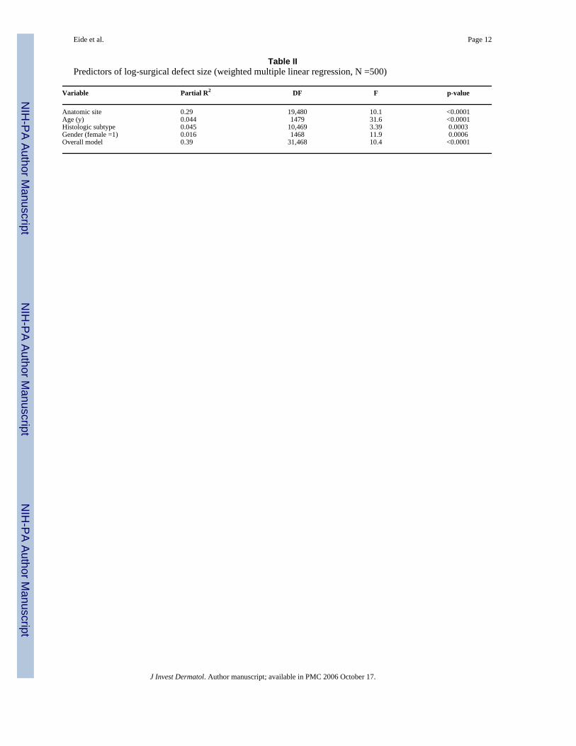
In the overall study population (Table II), a substantial portion of the variation in defect sizewas explained by histology, age, gender, and anatomic location (R2 =0.39, p<0.0001). No ageby gender interaction was identified, and the quadratic age term was not associated with defectsize. The majority of the variance was attributed to anatomic location (partial R2 =0.29). Largersurgical defects were associated with several anatomic sites, including pre- and post-auricular,chest, and the submandible. There was no association when BCC was compared with SCC(β=−0.16, 95% confidence interval (CI) −0.4 to 0.04, p =0.12). When more refined histologic
Eide et al. Page 2
J Invest Dermatol. Author manuscript; available in PMC 2006 October 17.
NIH
-PA Author Manuscript
NIH
-PA Author Manuscript
NIH
-PA Author Manuscript
subtype was entered into the model, superficial, morpheaform, and infiltrating BCC wereassociated with larger defects as were older age and male gender. Similar findings were alsoobserved in the analysis of interviewed participants (R2 =0.47, anatomic site partial R2 =0.37).
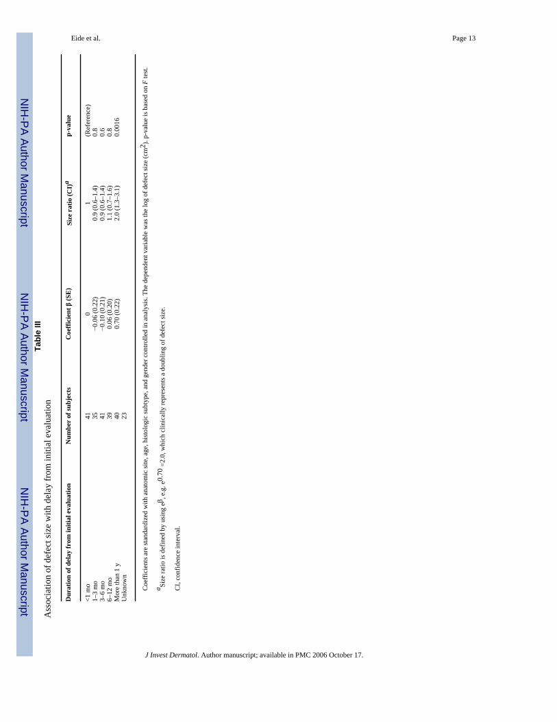
Controlling for anatomic location, histology, age and gender, all time-delay intervals wereassociated with defect size when examined individually (p-values from partial F test: discovery(p =0.034, median =194 d), suspicion (p =0.014, median =138 d), initial evaluation (p =0.004,median =107 d), and biopsy (p =0.025, median =90 d)). When an additional time-delay intervalwas, however, entered into a model already containing one of the time-delay intervals,significance for both time-delay intervals was lost, with one exception. Consistently, only delayfrom initial evaluation (the duration of time between initial provider evaluation until MMS)was associated with larger defects (partial R2 =0.018, p =0.004) whereas all other time-delayintervals failed to maintain a significant association with size. We evaluated the duration–response relationship between delay from initial evaluation and defect size (Table III). Thesize-delay association was seen only in the last quintile of time delay, i.e. greater than 1 year,which was associated with a doubling of defect size (size ratio (eβ) =2.0; 95% CI 1.3–3.1).
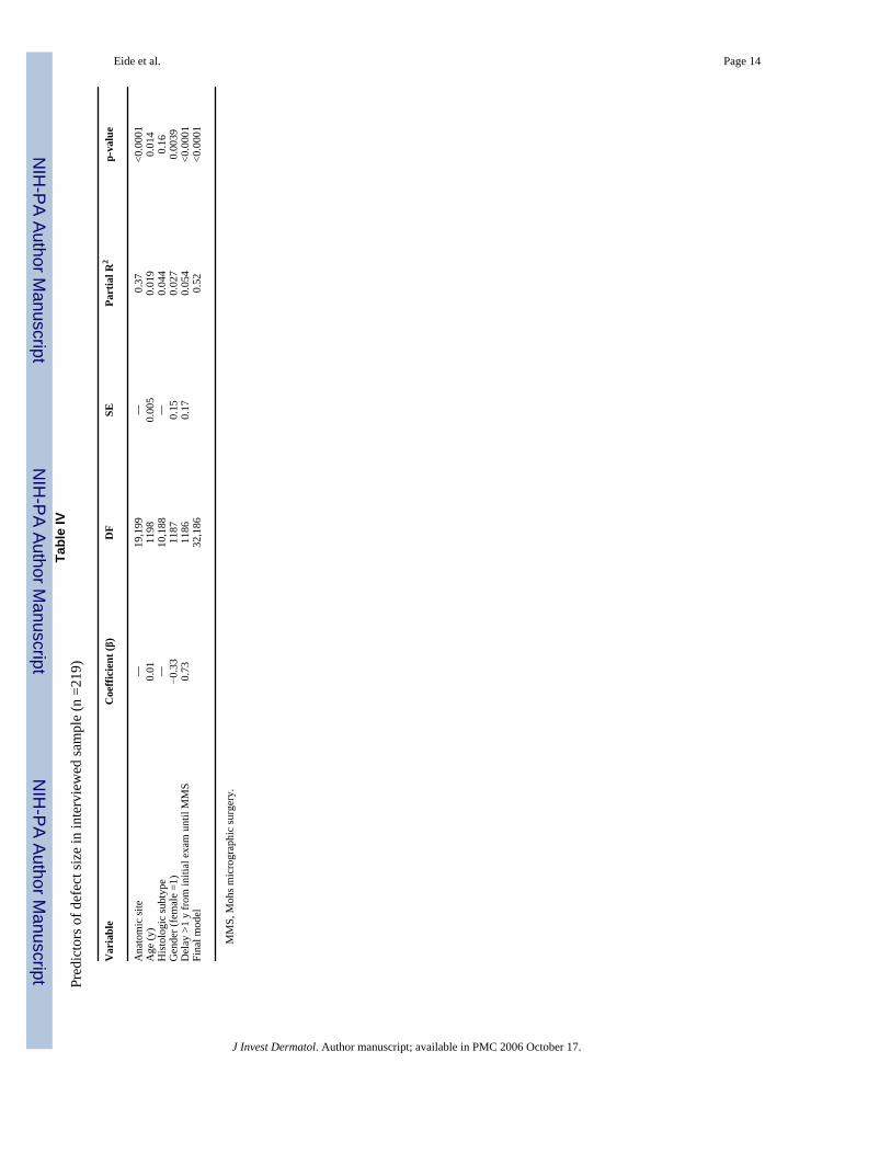
Controlling for anatomic location, gender, age, and histologic subtype as well as with or withoutdelay (Table IV), other factors were then examined to further explain variation in defect size.Size variation was not explained by personal or family history of skin cancer or by specialtyof initial care provider. No variation was attributable to marital status, education level,smoking, provider skin examination, personal history of previous cancer other than skin,number of provider visits in the 2 years prior to surgery, treatment by initial health provider,if skin cancer was diagnosed initially, the Whitley index, the attitudinal variables, medicalcosts, or insurance concerns.
Defect size (p-value based on partial F value) was associated with who discovered lesion (self,spouse, or a clinician; p =0.014), skin self-examination (SSE) (p =0.035), and the number ofprior removals (p =0.03). All of these factors, however, failed to maintain the significance incontrolling for anatomic location, histologic subtype, age, gender, and time interval greaterthan 1 years between provider evaluation and surgery. Out-of-pocket cost of diagnosis had amarginal association with larger lesions (β=0.005, p =0.049).
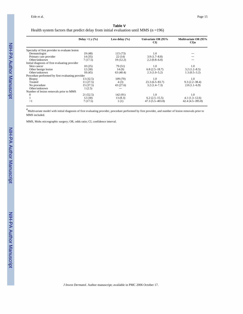
In separate logistic regression analyses, we examined factors associated with long delay(dependent variable: greater than 1 y from evaluation until MMS). The initial provider seenwas a dermatologist for 68% of patients whereas most of the remaining patients saw a primaryprovider first (Table V). Patient who saw a primary care provider first, compared with thosewho saw a dermatologist, were more likely to have a long delay (unadjusted odds ratio (OR)3.9; 95% CI 1.7–8.8). Delay was not associated with reason for initial office visit. Theprovider’s initial diagnostic impression of the lesion at the time of initial evaluation, however,differed in those with more than a year delay compared with those with shorter delay. If theskin cancer was misdiagnosed initially (unadjusted OR 6.8; 95% CI 2.5–18.7) or if the lesionwas treated as opposed to biopsied on the initial evaluation (unadjusted OR 23.3; 95% CI 6.5–83.7), the patient was more likely to have delay. Patients who had one (unadjusted OR 6.2;95% CI 2.5–15.5) or more prior surgeries (unadjusted OR 47.3; 95% CI 5.5–403.8) beforeMMS were also more likely to have a long delay. Using multivariate logistic regression, delayof more than 1 y from initial evaluation until MMS was associated with initial misdiagnosis,initial treatment, and having one or more removals prior to MMS (Table V). No other variables,including anatomic location, histologic subtype, age, and gender were significantly associatedwith delay.
Eide et al. Page 3
J Invest Dermatol. Author manuscript; available in PMC 2006 October 17.
NIH
-PA Author Manuscript
NIH
-PA Author Manuscript
NIH
-PA Author Manuscript

DiscussionThis study suggests that there are several factors that are associated with the size of MMSdefects in the treatment of KC. Delay of more than 1 y between initial provider evaluation ofa KC and its surgical removal with MMS was associated with surgical defects that were twiceas large as those where delay was shorter. We note that important predictors of this delayinclude initial misdiagnosis, initial procedure performed, and the number of surgical removalsprior to MMS. We also found important effects of anatomic site, age, histologic subtype, andgender on defect size, with the largest portion of the variation in size explained by anatomicsite. This is consistent with previous findings in which both anatomic location and histologicsubtype have been noted to be associated with the extent of infiltration and tissue invasion inKC (Batra and Kelley, 2002). In our study, a woman’s surgical defect size was approximatelyhalf the size of a man’s defect.
There are several limitations of our study. One limitation is the absence of data from referringproviders and the uncertainty of detailed explanations for the long delays (greater than 1 y)between initial evaluation and MMS. We do not have detailed information on prior medicalcare, including prior treatment with electrodessication and curettage, a relatively commontreatment method for KC (Silverman et al, 1991). This study did not examine adequacy ofpathology specimens (Rampen et al, 1989) or result follow-up (Cassileth et al, 1988). We donot know if there were difficulties surrounding the reason for the referral of suspected skinlesions (Dunkley and Morris, 1991) or misunderstandings of the urgency of scheduling thereferral evaluations (Dunkley and Morris, 1991; Brochez et al, 2001). Furthermore, most ofour data was collected by self-report, including the information relating to dates andprogression through the medical system prior to MMS. Imprecision in the assessment of delayis possible because of inaccurate or non-specific recall of dates. To minimize potential errorintroduced by recall bias, the time intervals used the date of MMS, a date available in themedical record.
Another limitation of this study is that some pathology reports were incomplete or missing.Histologic-subtype identification and verification were not available for all tumors. Ideally thisinformation would have been present for all individuals. Furthermore, we have noted anassociation with diagnostic suspicion of lesion on first evaluation, initial care plan and numberof prior surgical removals with delay. There may, however, be other sources of delay that wedid not adequately assess, including severe unrelated illness in the interval between initialevaluation and MMS, patient’s personal priorities, problems with referring or schedulingappointments, or additional visits related to ‘‘following’’/observing lesion.
A strength of this study is that it examines factors associated with defect size itself, a measureof KC morbidity. The use of surgical defect margins from MMS cases allows a precise estimateof tumor size. This study demonstrates evidence of the association between treatment delayand KC outcomes. All cases were obtained from the practice of a single Mohs surgeon. At thetime of this study, a majority of all MMS cases in the state of Rhode Island were treated bythis dermatologic surgeon. The use of a single surgical provider reduces provider-associatedvariability and cases are felt to be representative of Rhode Island KC patients. It is unclear ifthese results are generalizable outside of this population, including patients not referred forMohs surgery.
A review of patient care delay for cancer symptoms shows that cancer patients with co-paidfee-for-service coverage waited longer to seek healthcare than patients enrolled in healthmaintenance organizations (Love, 1991). Although almost all study participants had medicalcoverage, co-payments and fear of non-covered expenses could conceivably have contributedto patient delay in this study population. Participants were, however, asked specifically if the
Eide et al. Page 4
J Invest Dermatol. Author manuscript; available in PMC 2006 October 17.
NIH
-PA Author Manuscript
NIH
-PA Author Manuscript
NIH
-PA Author Manuscript
cost of seeing a health provider was a barrier to care, and we found no significant associationbetween response to this question and defect size. The meaning of the observed borderlineassociation found between out of pocket expense and surgical defect size is unclear.
There has been limited investigation of the relevance of treatment delay and KC size (Kirkupand De Berker, 1999; Bandaranayake, 2002). Kirkup and De Berker (1999) examined 50patients with BCC on the face who were identified as suitable for elective excision. Major andminor diameters (i.e. horizontal and vertical dimensions of lesion) were measured atpresentation and immediately before excision. Delay time ranged from 3 wk to 6 mo (mean of10 wk), and no correlation was found with change in size and time to surgery. The authors didnote, that because of the poor correlation between growth and time, that it was difficult topredict the effect of longer delays. Furthermore, it should be noted that if our study had usedthe same delay period of 6 mo or less, we too would have found no association between delayand size.
A recent doctoral dissertation examined the relationship of delay to KC size (Bandaranayake,2002). Although this Australian case–control study included people with smaller lesions andshorter duration of delay, her results were consistent with our findings. Bandaranayake notedlonger delay between the final physician evaluation and definitive treatment in those with largerlesions (cases: greater than 1–2.25 cm2). Controlling for sex, age, and histology, patients withlonger delay (more than 61 d) between consultation with final physician until definitivetreatment were more likely to have larger lesions than those treated within 2 wk (OR 1.96; 95%CI 1.42–2.71). She, however, found evidence that the delay was attributable to the morecomplex procedures required for treatment of the larger lesions.
Patient and tumor factors that relate to subclinical extension of non-melanoma skin cancer wereexamined in a study of 1131 MMS patients using a dependent variable of 3 or more MMSlayers during surgery. The authors noted an interaction between anatomic location of the skincancer, histologic classification, and sex that was predictive of the extent of subclinicalinvasion. Tumors located on the eyelids, temple, or ear helix; basosquamous, morpheaform,nodular, and recurrent BCC on the nose; and morpheaform BCC on the cheek were found tobe important predictors of extension. Pre-operative size greater than 10 mm and recurrentmalignancies or those on the neck in men also were predictive of infiltration (Batra and Kelley,2002). Although our study differed substantially in methods, the results were broadlyconsistent.
Previous studies have focused on the examination of factors contributing to health-care delayand thickness of melanoma. The association between delay and size has been inconsistent(Cassileth et al, 1988; Krige et al, 1991; Schmid-Wendtner et al, 2002). Temoshok et al1985 found that the most significant variable in a hierarchical multiple regression analysis ofmelanoma thickness was delay (p<0.0005), defined as time from patient first becomingsuspicious of lesion until evaluation by a physician specifically for that reason. Other than skintype, no significance was found for other variables including previous knowledge,understanding of treatment, age, faith, and personality.
Richard et al (1999, 2000a, b) investigated delay and melanoma thickness using five dates,similar to those used in this study: when the lesion was first noticed, first suspicion of lesion,first examination by a provider, first proposed removal, and melanoma resection. Her findings,suggested that in some groups, patient and provider delay may be associated with melanomadepth (Richard et al, 1999), although the relationships, when present, were complex.
Some prior studies examining melanoma thickness have found an association between initialexamining physician specialty and melanoma thickness (Richard et al, 2000a, b) whereasothers have failed to find an association (Brochez et al, 2001). In our data, it does not appear
Eide et al. Page 5
J Invest Dermatol. Author manuscript; available in PMC 2006 October 17.
NIH
-PA Author Manuscript
NIH
-PA Author Manuscript
NIH
-PA Author Manuscript

that the specialty of the initial provider (primary care provider or a dermatologist) wasassociated with size or delay, after controlling for other factors. We also did not find the numberof prior surgical removals to be associated with the lesion size. This is consistent with a studyby Brochez et al 2001 that found no association of prior treatment with melanoma thickness.Unlike Brochez, we, however, found an association with prior removals and delay.
We were able to explore the relationship between diagnostic and treatment delay and KC sizein greater depth than other studies. Although the results from this study do not prove that delaycauses larger surgical defects, it does suggest an important association. Besides the number ofprior removals, we note several aspects of the initial provider evaluation that relate to this delay,including the initial diagnostic impression and type of procedures performed at this evaluation.Some have speculated about the role of medical providers in prolonged delay (Cassileth etal, 1988; Dunkley and Morris, 1991; Blum et al, 1999; MacKie, 1999; Richard et al, 1999).Many of the clinicians that treat BCC and SCC in their practices have little formal training indermatology (only 60% of skin cancer treatments are provided by dermatologists) (Joseph etal, 2001). In our study, almost half of the participants with more than 1 y delay were firstevaluated by a dermatologist.
On the basis of these results, we suggest that for KC, delay after presentation to a health-careprovider may be more important than delay before initially presenting for care. This should beevaluated in other settings and locations. Further study to investigate mediating variables inhealth system delay in the diagnosis and treatment of KC is also necessary. This inquiry shouldinclude issues of misdiagnosis, inadequate follow-up efforts, inadequate initial treatment,interim events during the course of medical care, and cost-effectiveness of available therapeuticoptions. KC causes substantial morbidity, and optimal management of this public healthproblem requires both primary prevention and efficient medical management.
MethodsSubjects were selected from all cases of KC treated with MMS at an academic dermatologicalsurgery practice between September 12, 2000 and September 12, 2001 (n =1123 surgical cases).These MMS cases represented 892 different individuals. Of the surgical patients who had morethan one procedure, the surgical case with the largest final defect size was selected.Immunologically compromised patients were excluded. There were 20 surgical cases for whichit was not possible to calculate final defect size and 12 surgical cases in which the diagnoseswere not KC; all of which were excluded. Hence, 860 unique MMS patients were eligible forinclusion. Cases were then stratified by size of defect into quartiles. A random sample of 100cases was selected using computer-generated random numbers (Microsoft Foxpro Version 2.6,Redmond, Washington) from each quartile except the largest, from which 200 cases wereselected. (Patients with large defect size were over-sampled because factors contributing tothose with more morbidity were of special interest, and we wished to ensure statistical stabilityfor analysis related to these large lesions.) Approximate quartile parameters were: <1, 1–2, 2–4 cm2, and greater than 4 cm2 (maximum size: 43 cm2). Eighty percent of each quartile (n=400) were chosen for interview because of resource constraints.
Of these 400 subjects, 28 individuals could not be reached for interview. An additionalsubgroup was ineligible to participate because of communication barriers, including notspeaking English (n=32), other communication issues (e.g. hearing loss, dementia) (n =43), ordeath (n =5). Of those remaining, 71 people (24%) refused participation, and two completedonly partial interviews. Hence, after giving verbal consent, 219 people completed the telephoneinterview.
Eide et al. Page 6
J Invest Dermatol. Author manuscript; available in PMC 2006 October 17.
NIH
-PA Author Manuscript
NIH
-PA Author Manuscript
NIH
-PA Author Manuscript
Surgical records were used to obtain information recorded at time of MMS. The remainingvariables were obtained through computer-assisted telephone interviews, which wereconducted between January 2, 2002 and October 2, 2002 and were approximately 25 min induration.
Analysis utilized SAS System software, release 8.2, Enterprise Guide Version 2.0 (SASInstitute, Cary, North Carolina). After weighting the quartiles 1, 2, and 3 by a factor of 2 toreflect the sampling method, size was transformed, using a log transformation, to graphicallyapproximate a normal distribution.
Weighted linear regression was used to model the transformed-dependent variable, log finalsize of surgical defect. Residuals were checked using a Q–Q plot, and co-linearity was assessedusing variance inflation factors. The following variables were evaluated using linear regressionto establish the base model in the overall sample of 500 subjects: age, gender, histologicsubtype, anatomic location, an age × gender interaction, and age-squared. Controlling for thebase model variables, remaining variables were then entered into the model individually. Inseparate analyses, predictors of delay were examined using logistic regression and χ2 test (orFisher’s exact test, if appropriate).
Twenty different anatomic locations were defined a priori using information recorded at thetime of surgery. These anatomic areas were obtained from the chart (Table I). Comparison wasto the location of the nose, the most common location of lesions. BCC and SCC histologicsubtypes were obtained from records. Because of referral sources, 33% of charts did not havepathology reports with information on histologic subtype (32% of the interviewed and 34% ofnon-interviewed subjects). Review of the medical charts of the interviewed participantsreduced the number of missing subtypes to 20%.
Four time intervals, considered potential sources of treatment delay, were evaluated (Fig 1).These intervals were measured and analyzed in days (continuous variable), then by the numberof the quintile of delay (ordinal variable), and finally with indicator variables to represent eachof the individual quintiles (dummy variables; the first quintile was the comparison) to examineany possible gradient. Quintile intervals for delay from initial evaluation approximated <1, 1–3, 3–6, 6 mo to 1 y, and greater than 1 y between reference date and date of MMS. All dateswere available for 196 patients (89.5%).
We hypothesized that demographic, socio-economic, health-care resources, and psychosocialvariables may be related to delay, and hence defect size. Other factors analyzed were: maritalstatus, living alone, education level completed (a 1–6 scale, ranging from less than ninth gradeto graduate school degree), smoking status, provider skin examination (both as dichotomousvariable representing having had examination that year and as a variable with a 1–5 scale,ranging from never to monthly examination), SSE (both as dichotomous variable representinghaving had examination that year and as a variable with a 1–5 scale, ranging from never tomonthly exam), previous history of skin cancer, family history of skin cancer, personal historyof any cancer other than skin cancer, who discovered lesion (self, spouse, provider), specialtyof first provider to evaluate, number of visits to a primary provider or dermatologist in 2 y priorto surgery, clinician diagnostic and therapeutic management on first evaluation, and numberof treatments of lesion with the intention of cure prior to surgery. The Whitley index (Pilowsky,1967) (scale: 0–14) was assessed, followed by assessment of three of the subcomponents ofthe scale: bodily pre-occupation (scale: 0–3), disease phobia (scale: 0–4), and hypochondriasis(scale: 0–3). A summary variable was also created that utilized five-point Likert responses toa series of questions on excuses for not seeking care (e.g. hoping spot would clear on its own).Attitudinal Likert variables assessing confidence in detecting skin cancer, being too busy toseek medical care, ease of obtaining an appointment, faith in health care, cost of health care as
Eide et al. Page 7
J Invest Dermatol. Author manuscript; available in PMC 2006 October 17.
NIH
-PA Author Manuscript
NIH
-PA Author Manuscript
NIH
-PA Author Manuscript

a problem, and tendency to wait and hope problems will go away on their own were alsoexamined. Out-of-pocket cost of diagnosis (in dollars), out-of-pocket cost of surgery (indollars), and self-report of concerns of insurance status resulting in delay in seeking health carewere also assessed. The racial and ethnic background of the study population washomogeneous, 99% white by self-report, and 99% also reported having insurance at time ofboth diagnosis and surgery. Hence, these variables were not analyzed. Declaration of Helsinkiguidelines were adhered to, and the study was approved by the Committees on Human Subjectsfor Lifespan Hospitals and for the Providence VA Medical Center (Providence, Rhode Island).
ReferencesBandaranayake, DM. Doctor of philosophy dissertation. University of Newcastle; Newcastle: 2002. Why
do some non-melanocytic skin cancers reach an advanced stage before they are treated? The effect ofdelay and predictors of delay in presenation, referral and treatment of NMSC.
Batra RS, Kelley LC. Predictors of extensive subclinical spread in nonmelanoma skin cancer treated withMohs micrographic surgery. Arch Dermatol 2002;138:1043–1051. [PubMed: 12164742]
Blum A, Brand CU, Ellwanger U, Schlagenhauff B, Stroebel W, Rassner G, Garbe C. Awareness andearly detection of cutaneous melanoma: An analysis of factors related to delay in treatment. Br JDermatol 1999;141:783–787. [PubMed: 10583157]
Brochez L, Verhaeghe E, Bleyen L, Naeyert J-M. Time delays and related factors in the diagnosis ofcutaneous melanoma. Eur J Cancer 2001;37:843–848. [PubMed: 11313171]
Cassileth BR, Temoshok L, Frederick BE, et al. Patient and physician delay in melanoma diagnosis. JAm Acad Dermatol 1988;18:591–598. [PubMed: 3351022]
Dunkley MP, Morris AM. Cutaneous malignant melanoma: Audit of the diagnostic process. Ann R CollSurg Engl 1991;73:248–252. [PubMed: 1863047]
Hennrikus D, Girgis A, Redman S, Sanson-Fisher RW. A community study of delay in presenting withsigns of melanoma to medical practitioners. Arch Dermatol 1991;127:356–361. [PubMed: 1998366]
Jemal A, Murray T, Samuels A, Ghafoor A, Ward E, Thun MJ. Cancer Statistics, 2003. CA Cancer JClin 2003;53:5–26. [PubMed: 12568441]
Joseph AK, Mark TL, Mueller C. The period prevalence and costs of treating nonmelanoma skin cancersin patients over 65 years of age covered by medicare. Dermatol Surg 2001;27:955–959. [PubMed:11737130]
Kirkup ME, De Berker DAR. Clinical measurement of dimensions of basal cell carcinoma: Effect ofwaiting for elective surgery. Br J Dermatol 1999;141:876–879. [PubMed: 10583170]
Krige JEJ, Isaacs S, Hudson DA, King HS, Strover RM, Johnson CA. Delay in the diagnosis of cutaneousmalignant melanoma. Cancer 1991;68:2064–2068. [PubMed: 1913555]
Levine J, Kopf AW, Rigel DS, Bart RS, Hennessey P, Friedman RJ, Mintzis MM. Correlation ofthicknesses of superficial spreading malignant melanomas and ages of patients. J Dermatol SurgOncol 1981;7:311–316. [PubMed: 7240532]
Love N. Why patients delay seeking care for cancer symptoms. Postgrad Med 1991;89:155–158.MacKie RM. Thickness and delay in diagnosis of melanoma: How far can we go? Arch Dermatol
1999;135:339–340. [PubMed: 10086457]McKenna DB, Lee RJ, Prescott RJ, Doherty VR. The time from diagnostic excision biopsy to wide local
excision for primary cutaneous malignant melanoma may not affect patient survival. Br J Dermatol2002;147:48–54. [PubMed: 12100184]
Miller SJ. Biology of basal cell carcinoma (part i). J Am Acad Dermatol 1991;24:1–13. [PubMed:1999506]
Oliveria SA, Christos PJ, Halpern AC, Fine JA, Barnhill RL, Berwick M. Patient knowledge, awareness,and delay in seeking medical attention for malignant melanoma. J Clin Epidemiol 1999;52:1111–1116. [PubMed: 10527006]
Osborne JE, Hutchinson PE. Clinical correlates of breslow thickness of malignant melanoma. Br JDermatol 2001;144:476–483. [PubMed: 11260002]
Pilowsky I. Dimensions of hypochondriasis. Br J Psychiatry 1967;113:89–93. [PubMed: 6029373]
Eide et al. Page 8
J Invest Dermatol. Author manuscript; available in PMC 2006 October 17.
NIH
-PA Author Manuscript
NIH
-PA Author Manuscript
NIH
-PA Author Manuscript
Rampen FHJ, Rumke P, Hart AAM. Patients’ and doctors’ delay in the diagnosis and treatment ofcutaneous melanoma. Eur J Surg Oncol 1989;15:143–148. [PubMed: 2703058]
Randle HW. Basal cell carcinoma. Identification and treatment of the high-risk patient. Dermatol Surg1996;22:255–261. [PubMed: 8599737]
Richard MA, Grob JJ, Avril MF, et al. Melanoma and tumor thickness: Challenges of early diagnosis.Arch Dermatol 1999;135:269–274. [PubMed: 10086447]
Richard MA, Grob JJ, Avril MF, et al. Delays in diagnosis and melanoma prognosis (i): The role ofpatients. Int J Cancer 2000a;89:271–279. [PubMed: 10861504]
Richard MA, Grob JJ, Avril MF, et al. Delays in diagnosis and melanoma prognosis (ii): The role ofdoctors. Int J Cancer 2000b;89:280–285. [PubMed: 10861505]
Rowe DE, Carroll RJ, Day CL. Long-term recurrence rates in previously untreated (primary) basal cellcarcinoma: Implications for patient follow-up. J Dermatol Surg Oncol 1989;15:424–431. [PubMed:2925988]
Schmid-Wendtner MH, Baumert J, Stange J, Vokenandt M. Delay in the diagnosis of cutaneousmelanoma: An analysis of 233 patients. Melanoma Res 2002;12:389–394. [PubMed: 12170189]
Silfen R, Amir A, Regev D, Hauben DJ. Role of physicians and patients in the diagnostic delay ofcutaneous malignant melanoma. Ann Plast Surg 2002;49:439–442. [PubMed: 12370654]
Silverman MK, Kopf AW, Grin CM, Bart RS, Levenstein MJ. Recurrence rates of treated basal cellcarcinomas. J Dermatol Surg Oncol 1991;17:713–718. [PubMed: 1890243]
Smeets NW, Kuijpers DI, Nelemans P, Ostertag JU, Verhaegh ME, Krekels GA, Neumann HA. Mohs’micrographic surgery for treatment of basal cell carcinoma of the face—results of a retrospectivestudy and review of the literature. Br J Dermatol 2004;151:141–147. [PubMed: 15270883]
Temoshok L, DiClemente RJ, Sweet DM, Blois MS, Sagebiel RW. Prognostic and psychosocial factorsrelated to delay behavior in patients with cutaneous malignant melanoma. Prog Clin Biol Res1984;156:168–179.
Temoshok L, Heller BW, Sagebiel RW, Blois MS, Sweet DM, DiClemente RJ, Gold ML. The relationshipof psychosocial factors to prognostic indicators in cutaneous malignant melanoma. J Psychosom Res1985;29:139–153. [PubMed: 4009515]
Weinstock MA, Bingham SF, Cole GW, et al. Reliability of counting actinic keratoses before and afterbrief consensus discussion. Arch Dermatol 2001;137:1055–1058. [PubMed: 11493098]
Zitelli JA, Brown CD, Hanusa BH. Surgical margins for excision of primary cutaneous melanoma. J AmAcad Dermatol 1997;37:422–429. [PubMed: 9308558]
Eide et al. Page 9
J Invest Dermatol. Author manuscript; available in PMC 2006 October 17.
NIH
-PA Author Manuscript
NIH
-PA Author Manuscript
NIH
-PA Author Manuscript
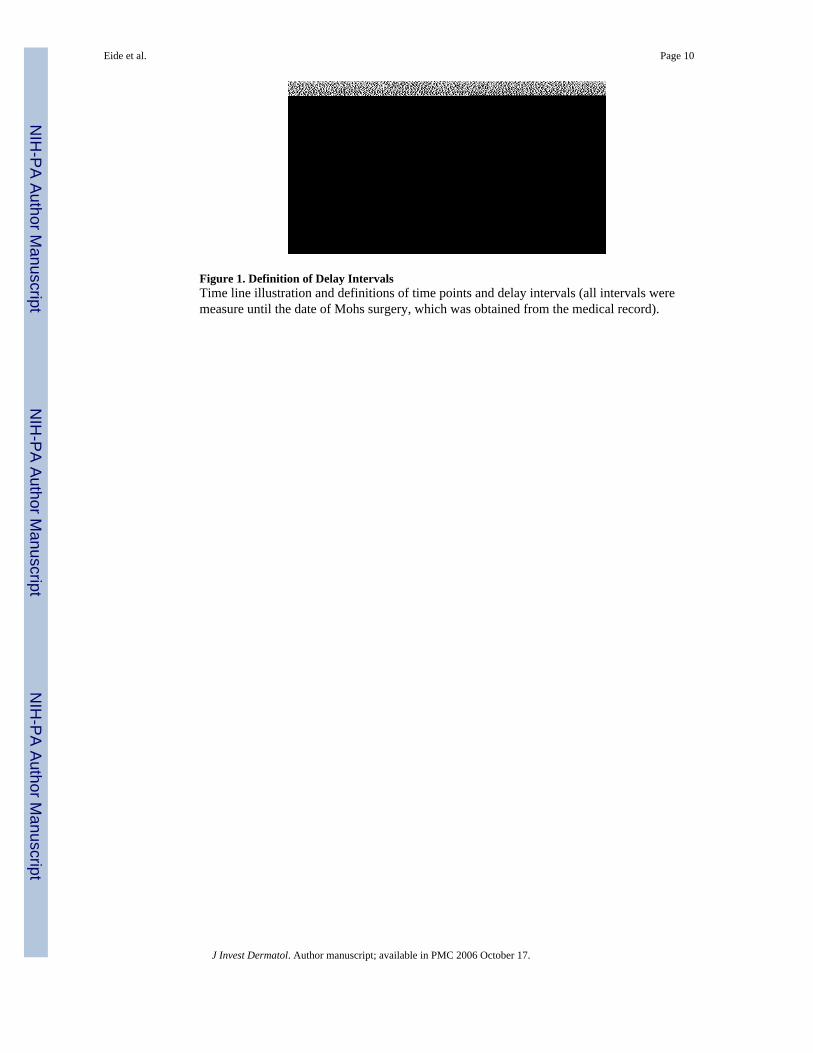
Figure 1. Definition of Delay IntervalsTime line illustration and definitions of time points and delay intervals (all intervals weremeasure until the date of Mohs surgery, which was obtained from the medical record).
Eide et al. Page 10
J Invest Dermatol. Author manuscript; available in PMC 2006 October 17.
NIH
-PA Author Manuscript
NIH
-PA Author Manuscript
NIH
-PA Author Manuscript

NIH
-PA Author Manuscript
NIH
-PA Author Manuscript
NIH
-PA Author Manuscript
Eide et al. Page 11
Table IComparison of interviewed and non-interviewed subjects n =500 (%)
Interviewedparticipants (%)
Non-interviewed (%) Total (%)
Anatomic sitea Upper limb 4 (1.8) 1 (0.4) 5 (1.0) Back 1 (0.5) 1 (0.4) 2 (0.4) Chest 3 (1.4) 2 (0.7) 5 (1.0) Posterior ear 2 (0.9) 5 (1.8) 7 (1.4) Anterior ear 18 (8.2) 27 (9.6) 45 (9.0) Eye 17 (7.8) 23 (8.2) 40 (8.0) Neck 6 (2.7) 5 (1.8) 11 (2.2) Submandible 1 (0.5) 0 (—) 1 (0.2) Jawline 1 (0.5) 2 (0.7) 3 (0.6) Chin 5 (2.3) 3 (1.1) 8 (1.6) Vermillion lip 6 (2.7) 5 (1.8) 11 (2.2) Anterior lip 7 (3.2) 11 (3.9) 18 (3.6) Alar lip 5 (2.3) 14 (5.0) 19 (3.8) Nose 39 (17.8) 66 (23.5) 105 (21.0) Nasofacial fold 20 (9.1) 17 (6.1) 37 (7.4) Cheek 26 (11.9) 31 (11.0) 57 (11.4) Pre-auricular 4 (1.8) 6 (2.1) 10 (2.0) Post-auricular 3 (1.4) 3 (1.1) 6 (1.2) Forehead 26 (11.9) 20 (7.1) 46 (9.2) Scalp 25 (11.4) 39 (13.9) 64 (12.8)Age (y)b <40 8 (4) 3 (1) 11 (2) 41–50 29 (13) 15 (5) 44 (9) 51–60 36 (16) 47 (17) 83 (17) 61–70 43 (20) 36 (13) 79 (16) 71–80 80 (37) 87 (31) 167 (33) 81–90 22 (10) 83 (30) 105 (21) >90 1 (0) 10 (4) 11 (2)Histologic subtypec Morpheaform BCC 10 (4.6) 13 (4.6) 23 (4.6) Infiltrating BCC 22 (10.0) 27 (9.6) 49 (9.8) Superficial BCC 6 (2.7) 6 (2.1) 12 (2.4) Basosquamous BCC 4 (1.8) 10 (3.6) 14 (2.8) Nodular BCC 60 (27.4) 77 (27.4) 137 (27.4) SCC in situ 13 (5.9) 17 (6.0) 30 (6.0) Moderately differentiated SCC 5 (2.3) 6 (2.1) 11 (2.2) Well-differentiated SCC 18 (8.2) 16 (5.7) 34 (6.8) Superficial SCC 4 (1.8) 5 (1.8) 9 (1.8) Other types of SCC 8 (3.7) 8 (2.8) 16 (3.2) No histological subtype available 69 (31.5) 96 (34.1) 165 (33.0)Genderd Females 94 (42.9) 110 (39.1) 204 (40.8) Males 125 (57.1) 171 (60.9) 296 (59.2)Total subjects 219 281 500
aLogistic regression Wald χ2 =15.46, p =0.7.
bLogistic regression Wald χ2 =28.55, p<0.0001.
cLogistic regression Wald χ2 =3.149, p =1.0.
dχ2 =0.726, p =0.4.
BCC, basal cell carcinoma; SCC, squamous cell carcinoma.
J Invest Dermatol. Author manuscript; available in PMC 2006 October 17.
NIH
-PA Author Manuscript
NIH
-PA Author Manuscript
NIH
-PA Author Manuscript
Eide et al. Page 12
Table IIPredictors of log-surgical defect size (weighted multiple linear regression, N =500)
Variable Partial R2 DF F p-value
Anatomic site 0.29 19,480 10.1 <0.0001Age (y) 0.044 1479 31.6 <0.0001Histologic subtype 0.045 10,469 3.39 0.0003Gender (female =1) 0.016 1468 11.9 0.0006Overall model 0.39 31,468 10.4 <0.0001
J Invest Dermatol. Author manuscript; available in PMC 2006 October 17.
NIH
-PA Author Manuscript
NIH
-PA Author Manuscript
NIH
-PA Author Manuscript
Eide et al. Page 13Ta
ble
IIIA
ssoc
iatio
n of
def
ect s
ize
with
del
ay fr
om in
itial
eva
luat
ion
Dur
atio
n of
del
ay fr
om in
itial
eva
luat
ion
Num
ber
of su
bjec
tsC
oeffi
cien
t β (S
E)
Size
rat
io (C
I)a
p-va
lue
<1 m
o41
01
(Ref
eren
ce)
1–3
mo
35−0
.06
(0.2
2)0.
9 (0
.6–1
.4)
0.8
3–6
mo
41−0
.10
(0.2
1)0.
9 (0
.6–1
.4)
0.6
6–12
mo
390.
06 (0
.20)
1.1
(0.7
–1.6
)0.
8M
ore
than
1 y
400.
70 (0
.22)
2.0
(1.3
–3.1
)0.
0016
Unk
now
n23
Coe
ffic
ient
s are
stan
dard
ized
with
ana
tom
ic si
te, a
ge, h
isto
logi
c su
btyp
e, a
nd g
ende
r con
trolle
d in
ana
lysi
s. Th
e de
pend
ent v
aria
ble
was
the
log
of d
efec
t siz
e (c
m2 )
. p-v
alue
is b
ased
on
F te
st.
a Size
ratio
is d
efin
ed b
y us
ing
eβ, e
.g. e
0.70
=2.
0, w
hich
clin
ical
ly re
pres
ents
a d
oubl
ing
of d
efec
t siz
e.
CI,
conf
iden
ce in
terv
al.
J Invest Dermatol. Author manuscript; available in PMC 2006 October 17.
NIH
-PA Author Manuscript
NIH
-PA Author Manuscript
NIH
-PA Author Manuscript
Eide et al. Page 14Ta
ble
IVPr
edic
tors
of d
efec
t siz
e in
inte
rvie
wed
sam
ple
(n =
219)
Var
iabl
eC
oeffi
cien
t (β)
DF
SEPa
rtia
l R2
p-va
lue
Ana
tom
ic si
te—
19,1
99—
0.37
<0.0
001
Age
(y)
0.01
1198
0.00
50.
019
0.01
4H
isto
logi
c su
btyp
e—
10,1
88—
0.04
40.
16G
ende
r (fe
mal
e =1
)−0
.33
1187
0.15
0.02
70.
0039
Del
ay >
1 y
from
initi
al e
xam
unt
il M
MS
0.73
1186
0.17
0.05
4<0
.000
1Fi
nal m
odel
32,1
860.
52<0
.000
1
MM
S, M
ohs m
icro
grap
hic
surg
ery.
J Invest Dermatol. Author manuscript; available in PMC 2006 October 17.
NIH
-PA Author Manuscript
NIH
-PA Author Manuscript
NIH
-PA Author Manuscript
Eide et al. Page 15
Table VHealth system factors that predict delay from initial evaluation until MMS (n =196)
Delay >1 y (%) Less delay (%) Univariate OR (95%CI)
Multivariate OR (95%CI)a
Specialty of first provider to evaluate lesion Dermatologist 19 (48) 115 (73) 1.0 — Primary care provider 14 (35) 22 (14) 3.9 (1.7–8.8) — Other/unknown 7 (17.5) 19 (12.2) 2.2 (0.8–6.0) —Initial diagnosis of first evaluating provider Skin cancer 10 (25) 79 (51) 1.0 1.0 Other benign lesion 12 (30) 14 (9) 6.8 (2.5–18.7) 3.2 (1.2–8.5) Other/unknown 18 (45) 63 (40.4) 2.3 (1.0–5.2) 1.3 (0.5–3.2)Procedure performed by first evaluating provider Biopsy 13 (32.5) 109 (70) 1.0 1.0 Treated 11 (27.5) 4 (3) 23.3 (6.5–83.7) 9.3 (2.2–38.4) No procedure 15 (37.5) 43 (27.6) 3.2 (1.4–7.3) 2.8 (1.1–6.9) Other/unknown 1 (2.5) —Number of lesion removals prior to MMS 0 21 (52.5) 142 (91) 1.0 1.0 1 12 (30) 13 (8.3) 6.2 (2.5–15.5) 4.1 (1.3–12.6) >1 7 (17.5) 1 (1) 47.3 (5.5–403.8) 42.4 (4.5–395.8)
aMultivariate model with initial diagnosis of first evaluating provider, procedure performed by first provider, and number of lesion removals prior to
MMS included.
MMS, Mohs micrographic surgery; OR, odds ratio; CI, confidence interval.
J Invest Dermatol. Author manuscript; available in PMC 2006 October 17.
Copyright © 2022 FDOKUMEN