Matrix Protein 2 Vaccination and Protection against Influenza Viruses, Including Subtype H5N1
Upload
independentCategory
view
1download
0
Chromosome 14q loss defines a molecularsubtype of clear-cell renal cell carcinomaassociated with poor prognosis
Federico A Monzon1,2, Karla Alvarez1, Lief Peterson3, Luan Truong1, Robert J Amato4,Joan Hernandez-McClain4, Nizar Tannir5, Anil V Parwani6 and Eric Jonasch5
1Department of Pathology, The Methodist Hospital Research Institute, Houston, TX, USA; 2Department of
Pathology, Weill-Cornell Medical College, New York, NY, USA; 3Center for Biostatistics, The Methodist
Hospital Research Institute, Houston, TX, USA; 4Division of Oncology, The University of Texas Medical
School, Houston, TX, USA; 5Department of Genitourinary Oncology, The University of Texas MD Anderson
Cancer Center, Houston, TX, USA and 6Department of Pathology, University of Pittsburgh Medical Center,
Pittsburgh, PA, USA
Loss of chromosome 14 has been associated with poor outcomes in clear-cell renal cell carcinoma. Expression of
HIFa isoforms has been linked to distinct molecular phenotypes of clear-cell renal cell carcinoma.
We hypothesized that chromosome 14 loss could lead to a decrease in HIF1a levels, as its gene (HIF1A) resides
in this chromosome. We analyzed 112 archival clear-cell renal cell carcinoma tumor specimens with 250K SNP
microarrays. We also evaluated expression of HIFa isoforms by qPCR and immunohistochemistry in a subset of 30
patients. Loss of chromosome 14q was associated with high stage (III–IV, P¼ 0.001), high risk for recurrence
(P¼ 0.002, RR 2.78 (1.506–5.153)) and with decreased overall survival (P¼ 0.030) in non-metastatic clear-cell renal
cell carcinoma. HIF1a mRNA and protein expression was reduced in specimens with loss of 14q (P¼ 0.014)
whereas HIF2a was not. Gain of 8q was associated with decreased overall survival (Po0.0001). Our studies confirm
an association between 14q loss and clinical outcome in non-metastatic clear-cell renal cell carcinoma patients
and that 8q gain is a candidate prognostic marker for decreased overall survival and appears to further decrease
survival in patients with 14q loss. We have also identified that differential expression of HIF1a is associated with
14q loss. Further exploration of 8q gain, 14q loss, MYC, HIF1A and EPAS1 (HIF2a) as molecular markers of tumor
behavior and prognosis could aid in personalizing medicine for patients with clear-cell renal cell carcinoma.Modern Pathology (2011) 24, 1470–1479; doi:10.1038/modpathol.2011.107; published online 1 July 2011
Keywords: angiogenesis; chromosomal imbalances; EPAS1; HIF1A; hypoxia-inducible factor; prognosis; renalcell carcinoma
Kidney and upper urinary tract cancers account forB54 000 cases annually in the United States, andrepresent B3.7% of adult malignancies with morethan 13 000 annual deaths.1,2 Renal cell carcinoma isthe most common renal malignancy and nearly 50%of these patients will eventually develop metastaticdisease. Although mortality of renal cell cancer hasshown an overall decrease due to early diagnosisand improved surgical techniques, as many as
20–30% of surgically treated patients still developrecurrence3 and there is no reliable biomarker thatcan predict metastatic or local recurrence in patientswith organ-confined tumors.4
The histologic subtypes of renal cell tumors havemarkedly different 5-year disease-specific survivalrates.5 However, a significant association betweenhistologic subtype and prognosis has not beenconfirmed by multivariate analysis, where TNMstage, nuclear grade and necrosis are the bestoutcome predictors.4,6,7 The lack of associationbetween histologic subtypes and prognosis in multi-variate analyses8 could reflect their intrinsic geneticheterogeneity. For clear-cell renal cell carcinoma,loss of the p arm of chromosome 9, loss of 14q andother non-characteristic copy number alterations are
Received 12 February 2011; revised 13 May 2011; accepted 18 May2011; published online 1 July 2011
Correspondence: Dr FA Monzon, MD, Department of Pathology,The Methodist Hospital, 6565 Fannin St, MS205, Houston, TX,USA.E-mail: [email protected]
Modern Pathology (2011) 24, 1470–1479
1470 & 2011 USCAP, Inc. All rights reserved 0893-3952/11 $32.00
www.modernpathology.org

seen frequently but not universally.9,10 Some of thesechromosomal aberrations have been associated withpoor prognosis (9p/14q deletions) (Table 2) whereasonly one, 5q gain, has been associated with goodprognosis.10–14 Two recent reports underscore theassociation between loss of 9p and unfavorableclinical outcome in clear-cell renal cell carcinomapatients.15,16
We hypothesize that outcome heterogeneity inclear-cell renal cell carcinoma could be a reflectionof its intrinsic molecular heterogeneity. Recentdata suggest that levels of hypoxia-inducible fac-tor-a isoforms may identify distinct molecularsubtypes of clear-cell renal cell carcinoma.17 Gordanet al17 identified two clear-cell renal cell carcinomasubpopulations: HIF1a/HIF2a codominant withenhanced Akt/mTOR and ERK/MAPK signaling,and HIF2a dominant with increased c-Myc activityand enhanced proliferation and resistance toreplication stress. The gene for HIF1a resides onchromosome 14q23.2, which is often lost in high-grade clear-cell renal cell carcinoma and associatedwith poor outcome in surgically treated patients.18
In addition, Arai et al19 reported that hierarchicalclustering of clear-cell renal cell carcinoma basedon patterns of chromosomal imbalances canidentify two groups of patients with markedlydifferent outcomes. We hypothesized that genomicdifferences in chromosomes carrying HIF1a orHIF2a genes (HIF1A and EPAS1) may be responsiblefor these phenotypes and impact patient prognosis.Therefore, we used virtual karyotyping with SNParrays to explore whether these chromosomalimbalances were associated with outcome in acohort of clear-cell renal cell carcinoma patientsand whether 14q loss is associated with changes inlevels of HIF isoforms.
Materials and methods
Source of Material
As part of institutionally review board approvedprotocols, we obtained archival formalin-fixedparaffin-embedded primary tumor samples from112 patients with clear-cell renal cell carcinomatreated at the participating institutions. Of these, 28were stage I–II on diagnosis and 84 were stage III–IV.Twenty-four samples were from patients enrolled ina sorafenib versus sorafenib plus interferon alphatrial and 31 patients with metastatic clear-cell renalcell carcinoma were from a presurgical bevacizumabtrial. Design and outcomes of these clinical trialshave been previously reported.20,21
Virtual Karyotyping
An area with at least 80% tumor content wasmanually microdissected from unstained slides andwas analyzed with the Affymetrix 250K Nsp SNP
Genotyping arrays (Affy250KNsp, Affymetrix, SantaClara, CA, USA) following the modified protocolreported before.22 Loss of heterozygosity and copynumber estimates were obtained using a publiclyavailable analysis package: Copy Number Analyzerfor Affymetrix GeneChip arrays (CNAG v3.0).23
Immunohistochemistry
Tissue microarrays from tumors samples from thebevacizumab trial have been described previously.21
Immunohistochemistry stains for HIF1a and HIF2awere performed with antibodies NB100-123 (1:1500)and NB100-122 (1:500) (Novus Biologicals, Littleton,CO, USA) respectively, using citrate buffer antigenretrieval and the CSAII Biotin Free Tyramide SignalAmplification System (Dako, Carpinteria, CA, USA)and appropriate secondary antibodies on a DakoAutostainer S3400 with a 60 min incubation forthe primary antibody. Nuclear staining intensity forboth proteins was evaluated by a pathologist (FAM)and scores for 3 cores from each sample wereaveraged.
Transcriptomic Analysis
RNA was extracted using the RNEasy kit (Qiagen,Valencia, CA, USA). RNA quality was evaluated byOD 260/OD 280 on a spectrophotometer (260/280ratio 41.8). Real-time qRT-PCR was performed todetermine levels of HIF1A and EPAS1 (HIF2A)transcripts in tumor samples. Real-time qPCR assaysfor HIF1A were performed in the QuantitativeGenomics Core Laboratory at The University ofTexas Health Sciences Center in Houston, TX, usinga publicly available primer set (RTPrimerDB ID:4048).24 For EPAS1 expression, RNA from eachsample was subjected to reverse transcription usingthe High Capacity cDNA Reverse Transcription kit(Applied Biosystems, Foster City, CA, USA) andTaqMan Gene Expression assays for EPAS1(Hs01026149_m1) and 18S rRNA (Hs03928990_g1)transcripts using the TaqMan Universal PCR MasterMix (Applied Biosystems). Relative expression oftranscripts was evaluated with the Miner software.25
Data from both HIF1A and EPAS1 transcripts werenormalized for RNA input with 18S rRNA levels.
Statistical Analysis
Determination of gene expression and proteindifferences between tumors with and withoutspecific chromosomal alterations was performedby Fisher’s exact test. Staining intensity for eachmarker and marker combinations was correlatedwith chromosome copy number changes in the sametumors and with the available outcome data.Non-parametric Mann–Whitney tests were used todetermine significant differences between stainingpatterns of tumors with and without specific
14q loss and outcome in renal cell carcinoma
FA Monzon et al 1471
Modern Pathology (2011) 24, 1470–1479

chromosomal alterations using log transformed andmean centered data. Association with outcomes wasanalyzed with Kaplan–Meir survival analysis(logrank test) and multivariate Cox proportionalhazard’s model. Patients treated with antiangiogenicagents (sorafenib and bevacizumab) were analyzedas a single cohort in this study.
Results
Frequency of Chromosomal Imbalances
As expected in clear-cell renal cell carcinoma, weidentified loss of 3p in 99% of samples; with lossesof 14q and 9p being the second and third mostfrequent chromosomal losses (55 and 54%, respec-tively) (Table 1). 5q gain was the most frequentchromosomal gain (51%), and gains in 5p andtrisomy 7 were seen in 29 and 23% of tumors,respectively. The frequency of 9p and 14q losses inour tumor population was considerably higher thanthat seen in unselected patient populations of clear-cell renal cell carcinoma10 but consistent with thatreported in association with high-grade and/or high-stage tumors, which comprised 75% of our tumorspecimens.18 When we evaluated low-stage (stageI–II) and high-stage (stage III–IV) tumors separately,we identified a statistically significant increasein the frequency of these two chromosomal imbal-ances in high-stage tumors (Table 1 and Figure 1).Concurrent loss of 9p and 14q was seen in 5% oflow-stage and 21% of high-stage tumors (P40.05).Other chromosomal arm imbalances associatedwith tumor stage are shown in Table 1. Whenwe evaluated the percent aneuploid genome (eitherby percentage of number of chromosomal arms orpercentage of probesets with gain or loss) weconfirmed that high-stage tumors have a greaterpercentage of their genome affected by imbalances,and that this difference is dependent on anincreased frequency of chromosomal losses inhigh-stage tumors (Figure 2). When we groupednon-metastatic tumors at diagnosis (stage I–III)versus metastatic (stage IV), 9p and 8q losses weresignificantly more frequent in stage IV tumors(P¼ 0.008 and P¼ 0.02, respectively). Given the highfrequency of 14q loss in our stage III tumors (and thefact that stage III tumors account for more than halfof the stage I–III group, there was no difference in thefrequency of this chromosomal lesion when stage I–III and stage IV tumors were compared.
Association of Chromosomal Imbalances withPatient Outcome
From our 112 clear-cell renal cell carcinoma tumorspecimens, 85 patients had associated outcomeinformation (13 stage I, 7 stage II, 20 stage III and45 stage IV) with a median follow-up of 29 months(range 6 to 101 months). As expected, Kaplan–Meier
analysis showed that overall survival was signifi-cantly associated with clinical stage (logrank testP¼ 0.006, Figure 3a) and nuclear grade (P¼ 0.003)(Supplementary Figure 1, Table 2). We evaluatedall chromosomal imbalances that had statisticallydifferent frequencies between low- and high-stagetumors (Table 1) for their association with patientoutcome. Loss of 9p showed a trend towardsdecreased overall survival but did not reach
Table 1 Frequency of chromosomal imbalances in ccRCCsamples (n¼112)
AllccRCC
StageI–II
StageIII–IV
Chr.arm
Loss(%)
Gain(%)
Loss(%)
Gain(%)
Loss(%)
Gain(%) P-valuea
1p 30 1 25 4 32 NS1q 6 11 11 4 5 13 NS
2p 4 5 11 6 4 NS2q 13 9 14 16 7 NS
3p 99 100 98 NS3q 14 13 25 11 11 14 NS
4p 21 1 7 25 1 NS4q 17 1 7 20 1 NS
5p 5 29 4 32 6 27 NS5q 7 51 57 9 48 NS
6p 26 25 26 NS6q 27 25 27 NS
7p 1 26 43 1 20 0.0307b
7q 3 23 36 4 19
8p 40 1 14 48 1 0.00038q 17 13 18 16 16 0.0195
9p 54 3 32 60 4 0.01529q 39 1 18 46 1 0.0077
10p 18 1 18 18 1 NS10q 24 2 21 25 2 NS
11p 12 1 4 14 1 NS11q 16 2 11 18 2 NS
12p 1 21 18 1 22 NS12q 3 18 18 4 18 NS
13q 23 4 7 7 28 4 0.0205
14q 55 1 29 64 1 0.0018
15q 18 1 4 22 1 0.0228
16p 13 12 18 18 9 0.020516q 17 9 7 11 20 8 NS
17p 18 4 4 22 5 0.022817q 12 6 15 8 0.0358
18p 23 1 4 29 1 0.0037c
18q 24 4 31
19p 28 2 4 36 1 0.0002c
19q 24 2 4 32 1
20p 6 13 4 8 16 0.012820q 4 14 7 5 16 NS
21q 11 10 7 7 12 11 NS22q 10 1 4 13 NS
Numbers in bold indicate percentage of cases that are significantlydifferent between stage groups..aFisher’s exact test.
bTrisomy.
cMonosomy.
14q loss and outcome in renal cell carcinoma
1472 FA Monzon et al
Modern Pathology (2011) 24, 1470–1479
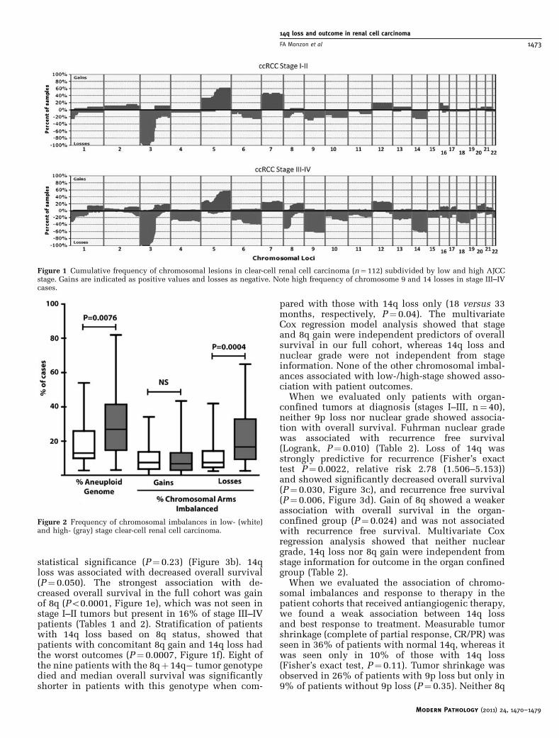
statistical significance (P¼ 0.23) (Figure 3b). 14qloss was associated with decreased overall survival(P¼ 0.050). The strongest association with de-creased overall survival in the full cohort was gainof 8q (Po0.0001, Figure 1e), which was not seen instage I–II tumors but present in 16% of stage III–IVpatients (Tables 1 and 2). Stratification of patientswith 14q loss based on 8q status, showed thatpatients with concomitant 8q gain and 14q loss hadthe worst outcomes (P¼ 0.0007, Figure 1f). Eight ofthe nine patients with the 8qþ 14q� tumor genotypedied and median overall survival was significantlyshorter in patients with this genotype when com-
pared with those with 14q loss only (18 versus 33months, respectively, P¼ 0.04). The multivariateCox regression model analysis showed that stageand 8q gain were independent predictors of overallsurvival in our full cohort, whereas 14q loss andnuclear grade were not independent from stageinformation. None of the other chromosomal imbal-ances associated with low-/high-stage showed asso-ciation with patient outcomes.
When we evaluated only patients with organ-confined tumors at diagnosis (stages I–III, n¼ 40),neither 9p loss nor nuclear grade showed associa-tion with overall survival. Fuhrman nuclear gradewas associated with recurrence free survival(Logrank, P¼ 0.010) (Table 2). Loss of 14q wasstrongly predictive for recurrence (Fisher’s exacttest P¼ 0.0022, relative risk 2.78 (1.506–5.153))and showed significantly decreased overall survival(P¼ 0.030, Figure 3c), and recurrence free survival(P¼ 0.006, Figure 3d). Gain of 8q showed a weakerassociation with overall survival in the organ-confined group (P¼ 0.024) and was not associatedwith recurrence free survival. Multivariate Coxregression analysis showed that neither nucleargrade, 14q loss nor 8q gain were independent fromstage information for outcome in the organ confinedgroup (Table 2).
When we evaluated the association of chromo-somal imbalances and response to therapy in thepatient cohorts that received antiangiogenic therapy,we found a weak association between 14q lossand best response to treatment. Measurable tumorshrinkage (complete of partial response, CR/PR) wasseen in 36% of patients with normal 14q, whereas itwas seen only in 10% of those with 14q loss(Fisher’s exact test, P¼ 0.11). Tumor shrinkage wasobserved in 26% of patients with 9p loss but only in9% of patients without 9p loss (P¼ 0.35). Neither 8q
Figure 1 Cumulative frequency of chromosomal lesions in clear-cell renal cell carcinoma (n¼ 112) subdivided by low and high AJCCstage. Gains are indicated as positive values and losses as negative. Note high frequency of chromosome 9 and 14 losses in stage III–IVcases.
Figure 2 Frequency of chromosomal imbalances in low- (white)and high- (gray) stage clear-cell renal cell carcinoma.
14q loss and outcome in renal cell carcinoma
FA Monzon et al 1473
Modern Pathology (2011) 24, 1470–1479

gain, 9p loss or 14q loss showed association withprogression free survival or overall survival in theKaplan–Meier survival analysis (SupplementaryFigures 1b–d) or Cox proportional hazards model.However, when we evaluated only patients with 14qloss, patients with concomitant 14q loss and 8qgain had a significantly shorter survival than thosewith 14q loss and either loss or normal 8q(P¼ 0.005, Supplementary Figure 1e).
Association of HIF1a Levels with Chromosomal Loss
We evaluated protein and mRNA levels of HIF1a inspecimens from the bevacizumab-treated cohort(n¼ 30).20 A tissue microarray (for immunohisto-
chemistry) and/or paraffin blocks (for RNA extrac-tion) were not available for all other samples inthe study. HIF1a protein levels, as measured byimmunohistochemistry, showed significant correla-tion with 14q status, with a decrease in HIF1a levelsin tumors with loss of 14q compared with tumorsharboring normal 14q (P¼ 0.0148, Student’s t-test)(Figure 4b). Decreased expression of HIF1A mRNAin samples with 14q loss was confirmed by qPCR(0.0019±0.0014 versus 0.0036±0.0032, P¼ 0.038,Student’s t-test) (Figure 4a). HIF1a protein andmRNA levels did not correlate with other chromo-somal imbalances (data not shown). Cox regressionresults indicated that HIF1A mRNA levels are asignificant predictor of progression-free survival inthe bevacizumab-treated cohort (HR¼ 2.29, 95%
Figure 3 Kaplan–Meier survival analyses in clear-cell renal cell carcinoma patients. (a) Overall survival by AJCC Stage; (b) overallsurvival by 9p loss; (c) overall survival by 14q loss; (d) recurrence free survival by 14q loss (stage I–III patients only); (e) overall survivalby 8q status and (f) overall survival by 8q status in patients with 14q loss.
14q loss and outcome in renal cell carcinoma
1474 FA Monzon et al
Modern Pathology (2011) 24, 1470–1479

Table 2 Association with outcome in patients with CRCC
Variable Overall survival
Logrank—univariate Cox—multivariate
P value P value Relative risk, 95% CI
All stagesAJCC Stage (I–II versus III–IV) 0.001 0.039 9.01, 1.19–67.76Fuhrman nuclear gradea 0.003 n.s.9 p loss n.s.14q loss 0.050 n.s.8q gain o0.0001 0.006 3.43, 1.50–7.83
Stages I–IIIAJCC Stage (I–II versus III) 0.022 0.075Fuhrman nuclear gradea n.s.9 p loss n.s.14q loss 0.030 n.s.8q gain 0.024 n.s.
Recurrence free survival
Logrank—univariate Cox—multivariate
Stages I–IIIAJCC Stage (I–II versus III) o0.0001 0.005 4.74, 1.57–14.26Fuhrman nuclear gradea 0.010 n.s9 p loss n.s. n.s.14q loss 0.0006 n.s.8q gain n.s. n.s.
aGrade I versus grades 3–4.
a
b
mRNA Expression
Protein Expression
HIF1A HIF2A0.0060 0.3500
0.3000
0.2500
0.2000
0.1500
0.1000
0.0500
0.0000
0.0050
0.0040
0.0030
0.0020
0.0010HIF
1A m
RN
A/1
8S r
RN
A
HIF
2A m
RN
A/1
8S r
RN
A
0.0000
14q loss 14q normal 14q loss 14q normal
2.000 1.000
0.750
0.500
0.250
0.000
HIF
-1al
ph
a IH
C s
core
HIF
-2al
ph
a IH
C s
core1.500
1.000
0.500
0.000
14q loss 14q loss
HIF1A HIF2A
14q normal 14q normal
Figure 4 Expression levels (a, mRNA expression; b, protein expression) of HIF1A and EPAS1 (HIF2a) in clear-cell renal cell carcinomatumor specimens with and without 14q loss.
14q loss and outcome in renal cell carcinoma
FA Monzon et al 1475
Modern Pathology (2011) 24, 1470–1479

CI¼ 1.01–5.16, P¼ 0.045). However HIF1a expres-sion as measured by immunohistochemistry wasnot significantly associated with progression-freesurvival (HR¼ 1.69, 95% CI¼ 0.46–6.10, P¼ 0.42).Neither protein nor mRNA expression of EPAS1were associated with 14q chromosomal imbalanceor outcome, consistent with the absence of recurrentloss of 2p21 (EPAS1 locus) (Figure 1). HIF1a andEPAS1 protein and mRNA levels were not correlatedwith survival outcomes, consistent with 14q loss notbeing predictive for outcome in this cohort ofpatients with metastatic disease.
Discussion
A significant issue in the management of patientswith clear-cell renal cell carcinoma is the identifica-tion of patients that are at higher risk for recurrence.Although mortality of renal cell cancer has shownan overall decrease due to early diagnosis andimproved surgical techniques there is no reliablebiomarker for prognosis in patients with organ-confined tumors (low TNM stage) of which as manyas 20–30% will recur.3,4 We and others havehypothesized that the variability in clear-cell renalcell carcinoma patient outcomes might reflectthe intrinsic genetic heterogeneity of this tumor.18
In this study, we focused on studying the role of 9pand 14q losses, which are two genetic lesionsfrequently associated with clear-cell renal cellcarcinoma progression and on other imbalanceswith increased frequency in high-stage tumors.Our results show that chromosome 14q loss isnegatively associated with outcome in patientswith non-metastatic clear-cell renal cell carcinoma(stages I–III) and indicate that this loss leads to amolecularly distinct phenotype of clear-cell renalcell carcinoma with decreased HIF1a. Furthermore,by using whole genome analysis, we have identified8q gain as a genetic lesion also associated with pooroutcomes.
In this study, loss of chromosome 14q wasassociated with decreased overall survival in clear-cell renal cell carcinoma patients, in particular thosepatients with non-metastatic disease (Figures 2c andd). As described above, the gene for HIF1a (HIF1A)resides in chromosome 14, and this transcriptionfactor is a key regulator of vascular endothelialgrowth factor A, platelet-derived growth factor b andtransforming growth factor a expression, all keyregulators of angiogenesis and proliferation.26–28
Our results further indicate that loss of 14q leadsto a decrease in HIF1A mRNA and reduced nuclearHIF1a protein expression and suggest a mechanismfor explaining the relationship with poor outcome.Importantly, the association between decreasedHIF1a expression and tumor aggressivenesshas been reported previously.29 In addition, otherinvestigators have shown that tumors with 14qloss show decreased expression of several HIF1a
regulated genes (William G Kaelin Jr, personalcommunication). We hypothesize that loss of 14qcould lead to an imbalance in HIF1a/EPAS1 activity,and that this imbalance leads to improved tumorcell viability. This hypothesis is also supported byrecent work from Gordan et al, who demonstratedthat HIF2a promotes cell cycle progressionin hypoxic renal cell carcinoma cell lines and isassociated with enhanced MYC promoter binding,with transcriptional effects on both activated andrepressed target genes. In their work, it is shown thatwhen both HIF1a and HIF2a are expressed, HIF1aacts in a dominant negative fashion, and MYCfunction is repressed.30 Furthermore, Toschi et alobserved that downregulation of HIF1a and HIF2aoccurs through differential regulation of specificmTOR complex components Raptor and Rictor,which in turn have differential effects on down-stream targets of S6 kinase and AKT, respectively.31
These studies emphasize the distinct function andregulation of the two HIFa isoforms. IncreasedEPAS1 (HIF2a) activity in tumors with decreasedHIF1a could lead to enhanced MYC expression,improving tumor cell viability by altering DNAdamage repair mechanisms, and by upregulatingvarious pro-survival pathways downstream ofAKT.17 The fact that EPAS1 protein and transcriptlevels were not affected by 14q loss in our samplessupports the hypothesis that an imbalance in HIFisoforms could define a specific clear-cell renal cellcarcinoma phenotype with aggressive behavior.
Loss of the p arm of chromosome 9 has beenrecently been associated with unfavorable clinicaloutcome in clear-cell renal cell carcinomapatients.13,15 Although the genes responsible for thisassociation have not been identified, several poten-tial candidates (such as CDKN2A (p16), CAIX andthe interferon genes INFA1/INFB1) reside in thischromosomal region.15 Interestingly, in our patientcohort 9p loss was neither associated with recur-rence, or response to therapy, but showed a trendtowards decreased overall survival. The lack ofstatistically significant association between 9q lossand survival in our study could be explained bythe fact this association has been established inunselected patient populations, with few of thepatients showing metastasis at presentation.13,15
Recently, La Rochelle et al16 indicated that 9p losswas specifically associated with poor outcomein patients with localized, small renal masses.Our cohort includes a significant proportion ofpatients with metastatic disease and includes veryfew patients with small, localized tumors. There-fore, our results and La Rochelle’s suggest that 9pdoes not influence the prognosis of large (advanced)or already metastatic tumors.
We also found that 8q gain was strongly prog-nostic for overall survival in clear-cell renal cellcarcinoma patients and seems to identify a subgroupof patients with 14q loss with significantly poorsurvival. Interestingly, 8q gain appears to be an early
14q loss and outcome in renal cell carcinoma
1476 FA Monzon et al
Modern Pathology (2011) 24, 1470–1479

event in hepatocellular carcinoma development,32
but our data suggests that in clear-cell renal cellcarcinoma 8q gain is a genetic abnormality acquiredduring tumor progression. Previous investigators inclear-cell renal cell carcinoma have postulated MYCas a candidate target for 8q amplification; however,data from hepatocellular carcinoma suggest thatCOPS5 (also known as JAB1) could be responsiblefor a proliferative advantage in hepatocellularcarcinoma.33 Interestingly, it has been shown thatCOPS5 regulates HIF1a activity by direct interactionand modulation of its stability.34 Thus, both ofthese 8q genes are functionally linked to the HIFpathway; MYC as a downstream effector of HIF2a asdescribed above and COPS5 as a regulator of HIF1afunction. To our knowledge, this is the first report ofthe association of 8q gain with outcome in patientswith clear-cell renal cell carcinoma and as such itshould be confirmed in other studies. If this associa-tion is confirmed, further work on the role of 8qgenes and the MYC pathway in clear-cell renal cellcarcinoma tumor behavior is therefore warranted.
Another significant problem in the managementof patients with metastatic renal cell carcinoma isdemonstration of innate or acquired resistance tomolecularly targeted therapies, including those thattarget angiogenesis. Our results indicate that neither9p nor 14q loss were significantly associated withresponse to the antiangiogenic agents sorafenib orbevacizumab. Measurable tumor response to theseagents was more frequent in patients with normal 9pand 14q, but this difference did not reach statisticalsignificance. The lack of significance is either due tothe lack of a relationship between these chromoso-mal abnormalities and clinical outcome in meta-static clear-cell renal cell carcinoma patients treatedwith antiangiogenic agents, due to limitations insample size, or due to the fact that we combined twotreatment cohorts. Interestingly, the presence of 8qgain seems to separate patients with 14q loss intodifferent subgroups of patients with markedlydifferent overall survival (Supplementary Figure1e) when treated with antiangiogenic agents. Thesefindings need to be corroborated in larger cohorts ofhomogenously treated patients.
Our results suggest a role for 14q loss and HIF1alevels in clear-cell renal cell carcinoma prognosis, aswell as a possible role for the MYC axis; however,there are a few shortcomings to our findings. First,our analysis is retrospective, and the sample hasoverrepresentation of high-stage tumors. Given thatthe association between 14q loss and outcome wasstronger in stage I–III patients, these results need tobe corroborated in a larger cohort of patients with nometastasis at diagnosis. Second, we were only ableto establish a correlation between 14q loss andHIF1a protein and mRNA levels in samples frommetastatic clear-cell renal cell carcinoma patients. Itis thus necessary to corroborate this associationin non-metastatic tumors, which are those in whichwe identified 14q to be of prognostic significance.
However, the fact that 14q loss has been previouslyassociated with high grade/stage tumors and tumor-specific survival,35,36 strongly support its role indefining an aggressive tumor phenotype. Third, theassociation of 14q loss and outcome does not seem tobe independent from stage. This needs to be investi-gated in a larger cohort with a larger representation oflow-stage tumors. However, the fact that 14 loss (andpossibly lower levels of HIF1a) is coupled tightlywith tumor progression strongly suggests that thischromosomal imbalance is part of the pro-survivaland pro-metastatic processes that allow clear-cellrenal cell carcinoma progression.
In summary, our studies suggest an associationbetween loss of chromosome 14q and clinical out-come in patients with non-metastatic renal cellcarcinoma. Furthermore, we have identified differ-ential expression of HIF1a associated with thischromosomal imbalance. This association givessupport to the notion that there are distinctmolecular phenotypes of clear-cell renal cell carci-noma characterized by predominance of differentHIFa subunits. Our results also suggest that geneslocated in 8q (such as COPS5 and MYC) could beparticipating in clear-cell renal cell carcinoma’saggressive phenotype. Validation studies to confirmthese associations are required, as is the explorationof the role of EPAS1 and MYC modulation as ameans to possibly overcome clear-cell renal cellcarcinoma aggressiveness. As has been shown withother tumor types such as leukemias and malignantgliomas, molecular classification of disease based onchromosomal alterations has led to better prognosticmarkers and implementation of more effectivetargeted therapies.37,38 Further exploration of 8qgain, 14q loss, HIF1a and EPAS1 as molecularmarkers of tumor behavior and prognosis have thepotential to aid in personalizing medicine forpatients with clear-cell renal cell carcinoma.
Acknowledgements
We wish to acknowledge the support from theResearch Pathology Core at The Methodist HospitalResearch Institute for histology services and im-munohistochemistry and the Quantitative GenomicsCore at the University of Texas Health ScienceCenter in Houston for analysis of HIF1A transcripts(with special thanks to Greg Shipley for helpfulsuggestions). This study was supported by a Scho-lars Research Award from The Methodist HospitalResearch Institute (FAM) and a grant from theInstitute for Personalized Cancer Therapy (IPCT)of the University of Texas MD. Anderson CancerCenter (EJ).
Disclosure/conflict of interest
The authors declare no conflict of interest.
14q loss and outcome in renal cell carcinoma
FA Monzon et al 1477
Modern Pathology (2011) 24, 1470–1479

References
1 American Cancer Society. Cancer Facts & Figures 2008American Cancer Society: Atlanta 2008.
2 DeVita VT, Hellman S, Rosenberg SA. Cancer, Princi-ples and Practice of Oncology, 6th edn. Lippincott,Williams & Wilkins: Philadelphia, 2001.
3 Cohen HT, McGovern FJ. Renal-cell carcinoma. N EnglJ Med 2005;353:2477–2490.
4 Lam JS, Shvarts O, Leppert JT, et al. Renal cellcarcinoma 2005: new frontiers in staging, progno-stication and targeted molecular therapy. J Urol2005;173:1853–1862.
5 Amin MB, Amin MB, Tamboli P, et al. Prognosticimpact of histologic subtyping of adult renal epithelialneoplasms: an experience of 405 cases. Am J Surg Path2002;26:281–291.
6 Delahunt B, Bethwaite PB, Nacey JN. Outcome predic-tion for renal cell carcinoma: evaluation of prognosticfactors for tumours divided according to histologicalsubtype. Pathology 2007;39:459–465.
7 Motzer RJ, Bacik J, Mazumdar M. Prognostic factors forsurvival of patients with stage IV renal cell carcinoma:memorial sloan-kettering cancer center experience.Clin Cancer Res 2004;10(Pt 2):6302S–6303S.
8 Amin MB, Tamboli P. Impact of histologic subtyping ofrenal epithelial neoplasms: authors’ reply. Am J SurgPath 2003;27:1022–1024.
9 Monzon FA, Hagenkord J, Lyons-Weiler M, et al.Whole genome SNP arrays as a potential diagnostictool for the detection of characteristic chromosomalaberrations in renal epithelial tumors. Mod Pathol2008;21:1–10.
10 Yoshimoto T, Matsuura K, Karnan S, et al. High-resolution analysis of DNA copy number alterationsand gene expression in renal clear cell carcinoma.J Pathol 2007;213:392–401.
11 Kardas I, Mrozek K, Babinska M, et al. Cytogenetic andmolecular findings in 75 clear cell renal cell carci-nomas. Oncol Rep 2005;13:949–956.
12 Gunawan B, Huber W, Holtrup M, et al. Prognosticimpacts of cytogenetic findings in clear cell renal cellcarcinoma: gain of 5q31-qter predicts a distinct clinicalphenotype with favorable prognosis. Cancer Res2001;61:7731–7738.
13 Brunelli M, Eccher A, Gobbo S, et al. Loss ofchromosome 9p is an independent prognosticfactor in patients with clear cell renal cell carcinoma.Mod Pathol 2008;21:1–6.
14 Moch H, Presti Jr JC, Sauter G, et al. Geneticaberrations detected by comparative genomic hybridi-zation are associated with clinical outcome in renalcell carcinoma. Cancer Res 1996;56:27–30.
15 Klatte T, Rao PN, de Martino M, et al. Cytogeneticprofile predicts prognosis of patients with clearcell renal cell carcinoma. J Clin Oncol 2009;27:746–753.
16 La Rochelle J, Klatte T, Dastane A, et al. Chromosome9p deletions identify an aggressive phenotype ofclear cell renal cell carcinoma. Cancer 2010;116:4696–4702.
17 Gordan JD, Lal P, Dondeti VR, et al. HIF-alpha effectson c-Myc distinguish two subtypes of sporadicVHL-deficient clear cell renal carcinoma. Cancer Cell2008;14:435–446.
18 Kim HJ, Shen SS, Ayala AG, et al. Virtual-karyotypingwith SNP microarrays in morphologically challenging
renal cell neoplasms: a practical and useful diagnosticmodality. Am J Surg Pathol 2009;33:1276–1286.
19 Arai E, Ushijima S, Tsuda H, et al. Genetic clustering ofclear cell renal cell carcinoma based on array-com-parative genomic hybridization: its association withDNA methylation alteration and patient outcome. ClinCancer Res 2008;14:5531–5539.
20 Jonasch E, Wood CG, Matin SF, et al. Phase IIpresurgical feasibility study of bevacizumab in un-treated patients with metastatic renal cell carcinoma.J Clin Oncol 2009;27:4076–4081.
21 Jonasch E, Corn P, Pagliaro LC, et al. Upfront,randomized, phase 2 trial of sorafenib versus sorafeniband low-dose interferon alfa in patients with advancedrenal cell carcinoma: clinical and biomarker analysis.Cancer 2010;116:4–7.
22 Lyons-Weiler M, Hagenkord J, Sciulli CM, et al.Optimization of the Affymetrix GeneChip mapping10K 2.0 assay for routine clinical use on formalin fixedparaffin embedded tissues. Diagn Mol Pathol 2008;17:3–13.
23 Yamamoto G, Nannya Y, Kato M, et al. Highly sensitivemethod for genomewide detection of allelic composi-tion in nonpaired, primary tumor specimens byuse of affymetrix single-nucleotide-polymorphismgenotyping microarrays. Am J Hum Genet 2007;81:114–126.
24 Lefever S, Vandesompele J, Speleman F, et al.RTPrimerDB: the portal for real-time PCR primersand probes. Nucleic Acids Res 2009;37, Database issueD942–D945.
25 Zhao S, Fernald RD. Comprehensive algorithmfor quantitative real-time polymerase chain reaction.J Comput Biol 2005;12:1047–1064.
26 Motzer RJ, Bander NH, Nanus DM. Renal-cell carci-noma. N Engl J Med 1996;335:865–875.
27 Liao D, Johnson R. Hypoxia: a key regulator ofangiogenesis in cancer. Cancer Met Reviews 2007;26:281–290.
28 Young AN, Master VA, Paner GP, et al. Renal epithelialneoplasms: diagnostic applications of gene expressionprofiling. Adv Anat Pathol 2008;15:28–38.
29 Lidgren A, Hedberg Y, Grankvist K, et al. Hypoxia-inducible factor 1[alpha] expression in renalcell carcinoma analyzed by tissue microarray. Eur Urol2006;50:1272–1277.
30 Gordan JD, Bertout JA, Hu C-J, et al. HIF-2a promoteshypoxic cell proliferation by enhancing c-Myctranscriptional activity. Cancer Cell 2007;11:335–347.
31 Toschi A, Lee E, Gadir N, et al. Differential depen-dence of hypoxia-inducible factors1a and 2a onmTORC1 and mTORC2. J Biol Chem 2008;283:34495–34499.
32 Moinzadeh P, Breuhahn K, Stutzer H, et al. Chromo-some alterations in human hepatocellular carcinomascorrelate with aetiology and histological grade—resultsof an explorative CGH meta-analysis. Br J Cancer2005;92:935–941.
33 Patil MA, Gutgemann I, Zhang J, et al. Array-basedcomparative genomic hybridization reveals recurrentchromosomal aberrations and Jab1 as a potentialtarget for 8q gain in hepatocellular carcinoma.Carcinogenesis 2005;26:2050–2057.
34 Bae MK, Ahn MY, Jeong JW, et al. Jab1 interactsdirectly with HIF-1alpha and regulates its stability.J Biol Chem 2002;277:9–12.
14q loss and outcome in renal cell carcinoma
1478 FA Monzon et al
Modern Pathology (2011) 24, 1470–1479

35 Alimov A, Sundelin B, Wang N, et al. Loss of14q31-q32. 2 in renal cell carcinoma is associatedwith high malignancy grade and poor survival. Int JOncol 2004;25:179–185.
36 Mitsumori K, Kittleson JM, Itoh N, et al. Chromosome14q LOH in localized clear cell renal cell carcinoma. JPathol 2002;198:110–114.
37 Louis DN. Molecular pathology of malignant gliomas.Ann Rev Pathol: Mech Dis 2006;1:97–117.
38 Jaffe ES. Pathology and Genetics of Tumours ofHaematopoietic and Lymphoid Tissues. IARC Press;Oxford University Press (distributor): Lyon, Oxford,2001.
Supplementary Information accompanies the paper on Modern Pathology website (http://www.nature.com/modpathol)
14q loss and outcome in renal cell carcinoma
FA Monzon et al 1479
Modern Pathology (2011) 24, 1470–1479
Copyright © 2022 FDOKUMEN