Yeast 18S rRNA Dimethylase Dim1p: a Quality Control Mechanism in Ribosome Synthesis?
Upload
independentCategory
view
2download
0
Molecular Biology of the CellVol. 13, 2016–2030, June 2002
The RNA Binding Activity of a Ribosome BiogenesisFactor, Nucleophosmin/B23, Is Modulated byPhosphorylation with a Cell Cycle-dependent Kinaseand by Association with Its SubtypeMitsuru Okuwaki,*† Masafumi Tsujimoto,† and Kyosuke Nagata*‡
*Department of Infection Biology, Institute of Basic Medical Sciences, University of Tsukuba, 1-1-1Tennohdai, Tsukuba 305-8575, Japan; and †Laboratory of Cellular Biochemistry, RIKEN (The Instituteof Physical and Chemical Research), 2-1 Hirosawa, Wako 351-0198, Japan
Submitted November 8, 2001; Revised February 22, 2002; Accepted March 12, 2002Monitoring Editor: Joseph Gall
Nucleophosmin/B23 is a nucleolar phosphoprotein. It has been shown that B23 binds to nucleicacids, digests RNA, and is localized in nucleolar granular components from which preribosomalparticles are transported to cytoplasm. The intracellular localization of B23 is significantlychanged during the cell cycle. Here, we have examined the cellular localization of B23 proteinsand the effect of mitotic phosphorylation of B23.1 on its RNA binding activity. Two splicingvariants of B23 proteins, termed B23.1 and B23.2, were complexed both in vivo and in vitro. TheRNA binding activity of B23.1 was impaired by hetero-oligomer formation with B23.2. Bothsubtypes of B23 proteins were phosphorylated during mitosis by cyclin B/cdc2. The RNA bindingactivity of B23.1 was repressed through cyclin B/cdc2-mediated phosphorylation at specific sitesin B23. Thus, the RNA binding activity of B23.1 is stringently modulated by its phosphorylationand subtype association. Interphase B23.1 was mainly localized in nucleoli, whereas B23.2 andmitotic B23.1, those of which were incapable of binding to RNA, were dispersed throughout thenucleoplasm and cytoplasm, respectively. These results suggest that nucleolar localization ofB23.1 is mediated by its ability to associate with RNA.
INTRODUCTION
The cell nucleolus is the place for ribosome biogenesis,which is the synthesis and processing of a precursor rRNA(pre-rRNA), and the assembly of ribosomal proteins on rR-NAs to form premature ribosome. rRNA is first transcribedby RNA polymerase I as a pre-rRNA of �13,500 nucleotides.To produce mature rRNAs (18S, 5.8S, and 28S rRNA), exter-nal and internal spacer sequences (ETS and ITS, respec-tively) in the long pre-rRNA are removed sequentially whilenucleotide modifications such as pseudouridylation and 2�-O-methylation occur concurrently (Maden, 1990). For suchcomplex processes, it is suggested that nonribosomal pro-teins and small nucleolar RNAs are multifunctional (Srivas-tava and Pollard, 1999; Peculis, 2000). One such nucleolarprotein is the well-characterized and abundant nucleophos-min/B23. It is highly conserved in vertebrates. In rat andhuman cells, at least two isoforms of nucleophosmin/B23,
termed B23.1 and B23.2, are expressed (Chang and Olson,1990; Okuwaki et al., 2001a). These two proteins are identicalexcept that the C-terminal 35 amino acids observed in B23.1are absent in B23.2. Nucleophosmin/B23 functions to bindnucleic acids (Dumbar et al., 1989), to endonucleolyticallycleave RNA preferentially within ITS2 in pre-rRNA (Savkurand Olson, 1998), to suppress the aggregation of proteins(Szebeni and Olson, 1999), and to bind peptides containingnuclear localization signals for their nuclear import (Szebeniet al., 1995). Recently, phosphorylation of nucleophosmin/B23 by cyclin E/cdk2, a G1 cyclin-dependent kinase, wasshown to be essential for centrosome duplication in fibro-blast cells (Okuda et al., 2000). Furthermore, it is shown thatB23 protein binds a wide diversity of proteins, such asnucleolar phosphoproteins, nucleolin and p120 (Durban etal., 1995; Li et al., 1996), HIV1-Rev protein (Fankhauser et al.,1991), retinoblastoma protein (Takemura et al., 1999) to stim-ulate the DNA polymerase � activity (Umekawa et al., 2001),and hepatitis delta virus delta antigens (Huang et al., 2001).Because the B23-nucleic acid binding and RNase activitydomains are mapped in the C-terminal region present onlyin B23.1 (Hingorani et al., 2000), the C-terminal region ofB23.1 is likely to be important for ribosome biogenesis. The
Article published online ahead of print. Mol. Biol. Cell 10.1091/mbc.02–03–0036. Article and publication date are at www.molbiol-cell.org/cgi/doi/10.1091/mbc.02–03–0036.
‡ Corresponding author. E-mail address: [email protected].
2016 © 2002 by The American Society for Cell Biology
function of B23.1 has been well studied and characterized,whereas that of B23.2 is not well understood.
Eukaryotic gene expression is regulated through the cellcycle, partly by the phosphorylation and dephosphorylationof proteins involved in this process (Gottesfeld and Forbes,1997). In particular, transcription is significantly repressedduring mitosis. This also applies to the transcription systemmediated by RNA polymerase I (pol I). Several factors in-volved in pol I transcription are found phosphorylated andinactivated during mitosis. The promoter selectivity factorSL1 is phosphorylated during mitosis, which inactivates itfor transcription from the pol I promoter (Heix et al., 1998).During mitosis, not only transcription machinery but alsofactors involved in mRNA biogenesis (Colgan et al., 1998)and in chromatin remodeling (Sif et al., 1998) are phosphor-ylated and inactivated. On the contrary, the activity of sev-eral proteins is stimulated by phosphorylation during mito-sis. For example, a component of the chromosomecondensation machinery, condensin, becomes activatedthrough phosphorylation by cdc2 kinase during mitosis(Kimura et al., 1998). Nucleophosmin/B23 is also phosphor-ylated by cyclin B/cdc2 during mitosis (Peter et al., 1990),thereafter changing its cellular localization drastically. Thelocalization of nucleophosmin/B23 in mitotic cells can befound in three locations; throughout cytoplasm, on the mi-totic apparatus, or at nucleolus-derived foci (NDF) (Zat-sepina et al., 1997, 1999; Dundr et al., 2000). At NDF, B23remains associated with pre-rRNA and rRNA processingfactors such as nucleolin, fibrillalin, and snoRNP (Dundrand Olson, 1998; Pinol-Roma, 1999). However, it is notknown if phosphorylation of B23 proteins during mitosishas any effect on its function in ribosome biogenesis.
We previously identified nucleophosmin/B23 to be a ma-jor component of template activating factor (TAF)-III (Oku-waki et al., 2001a). TAF-III can stimulate cell-free replicationfrom the adenovirus genome complexed with viral basiccore proteins, thus forming a chromatin-like structure. Be-cause nucleophosmin/B23 binds directly to core histonesand mediates assembly of nucleosome in vitro, it is sug-gested that nucleophosmin/B23 functions as a histone chap-erone (Okuwaki et al., 2001b). Here, we have shown that theB23 subtypes were complexed with each other in vivo and invitro, and that the RNA binding activity of B23.1 was mod-ulated through its complex formation with B23.2. Greenfluorescent protein (GFP)-tagged B23.2 was dispersedthroughout nucleoplasm, whereas a major fraction of GFP-tagged B23.1 was concentrated in nucleoli as endogenousB23.1. In addition, B23.2 was extracted with detergent,whereas B23.1 remained tightly associated with nuclearstructure in the presence of detergent. These in vitro and invivo properties of both subtypes of B23 proteins suggest thatB23.2 could modulate the ribosome biogenesis function ofB23.1 as well as its intracellular localization. Furthermore,the RNA binding activity of B23.1 was completely disruptedby cyclin B/cdc2-mediated phosphorylation. A mutantB23.1 in which four threonines for phosphorylation weresubstituted by alanines was not phosphorylated by cyclinB/cdc2, and remained associated with RNA after phosphor-ylation reaction in the presence of cyclin B/cdc2. This resultstrongly suggests that the RNA binding activity of B23.1 isregulated partly by phosphorylation and dephosphoryla-tion. Thus, it is proposed that at least two mechanisms,
hetero-oligomerization with a modulator protein (e.g.,B23.2) and phosphorylation/de-phosphorylation of B23.1,contribute to regulating the involvement of B23 in ribosomebiogenesis.
MATERIALS AND METHODS
Plasmid ConstructionFor expression of N-terminal HA-epitope tag–fused hB23.1 in HeLacells, the pCHA vector (Nagata et al., 1998), which is driven by thecytomegalovirus immediate early gene enhancer, and chicken �-ac-tin promoter was used. pCHA-hB23.1 was prepared as described(Okuwaki et al., 2001a). To generate pCHA-hB23.2, a fragment ofhB23.2 cDNA was obtained by digestion of pET14b-hB23.2 (Oku-waki et al., 2001a) with NdeI and BamHI. The fragment was thencloned in-frame into BstEII- and BglII-digested pCHA. To generatemutant B23.1 proteins, an appropriate set of oligonucleotide primerswere used for site-directed amino acid substitutions (threonine toalanine). To generate a T199A expression vector, cDNA fragmentscontaining a mutation at T199 were amplified by polymerase chainreaction (PCR) using the N-terminal primer for B23.1 and 5�-tttg-gctggagcatctcgtat-3�, or C-terminal primer for B23.1 and 5�-atac-gagatgctccagccaaa-3�, with pET14b-hB23.1 as a template. Two kindsof amplified cDNA fragments were purified and used as templatesfor the second PCR to amplify a full-length cDNA fragment forT199A using N- and C-terminal primers of B23.1. The same protocolwas applied to construct T219A and T234/237A expression vectors.Primer sets for T219A and T234/237A were 5�-ccatcatcagcaccaa-gatca-3� and 5�-tgatcttggtgctgatgatgg-3�, and 5�-caggaaaaagctccta-aagcaccaaaagga-3� and 5�-tccttttggtgctttaggagctttttcctg-3�, respec-tively. The mutated cDNA fragments were then digested with NdeIand BamHI, and then cloned into NdeI- and BamHI-digested pET14b (Novagen, Madison, WI). Triple mutant proteins T199/234/237A and T219/234/237A were prepared by PCR using the appro-priate primer sets described above and pET14b-T234/237A as atemplate. Recombinant proteins were expressed and purified asdescribed by Okuwaki et al. (2001a). For expression of Flag-taggedwild-type and mutant B23.1 proteins in HeLa cells, each cDNAfragment digested with NdeI and BamHI was subcloned into NdeI-and BamHI-digested pBS-Falg vector to generate pBS-Flag-B23.1,-T199A, -T219/23 4/237A, and -T4A. Each cDNA attached by theFlag-tag at its 5� terminus was cut out from pBS-Flag vector bydigestion with BamHI, and then subcloned into BglII-digestedpCAGGS vector. To express GFP-tagged B23.1 or B23.2, cDNAfragments attached by the Flag-tag were cut out from pBS-Flagvector by BamHI, and then subcloned into BamHI-digested pEG-FPC1 vector (CLONTECH, Palo Alto, CA). Transient transfection ofeach plasmid to HeLa cells was performed by the calcium phos-phate precipitation method (Graham and Eb, 1973).
Immunoprecipitation AnalysisHeLa cells expressing HA-tagged hB23.1 or hB23.2 were lysed inbuffer A (50 mM Tris, pH 7.9, 0.1 mM EDTA, and 0.1% Nonidet P40)containing 150 mM NaCl on ice for 10 min and were then disruptedby extensive sonication. Cell extracts recovered by centrifugationwere mixed with anti-HA antibody (3F10; Roche, Indianapolis, IN)and incubated on ice for 30 min. Then, protein A Sepharose beads (5�l of resin; Amersham Pharmacia, Piscataway, NJ) were added andfurther incubated for 2 h with gentle agitation. Proteins bound toresins were eluted by an SDS sample buffer, boiled, separated on a10% SDS-PAGE, and transferred to a polyvinylidene difluoride(PVDF) membrane. Endogenous B23.1 was detected by westernblotting with anti-B23.1 antibody (C19; Santa Cruz Biotechnology,Santa Cruz, CA).
Regulation of the RNA Binding Activity of B23
Vol. 13, June 2002 2017

In Vitro Phosphorylation of B23 Proteins by Cyclin-dependent KinasesTo prepare cyclin E/cdk2, NIH3T3 cells were first grown in DMEMsupplemented with 0.1% calf serum for 48 h. The medium waschanged to DMEM containing 10% calf serum, and cells were fur-ther incubated for 14 h. Cells were lysed in buffer A containing 150mM NaCl, 10 mM �-d-glycerophosphate, 1 mM NaF, 1 mM NaVO4, and 0.5 mM phenylmethylsulfonyl fluoride (PMSF), and dis-rupted by extensive sonication. Cell extracts (200 �g) recovered bycentrifugation were mixed with anti-cyclin E antibody (M20; SantaCruz Biotechnolgy). The cyclin E-associated kinase bound to theantibody was purified using protein A Sepharose beads. The beadswere washed extensively with buffer A containing 150 mM NaCl,and were suspended in a kinase reaction buffer (20 mM Tris, pH 7.9,10 mM MgCl 2, 10 mM �-d-glycerophosphate, 1 mM NaF, 1 mMNaVO 4, and 0.5 mM PMSF). For preparation of cyclin B/cdc2, HeLacells were arrested at prometaphase by incubation with nocodazole,a microtubule destabilizing reagent, at a final concentration of 50ng/ml for 12 h. Mitotic cells were collected by shaking dishesgently. Cell extracts were prepared as described (Pinol-Roma, 1999).In standard experiments, cyclin B/cdc2 kinase was purified from500 �g of mitotic extracts using anti-cyclin B antibody (GNS1; SantaCruz Biotechnology, 500 ng). The cyclin B/cdc2-immobilized pro-tein A beads were then suspended in 100 �l of buffer H (20 mMHEPES-NaOH, 0.5 mM EDTA, 50 mM NaCl, 10% glycerol, and 0.5mM PMSF) and used as a kinase. The phosphorylation reaction wasperformed as follows: Recombinant B23 proteins (5 �g) were mixedwith 20 �l of a kinase fraction in the presence of 1 mM ATP in 20mM Tris, pH 7.4, 10 mM MgCl 2, 50 mM NaCl, 5 mM �-d-glycero-phosphate, and 0.5 mM PMSF. The mixture was incubated at 30°Cfor 30 min. Under these reaction conditions, 5 �g of B23.1 wascompletely phosphorylated. Phosphorylated B23.1 was separablefrom nonphosphorylated protein by SDS-PAGE (see Figure 7A).Note that for results shown in reactions of Figures 6, C through E,and 8B, ATP concentration was adjusted to 1 �M, and 1 �Ci of[�-32P]ATP (Amersham Pharmacia) was supplemented.
Cell Fractionation ExperimentsCell fractionation experiments were performed essentially as de-scribed (He et al., 1990). HeLa cells grown in Eagle’s minimumessential medium supplemented with 10% fetal bovine serum werewashed with ice-cold phosphate-buffered saline, and then lysed onice for 5 min in cytoskeletal (CSK) buffer containing 0.5% TritonX-100. Then, supernatant was collected and the cell pellet waswashed with the CSK buffer. The cell pellet that contained thechromatin DNA was digested by 2 units/�l RNase-free DNase I(Invitrogen, Carlsbad, CA) at 37°C for 15 min and was extractedwith (NH 4)2SO 4 at a final concentration of 0.25 M in CSK buffer.The supernatant was collected while the pellet was further extractedon ice for 5 min with 2 M NaCl in CSK buffer. Remaining insolublefractions were dissolved in 8 M urea. For Figure 5, a part of TritonX-100–extracted cell pellet was digested with 0.1 mg/ml RNase Ainstead of DNase I. Extracted proteins at each step were separatedon a 10% SDS-PAGE and were subjected to western blotting usinganti-B23 polyclonal (C19) or monoclonal (NB23) antibodies, antito-poisomerase II� (8D2), and anticyclin B (GNS1) antibodies. To de-tect HA- and Flag-tagged B23 proteins, anti-HA (12CA5; Roche) andanti-Flag (M2; Sigma, St. Louis, MO) antibodies, respectively, wereused. For experiments in Figure 8, HeLa cells were arrested atprometaphase in the presence of 50 ng/ml nocodazole for 12 h, andthe mitotic cells were then collected. Mitotic cells were subjected tocell fractionation experiment as described above.
RNA Binding AssayRNA was extracted from HeLa cells using guanidium thiocyanate.Recombinant B23 proteins (5 �g) were mixed with 5 �g of RNA,incubated at room temperature for 30 min, and then loaded onto a
15–40% sucrose gradient in 20 mM Tris, pH 7.4, 50 mM NaCl, 0.5mM PMSF, and 1 mM dithiothreitol. Samples were centrifuged at54,000 rpm for 2 h in a TLS55 rotor (Beckman Instruments, Fuller-ton, CA), and fractions (150 �l) were collected from the top. Foranalysis of phosphorylated proteins, 10 mM �-d-glycerophosphate,1 mM NaF, and 0.1 mM NaVO 4 were added to inhibit the poten-tially contaminating phosphatase activity. Proteins in each fractionwere separated by SDS-PAGE and were visualized by staining withCBB.
For nitrocellulose filter binding assay, RNA (5 �g) extracted fromHeLa cells were end-labeled by polynucleotide kinase (Toyobo) inthe presence of 30 �Ci of [�-32P]ATP (3000 Ci/mmol). To form B23complexes in which the ratio of B23.1 to B23.2 was 1:0, 1:0.2, 1:0.5,1:1, or 1:2, B23 proteins were mixed and subjected to the denature-renature protocol (Hager and Burgess, 1980). [32P]-labeled RNA (100ng, specific activity was �500 cpm/ng) was incubated in the pres-ence or absence of B23 complexes at 30°C for 30 min in 20 mMHEPES-NaOH, pH 7.9, 150 mM NaCl, and 0.5 mM EDTA. TheRNA–protein complexes were filtrated through a nitrocellulose fil-ter (Hybond N; Amersham Pharmacia). The radioactivity retained
Figure 1. B23.1 and B23.2 are complexed in vivo and in vitro. (A)Immunoprecipitation of HA-tagged B23 proteins from cell extracts.Cell extracts prepared from HeLa cells transfected with pCHA (lane1), pCHA-hB23.1 (lane 2), or pCHA-hB23.2 (lane 3) were subjectedto immunoprecipitation analysis using anti-HA (3F10) antibody.Proteins associated with anti-HA were separated on a 10% SDS-PAGE, transferred to a PVDF membrane, and endogenous andHA-tagged B23.1 proteins were detected with anti-B23.1 antibody(C19). Positions of HA-tagged and endogenous B23.1 are indicatedby arrowheads at the right side of the panel. (B) Direct interactionbetween B23.1 and B23.2. Either recombinant (r) B23.1 alone (2 �g)(lanes 1 and 2), rB23.2 alone (2 �g) (lanes 5 and 6), and a mixture ofrB23.1 and rB23.2 (2 �g each) (lanes 3 and 4) were subjected to thedenature-renature protocol (Hager and Burgess, 1980). Renaturedproteins were immunoprecipitated with the anti-B23.1 (C19) anti-body that specifically recognizes the C-terminal region of B23.1.Immunoprecipitated proteins (“P,”, lanes 2, 4, and 6) were sepa-rated on a 10% SDS-PAGE and visualized via silver staining. Posi-tions of rB23.1 and rB23.2 are indicated by arrowheads at the rightside of the panel. “In” indicates 10% of input proteins (lanes 1, 3,and 5). Protein bands indicated by “H” and “L” shown at the rightside of the panel correspond to heavy and light chains of immuno-globulin, respectively.
Okuwaki et al.
Molecular Biology of the Cell2018

on the filter was measured by a FUJIX BAS 2000 image analyzingsystem.
Indirect Immunofluorescence AnalysisHeLa cells grown on coverslips were transiently transfected withpCAGGS-Flag B23.1 (5 �g) by the calcium phosphate precipitationmethod. Twenty-four hours after transfection, cells were fixed byice-cold acetone-methanol (1:1) at �30° for 10 min, and Flag-taggedB23.1 was bound by the primary anti-Flag (M2) antibody and de-tected by a secondary antibody consisting of anti-mouse IgG con-jugated with Alexa564 (Molecular Probes, Eugene, OR).
RESULTS
Homo- and Hetero-Oligomerization and CellularDistribution of Splicing Variants ofNucleophosmin/B23The C-terminal region specific to B23.1 is suggested to beinvolved in several important functions of B23 in ribosomebiogenesis, whereras the function of B23.2, which lacks thisC-terminal tail, is totally unknown. B23.1 and B23.2 cofrac-tionated after five steps of column chromatography, includ-ing ion-exchange and gel filtration columns (Okuwaki et al.,2001a). In addition, several previous reports have suggestedthat the N-terminal portion of B23 protein is involved inoligomer formation (Yung and Chan, 1987; Herrera et al.,1996; Dutta et al., 2001). Therefore, B23.1 and B23.2 could becomplexed in vivo. We first decided to examine this possi-bility by immunoprecipitation and by looking at cellularlocalization of both proteins. We used anti-HA antibody tosee if endogenous B23.1 from cell extracts coimmunoprecipi-tates transiently expressed HA-tagged B23.1 or B23.2. Cellextracts prepared from HeLa cells expressing either HA-tagged B23.1 or B23.2 were prepared and subjected to im-munoprecipitation analyses. Coimmunoprecipitation of en-dogenous B23.1 by anti-HA antibody was detected bywestern blotting with anti-B23.1 antibody (Figure 1A). En-dogenous B23.1 was coimmunoprecipitated with both HA-tagged B23.1 and B23.2 (Figure 1A, lanes 2 and 3), indicatingthat both subtypes are complexed in vivo. To confirm that
B23.1 and B23.2 directly interact with each other, recombi-nant B23.1 and B23.2 proteins were mixed and subjected toimmunoprecipitation analysis using a B23.1-specific anti-body. To reconstitute the complex, recombinant His-taggedB23.1 and B23.2 were mixed and codenatured/renaturedbefore immunoprecipitation analysis. Recombinant B23.1 in-deed was precipitated with anti-B23.1 antibody, whereasB23.2 alone was not (Figure 1B, compare lanes 2 and 6). Onthe other hand, when B23.2 was corenatured with B23.1, itwas coimmunoprecipitated with B23.1 with the same anti-B23.1 antibody (Figure 1B, lane 4). From these results, weconcluded that the B23 subtypes directly interact in vivo andin vitro. This is also consistent with a previous report thatB23 forms an oligomer in solution (Yung and Chan, 1987).
Next, the subcellular localization of these proteins wasexamined. B23 proteins fused with GFP-tag at their N ter-mini were expressed in HeLa cells. The localization of GFP-tagged proteins was visualized under a fluorescent micro-scope (Figure 2). GFP-tagged B23.1 was mainly localized innucleoli in HeLa cells. This localization pattern is identical tothat of endogenous B23.1 (unpublished results). In contrast,GFP-tagged B23.2 was localized not only in nucleoli, but wasalso dispersed throughout the nucleoplasm. These observa-tions support the previous reports that the C-terminal regionspecific to B23.1 plays an important role in nucleolar local-ization (Peculis and Gall, 1992; Wang et al., 1993; Zirwes etal., 1997a). Next, cells on coverslips were sequentially ex-tracted in situ as follows. Cells on coverslips were exposedto detergent (0.5% Triton X-100, Figure 2, second column),and then digested with DNase I followed by extraction with0.25 M ammonium sulfate (Figure 2, third column) or RNaseA (Figure 2, fourth column) followed by extraction with 0.25M ammonium sulfate. The majority of B23.1 was resistant todetergent extraction and a fraction of B23.1 was detected innuclei even after treatment with DNase I or RNase A. Thus,it is possible that a fraction of B23.1 is associated withprotein(s) that remains in nuclease-treated nuclei. On thisline, it is reported that B23.1 is a nuclear matrix–associatedprotein (Feuerstein and Mond, 1987). On the other hand,nucleoplasmic GFP-tagged B23.2 was released by detergent
Figure 2. Cellular localization ofB23 splicing variants. HeLa cellsgrown on coverslips were trans-fected with pEGFPC1-hB23.1 (up-per panels) or -hB23.2 (bottom pan-els). Thirty hours after transfection,cells on coverslips were fixed with3% paraformaldehyde (first col-umn) or were treated with 0.5% Tri-ton X-10 0 followed by fixation with3% paraformaldehyde (second col-umn). Triton X-100–treated cellswere further digested with DNase I(third column) or RNase A (fourthcolumn) followed by extractionwith 0.25 M ammonium sulfate be-fore fixation with paraformalde-hyde. DNA stained with Hoechst33258 (blue) and GFP-tagged pro-teins (green) were visualized undera fluorescent microscope. The bar atthe right bottom indicates 10 �m.
Regulation of the RNA Binding Activity of B23
Vol. 13, June 2002 2019

extraction, whereas a trace amount of B23.2 remained innucleoli (Figure 2, second column in bottom panels). GFP-tagged B23.2 was not qualitatively detected after nucleasetreatment (Figure 2, third and fourth columns in bottompanels). To further examine these properties of B23 sub-
types, protein composition in the fractions collected aftereach treatment was analyzed with SDS-PAGE followed bywestern blotting (Figure 3). Core histones (major chromatinproteins) fractionated mainly in the “DNase I” fraction (Fig-ure 3A, upper panel), whereas TAF-I, a nuclear proteininvolved in chromatin assembly and remodeling (Nagata etal., 1995), was mainly detected in the “Detergent” fraction(unpublished results). The majority of B23.1 was recoveredin both “DNase I” and “Detergent” fractions. A low butdistinct amount of B23.1 was in the “Nuclear Matrix” frac-tions. In contrast, B23.2 mainly fractionated in the “Deter-gent” fraction, whereas only a part of B23.2 was detected inthe “DNase I” fraction. To further confirm these B23 proteinproperties, HA-tagged B23.1 or B23.2 were expressed inHeLa cells and the cells were then subjected to fractionationas described above. HA-tagged B23.1 was recovered mainlyin the “Detergent” and “DNase I” fractions as endogenousprotein, whereas HA-tagged B23.2 is mainly in the “Deter-gent” fraction (Figure 3B). Figures 2 and 3 suggest that B23.1,but not B23.2, is a nuclear matrix–associated protein, andB23.2 is less tightly associated with nuclear structures suchas chromatin and nuclear matrix relative to B23.1.
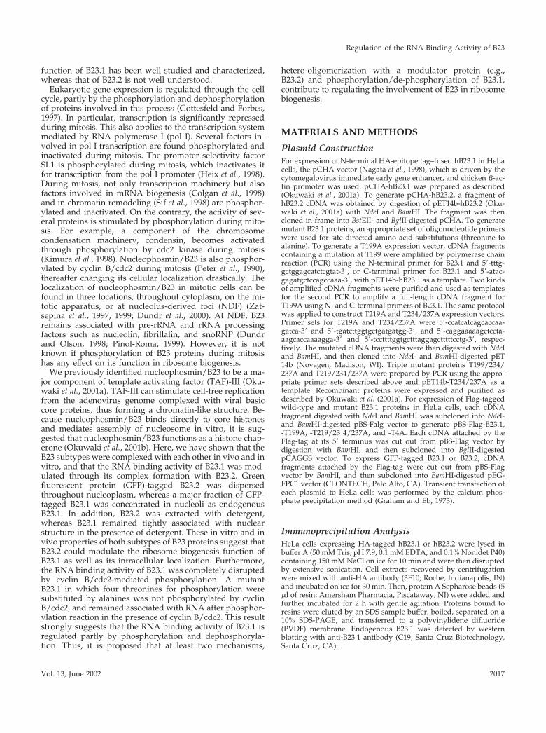
RNA Binding Activity of B23.1 Is Modulated byHetero-Oligomer Formation with B23.2The observations that both subtypes of B23 proteins aredirectly complexed in vivo and that their localization pat-terns are different from each other led us to hypothesize thathetero-oligomer formation of B23.1 and B23.2 may impairthe C-terminal–specific function of B23.1 such as nucleic acidbinding and RNase function. To address this, the RNAbinding activity of B23 proteins was examined in a sucrosegradient sedimentation assay. B23 complex was mixed withtotal RNA purified from HeLa cells, incubated, and thenloaded onto the sucrose gradient. B23.1 or RNA alone wasrecovered in relatively low and high density fractions (Fig-ure 4A, lanes 2–4 or lanes 8–10), respectively. B23.2 alonewas also recovered in the low density fractions (unpublishedresults). In sharp contrast, B23.1 preincubated with RNAwas sedimented in the high density fractions (Figure 4A,lanes 8–11) and cofractionated with RNA containing 18Sand 28S rRNA (Figure 4A, fourth panel), whereas B23.2 wasdetected in low density fractions even after mixing withRNA (Figure 4A, seventh panel). This is consistent with theprevious observation that rat B23.1 but not B23.2 bindsnucleic acids (Wang et al., 1994). Although the peak positionof 28S rRNA is slightly shifted from fraction 8 to fraction 9in the presence of B23.1, we cannot conclude that B23.1binds directly to 28S rRNA or which RNA is a target of B23.1at present. To evaluate the effect of B23.2 on the RNA bind-ing activity of B23.1, both proteins were mixed at a ratio of1:1 or 1:3 and then subjected to the denature-renature pro-tocol to form hetero-oligomer as shown in Figure 1B. Rena-tured complexes were tested for the RNA binding activity asabove. Interestingly, B23.1 complexed with B23.2 was recov-ered mainly in the low density fractions (Figure 4A, lanes2–4), whereas only a part of B23.1 sedimented to the RNAcontaining fractions with a small amount of B23.2 (Figure4A, lanes 8–10). When the ratio of B23.2 to B23.1 was in-creased in the complex, the RNA binding activity of B23.1significantly decreased (Figure 4A, compare fourth, fifth,and sixth panels), suggesting that B23.2 impairs the RNA
Figure 3. Cell fractionation and diverse distribution of B23 sub-types. (A) Cell fractionation experiments. Exponentially growingHeLa cells were fractionated sequentially as described in the “Ma-terials and Methods.” Proteins in “Detergent,” “DNase I,” and“nuclear matrix” (lanes 1–3, respectively) fractions (1 � 105 cells)were separated on a 12.5% SDS-PAGE and visualized with CBBstaining (upper panel) or transferred to a PVDF membrane andsubjected to western blotting with anti-B23 (NB23 that recognizesboth subtypes of B23 proteins) (bottom panel). Positions of theproteins are indicated on the right side of the panels. Lane M in theupper panel shows standard molecular weight markers. Positions ofcore histones are also indicated with a bracket. (B) Fractionation ofHeLa cells expressing HA-tagged B23 proteins. HeLa cells weretransiently transfected with 500 ng of pCHA-hB23.1 (lanes 1–3) or-hB23.2 (lanes 4–6) and were incubated at 37°C for 30 h. Cellfractionation experiments were carried out as described in A. Posi-tions of HA-tagged B23.1 and B23.2 are indicated at the right side ofthe panel.
Okuwaki et al.
Molecular Biology of the Cell2020
binding activity of B23.1. To quantitatively estimate the ef-fect of B23.2 on the RNA binding activity of the B23 com-plex, nitrocellulose filter binding assays were carried out.Total RNA prepared from HeLa cells was end-labeled andused for the filter binding assay. B23.1 were mixed withB23.2 at a ratio (B23.1:B23.2) of 1:0, 1:0.2, 1:0.5, 1:1, or 1:2, andthen subjected to the denature-renature protocol (Figure 4B,upper panel). Renatured B23 complexes were incubatedwith the end-labeled RNA, followed by filtration through anitrocellulose filter. Radioactive RNA retained on the nitro-cellulose filter was quantified (Figure 4B, left plot). RNAalone passed through the filter and the radioactive RNA wasnot retained. Homo-oligomer of B23.1 efficiently bound toRNA in a B23.1 dose-dependent manner, whereas B23.2
homo-oligomer did not (Figure 4B and unpublished results).This is consistent with the results of sucrose gradient sedi-mentation analyses. The RNA binding ability of the B23complex was decreased with the increasing ratio of B23.2 toB23.1 in the complex (Figure 4B, left and right plots). Theamount of B23.1 recovered in high density fractions (Figure4A, fourth panel, lanes 8–11) was significantly decreasedwith increasing amounts of B23.2 (Figure 4A, fifth and sixthpanels) under a sucrose gradient assay condition, whereasunder the filter binding assay condition, the RNA bindingactivity of the 1:2-ratio–B23.1:B23.2 complexes was reduced�50%. This difference of the effect of B23.2 on the RNAbinding activity of B23 complex could be explained by ob-serving that the B23-RNA complexes recovered in middle
Figure 4. The RNA binding activity of B23.1 complexed with B23.2. (A) Sucrose gradient analyses of the B23–RNA complex. rB23.1 (5 �g),rB23.2 (5 �g), and a 1:1 ratio mixture (5 �g each), and a 1:3 mixture (5 �g and 15 �g, respectively) of B23.1 and B23.2 were subjected to thedenature-renature protocol (Hager and Burgess, 1980). Renatured proteins were incubated in the absence or presence of RNA purified fromHeLa cells as indicated at the left side of the panels and were loaded on a 15–40% sucrose gradient. Proteins in fractions collected from thetop were separated on a 10% SDS-PAGE and were visualized by staining with CBB (third through seventh panels). RNA was extracted fromeach fraction and was separated on a 1% agarose gel containing 6.3% formaldehyde in MOPS buffer [3-(N-morpholino)-propanesulfonic acid]and was visualized by staining with ethidium bromide. (Top and second panels) Positions of 18S and 28S rRNA, and rB23.1 and rB23.2 areindicated at the right side of the panels. Lane M indicates molecular weight markers. (B) Filter binding assay. The B23.1 and B23.2hetero-oligomers were reconstituted and subjected to nitrocellulose filter RNA binding assays. The reconstituted B23 complexes (B23:1:B23.2 � 1:0, 1:0.2, 1:0.5, 1:1, and 1:2 for lanes 1–5, respectively; each lane contains 100 ng of B23.1) were separated on a 10% SDS-PAGE andstained with CBB (upper panel). The B23 complexes (a ratio of B23.1 to B23.2 � 1:0 (�), 1:0.2(f), 1:0.5 (Œ), 1:1 (�), or 1:2 (E)) containing 50,100, 200, and 400 ng of B23.1 were mixed and incubated with [32P]-end-labeled RNA (100 ng), followed by filtration through nitrocellulosefilters. The radioactivity of RNA retained on the filter was quantified by a FUJIX BAS2000 image analyzing system. The amount of RNAbound on the filter is plotted as a function of the amount of B23. 1 in the B23 complexes (left plot). The efficiency of the RNA binding activityof the B23 complexes as a function of the ratio of B23.2 to B23.1 is calculated and summarized at the right plot. The RNA binding activityof B23.1 homo-oligomer is represented as 100%. Shown are the average of a series of experiments where different amounts of B23.1 (50, 100,200, and 400 ng) in the B23 complexes were used for filter binding assays. (C) Sucrose density gradient of cell extracts. Cell extracts preparedfrom HeLa cells as described by Manley et al. (1980) were subjected to sucrose gradient sedimentation assays. Cell extracts (100 �g) treatedwithout (upper panel) or with (bottom panel) RNase A (0.2 mg/ml) were loaded on a 15–40% sucrose density gradient and centrifuged asin A. Fractions (150 �l) were collected from the top, and proteins in each fraction were analyzed by SDS-PAGE. B23 proteins were detectedby western blotting with anti-B23 antibody (NB23). Positions of B23.1 and B23.2 are shown at the right side of the panels. Lane M showsmolecular weight markers.
Regulation of the RNA Binding Activity of B23
Vol. 13, June 2002 2021

density fractions (Figure 4A, fifth and sixth panels, lanes4–7) were collected on filters.
Next, to examine the relationship between oligomeriza-tion form and the RNA binding activity of B23 proteins invivo, whole cell extracts prepared from HeLa cells weresubjected to the sucrose gradient analysis (Figure 4C). Frac-tionation patterns of B23.1 and B23.2 were analyzed bywestern blotting using anti-B23 antibody, which recognizesboth subtypes. B23.1 sedimented in low (Figure 4C, fractions2–4) and high (Figure 4C, fractions 11–14) density fractions,whereas B23.2 mainly fractionated in low density fractions(Figure 4C, fractions 2–4). These fractionation patterns arequite similar to a mixed pattern of in vitro reconstitutedB23–RNA complexes. When cell extracts were treated withRNase A before loading on a sucrose gradient, B23.1 in thehigh density fractions disappeared (Figure 4C, bottom pan-el). Thus, it is likely that B23.1 recovered in the high densityfractions is associated with RNA. This observation supportsthe idea that B23.2 modulates the RNA binding activity ofB23.1 in a cell. In addition, B23.1 present in cell extracts wasrecovered in higher density fractions than the in vitro recon-stituted B23.1–RNA complex. Therefore, B23.1 would beintegrated into large complex containing RNA and otherproteins associated with RNA and/or B23 in cell extracts.From results in Figure 4, it is strongly suggested that theratio of B23.2 to B23.1 in B23 oligomer is important indetermining the RNA binding activity and that B23.2 mod-ulates the RNA binding ability of B23.1 by hetero-oligomerformation.
The different intracellular localization and the RNA bind-ing ability of each B23 subtype allowed us to hypothesizethat nucleolar localization of B23.1 is partly mediated by itsRNA binding activity. Because there are abundant rRNAs
and small nucleolar RNAs in nucleoli, it is possible thatB23.1 is recruited to nucleoli by association with these RNAmolecules. To address this, exponentially growing HeLacells were subjected to cell fractionation experiments (sum-marized in Figure 5A, upper panel). Cells were extracted byTriton X-100, followed by digestion with RNase A or DNaseI. B23.1 was not released from nucleoli by DNase I digestionalone (Figure 5, lane 6). To release B23.1 from DNase I-di-gested nuclear structure, extraction of DNase I-treated nu-clei with ammonium sulfate was required (Figure 5A, lane7). This process was also applied for experiments in Figures2 and 3. In contrast, B23.1 was released only by RNase Adigestion of nuclei (Figure 5A, lane 3). The amounts of B23.1increased proportionally to the increasing amounts of RNaseA (Figure 5B, lanes 2–5), whereas a small but distinct level ofB23.1 remained on nuclear structure after RNase A treat-ment (Figure 5A, lane 5 and 5B, lane 5). These observationssupport that nucleolar localization of B23.1 is partly medi-ated by its RNA binding. However, it should be noted thata trace but detectable amount of B23.1 remains in RNaseA-treated nuclei (Figures 2A and 5A). This could be inter-preted that a part of B23.1 is tightly associated with nuclear(nucleolar) structure through protein–protein interaction, al-though a majority of B23.1 is retained in nuclei (nucleoli) byassociation with RNA molecule.
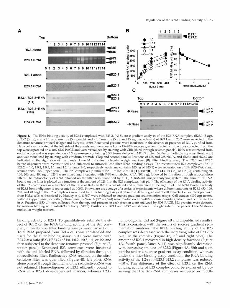
Mitotic Phosphorylation of B23.1B23 protein is hyperphosphorylated during mitosis (Figure6A). A candidate kinase responsible for B23 phosphorylationis reported to be cyclin B/cdc2 kinase (Peter et al., 1990).There are several patches of serine (S) or threonine (T)residues similar to the consensus sequences that are poten-
Figure 5. B23.1 is released fromnuclear structure by RNase treat-ment. (A) Cell fractionation ex-periment using RNase A orDNase I. The experimentalscheme is shown in the upperpanel. Exponentially growingHeLa cells were first treated with0.5% Triton X-100 in CSK bufferand were then subjected to diges-tion with DNase I (3 unit/�l) orRNase A (200 �g/ml). Releasedproteins by nuclease treatmentwere recovered by centrifugationas supernatant fraction, and thecell pellet was further extractedwith 0.25 M ammonium sulfateon ice for 5 min. Remaining in-soluble proteins were dissolvedin 8 M urea. Proteins in each frac-tion were separated on a 10%SDS-PAGE, and B23 proteinswere detected by western blot-ting with anti-B23 antibody(NB23). (B) RNA-dependent nu-
clear retention of B23.1. Cells treated with Triton X-100 were digested with increasing amounts of RNase A (0, 2, 20, and 200 �g/ml for lanes2–5, respectively) or DNase I (3 units/�l for lane 6) at 37°C for 20 min. Proteins were separated from insoluble materials by centrifugation,and the remaining pellet was dissolved in 8 M urea. Insoluble (Ppt, upper panel) and released (Sup, bottom panel) proteins were separatedon a 10% SDS-PAGE, and B23 proteins were detected by western blotting. Insoluble proteins after treatment with Triton X-100 are shown inlane 1 (Input). Positions of B23.1 are indicated by arrowheads at the left sides of panels.
Okuwaki et al.
Molecular Biology of the Cell2022
tially targeted by CDK (Figure 6B). It has been shown pre-viously that threonine(s) is a B23 phosphorylation targetduring mitosis (Peter et al., 1990). In fact, the 234th and 237ththreonine residues of glutathione S-transferase (GST)-
tagged B 23.1 were found to be phosphorylated by cyclinB/cdc2 kinase in vitro (Tokuyama et al., 2001). To confirmthese observations, we examined the in vitro level of cyclinB/cdc2-derived phosphorylation using a series of C-termi-
Figure 6. Phosphorylation sites of B23.1 by cyclin B/cdc2. (A) Hyperphosphorylation of B23.1 during mitosis. Cell extracts (20 �g) preparedfrom interphase (lane 1) or prometaphase (lane 2) HeLa cells were separated on a 10% SDS-PAGE, and B23.1 was detected by western blottingwith anti-B23.1 antibody (C19). Positions of B23.1 are indicated with a bracket at the right side of the panel. (B) Schematic representation ofmutant proteins used in this study. Amino acids are indicated with single letter abbreviations. Threonine residues (T) replaced by alanine(A) are indicated by gray letters. (C) Phosphorylation of B23.1 by cyclin E/cdk2. Cyclin E/cdk2 was purified from NIH3T3 cells without (lane1) or with (lanes 2–4) anticyclin E antibody (M20). Wild-type B23.1 (lanes 1 and 2), T199A (lane 3), and T4A (lane 4) (200 ng) werephosphorylated by purified cyclin E/cdk2 in the presence of histone H1 (100 ng) and [�-32P]ATP at 30°C for 30 min. (D) In vitrophosphorylation of wild-type and mutant B23.1 proteins by cyclin B/cdc2 kinase. Cyclin B/cdc2 was purified from mitotic HeLa cell extracts.B23.1 and point mutant proteins (200 ng) as indicated above each lane were phosphorylated by 1 (lanes 1–5), 3 (lanes 6–10), and 10 (lanes11–15) �l of cyclin B/cdc2 kinase (see “Materials and Methods”) in the presence of [�-32P]ATP at 30°C for 30 min. (E) Threonine residuesphosphorylated by cyclin B/cdc2. Wild-type (WT, lane 1) and mutant B23.1 (T199/234/237A and T219/234/237A, lanes 2 and 3, respectively)were phosphorylated by cyclin B/cdc2 and separated on a 10% SDS-PAGE. In C through E, proteins were stained with CBB, andphosphorylated proteins were detected by autoradiography (upper and bottom panels, respectively). (F) Phosphorylation of Flag-taggedwild-type and mutant B23.1 proteins in vivo. Cell extracts (30 �g) prepared from exponentially growing (lanes 1, 3, 5, and 7) ornocodazole-arrested (lanes 2, 4, 6, and 8) HeLa cells expressing either Flag-tagged B23.1 (lanes 1 and 2), T199A (lanes 3 and 4), T219/234/237A(lanes 5 and 6), or T4A (lanes 7 and 8) were separated on a 8% SDS-PAGE and subjected to western blotting with anti-Flag antibody. Proteinbands shown by an asterisk are nonspecific background bands that reacted with the secondary antibody (biotinylated anti-mouse IgG).
Regulation of the RNA Binding Activity of B23
Vol. 13, June 2002 2023

nal deletion mutants of B23.1. Cyclin B/cdc2 kinase waspurified from mitotic HeLa cell extracts using anti-cyclin Bantibody as described in “Materials and Methods.” Wild-type full-length B23.1 was efficiently phosphorylated (samelevels in Figure 6 and unpublished results). B23.1(1–257),which contains 257 amino acids from the N-terminus ofB23.1, was phosphorylated as efficiently as the full-lengthB23.1, whereas the more truncated B23.1(1–160) was notefficiently phosphorylated by cyclin B/cdc2 in vitro (unpub-lished results). This indicates that a target residue(s) forphosphorylation by cyclin B/cdc2 resides between the 160thand 257th amino acids in the B23.1 protein sequence. Weconstructed point mutant proteins that contained amino acidsubstitutions for potential cyclin B/cdc2 kinase target sitesas shown in Figure 6B in an effort to determine the precisephosphorylation site(s) by cyclin B/cdc2 kinase. His-taggedwild-type and mutant B23.1 proteins, WT, T199A, T219A,T234/237A, T199/234/234/A, T219/234/237A, and T4A(see Figure 6B) were generated in Escherichia coli and puri-fied. Wild-type and mutant B23.1 proteins were subjected tophosphorylation by cyclin E/cdk2 or cyclin B/cdc2 kinase inthe presence of [�-32P]ATP. Phosphorylated proteins wereseparated by SDS-PAGE and were detected by autoradiog-raphy. Before examining phosphorylation by cyclin B/cdc2kinase in detail, we performed phosphorylation reactionsusing purified cyclin E/cdk2 to confirm that T199 is a phos-phorylation target of cyclin E/cdk2 (Tokuyama et al., 2001).Wild-type B23.1 and histone H1 were efficiently phosphor-ylated by cyclin E/cdk2 purified from NIH3T3 cells (Figure6C, lane 2), although T199A and T4A, in which known cyclinE/cdk2-target threonine residues are substituted by alanine(Figure 6B), were not efficiently phosphorylated (Figure 6C,lanes 3 and 4). This is consistent with the previous reportthat T199 in B23.1 is phosphorylated by cyclin E/cdk2 (To-kuyama et al., 2001). Wild-type B23.1 was efficiently phos-phorylated by cyclin B/cdc2 in a kinase dose-dependentmanner (Figure 6D, lanes 1, 6, and 11). Although Tokuyamaet al. (2001) reported that a GST-tagged T234/237A mutant isnonphosphorylatable by cyclin B/cdc2, His-tagged T234/
237A mutant protein was phosphorylated as efficiently aswild-type protein by cyclin B/cdc2 in our study. On theother hand, the T199A mutant showed decreased phosphor-ylation efficiency compared with wild-type protein, and theT4A mutant was hardly phosphorylated by cyclin B/cdc2 invitro. To confirm that T199 is one of the most accessibletargets of cyclin B/cdc2, the mutant proteins T199/234/237A and T219/234/237A, in which three threonine resi-dues were substituted by alanine, were also tested for phos-phorylation susceptibility (Figure 6E). As expected, thephosphorylation efficiency of the mutant protein T199/234/237A (i.e., T219 intact) was significantly decreased, whereasthe T219/234/237A (i.e., T199 intact) is efficiently phosphor-ylated. The T199/234/237A was inefficiently but distinctlyphosphorylated by cyclin B/cdc2 in vitro, whereas the T4Amutant was hardly phosphorylated. Therefore, T219 mayalso be a potential target of cyclin B/cdc2, but with lessaccessibility than the T199. To examine the phosphorylationsite(s) of B23.1 proteins in vivo, Flag-tagged wild-type andmutant B23.1 proteins were transiently expressed in HeLacells. Cell extracts prepared from cells grown in the presenceor absence of nocodazole were subjected to western blottingusing anti-Flag antibody. Separation by SDS-PAGE showsthat relative to endogenous B23.1 from interphase cells,B23.1 from mitotic cell extracts migrated slower due to hy-perphosphorylation (Figure 6A). The band position of tran-siently expressed mitotic Flag-tagged B23.1 on SDS-PAGE isshifted from that of the interphase protein, suggesting thatFlag-tagged B23.1 is phosphorylated during mitosis (Figure6F, lanes 1 and 2). Because phosphatase-treated mitotic Flag-tagged B23.1 migrated faster than mock-treated protein onSDS-PAGE, the band shift of mitotic Flag-tagged B23.1 onSDS-PAGE was confirmed to be due to phosphorylation(unpublished results). Migration positions of Flag-taggedT199A and T219/234/237A are also shifted from those ofinterphase proteins, although the band shift of these mutantproteins in mitotic extracts was less than that of the wildtype. In addition, the band shift of T4A mutant in mitoticextracts on SDS-PAGE was decreased significantly, indicat-
Figure 7. Inactivation of theRNA binding activity of B23.1 bycyclin B/cdc2-mediated phos-phorylation. (A) Phosphorylationof rB23.1 proteins by purified cy-clin B/cdc2 kinase. rB23.1 (lanes1 and 2) or T4A mutant (lanes 3and 4) proteins (5 �g each) wereincubated in the absence (lanes 1and 3) or presence (lanes 2 and 4)of purified cyclin B/cdc2 kinase.A part of the protein solution(200 ng) was separated on a 10%SDS-PAGE and stained withCoomassie Brilliant Blue (CBB).Lane M indicates molecularweight markers. (B) RNA bind-ing activity of phosphorylatedB23.1. Wild-type (upper two pan-els) or T4A mutant B23.1 (bottomtwo panels) proteins phosphory-
lated in the absence (first and third panels) or presence (second and fourth panels) of cyclin B/cdc2 kinase were examined for the RNAbinding activity by a sucrose gradient sedimentation analysis as in Figure 4. Proteins in each fraction collected from the top were separatedon a 10% SDS-PAGE and stained with CBB.
Okuwaki et al.
Molecular Biology of the Cell2024

ing that this mutant protein is not efficiently phosphorylatedduring mitosis. These observations support our results ofthe in vitro phosphorylation assay using purified cyclinB/cdc2. From these in vitro and in vivo observations, it isstrongly suggested that at least four threonine residues,T199, T219, T234, and T237, are the potential phosphoryla-tion targets during mitosis possibly by cyclin B/cdc2.
Mitotic Inactivation of the RNA Binding Activityof B23.1Next, we examined if phosphorylation of B23 protein duringmitosis affects its RNA binding activity. Recombinant B23.1preincubated in the presence or absence of cyclin B/cdc2and ATP was mixed with RNA and was then subjected to asucrose gradient sedimentation assay. As shown in Figure7A, phosphorylated B23.1 migrated slower than nonphos-phorylated B23.1 on SDS-PAGE (Figure 7A, compare lanes 1
and 2), suggesting that recombinant B23.1 is completelyphosphorylated by cyclin B/cdc2. NonphosphorylatedB23.1 bound to RNA and was recovered in high densityfractions (Figure 7B, first panel, lanes 8–11) as in Figure 8. Incontrast, B23.1 phosphorylated by cyclin B/cdc2 was recov-ered in low density fractions (Figure 7B, second panel, lanes2–5), even after incubation with RNA, indicating that phos-phorylated B23.1 lost the ability to bind RNA. To confirmthat B23.1 phosphorylation is critical for inactivation of theRNA binding activity, T4A mutant protein (a nonphosphor-ylatable mutant protein by cyclin B/cdc2) was subjected tothe RNA binding assay. Before conducting this assay, it wasfirst established that T4A mutant bound RNA similarly towild-type protein (Figure 7B, third panel), meaning that theT to A substitutions in mutant B23.1 have no effect on itsRNA binding activity. Although the phosphorylation reac-tion was performed under the same conditions as for wild-
Figure 8. Phosphorylation of B23.1-RNA complex in vitro. (A) Release of B23.1 from RNA by cyclin B/cdc2-mediated phosphorylation.rB23.1 (5 �g) preincubated with RNA (5 �g) was phosphorylated by cyclin B/cdc2 kinase in the absence (upper panel) or presence (bottompanel) of ATP, and was then subjected to sucrose gradient sedimentation analyses as shown in Figures 4 and 7. Proteins in each fraction wereseparated on a 10% SDS-PAGE and stained with CBB. (B) Inhibition of cyclin B/cdc2-mediated phosphorylation of B23.1 through interactionwith RNA. B23.2 (lanes 1–3), B23.1 (lanes 4–6), and T199A (lanes 7–9) (200 ng each) were preincubated in the absence (lanes 1, 4, and 7) orpresence of 50 ng (lanes 2, 5, and 8) or 200 ng (lanes 3, 6, and 9) of RNA purified from HeLa cells and were phosphorylated by cyclin B/cdc2in the presence of [�- 32P]ATP. After the phosphorylation reaction, proteins and phosphorylated proteins were separated on a 10% SDS-PAGEand stained with CBB (upper panel) or autoradiographed (bottom panel), respectively. The phosphorylation efficiency was quantified usinga FUJIX BAS2000 image analyzer system and is shown in the bottom graph.
Regulation of the RNA Binding Activity of B23
Vol. 13, June 2002 2025
type protein, a band shift of T4A mutant was not detected(Figure 7A, compare lanes 3 and 4). In addition, nonphos-phorylatable B23.1 mutant T4A protein remained associatedwith RNA even after the phosphorylation reaction beforethe RNA binding assay (Figure 7B, fourth panel). Theseresults strongly support the concept that the RNA bindingactivity of B23.1 is inactivated by cyclin B/cdc2-mediatedphosphorylation.
This concept lead us to propose that B23.1 is associatedwith RNA in interphase cells, whereas it is released fromRNA by phosphorylation when cells enter mitosis. In fact,B23 proteins are mainly dispersed throughout the cytoplasmduring mitosis (see Figure 9A and “Discussion” below),whereas some are reported to be concentrated in NDF andcomplexed with pre-rRNA and rRNA processing factorsduring mitosis (Dundr and Olson, 1998). Therefore, we sup-posed that a fraction of B23.1 remains associated with RNAas a nonphosphorylated form. We tested if the preformedB23.1–RNA complex is disrupted by cyclin B/cdc2-medi-ated phosphorylation in vitro (Figure 8). B23.1 was mixedand incubated with RNA to form a complex, followed byphosphorylation with cyclin B/cdc2 kinase in the absence orpresence of ATP. The phosphorylation conditions used herewere essentially the same as in Figure 7, where the free formof B23.1 was completely phosphorylated by cyclin B/cdc2.The RNA binding status of B23.1 was monitored by sucrosegradient sedimentation. The B23.1–RNA complex incubatedwith cyclin B/cdc2 in the absence of ATP remained associ-ated with RNA as indicated by being recovered in highdensity fractions (Figure 8A, upper panel). In contrast, whenthe B23.1–RNA complex was incubated with cyclin B/cdc2in the presence of ATP, some of B23.1 was released fromRNA and was found in low density fractions (Figure 8A,bottom panel). These results suggest that the interactionbetween B23.1 and RNA is somewhat disrupted duringmitosis by cyclin B/cdc2-mediated phosphorylation,whereas a distinct level of B23.1 remains associated withRNA by escaping from phosphorylation. Because com-pletely phosphorylated B23.1 looses its ability to associatewith RNA (Figure 7), it is speculated that phosphorylation ofRNA-associated B23.1 is inhibited through its interactionwith RNA. To examine this, the phosphorylation efficiencyof the B23.1–RNA complex by cyclin B/cdc2 kinase wasquantified relative to those of B23.2 and T199A protein–RNA complexes. Recombinant B2 3.1, B23.2, and T199Awere mixed with increasing amounts of RNA, and were thenphosphorylated by a fixed amount of cyclin B/cdc2 kinasein the presence of [�- 2P]ATP (Figure 8B). As predicted, thephosphorylation efficiency of B23.1 decreased when B23.1was preincubated with increasing amounts of RNA,whereas B23.2, which lacks RNA binding activity, was effi-ciently phosphorylated, even in the presence of RNA. Theseresults indicate that phosphorylation of B23.1 is negativelyregulated by its complex formation with RNA. Phosphory-lation of the mutant B23.1 protein, T199A, wherein phos-phorylation accessibility was much less than that of wild-type protein, showed a pattern similar to B23.1 (Figure 8B,lanes 7–9). Results shown here suggest that the B23.1 proteinassociated with RNA is resistant to phosphorylation andremains associated with RNA, whereas protein free fromRNA is phosphorylated and thus restricted in accession oftarget RNA during mitosis. Because partially phosphory-
lated B23.1 is capable of associating with RNA (unpublishedresults), the phosphorylation status of B23.1 oligomers suchas the ratio of nonphosphorylated to phosphorylated B23.1may be a key determinant in regulation of the RNA bindingactivity of B23.1 during mitosis.
To confirm the localization of B23.1 during mitosis, expo-nentially growing HeLa cells expressing Flag-tagged B23.1were subjected to indirect immunofluorescence analysis us-ing anti-Flag antibody. As shown in Figure 9A, Flag-taggedB23.1 was localized in nucleoli of interphase cells as theGFP-tagged protein (Figure 2). In contrast, mitotic Flag-tagged B23.1 was dispersed throughout cells, and partiallyconcentrated in the periphery of chromosomes and NDF(Figure 9A). Before the end of mitosis, NDF is assembledinto nuclei and nucleoli are reconstituted. This localizationpattern of B23 during mitosis supports previous reports(Zatsepina et al., 1997, 1999; Dundr and Olson, 1998; Dundret al., 2000). To further confirm that B23 is released fromnuclear structure during mitosis by phosphorylation, cellfractionation experiments were performed using exponen-tially growing or mitotic HeLa cells (Figure 9B). The expres-sion level of cyclin B in mitotic cells was much higher thanthat in asynchronous cells, and cyclin B was recovered in the“Detergent” and “DNase I” fractions (Figure 9B, middlepanel, compare lanes 1 and 2, and 4 and 5). This is in goodagreement with the fact that cyclin B accumulates duringG2/M phase and is translocated to nuclei at the beginning ofmitosis (Pines and Hunter, 1991). It is noted that the migra-tion of cyclin B detected in the “DNase I” fraction is slowerthan that in the “Detergent” fraction (Figure 9B, lanes 4 and5). Because cyclin B is phosphorylated by a polo-like kinaseand translocated to nuclei (Toyoshima-Morimoto et al.,2001), cyclin B recovered in the “DNase I” fraction should bephosphorylated. Topoisomerase II�, which is also phos-phorylated during mitosis (Heck et al., 1989; Kimura et al.,1996) and binds tightly to chromatin, was detected mainly inthe “DNase I” fraction (Figure 9B, upper panel). The migra-tion position of mitotic topoisomerase II� was also slightlyshifted from that of interphase protein (Figure 9B, comparelanes 2 and 5). Interestingly, interphase B23.1 fractionatedmainly in the “DNase I” fraction, whereas the majority ofmitotic B23.1, which was supposed to be phosphorylated,fractionated in the “Detergent” fraction (Figure 9B, bottompanel). A minor potion of mitotic B23.1 was also detected inthe “DNase I” fraction, but the B23.1 in this fraction showedfaster mobility on SDS-PAGE. From these observations, it issuggested that B23.1 in interphase cells associates with nu-clear structures with the help of RNA, whereas B23.1 phos-phorylated by cyclin B/cdc2 during mitosis looses its RNAbinding activity and is released from nuclei.
DISCUSSION
As evidenced through the RNA binding and cellular local-ization of ribosome biogenesis factor B23, we have shown itscell cycle-related regulation mechanism. Both splicing vari-ants of B23 proteins, B23.1 and B23.2, are complexed in vivo(Figure 1), and the RNA binding activity of B23.1 is modu-lated by hetero-oligomerization with B23.2 (Figure 4). Fur-thermore, phosphorylation of B23.1 by cyclin B/cdc2 inac-tivates its RNA binding activity (Figure 7). It is possible thatthese regulation mechanisms of the RNA binding activity of
Okuwaki et al.
Molecular Biology of the Cell2026

B23.1 are important for regulation of ribosome biogenesisand possibly cell cycle progression.
In rat and human cells, both subtypes of B23 proteins areexpressed, although the expression level of B23.2 is muchlower than that of B23.1. As shown here, the cellular local-izations of B23.1 and B23.2 are not exactly identical (Figure2). B23.1 is mainly localized in nucleoli, whereas B23.2 islocalized not only in nucleoli but also throughout nucleo-plasm. From these observations, it is presumed that B23.1 istargeted to rRNA rich-nucleoli, possibly due to its RNAbinding activity. This is supported by the facts that B23.2,which is incapable of associating with RNA, fails to localizenucleoli (Figure 2), that a majority of B23.1 is released byRNase A digestion of nuclei (Figure 5), and that mitoticB23.1 phosphorylated by cyclin B/cdc2, which is incapableof binding to RNA, is dispersed throughout cells (Figure 9).In addition, B23.2 and mitotic B23.1 easily leak out of nucleiafter detergent treatment (Figures 2, 3, and 9), indicating thatthese proteins are less tightly associated with nuclear struc-tures compared with interphase B23.1. However, when cellsoverexpressing HA-tagged B23.2 were subjected to cell frac-tionation experiments as in Figures 2 and 3, distinct levels ofHA-tagged B23.2 were detected in detergent- or nuclease-treated cell nuclei (Figure 3 and unpublished results), al-though GFP-tagged B23.2 was not detected after DNase I orRNase A digestion (Figure 2). B23.2 may have a potentialability to associate with nuclear structure. Because B23.2complexed with B23.1 can bind to RNA in vitro (Figure 4), adetergent- and nuclease resistant-nucleolar B23.2 is probablycomplexed with B23.1. This is supported by the fact thatHA-tagged B23.2 in detergent-extracted HeLa cells copre-cipitates with endogenous B23.1. When the ratio of B23.2 toB23.1 is increased, the RNA binding activity of the complex
decreased even in the presence of constant amounts of B23.1(Figure 4). Therefore, the ratio of B23.1 to B23.2 is likelyimportant in determining the RNA binding activity of theB23 complex. B23 proteins are suggested to form hexamer orpentamer/decamer in solution (Yung and Chan, 1987; Duttaet al., 2001). Therefore, there are several different combina-tions of B23.1/B23.2 complexes that could be formed. Het-ero-oligomer formation of B23.1 and B23.2 is a possible RNAbinding modulation mechanism that may be involved innucleolar targeting and ribosome biogenesis function. Theexpression level of B23.1 is closely correlated with the cellgrowth rate, whereas that of B23.2 is relatively constant(Wang et al., 1993). Therefore, it is possible that the ribosomebiogenesis function of B23.1 in slowly growing cells could beimpaired through two distinct pathways: 1) expression ofB23.1 is down-regulated, and 2) the low level of B23.1 istrapped by B23.2 and its RNA binding activity is thus sup-pressed, thereby resulting in inhibition of ribosome biogen-esis, including rRNA processing and ribosome assembly.This could act to control the rate of ribosome biogenesis.
Although the N-terminal portion of B23 proteins is shownto be involved in oligomer formation (Yung and Chan, 1987;Herrera et al., 1996; Dutta et al., 2001), the interaction forcefor oligomer formation is unknown. Cellular B23 oligomercan be seen even in the presence of SDS or a reducing agentsuch as dithiothreitol, but is disrupted by extensive boiling(unpublished results). The N-terminal region is highly con-served in nucleoplasmin family proteins (Schmidt-Zach-mann et al., 1987), including nucleoplasmin, B23/NO38,NO29 (Zirwes et al., 1997b), and mitotic apparatus proteinp62 (Ye and Sloboda, 1997). In B23 proteins, oligomer for-mation is critical for its molecular chaperone activity (Hin-gorani et al., 2000) and RNA binding activity (in this study).
Figure 9. Dissociation of B23from nuclear structure duringmitosis. (A) Cellular localizationof Flag-tagged B23.1. Asynchro-nous HeLa cells expressing Flag-tagged B23.1 were fixed with anacetone:methanol (1:1) solutionat �30°C for 10 min, and werethen immunostained with anti-Flag antibody. Flag-tagged pro-teins were visualized by second-ary antibody against mouse IgGconjugated with Alexa564. Im-ages of B23.1 in a HeLa cell ininterphase, prometaphase, meta-phase, anaphase, and telophaseon a coverslip are arranged asindicated. DNA was stained withHoechst33258. The bar corre-sponds to 10 �m. (B) Cell frac-tionation of mitotic HeLa cells.Exponentially growing (lanes1–3) or nocodazole-arrested(lanes 4–6) HeLa cells were sub-jected to cell fractionation exper-iments as shown in Figure 3. Pro-teins in each fraction (1 � 10 5
cells equivalent) were separatedon a 8% SDS-PAGE followed bywestern blotting using anti-Topo II� (8D2) (upper panel), anticyclin B (GNS1) (middle panel), or anti-B23.1 (C19) (bottom panel) antibodies.Positions of proteins are indicated at the right side of the panels.
Regulation of the RNA Binding Activity of B23
Vol. 13, June 2002 2027
However, it is shown that oligomer formation is dispensablefor B23 RNase activity (Hingorani et al., 2000), and thatRNase activity of B23.2 is about one-half of B23.1. Becausethe C-terminal region of B23.1 is essential to tether B23.1protein to the target RNA, hetero-oligomer formation ofB23.1 and B23.2 (which interferes the RNA binding activityof B23.1) would obstruct RNase function. Again, it is em-phasized that B23.2 modulates the ribosome biogenesisfunction of B23.1.
B23 protein is phosphorylated by cyclin B/cdc2 duringmitosis possibly at the 199th, 219th, 234th, or 237th threonineresidues. Previously, T234 and T237 were shown to be phos-phorylation sites (Tokuyama et al., 2001). However, underthe experimental conditions used here, the T199 residue wasthe most likely candidate to be phosphorylated by cyclinB/cdc2 (Figure 6). This difference could be explained by thestructure of recombinant proteins used as a substrate. A longN-terminal extra GST polypeptide may block the properstructure of B23 proteins and change the target residue ofcyclin B/cdc2, although we cannot exclude the possibilitythat the histidine-tag containing 20 amino acid residuesattached at the N-terminus may also have altered the struc-ture of B23 proteins. As shown here, because the T199A,T234/237A, and even T199/234/237A mutants are phos-phorylated by cyclin B/cdc2 kinase in vitro, four threonineresidues, T99, T219, T234, and T237, are possibly phosphor-ylated. Furthermore, T4A mutant is distinctly phosphory-lated by cyclin B/cdc2 in vivo and in vitro but at signifi-cantly low level (Figure 6, D and F). Thus, there could beanother potential target residue phosphorylated by cyclinB/cdc2 in B23. Nevertheless, an important point is thatphosphorylation of B23.1 by cyclin B/cdc2 leads to inacti-vation of its RNA binding activity. Two distinct mecha-nisms, hetero-oligomer formation and phosphorylation bycyclin B/cdc2, regulate the RNA binding activity of B23.1.Not only the RNA binding activity of B23.1, but also otherfunctions specific to the C-terminal region such as proteinbinding (Takemura et al., 1999) and RNase could be similarlyregulated by cyclin B/cdc2-mediated phosphorylation andhetero-oligomer formation with B23.2. On this line, it islikely that B23 localizes to centrosome through the C-termi-nal tail of B23.1, because B23 is released from centrosome bythe phosphorylation at T199 with cyclin E/cdk2 (Tokuyamaet al., 2001). Because release of B23.1 from centrosome byphosphorylation is indispensable for centrosome duplica-tion, it is important to identify the target protein of B23.1that localizes to centrosome in clarifying the mechanism ofcentrosome duplication. Furthermore, a fraction of B23.1 istightly associated with nuclear structures, possibly so-callednuclear matrix, whereas B23.2 is not (Figures 2 and 3).Because B23.1 associates with nuclear structure after RNasetreatment (Figures 2 and 5), it is presumed that B23.1 asso-ciates with nuclear matrix or residual nuclear materialsthrough protein–protein interaction between an unidentifiednuclear structural protein(s) and the C-terminal tail of B23.1.This interaction is also impaired by the phosphorylationduring mitosis and hetero-oligomer formation (Figure 9).
In mitotic cells, a part of B23 is associated with a largecomplex containing pre-rRNA, fibrillarin, nucleolin, and U3small nucleolar RNA (Pinol-Roma, 1999), and is localized incytoplasmic dots termed NDF (Dundr et al., 2000; Dundr andOlson, 1998; Zatsepina et al., 1999; Zatsepina et al., 1997). It
remains unclear how NDF is released from chromosomesduring mitosis and is reassembled to form nucleoli after celldivision. As shown in Figure 8, B23.1 complexed with RNAwas not efficiently phosphorylated, and a fraction of B23.1remains associated with RNA even after phosphorylation.Because NDF is suggested to be important to form nucleoliand restart ribosome biogenesis after cell division, it is pos-sible that the phosphorylation restriction of non-rRNA bind-ing proteins is essential to maintain NDF during mitosis.Protection from mitotic phosphorylation of RNA bindingproteins is one possible mechanism for preserving a “core”nucleolar structure for the next cell cycle. Nucleolin, a majorRNA binding nucleolar phosphoprotein, shows similar be-havior to B23; nucleolin is also phosphorylated during mi-tosis by cyclin B/cdc2 kinase and is dispersed throughoutcells, and some nucleolin can be found in NDF (Pinol-Roma,1999; Srivastava and Pollard, 1999). Therefore, mitotic inac-tivation of the RNA binding activity by phosphorylationmay also be significant to nucleolin. Alternatively, it is pos-sible that a factor(s) that connects NDFs or NDFs and chro-mosomes may exist whereby the interaction between theputative factor and the NDF or chromosome may be brokendown when cells enter mitosis. Because both B23 andnucleolin were shown to bind chromatin (Olson andThompson, 1983; Okuwaki et al., 2001a), these nonribosomalproteins may function as linker proteins between NDFs orbetween NDFs and chromosomes.
During mitosis, several cellular functions such as tran-scription and translation are silenced so that genetic materialcan be correctly condensed and segregated into daughtercells. Mitotic inactivation of these cellular processes is partlymediated through phosphorylation by mitotic kinases in-cluding cyclin B/cdc2. Most of transcription and chromatinbinding factors are phosphorylated and released from chro-mosomes, thereby silencing transcription (Gottesfeld andForbes, 1997). Mitotic release also functions in pre-rRNAprocessing components, whereas the pol I transcription ma-chinery, including RNA polymerase I, UBF, and DNA topo-isomerase I, remains associated with chromosomes duringmitosis (Weisenberger and Scheer, 1995; Roussel et al., 1996).Our findings demonstrate the first case where a factor in-volved in ribosome biogenesis is regulated through phos-phorylation and dephosphorylation during the cell cycle.
ACKNOWLEDGMENTS
We thank Dr. H. Umekawa (Mie University, Japan) for mAb for B23(NB23), Dr. A. Kikuchi (Nagoya University, Japan) for antitopoi-somerase II antibody (8D2), and Dr. K. Matsumoto (RIKEN, Japan)for useful discussion and critical comments on the manuscript. Wealso thank K. Pepin for proofreading of the manuscript. This workwas supported in part by a Grant-in-Aid from the Ministry ofEducation, Culture, Sports, Science and Technology of Japan and bya grant for Bioarchitect Research Program from RIKEN (to K.N.).M.O. is a Special Postdoctoral Researcher of RIKEN.
REFERENCES
Chang, J.H., and Olson, M.O. (1990). Structure of the gene for ratnucleolar protein B23. J. Biol. Chem. 265, 18227–18233.
Colgan, D.F., Murthy, K.G., Zhao, W., Prives, C., and Manley, J.L.(1998). Inhibition of poly(A) polymerase requires p34cdc2/cyclin B
Okuwaki et al.
Molecular Biology of the Cell2028
phosphorylation of multiple consensus and non-consensus sites.EMBO J. 17, 1053–1062.
Dumbar, T.S., Gentry, G.A., and Olson, M.O. (1989). Interaction ofnucleolar phosphoprotein B23 with nucleic acids. Biochemistry 28,9495–9501.
Dundr, M., Misteli, T., and Olson, M.O. (2000). The dynamics ofpostmitotic reassembly of the nucleolus. J. Cell Biol. 150, 433–446.
Dundr, M., and Olson, M.O. (1998). Partially processed pre-rRNA ispreserved in association with processing components in nucleolus-derived foci during mitosis. Mol. Biol. Cell 9, 2407–2422.
Durban, E., Valdez, B.C., Gustafson, W.C., Taylor, C.W., Cardellini,E., and Busch, H. (1995). Functional domains of nucleolar phospho-protein p120. Physiol. Chem. Phys. Med. NMR 27, 303–311.
Dutta, S., Akey, I.V., Dingwall, C., Hartman, K.L., Laue, T., Nolte,R.T., Head, J.F., and Akey, C.W. (2001). The crystal structure ofnucleoplasmin-core. Implications for histone binding and nucleo-some assembly. Mol. Cell 8, 841–853.
Fankhauser, C., Izaurralde, E., Adachi, Y., Wingfield, P., and Lae-mmli, U.K. (1991). Specific complex of human immunodeficiencyvirus type 1 rev and nucleolar B23 proteins: dissociation by the Revresponse element. Mol. Cell. Biol. 11, 2567–2575.
Feuerstein, N., and Mond, J.J. (1987). “Numatrin,” a nuclear matrixprotein associated with induction of proliferation in B lymphocytes.J. Biol. Chem. 262, 11389–11397.
Gottesfeld, J.M., and Forbes, D.J. (1997). Mitotic repression of thetranscriptional machinery. Trends Biochem. Sci. 22, 197–202.
Graham, F.L., and Eb, A.J.v.d. (1973). A new technique for the assayof infectivity of human adenovirus 5 DNA. Virology 52, 456–467.
Hager, D.A., and Burgess, R.R. (1980). Elution of proteins fromsodium dodecyl sulfate-polyacrylamide gels, removal of sodiumdodecyl sulfate, and renaturation of enzymatic activity: results withsigma subunit of Escherichia coli RNA polymerase, wheat germ DNAtopoisomerase, and other enzymes. Anal. Biochem. 109, 76–86.
He, D.C., Nickerson, J.A., and Penman, S. (1990). Core filaments ofthe nuclear matrix. J. Cell Biol. 110, 569–580.
Heck, M.M., Hittelman, W.N., and Earnshaw, W.C. (1989). In vivophosphorylation of the 170-kDa form of eukaryotic DNA topoisom-erase. II. Cell cycle analysis. J. Biol. Chem. 264, 15161–15164.
Heix, J., Vente, A., Voit, R., Budde, A., Michaelidis, T.M., andGrummt, I. (1998). Mitotic silencing of human rRNA synthesis:inactivation of the promoter selectivity factor SL1 by cdc2/cyclinB-mediated phosphorylation. EMBO J. 17, 7373–7381.
Herrera, J.E., Correia, J.J., Jones, A.E., and Olson, M.O. (1996). Sed-imentation analyses of the salt- and divalent metal ion-inducedoligomerization of nucleolar protein B23. Biochemistry 35, 2668–2673.
Hingorani, K., Szebeni, A., and Olson, M.O. (2000). Mapping thefunctional domains of nucleolar protein B23. J. Biol. Chem. 275,24451–24457.
Huang, W.H., Yung, B.Y., Syu, W.J., and Lee, Y.H. (2001). Thenucleolar phosphoprotein B23 interacts with hepatitis delta anti-gens, and modulates the hepatitis delta virus RNA replication.J. Biol. Chem. 276, 25166–25175.
Kimura, K., Hirano, M., Kobayashi, R., and Hirano, T. (1998). Phos-phorylation and activation of 13S condensin by Cdc2 in vitro. Sci-ence 282, 487–490.
Kimura, K., Nozaki, N., Enomoto, T., Tanaka, M., and Kikuchi, A.(1996). Analysis of M phase-specific phosphorylation of DNA topo-isomerase II. J. Biol. Chem. 271, 21439–21445.
Li, Y.P., Busch, R.K., Valdez, B.C., and Busch, H. (1996). C23 inter-acts with B23, a putative nucleolar-localization-signal-binding pro-tein. Eur. J. Biochem. 237, 153–158.
Maden, B.E. (1990). The numerous modified nucleotides in eukary-otic ribosomal RNA. Prog. Nucleic Acid Res. Mol. Biol. 39, 24 1–303.
Manley, J.L., Fire, A., Cano, A., Sharp, P.A., and Gefter, M.L. (1980).DNA-dependent transcription of adenovirus genes in a solublewhole-cell extract. Proc. Natl. Acad. Sci. USA 77, 3855–3859.
Nagata, K., Kawase, H., Handa, H., Yano, K., Yamasaki, M., Ishimi,Y., Okuda, A., Kikuchi, A., and Matsumoto, K. (1995). Replicationfactor encoded by a putative oncogene, set, associated with myeloidleukemogenesis. Proc. Natl. Acad. Sci. USA 92, 4279–4283.
Nagata, K., Saito, S., Okuwaki, M., Kawase, H., Furuya, A., Kusano,A., Hanai, N., Okuda, A., and Kikuchi, A. (1998). Cellular localiza-tion and expression of template-activating factor I in different celltypes. Exp. Cell Res. 240, 274–281.
Okuda, M., Horn, H.F., Tarapore, P., Tokuyama, Y., Smulian, A.G.,Chan, P.K., Knudsen, E.S., Hofmann, I.A., Snyder, J.D., Bove, K.E.,and Fukasawa, K. (2000). Nucleophosmin/B23 is a target of CDK2/cyclin E in centrosome duplication. Cell 103, 127–140.
Okuwaki, M., Iwamatsu, A., Tsujimoto, M., and Nagata, K. (2001a).Identification of nucleophosmin/B23, an acidic nucleolar protein, asa stimulatory factor for in vitro replication of adenovirus DNAcomplexed with viral basic core proteins. J. Mol. Biol. 311, 41–55.
Okuwaki, M., Matsumoto, K., Tsujimoto, M., and Nagata, K.(2001b). Function of nucleophosmin/B23, a nucleolar acidic protein,as a histone chaperone. FEBS Lett. 506, 272–276.
Olson, M.O., and Thompson, B.A. (1983). Distribution of proteinsamong chromatin components of nucleoli. Biochemistry 22, 3187–3193.
Peculis, B.A. (2000). RNA-binding proteins: if it looks like asn(o)RNA. Curr. Biol. 10, R916–R918.
Peculis, B.A., and Gall, J.G. (1992). Localization of the nucleolarprotein NO38 in amphibian oocytes. J. Cell Biol. 116, 1–14.
Peter, M., Nakagawa, J., Doree, M., Labbe, J.C., and Nigg, E.A.(1990). Identification of major nucleolar proteins as candidate mi-totic substrates of cdc2 kinase. Cell 60, 791–801.
Pines, J., and Hunter, T. (1991). Human cyclins A and B1 aredifferentially located in the cell and undergo cell cycle-dependentnuclear transport. J. Cell Biol. 115, 1–17.
Pinol-Roma, S. (1999). Association of nonribosomal nucleolar pro-teins in ribonucleoprotein complexes during interphase and mitosis.Mol. Biol. Cell 10, 77–90.
Roussel, P., Andre, C., Comai, L., and Hernandez-Verdun, D. (1996)The rDNA transcription machinery is assembled during mitosis inactive NORs, and absent in inactive NORs. J. Cell Biol. 133, 235–246.
Savkur, R.S., and Olson, M.O. (1998). Preferential cleavage in pre-ribosomal RNA byprotein B23 endoribonuclease. Nucleic Acids Res.26, 4508–4515.
Schmidt-Zachmann, M.S., Hugle-Dorr, B., and Franke, W.W. (1987).A constitutive nucleolar protein identified as a member of thenucleoplasmin family. EMBO J. 6, 1881–1890.
Sif, S., Stukenberg, P.T., Kirschner, M.W., and Kingston, R.E. (1998).Mitotic inactivation of a human SWI/SNF chromatin remodelingcomplex. Genes Dev. 12, 2842–2851.
Srivastava, M., and Pollard, H.B. (1999). Molecular dissection ofnucleolin’s role in growth and cell proliferation: new insights.FASEB J. 13, 1911–1922.
Szebeni, A., Herrera, J.E., and Olson, M.O. (1995). Interaction ofnucleolar protein B23 with peptides related to nuclear localizationsignals. Biochemistry 34, 8037–8042.
Regulation of the RNA Binding Activity of B23
Vol. 13, June 2002 2029

Szebeni, A., and Olson, M.O. (1999). Nucleolar protein B23 hasmolecular chaperone activities. Protein Sci. 8, 905–912.
Takemura, M., Sato, K., Nishio, M., Akiyama, T., Umekawa, H., andYoshida, S. (1999). Nucleolar protein B23.1 binds to retinoblastomaprotein and synergistically stimulates DNA polymerase � activity.J. Biochem. 125, 904–909.
Tokuyama, Y., Horn, H.F., Kawamura, K., Tarapore, P., and Fuka-sawa, K. (2001). Specific phosphorylation of nucleophosmin on Thr(199) by cyclin-dependent kinase 2-cyclin E, and its role in centro-some duplication. J. Biol. Chem. 276, 21529–21537.
Toyoshima-Morimoto, F., Taniguchi, E., Shinya, N., Iwamatsu, A.,and Nishida, E. (2001). Polo-like kinase 1 phosphorylates cyclin B1,and targets it to the nucleus during prophase. Nature 410, 215–220.
Umekawa, H., Sato, K., Takemura, M., Watanabe, Y., Usui, S.,Takahashi, T., Yoshida, S., Olson, M.O., and Furuichi, Y. (2001). Thecarboxyl terminal sequence of nucleolar protein B23.1 is importantin its DNA polymerase �-stimulatory activity. J. Biochem. 130,199–205.
Wang, D., Baumann, A., Szebeni, A., and Olson, M.O. (1994). Thenucleic acid binding activity of nucleolar protein B23.1 resides in itscarboxyl-terminal end. J. Biol. Chem. 269, 30994–30998.
Wang, D., Umekawa, H., and Olson, M.O. (1993). Expression andsubcellular locations of two forms of nucleolar protein B23 in rattissues and cells. Cell. Mol. Biol. Res. 39, 33–42.
Weisenberger, D., and Scheer, U. (1995). A possible mechanism forthe inhibition of ribosomal RNA gene transcription during mitosis.J. Cell Biol. 129, 561–575.
Ye, X., and Sloboda, R.D. (1997) Molecular characterization of p62,a mitotic apparatus protein required for mitotic progression. J. Biol.Chem. 272, 3606–3614.
Yung, B.Y., and Chan, P.K. (1987). Identification and characteriza-tion of a hexameric form of nucleolar phosphoprotein B23. Biochim.Biophys. Acta, 925, 74–82.
Zatsepina, O.V., Rousselet, A., Chan, P.K., Olson, M.O., Jordan,E.G., and Bornens, M. (1999). The nucleolar phosphoprotein B23redistributes in part to the spindle poles during mitosis. J. Cell Sci.112, 455–466.
Zatsepina, O.V., Todorov, I.T., Philipova, R.N., Krachmarov, C.P.,Trendelenburg, M.F., and Jordan, E.G. (1997). Cell cycle-dependenttranslocations of a major nucleolar phosphoprotein, B23, and somecharacteristics of its variants. Eur. J. Cell Biol. 73, 58–70.
Zirwes, R.F., Kouzmenko, A.P., Peters, J.M., Franke, W.W., andSchmidt-Zachmann, M.S. (1997a). Topogenesis of a nucleolar pro-tein: determination of molecular segments directing nucleolar asso-ciation. Mol. Biol. Cell 8, 231–248.
Zirwes, R.F., Schmidt-Zachmann, M.S., and Franke, W.W. (1997b).Identification of a small, very acidic constitutive nucleolar protein(NO29) as a member of the nucleoplasmin family. Proc. Natl. Acad.Sci. USA 94, 11387–11392.
Okuwaki et al.
Molecular Biology of the Cell2030
Copyright © 2022 FDOKUMEN