Mucin 16 (cancer antigen 125) expression in human tissues and cell lines and correlation with...
9
Original contribution Mucin 16 (cancer antigen 125) expression in human tissues and cell lines and correlation with clinical outcome in adenocarcinomas of the pancreas, esophagus, stomach, and colon ☆,☆☆ Mirte M. Streppel MD a,b,c , Audrey Vincent PhD a , Radha Mukherjee MSc a , Nathaniel R. Campbell MSc a , Shih-Hsun Chen MSc d , Konstantinos Konstantopoulos PhD d , Michael G. Goggins MD a,e , Isabelle Van Seuningen PhD f,g , Anirban Maitra MBBS a , Elizabeth A. Montgomery MD a, ⁎ a Department of Pathology, Johns Hopkins University, Baltimore, MD 21231-2410, USA b Department of Gastroenterology and Hepatology, University Medical Center Utrecht, Utrecht, The Netherlands c Department of Pathology, University Medical Center Utrecht, Utrecht, The Netherlands d Department of Chemical and Biomolecular Engineering, Johns Hopkins University, Baltimore, MD, USA e Department of Gastroenterology and Hepatology, Johns Hopkins University, Baltimore, MD, USA f Inserm, UMR837, Jean-Pierre Aubert Research Center, Team 5 “Mucins, Epithelial Differentiation and Carcinogenesis”, rue Polonovski, 59045 Lille cedex, France g Université Lille-Nord de France, 1 place de Verdun, 59045 Lille Cedex, France Received 18 October 2011; revised 11 January 2012; accepted 13 January 2012 Keywords: MUC16; CA125; Digestive tract adenocarcinoma Summary Mucin 16 (cancer antigen 125) is a cell surface glycoprotein that plays a role in promoting cancer cell growth in ovarian cancer. The aims of this study were to examine mucin 16 expression in a large number of digestive tract adenocarcinomas and precursors and to determine whether mucin 16 up- regulation is correlated with patient outcome. Tissue microarrays were constructed using surgical resection tissues and included pancreatic (115 normal, 29 precursors, 200 pancreatic ductal adenocarcinomas), esophageal (86 normal, 104 precursors, 95 esophageal adenocarcinomas, 35 lymph node metastases), gastric (211 normal, 8 precursors, 119 gastric adenocarcinomas, 62 lymph node metastases), and colorectal (34 normal, 17 precursors, 39 colorectal adenocarcinomas) tissues. Mucin 16 was detected in 81.5%, 69.9%, 41.2%, and 64.1% of the pancreatic ductal adenocarcinomas, esophageal adenocarcinomas, gastric adenocarcinomas, and colorectal adenocarcinomas, respectively. Mucin 16 was seen in a subset of the precursors. On multivariate analysis, moderate/diffuse mucin 16 in pancreatic ductal adenocarcinomas was strongly associated with poor survival (P b .001), independent of other prognosis predictors. A similar trend was observed for esophageal adenocarcinomas (P = .160) and ☆ Disclosure/conflict of interest: All authors declare to have no conflicts of interest. ☆☆ Funding sources supporting the work: This project was sponsored by the Jerry D'Amato Foundation, CA101135 (KK) and CA143868 (KK). MMS was supported by a Fulbright/Netherland America Foundation fellowship, the René Vogels foundation, and the Dutch Cancer society. ⁎ Corresponding author. E-mail address: [email protected] (E. A. Montgomery). www.elsevier.com/locate/humpath 0046-8177/$ – see front matter © 2012 Elsevier Inc. All rights reserved. doi:10.1016/j.humpath.2012.01.005 Human Pathology (2012) 43, 1755–1763
-
Upload
johnshopkins -
Category
Documents
-
view
0 -
download
0
Transcript of Mucin 16 (cancer antigen 125) expression in human tissues and cell lines and correlation with...
www.elsevier.com/locate/humpath
Human Pathology (2012) 43, 1755–1763
Original contribution
Mucin 16 (cancer antigen 125) expression in human tissuesand cell lines and correlation with clinical outcome inadenocarcinomas of the pancreas, esophagus, stomach,and colon☆,☆☆
Mirte M. Streppel MDa,b,c, Audrey Vincent PhDa, Radha Mukherjee MSc a,Nathaniel R. Campbell MSc a, Shih-Hsun Chen MSc d,Konstantinos Konstantopoulos PhDd, Michael G. Goggins MDa,e,Isabelle Van Seuningen PhD f,g, Anirban Maitra MBBS a, Elizabeth A. Montgomery MDa,⁎
aDepartment of Pathology, Johns Hopkins University, Baltimore, MD 21231-2410, USAbDepartment of Gastroenterology and Hepatology, University Medical Center Utrecht, Utrecht, The NetherlandscDepartment of Pathology, University Medical Center Utrecht, Utrecht, The NetherlandsdDepartment of Chemical and Biomolecular Engineering, Johns Hopkins University, Baltimore, MD, USAeDepartment of Gastroenterology and Hepatology, Johns Hopkins University, Baltimore, MD, USAfInserm, UMR837, Jean-Pierre Aubert Research Center, Team 5 “Mucins, Epithelial Differentiation and Carcinogenesis”,rue Polonovski, 59045 Lille cedex, FrancegUniversité Lille-Nord de France, 1 place de Verdun, 59045 Lille Cedex, France
Received 18 October 2011; revised 11 January 2012; accepted 13 January 2012
su
0d
Keywords:MUC16;CA125;Digestive tractadenocarcinoma
Summary Mucin 16 (cancer antigen 125) is a cell surface glycoprotein that plays a role in promotingcancer cell growth in ovarian cancer. The aims of this study were to examine mucin 16 expression in alarge number of digestive tract adenocarcinomas and precursors and to determine whether mucin 16 up-regulation is correlated with patient outcome. Tissue microarrays were constructed using surgicalresection tissues and included pancreatic (115 normal, 29 precursors, 200 pancreatic ductaladenocarcinomas), esophageal (86 normal, 104 precursors, 95 esophageal adenocarcinomas, 35 lymphnode metastases), gastric (211 normal, 8 precursors, 119 gastric adenocarcinomas, 62 lymph nodemetastases), and colorectal (34 normal, 17 precursors, 39 colorectal adenocarcinomas) tissues. Mucin 16was detected in 81.5%, 69.9%, 41.2%, and 64.1% of the pancreatic ductal adenocarcinomas, esophagealadenocarcinomas, gastric adenocarcinomas, and colorectal adenocarcinomas, respectively. Mucin 16was seen in a subset of the precursors. On multivariate analysis, moderate/diffuse mucin 16 in pancreaticductal adenocarcinomas was strongly associated with poor survival (P b .001), independent of otherprognosis predictors. A similar trend was observed for esophageal adenocarcinomas (P = .160) and
☆ Disclosure/conflict of interest: All authors declare to have no conflicts of interest.☆☆ Funding sources supporting the work: This project was sponsored by the Jerry D'Amato Foundation, CA101135 (KK) and CA143868 (KK). MMS was
pported by a Fulbright/Netherland America Foundation fellowship, the René Vogels foundation, and the Dutch Cancer society.⁎ Corresponding author.E-mail address: [email protected] (E. A. Montgomery).
046-8177/$ – see front matter © 2012 Elsevier Inc. All rights reserved.oi:10.1016/j.humpath.2012.01.005
1756 M. M. Streppel et al.
gastric adenocarcinomas (P = .080). Focal mucin 16 in colorectal adenocarcinomas was significantlycorrelated (P = .044) with a better patient outcome, when compared with mucin 16–negative cases.Using Western blot analysis, we found mucin 16 expression in 3 of 6 pancreatic ductal adenocarcinomaand 1 of 2 esophageal adenocarcinoma cell lines. We conclude that most of the digestive tractadenocarcinomas and a subset of their precursors express mucin 16. Mucin 16 expression is anindependent predictor of poor outcome in pancreatic ductal adenocarcinomas and potentially inesophageal adenocarcinomas and gastric adenocarcinomas. We propose that mucin 16 may function as aprognostic marker and therapeutic target in the future.© 2012 Elsevier Inc. All rights reserved.
1. Introduction
The digestive tract is the second most common siteaffected by cancer in the United States. Cancers of thedigestive tract cause approximately 140 000 deaths annually,which accounts for 25% of all cancer-related deaths.Colorectal, pancreatic, and esophageal cancer are amongthe top 10 deadliest cancer types [1]. Most patients arediagnosed at an advanced stage because of nonspecificclinical symptoms. There is a pressing need for discovery ofnew markers because such markers cannot only function asprognostic tools but can potentially also offer newtherapeutic targets for these aggressive tumors.
Mucins, encoded by MUC genes, are glycoproteins thatcontain a variable number of tandem repeat structures. Thesevariable number of tandem repeat structures constitute aprolines-, threonines-, and serines- (PTS) rich domain, which,in turn, carries the mucin characteristic O-glycosylation.Mucins can be divided into 4 categories: secreted gel forming(MUC2, -5AC, -5B, -6, and -19), secreted non-gel forming(MUC7, -8, and -9), transmembrane (TM) (MUC1, -3A, -3B,-4, -12, -13, -14, -15, -16, -17, -20, and -21), and unclassified(MUC11) mucins [2]. TM mucins are found on the apicalmembrane of epithelial cells, as well as in the mucus layers ofthe respiratory and gastrointestinal tracts [3]. Cell surfaceoverexpression of TM mucins has been identified in cancersincluding digestive tract lesions, and these mucins are believedto promote tumor cell growth and tumor cell survival [4-6].
Molecular cloning of the cancer antigen 125 [CA125] ledto the discovery of mucin 16 (MUC16) in 2001 [7]. MUC16is the largest TMmucin and is used worldwide for monitoringpatients with ovarian cancer [4,8]. Several research groupshave attempted to elucidate the role of MUC16 in promotingovarian cancer cell growth and survival [9-14]. At present, nostudies have been performed attempting to clarify mecha-nisms via whichMUC16 can encourage tumor progression incancers other than ovarian cancer.
MUC16 (CA125) was initially believed to be a specificbiomarker for ovarian cancer, but over the past 15 years, ithas become clear that this marker can also be detected in seraof patients with other types of cancers including gastric,colorectal, and pancreatic adenocarcinomas. Although it isalready known that MUC16 plays a role in at least a subset ofthe digestive tract adenocarcinomas and elevated serum
MUC16 levels are associated with poor survival [15-21], thepresence of MUC16 in digestive tract adenocarcinomatissues, precursor lesions, and normal digestive tract epitheliahas not been comprehensively evaluated.
In the present study, we show that MUC16 is up-regulatedin a large percentage of the digestive tract adenocarcinomasand in a subset of noninvasive precursor lesions. Furthermore,we propose that MUC16 may function as a prognostic markerand therapeutic target for pancreatic ductal adenocarcinomas(PDAC) and, potentially, for esophageal adenocarcinomas(EAC) and gastric adenocarcinomas (GAC) in the future.
2. Materials and methods
2.1. Patient material
Paraffin-embedded material obtained from digestive tractadenocarcinoma patients who underwent surgical resectionat the Johns Hopkins Hospital, Baltimore, Maryland,between the years 1984 and 2004 was used for this study.Tissue microarrays (TMA) were constructed using thesespecimens. Each cancer case was represented by three totwelve 1.5-mm cores on the TMAs to obtain adequaterepresentation of different regions of the tumor, precursorlesions, and normal epithelium.
Tissue cores included pancreatic (115 normal acinartissue, 76 obstructive ducts, 10 pancreatic intraepithelialneoplasia [PanIN], 200 PDACs), esophageal (86 normalsquamous epithelium, 37 nondysplastic Barrett esophagus[BE], 21 low-grade dysplasia [LGD], 46 high-grade dysplasia[HGD], 95 EACs, and 35 lymph nodemetastases [LNM], and3 distant metastases [DM]), gastric (176 normal columnarlining of the stomach, 35 normal gastric cardia, 8 HGDs, 119GACs, and 62 LNMs), colorectal (34 normal colonic mucosa,10 LGD adenomas, 7 HGD adenomas, and 39 colorectalcarcinomas [CRC]) tissues. Normal and precursor lesionswere primarily obtained from adjacent regions of the cancerresection specimen. In addition to the PanIN lesions presentin the TMAs, 19 low- and high-grade PanIN lesions wererandomly selected and also included in this study. All PanINlesions were categorized as follows: low-grade (PanIN IA andIB) and high-grade (PanIN II and III) PanINs. BEwas definedas nondysplastic columnar mucosa with intestinal metaplasia

1757MUC16 in digestive tract adenocarcinomas
(goblet cells) in the esophagus as per the guidelines of boththe American College of Gastroenterologists and theAmerican Gastroenterological Association [22,23]. Nondys-plastic columnar mucosa with gastric foveolar type surfacecells and gastric cardiac type glands without goblet cells wasclassified as cardiac mucosa.
Characteristics of the patients with cancer including age,sex, race, and survival time were collected. In addition,tumor features such as differentiation of the tumor, thepresence of LNMs and DMs, and angiolymphatic invasionwere, if available, obtained from the original pathologicsurgical resection reports.
2.2. Immunohistochemistry
Five-micrometer sections were deparaffinized, endog-enous peroxidase was blocked, and antigen retrieval wasperformed using routine techniques. After blocking ofnonspecific binding sites using serum free Protein-block(DAKO North America, Carpinteria, CA), the slides wereincubated with a primary monoclonal antibody againstMUC16 (dilution 1:200, 1 hour, room temperature [RT],X325; Abcam, Cambridge, MA). The slides weresubsequently incubated with Post-antibody blocking andPowerVision+ Poly-HRP-anti-mouse/rabbit IgG (LeicaBiosystems, Newcastle, United Kingdom) for 15 and30 minutes, respectively. 3,3′-diaminobenzidine tetrachlo-ride plus (Leica Biosystems) was used as the chromogenfor visualization. Finally, the slides were counterstainedwith hematoxylin.
2.3. Scoring of immunolabeling
Stainingpatternswere evaluated by3 authors (E.A.M.,A.M.,and M. M. S.). MUC16 protein was considered present ifepithelial cells exhibited brown membranous staining.Because cytoplasmic and nuclear staining were rarelypresent, only membranous staining was scored. Stainingwas categorized as follows: “negative” (no detectablelabeling), “focal labeling” (b25% of the epithelial cellsshowing positivity), “moderate labeling” (25%-50%), and“diffuse labeling” (N50%). Staining intensity was not scoredbecause no variability in staining intensity was observed.Normal tissues obtained from noncancerous patients wereincluded in all TMAs and were used as negative controls.Samples of nonneoplastic mucosa from the same patientswhose tumors appeared in the arrays were also included inthe TMAs. Randomly selected ovarian cancer specimensfunctioned as positive controls in this study.
2.4. Cell culture and protein extraction
Six PDAC cell lines (Panc3.014, Panc5.04, Panc8.13,Panc10.05, Panc10.7, and BxPC-3) and 2 EAC cell lines(OE33, JHesoAD1) were analyzed for the presence of
MUC16 protein. OVCAR3, a commonly used ovarian cancercell line in which MUC16 expression has been confirmed byGoodell et al [24], was used as a positive control. Humanpancreatic normal epithelial (HPNE) cells functioned as anegative control. OE33, JHesoAD1, andOVCAR3 cells weregrown in RPMI containing 15% to 20% fetal bovin serum,and the PDAC cell lines were all cultured in DMEMsupplemented with 10% to 20% fetal bovin serum.
Total protein was extracted from cell lines using lysisbuffer supplemented with protease and phosphatase in-hibitors. After undergoing 2 freeze thaw cycles, the lysateswere centrifuged for 10 minutes at full speed. Proteinconcentrations were measured following the bicinchoninicacid method, as described in the manufacturer's instructionmanual (Thermo Fisher Scientific, Landford, IL).
2.5. Western blots
Forty micrograms of total protein extracts were separatedon 3% to 8% Tris-acetate and 4% to 12% Bis-Tris gels fordetection of MUC16 (500-5000 kDa) and β-actin (42 kDa),respectively. Proteins were transferred to 0.45-μm nitrocel-lulose membranes. The membranes were subsequentlyincubated in 5% fat-free milk at 4°C overnight to blocknonspecific binding. A mouse monoclonal antibody againstMUC16 (M11, dilution 1:1000, 2 hours at RT; DAKO NorthAmerica) was used forMUC16 detection. Themembranewassubsequently incubated with an antimouse secondary anti-body (dilution 1:4000, 1 hour at RT; Sigma Aldrich, St.Louis, MO). The loading control β-actin was detected using agoat antibody against β-actin (dilution 1:1000, 1 hour, RT;Santa Cruz, Santa Cruz, CA), and a secondary antigoatantibody (dilution 1:2000, 1 hour, RT; Santa Cruz). Blotswere developed using PICO reagents (Bio-Rad).
2.6. Statistical analysis
Kaplan-Meier survival curves were prepared. Log-ranktests were used to determine whether MUC16 expressioncorrelated with survival. Univariate and multivariate analyseswere run using Cox regression analysis to examine whetherMUC16 was an independent prognostic indicator. P b .05was considered statistically significant.
3. Results
Clinicopathologic features of the patients with cancer aredetailed in Table 1.
3.1. PDAC
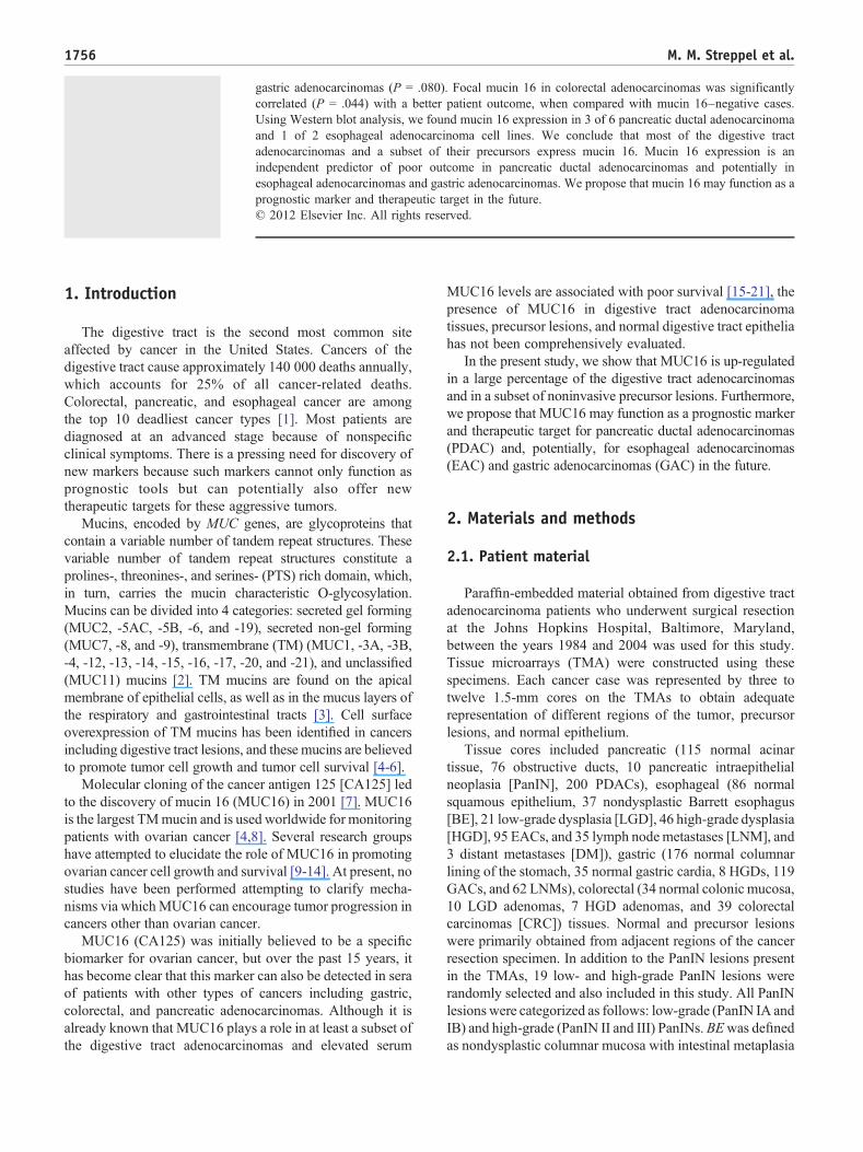
Examples of the presence of MUC16 in pancreatic tissuesare provided in Fig. 1 (row 1). Membranous MUC16 waspresent in 81.5% (163/200) of the surgically resected PDACs
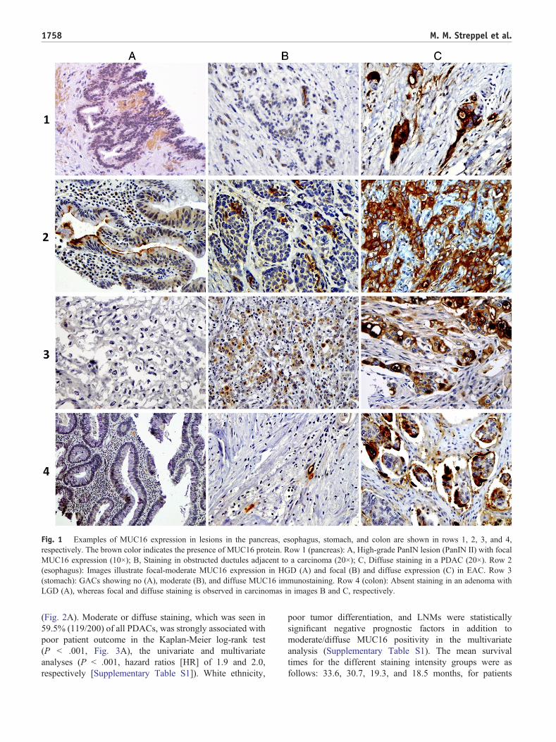
Fig. 1 Examples of MUC16 expression in lesions in the pancreas, esophagus, stomach, and colon are shown in rows 1, 2, 3, and 4,respectively. The brown color indicates the presence of MUC16 protein. Row 1 (pancreas): A, High-grade PanIN lesion (PanIN II) with focalMUC16 expression (10×); B, Staining in obstructed ductules adjacent to a carcinoma (20×); C, Diffuse staining in a PDAC (20×). Row 2(esophagus): Images illustrate focal-moderate MUC16 expression in HGD (A) and focal (B) and diffuse expression (C) in EAC. Row 3(stomach): GACs showing no (A), moderate (B), and diffuse MUC16 immunostaining. Row 4 (colon): Absent staining in an adenoma withLGD (A), whereas focal and diffuse staining is observed in carcinomas in images B and C, respectively.
1758 M. M. Streppel et al.
(Fig. 2A). Moderate or diffuse staining, which was seen in59.5% (119/200) of all PDACs, was strongly associated withpoor patient outcome in the Kaplan-Meier log-rank test(P b .001, Fig. 3A), the univariate and multivariateanalyses (P b .001, hazard ratios [HR] of 1.9 and 2.0,respectively [Supplementary Table S1]). White ethnicity,
poor tumor differentiation, and LNMs were statisticallysignificant negative prognostic factors in addition tomoderate/diffuse MUC16 positivity in the multivariateanalysis (Supplementary Table S1). The mean survivaltimes for the different staining intensity groups were asfollows: 33.6, 30.7, 19.3, and 18.5 months, for patients
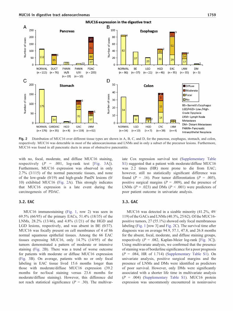
Fig. 2 Distribution of MUC16 over different tissue types are shown in A, B, C, and D, for the pancreas, esophagus, stomach, and colon,respectively. MUC16 was detectable in most of the adenocarcinomas and LNMs and in only a subset of the precursor lesions. Furthermore,MUC16 was found in all pancreatic ducts in areas of obstructive pancreatitis.
1759MUC16 in digestive tract adenocarcinomas
with no, focal, moderate, and diffuse MUC16 staining,respectively (P = .001, log-rank test [Fig. 3A]).Furthermore, MUC16 expression was observed in only2.7% (3/115) of the normal pancreatic tissues, and noneof the low-grade (0/19) and high-grade PanIN lesions (0/10) exhibited MUC16 (Fig. 2A). This strongly indicatesthat MUC16 expression is a late event during thecarcinogenesis of PDAC.
3.2. EAC
MUC16 immunostaining (Fig. 1, row 2) was seen in69.5% (66/95) of the primary EACs, 51.4% (18/35) of theLNMs, 28.2% (13/46), and 4.8% (1/21) of the HGD andLGD lesions, respectively, and was absent in BE (0/37).MUC16 was focally present on cell membranes of 4 of 86normal squamous epithelial tissues. Among the 66 EACtissues expressing MUC16, only 14.7% (14/95) of thetumors demonstrated a pattern of moderate or intensivestaining (Fig. 2B). There was a trend of worse outcomefor patients with moderate or diffuse MUC16 expression(Fig. 3B). On average, patients with no or only focallabeling in EAC tissue lived 15.6 months longer thanthose with moderate/diffuse MUC16 expression (39.2months for no/focal staining versus 23.6 months formoderate/diffuse staining). However, this difference didnot reach statistical significance (P = .30). The multivar-
iate Cox regression survival test (Supplementary TableS1) suggested that a patient with moderate/diffuse MUC16was 2.2 times (HR) more prone to die from EAC;however, still no statistically significant difference wasfound (P = .16). Poor tumor differentiation (P = .005),positive surgical margins (P = .009), and the presence ofLNMs (P = .023) and DMs (P b .001) were predictors ofpoor patient outcome in univariate analysis.
3.3. GAC
MUC16 was detected in a sizable minority (41.2%, 49/119) of the GACs and LNMs (40.3%, 25/62). Of theMUC16-positive tumors, 27 (55.1%) showed only focal membranouslabeling (Fig. 1 [row 3] and Fig. 2C). The survival time afterdiagnosis was on average 94.9, 57.1, 47.8, and 26.8 monthsfor the absent, focal, moderate, and diffuse staining groups,respectively (P = .082, Kaplan-Meier log-rank [Fig. 3C]).Using multivariate analysis, we confirmed that the presenceof staining was of borderline significance for a poor prognosis(P = .084, HR of 1.714) (Supplementary Table S1). Onunivariate analysis, positive surgical margins and thepresence of LNMs and DMs were identified as predictorsof poor survival. However, only DMs were significantlyassociated with a shorter life time in multivariate analysis(P = .004) (Supplementary Table S1). MUC16 proteinexpression was uncommonly encountered in noninvasive

Fig. 3 Kaplan-Meier survival curves of 4 digestive tract adenocarcinomas. PDAC patients whose tumors displayed no/focal MUC16expression lived significantly longer than patients with moderate/diffuse MUC16 expression (log-rank test, P b .001) (A). GAC (C) and EAC(B) patients with detectable MUC16 in their adenocarcinomas also tended to have a worse prognosis (log-rank test, P = .080 and P = .300,respectively). Notably, although not statistically significant (log-rank test, P = .130) (D), a trend of better patient outcome was seen forMUC16-positive CRC patients compared with MUC16-negative CRC patients.
1760 M. M. Streppel et al.
precursor lesions of GAC; only 25% (2/8) of the HGDtissues labeled with MUC16. Furthermore, MUC16 wasnot detected in normal gastric mucosa (Fig. 2C).
3.4. Colorectal adenocarcinoma
Among the 39 CRCs examined, 25 (64.1%) expressedMUC16. Fifteen of these cases showed focal staining (Fig.1 [row 4] and Fig. 2D). Direct comparison of the cases withabsent expression to those with focal expression revealed asignificant survival advantage for patients whose tumorsdisplayed focal MUC16 expression (P = .044, HR of 3.0).Patients whose tumors lacked staining (n = 14) had anaverage postoperative survival of 87.3 (95% CI, 34.0-140.5) months, compared with 182.6 (95% CI, 143.1-222.1) months for patients whose CRCs had focal staining(Kaplan-Meier log-rank, Fig. 3D). The mean survival timefor all MUC16-positive CRC patients was 157.5 months,which surprisingly did not reach statistical significancewhen compared with MUC16-negative CRC patients (P =.13, Fig. 3D). Among all possible predictors of prognosis,only the presence of DMs was significantly correlated withpoor patient outcome in the multivariate analysis (P = .008,HR of 46.4, Supplementary Table S1). In addition, MUC16was present in 42.9% (3/7) of the adenomas with HGD and10% (1/10) of the adenomas without HGD, reiterating thataberrant expression can be observed in noninvasive
precursor lesions in the colon. In contrast, normal colonmucosa lacked MUC16 expression (Fig. 2D).
3.5. MUC16 in pancreatic and esophageal cell lines
The Western blot results appear in Fig. 4. MUC16 wasdetected in the positive control OVCAR3. The size ofMUC16 in OVCAR3 was above the 500 kDa, which iswithin the range of the predicted size of MUC16 (500-5000kDa). MUC16 was undetectable in our negative controlHPNE. MUC16 was found in 3 of 6 examined PDAC celllines, with strong expression in Panc8.13 and Panc10.7 andweak expression in Panc5.04. In addition, MUC16 waspresent in JHesoAD1, whereas it was undetectable in OE33.β-actin was used as a control for presence of protein and wasstrongly expressed in all cell lines (Fig. 4).
4. Discussion
Herein, we provide an overview of MUC16 proteinexpression in normal epithelia, precursor lesions, adeno-carcinomas, and metastases of the digestive tract, includingthe esophagus and the stomach, which have not beenevaluated for this protein to date. MUC16 has beenpreviously studied in CRCs and PDACs; however,
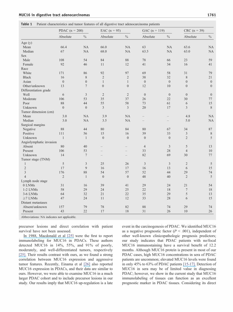
Table 1 Patient characteristics and tumor features of all digestive tract adenocarcinoma patients
PDAC (n = 200) EAC (n = 95) GAC (n = 119) CRC (n = 39)
Absolute % Absolute % Absolute % Absolute %
Age (y)Mean 66.4 NA 66.0 NA 63 NA 63.6 NAMedian 67 NA 68.0 NA 63.5 NA 63.0 NASexMale 108 54 84 88 78 66 23 59Female 92 46 11 12 41 34 16 41RaceWhite 171 86 92 97 69 58 31 79Black 16 8 2 2 38 32 8 21Asian 0 0 1 1 0 0 0 0Other/unknown 13 7 0 0 12 10 0 0Differentiation gradeWell 6 3 2 2 0 0 0 0Moderate 106 53 35 37 26 22 30 77Poor 88 44 55 58 73 61 6 15Unknown 0 0 3 3 20 17 3 8Tumor dimension (cm)Mean 3.0 NA 3.9 NA – – 4.8 NAMedian 3.0 NA 3.5 NA – – 5.0 NASurgical marginsNegative 88 44 80 84 80 67 34 87Positive 111 56 15 16 39 33 3 8Unknown 1 1 0 0 0 0 2 5Angiolymphatic invasionAbsent 80 40 – – 4 3 5 13Present 106 53 – – 33 28 4 10Unknown 14 7 – – 82 69 30 77Tumor stage (TNM)1 5 3 25 26 3 3 2 52 17 9 16 17 16 13 6 153 176 88 54 57 52 44 29 744 2 1 0 0 48 40 2 5
Lymph node stage0 LNMs 31 16 39 41 29 24 21 541-2 LNMs 58 29 24 25 22 18 7 183-6 LNMs 64 32 21 22 35 29 5 13≥7 LNMs 47 24 11 12 33 28 6 15Distant metastasesAbsent/unknown 157 79 78 82 88 74 29 74Present 43 22 17 18 31 26 10 26
Abbreviations: NA indicates not applicable.
1761MUC16 in digestive tract adenocarcinomas
precursor lesions and direct correlation with patientsurvival have not been assessed.
In 1988, Macdonald et al [25] were the first to reportimmunolabeling for MUC16 in PDACs. These authorsdetected MUC16 in 14%, 55%, and 91% of poorly,moderately, and well-differentiated tumors, respectively[25]. Their results contrast with ours, as we found a strongcorrelation between MUC16 expression and aggressivetumor features. Recently, Einama et al [26] also reportedMUC16 expression in PDACs, and their data are similar toours. However, we were able to examine MUC16 in a muchlarger PDAC cohort and to include precursor lesions in ourstudy. Our results imply that MUC16 up-regulation is a late
event in the carcinogenesis of PDAC. We identified MUC16as a negative prognostic factor (P b .001), independent ofother well-known clinicopathologic prognosis predictors;our study indicates that PDAC patients with no/focalMUC16 immunostaining have a survival benefit of 12.2months. Although MUC16 protein is present in most of ourPDAC cases, high MUC16 concentrations in sera of PDACpatients are uncommon; elevated MUC16 levels were foundin only 45% to 63% of PDAC patients [15-17]. Detection ofMUC16 in sera may be of limited value in diagnosingPDAC; however, we show in the current study that MUC16immunolabeling of tissues can function as an excellentprognostic marker in PDAC tissues. Considering its direct
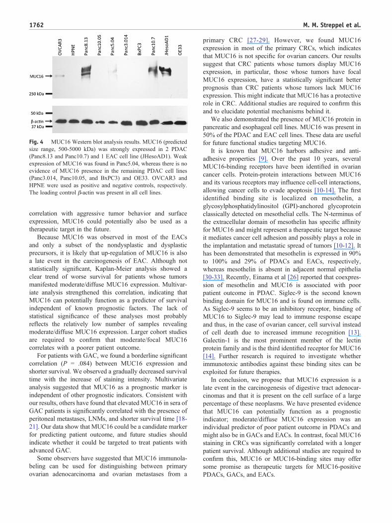
Fig. 4 MUC16 Western blot analysis results. MUC16 (predictedsize range, 500-5000 kDa) was strongly expressed in 2 PDAC(Panc8.13 and Panc10.7) and 1 EAC cell line (JHesoAD1). Weakexpression of MUC16 was found in Panc5.04, whereas there is noevidence of MUC16 presence in the remaining PDAC cell lines(Panc3.014, Panc10.05, and BxPC3) and OE33. OVCAR3 andHPNE were used as positive and negative controls, respectively.The loading control β-actin was present in all cell lines.
1762 M. M. Streppel et al.
correlation with aggressive tumor behavior and surfaceexpression, MUC16 could potentially also be used as atherapeutic target in the future.
Because MUC16 was observed in most of the EACsand only a subset of the nondysplastic and dysplasticprecursors, it is likely that up-regulation of MUC16 is alsoa late event in the carcinogenesis of EAC. Although notstatistically significant, Kaplan-Meier analysis showed aclear trend of worse survival for patients whose tumorsmanifested moderate/diffuse MUC16 expression. Multivar-iate analysis strengthened this correlation, indicating thatMUC16 can potentially function as a predictor of survivalindependent of known prognostic factors. The lack ofstatistical significance of these analyses most probablyreflects the relatively low number of samples revealingmoderate/diffuse MUC16 expression. Larger cohort studiesare required to confirm that moderate/focal MUC16correlates with a poorer patient outcome.
For patients with GAC, we found a borderline significantcorrelation (P = .084) between MUC16 expression andshorter survival. We observed a gradually decreased survivaltime with the increase of staining intensity. Multivariateanalysis suggested that MUC16 as a prognostic marker isindependent of other prognostic indicators. Consistent withour results, others have found that elevated MUC16 in sera ofGAC patients is significantly correlated with the presence ofperitoneal metastases, LNMs, and shorter survival time [18-21]. Our data show that MUC16 could be a candidate markerfor predicting patient outcome, and future studies shouldindicate whether it could be targeted to treat patients withadvanced GAC.
Some observers have suggested that MUC16 immunola-beling can be used for distinguishing between primaryovarian adenocarcinoma and ovarian metastases from a
primary CRC [27-29]. However, we found MUC16expression in most of the primary CRCs, which indicatesthat MUC16 is not specific for ovarian cancers. Our resultssuggest that CRC patients whose tumors display MUC16expression, in particular, those whose tumors have focalMUC16 expression, have a statistically significant betterprognosis than CRC patients whose tumors lack MUC16expression. This might indicate that MUC16 has a protectiverole in CRC. Additional studies are required to confirm thisand to elucidate potential mechanisms behind it.
We also demonstrated the presence of MUC16 protein inpancreatic and esophageal cell lines. MUC16 was present in50% of the PDAC and EAC cell lines. These data are usefulfor future functional studies targeting MUC16.
It is known that MUC16 harbors adhesive and anti-adhesive properties [9]. Over the past 10 years, severalMUC16-binding receptors have been identified in ovariancancer cells. Protein-protein interactions between MUC16and its various receptors may influence cell-cell interactions,allowing cancer cells to evade apoptosis [10-14]. The firstidentified binding site is localized on mesothelin, aglycosylphosphatidylinositol (GPI)-anchored glycoproteinclassically detected on mesothelial cells. The N-terminus ofthe extracellular domain of mesothelin has specific affinityfor MUC16 and might represent a therapeutic target becauseit mediates cancer cell adhesion and possibly plays a role inthe implantation and metastatic spread of tumors [10-12]. Ithas been demonstrated that mesothelin is expressed in 90%to 100% and 29% of PDACs and EACs, respectively,whereas mesothelin is absent in adjacent normal epithelia[30-33]. Recently, Einama et al [26] reported that coexpres-sion of mesothelin and MUC16 is associated with poorpatient outcome in PDAC. Siglec-9 is the second knownbinding domain for MUC16 and is found on immune cells.As Siglec-9 seems to be an inhibitory receptor, binding ofMUC16 to Siglec-9 may lead to immune response escapeand thus, in the case of ovarian cancer, cell survival insteadof cell death due to increased immune recognition [13].Galectin-1 is the most prominent member of the lectinprotein family and is the third identified receptor for MUC16[14]. Further research is required to investigate whetherimmunotoxic antibodies against these binding sites can beexploited for future therapies.
In conclusion, we propose that MUC16 expression is alate event in the carcinogenesis of digestive tract adenocar-cinomas and that it is present on the cell surface of a largepercentage of these neoplasms. We have presented evidencethat MUC16 can potentially function as a prognosticindicator; moderate/diffuse MUC16 expression was anindividual predictor of poor patient outcome in PDACs andmight also be in GACs and EACs. In contrast, focal MUC16staining in CRCs was significantly correlated with a longerpatient survival. Although additional studies are required toconfirm this, MUC16 or MUC16-binding sites may offersome promise as therapeutic targets for MUC16-positivePDACs, GACs, and EACs.
1763MUC16 in digestive tract adenocarcinomas
Supplementary data
Supplementary data to this article can be found onlineat doi:10.1016/j.humpath.2012.01.005.
References
[1] Jemal A, Siegel R, Xu J, Ward E. Cancer statistics, 2010. CA Cancer JClin 2010;60:277-300.
[2] Senapati S, Sharma P, Bafna S, Roy HK, Batra SK. The MUC genefamily: their role in the diagnosis and prognosis of gastric cancer.Histol Histopathol 2008;23:1541-52.
[3] Bafna S, Kaur S, Batra SK. Membrane-bound mucins: the mechanisticbasis for alterations in the growth and survival of cancer cells.Oncogene 2010;29:2893-904.
[4] Kufe DW. Mucins in cancer: function, prognosis and therapy. Nat RevCancer 2009;9:874-85.
[5] Jonckheere N, Van Seuningen I. The membrane-bound mucins: fromcell signaling to transcriptional regulation and expression in epithelialcancers. Biochimie 2010;92:1-11.
[6] Jonckheere N, Van Seuningen I. The membrane-bound mucins: howlarge O-glycoproteins play key roles in epithelial cancers and holdpromise as biological tools for gene-based and immunotherapies. CritRev Oncog 2008;14:177-96.
[7] Yin BW, Lloyd KO. Molecular cloning of the CA125 ovarian cancerantigen: identification as a new mucin, MUC16. J Biol Chem2001;276:27371-5.
[8] Yin BW, Dnistrian A, Lloyd KO. Ovarian cancer antigen CA125is encoded by the MUC16 mucin gene. Int J Cancer 2002;98:737-40.
[9] Hilkens J, Ligtenberg MJ, Vos HL, Litvinov SV. Cell membrane-associated mucins and their adhesion-modulating property. TrendsBiochem Sci 1992;17:359-63.
[10] Rump A, Morikawa Y, Tanaka M, et al. Binding of ovarian cancerantigen CA125/MUC16 to mesothelin mediates cell adhesion. J BiolChem 2004;279:9190-8.
[11] Kaneko O, Gong L, Zhang J, et al. A binding domain on mesothelin forCA125/MUC16. J Biol Chem 2009;284:3739-49.
[12] Gubbels JA, Belisle J, Onda M, et al. Mesothelin-MUC16 binding is ahigh affinity, N-glycan dependent interaction that facilitates peritonealmetastasis of ovarian tumors. Mol Cancer 2006;5:50.
[13] Belisle JA, Horibata S, Jennifer GA, et al. Identification of Siglec-9 asthe receptor for MUC16 on human NK cells, B cells, and monocytes.Mol Cancer 2010;9:118.
[14] Seelenmeyer C, Wegehingel S, Lechner J, Nickel W. The cancerantigen CA125 represents a novel counter receptor for galectin-1.J Cell Sci 2003;116:1305-18.
[15] Haglund C. Tumour marker antigen CA125 in pancreatic cancer:a comparison with CA19-9 and CEA. Br J Cancer 1986;54:897-901.
[16] Yue T, Goldstein IJ, Hollingsworth MA, et al. The prevalence andnature of glycan alterations on specific proteins in pancreatic cancerpatients revealed using antibody-lectin sandwich arrays. Mol CellProteomics 2009;8:1697-707.
[17] Haga Y, Sakamoto K, Egami H, et al. Clinical significance of serumCA125 values in patients with cancers of the digestive system. Am JMed Sci 1986;292:30-4.
[18] Zhang YH, Li Y, Chen C, Peng CW. Carcinoembryonic antigen levelis related to tumor invasion into the serosa of the stomach: study on166 cases and suggestion for new therapy. Hepatogastroenterology2009;56:1750-4.
[19] Fujimura T, Kinami S, Ninomiya I, et al. Diagnostic laparoscopy,serum CA125, and peritoneal metastasis in gastric cancer. Endoscopy2002;34:569-74.
[20] Fan B, Xiong B. Investigation of serum tumor markers in the diagnosisof gastric cancer. Hepatogastroenterology 2011;58:239-45.
[21] Webb A, Scott-Mackie P, Cunningham D, et al. The prognostic valueof serum and immunohistochemical tumour markers in advancedgastric cancer. Eur J Cancer 1996;32A:63-8.
[22] Wang KK, Sampliner RE. Updated guidelines 2008 for the diagnosis,surveillance and therapy of Barrett's esophagus. Am J Gastroenterol2008;103:788-97.
[23] American Gastroenterological Association, Spechler SJ, Sharma P,Souza RF, Inadomi JM, Shaheen NJ. American GastroenterologicalAssociation medical position statement on the management ofBarrett's esophagus. Gastroenterology 2011;140:1084-91.
[24] Goodell CA, Belisle JA, Gubbels JA, et al. Characterization of thetumor marker muc16 (ca125) expressed by murine ovarian tumor celllines and identification of a panel of cross-reactive monoclonalantibodies. J Ovarian Res 2009;2:8.
[25] Macdonald F, Downing R, Allum WH. Expression of CA125 inpancreatic carcinoma and chronic pancreatitis. Br J Cancer 1988;58:505-6.
[26] Einama T, Kamachi H, Nishihara H, et al. Co-expression of mesothelinand CA125 correlates with unfavorable patient outcome in pancreaticductal adenocarcinoma. Pancreas 2011;40:1276-82.
[27] Multhaupt HA, Arenas-Elliott CP, Warhol MJ. Comparison ofglycoprotein expression between ovarian and colon adenocarcinomas.Arch Pathol Lab Med 1999;123:909-16.
[28] Lagendijk JH, Mullink H, van Diest PJ, Meijer GA, Meijer CJ.Immunohistochemical differentiation between primary adenocarci-nomas of the ovary and ovarian metastases of colonic and breastorigin. Comparison between a statistical and an intuitive approach. JClin Pathol 1999;52:283-90.
[29] Brown RW, Campagna LB, Dunn JK, Cagle PT. Immunohistochem-ical identification of tumor markers in metastatic adenocarcinoma. Adiagnostic adjunct in the determination of primary site. Am J ClinPathol 1997;107:12-9.
[30] Argani P, Iacobuzio-Donahue C, Ryu B, et al. Mesothelin isoverexpressed in the vast majority of ductal adenocarcinomas of thepancreas: identification of a new pancreatic cancer marker by serialanalysis of gene expression (SAGE). Clin Cancer Res 2001;7:3862-8.
[31] Swierczynski SL, Maitra A, Abraham SC, et al. Analysis of noveltumor markers in pancreatic and biliary carcinomas using tissuemicroarrays. HUM PATHOL 2004;35:357-66.
[32] Hassan R, Laszik ZG, Lerner M, et al. Mesothelin is overexpressed inpancreaticobiliary adenocarcinomas but not in normal pancreas andchronic pancreatitis. Am J Clin Pathol 2005;124:838-45.
[33] Alvarez H, Rojas PL, Yong KT, et al. Mesothelin is a specificbiomarker of invasive cancer in the Barrett-associated adenocarcinomaprogression model: translational implications for diagnosis andtherapy. Nanomedicine 2008;4:295-301.