Pseudoprevotella muciniphila gen. nov., sp. nov., a mucin ...
18
RESEARCH ARTICLE Pseudoprevotella muciniphila gen. nov., sp. nov., a mucin-degrading bacterium attached to the bovine rumen epithelium Sang Weon Na ID 1 , Byung Hee Chun 2 , Seok-Hyeon Beak ID 1 , Shehzad Abid Khan 2 , Md. Najmul Haque 1 , Jae Sung Lee 1 , Che Ok Jeon 2 , Sang-Suk Lee 3 , Myunggi Baik ID 1 * 1 Department of Agricultural Biotechnology and Research Institute of Agriculture and Life Sciences, College of Agriculture and Life Sciences, Seoul National University, Seoul, Republic of Korea, 2 Department of Life Science, Chung-Ang University, Seoul, Republic of Korea, 3 Department of Animal Science & Technology, Sunchon National University, Sunchon, Republic of Korea * [email protected] Abstract A Gram-negative, strictly anaerobic mucin-degrading bacterium, which we designated strain E39 T , was isolated from the rumen epithelium of Korean cattle. The cells were non-motile and had a coccus morphology. Growth of strain E39 T was observed at 30–45˚C (optimum, 39˚C), pH 6.5–8.5 (optimum, pH 7.5), and in the presence of 0.0–1.0% (w/v) NaCl (optimum, 0.0–0.5%). Strain E39 T contained C 16:0 ,C 18:0 ,C 18:1 ω9c, iso-C 15:0 , and anteiso-C 15:0 as the major fatty acids. The major polar lipids were phosphatidylethanolamine, unidentified ami- nophospholipid, and unidentified lipids. The major respiratory isoprenoid quinones were MK-8 and MK-9. The major fermented end-products of mucin were acetate and succinate. The G+C content of the genomic DNA was 46.4 mol%. Strain E39 T was most closely related to Alloprevotella rava 81/4-12 T with an 87.3% 16S rRNA gene sequence similarity. On the basis of phenotypic, chemotaxonomic, and molecular properties, strain E39 T represents a novel genus of the family Prevotellaceae; as such, the name Pseudoprevotella muciniphila gen. nov., sp. nov. is proposed. A functional annotation of the whole genome sequences of P. muciniphila E39 T revealed that this bacterium has a putative mucin-degrading pathway and biosynthetic pathways of extracellular polymeric substances and virulence factors which enable bacteria to adhere to the epithelial cells and avoid the host’s immune responses. Introduction Rumen, a forestomach of ruminants, is a complex symbiotic ecosystem. In the rumen, various types of microorganisms (archaea, bacteria, protozoa, fungi, and virus) exist and they are known as the rumen microbiota. The rumen microbiota provides nutrients for the host rumi- nants by digestion of feed particles, especially indigestive fibrous material, and biosynthesis of metabolites like amino acids, lipids, and vitamins [1]. The nutrients including volatile fatty acids, minerals, and other metabolites are absorbed and transported into the host’s blood PLOS ONE PLOS ONE | https://doi.org/10.1371/journal.pone.0251791 May 20, 2021 1 / 18 a1111111111 a1111111111 a1111111111 a1111111111 a1111111111 OPEN ACCESS Citation: Na SW, Chun BH, Beak S-H, Khan SA, Haque M.N, Lee JS, et al. (2021) Pseudoprevotella muciniphila gen. nov., sp. nov., a mucin-degrading bacterium attached to the bovine rumen epithelium. PLoS ONE 16(5): e0251791. https:// doi.org/10.1371/journal.pone.0251791 Editor: Pankaj Kumar Arora, Babasaheb Bhimrao Ambedkar University, INDIA Received: September 18, 2020 Accepted: May 4, 2021 Published: May 20, 2021 Copyright: © 2021 Na et al. This is an open access article distributed under the terms of the Creative Commons Attribution License, which permits unrestricted use, distribution, and reproduction in any medium, provided the original author and source are credited. Data Availability Statement: All 16S gene and genome sequence data of stain E39 are available from the GenBank database (accession numbers MG763147 and CP033459). Other reference strains’ genome sequence data are available from the GenBank: A. tannerae ATCC 51259T (GenBank acc. no., ACIJ00000000), A. rava 81/4-12T (GenBank acc. no., ACZK00000000), P. clara YIT 11840T (GenBank acc. no., AFFY00000000), P. melaninogenica ATCC 25845T (GenBank acc. no., CP002122-3), and Bacteroides thetaiotaomicron VPI 5482T (GenBank acc. No., AE015928) were
-
Upload
khangminh22 -
Category
Documents
-
view
1 -
download
0
Transcript of Pseudoprevotella muciniphila gen. nov., sp. nov., a mucin ...

RESEARCH ARTICLE
Pseudoprevotella muciniphila gen. nov., sp.
nov., a mucin-degrading bacterium attached
to the bovine rumen epithelium
Sang Weon NaID1, Byung Hee Chun2, Seok-Hyeon BeakID
1, Shehzad Abid Khan2, Md.
Najmul Haque1, Jae Sung Lee1, Che Ok Jeon2, Sang-Suk Lee3, Myunggi BaikID1*
1 Department of Agricultural Biotechnology and Research Institute of Agriculture and Life Sciences, College
of Agriculture and Life Sciences, Seoul National University, Seoul, Republic of Korea, 2 Department of Life
Science, Chung-Ang University, Seoul, Republic of Korea, 3 Department of Animal Science & Technology,
Sunchon National University, Sunchon, Republic of Korea
Abstract
A Gram-negative, strictly anaerobic mucin-degrading bacterium, which we designated strain
E39T, was isolated from the rumen epithelium of Korean cattle. The cells were non-motile
and had a coccus morphology. Growth of strain E39T was observed at 30–45˚C (optimum,
39˚C), pH 6.5–8.5 (optimum, pH 7.5), and in the presence of 0.0–1.0% (w/v) NaCl (optimum,
0.0–0.5%). Strain E39T contained C16:0, C18:0, C18:1 ω9c, iso-C15:0, and anteiso-C15:0 as the
major fatty acids. The major polar lipids were phosphatidylethanolamine, unidentified ami-
nophospholipid, and unidentified lipids. The major respiratory isoprenoid quinones were
MK-8 and MK-9. The major fermented end-products of mucin were acetate and succinate.
The G+C content of the genomic DNA was 46.4 mol%. Strain E39T was most closely related
to Alloprevotella rava 81/4-12T with an 87.3% 16S rRNA gene sequence similarity. On the
basis of phenotypic, chemotaxonomic, and molecular properties, strain E39T represents a
novel genus of the family Prevotellaceae; as such, the name Pseudoprevotella muciniphila
gen. nov., sp. nov. is proposed. A functional annotation of the whole genome sequences of
P. muciniphila E39T revealed that this bacterium has a putative mucin-degrading pathway
and biosynthetic pathways of extracellular polymeric substances and virulence factors
which enable bacteria to adhere to the epithelial cells and avoid the host’s immune
responses.
Introduction
Rumen, a forestomach of ruminants, is a complex symbiotic ecosystem. In the rumen, various
types of microorganisms (archaea, bacteria, protozoa, fungi, and virus) exist and they are
known as the rumen microbiota. The rumen microbiota provides nutrients for the host rumi-
nants by digestion of feed particles, especially indigestive fibrous material, and biosynthesis of
metabolites like amino acids, lipids, and vitamins [1]. The nutrients including volatile fatty
acids, minerals, and other metabolites are absorbed and transported into the host’s blood
PLOS ONE
PLOS ONE | https://doi.org/10.1371/journal.pone.0251791 May 20, 2021 1 / 18
a1111111111
a1111111111
a1111111111
a1111111111
a1111111111
OPEN ACCESS
Citation: Na SW, Chun BH, Beak S-H, Khan SA,
Haque M.N, Lee JS, et al. (2021) Pseudoprevotella
muciniphila gen. nov., sp. nov., a mucin-degrading
bacterium attached to the bovine rumen
epithelium. PLoS ONE 16(5): e0251791. https://
doi.org/10.1371/journal.pone.0251791
Editor: Pankaj Kumar Arora, Babasaheb Bhimrao
Ambedkar University, INDIA
Received: September 18, 2020
Accepted: May 4, 2021
Published: May 20, 2021
Copyright: © 2021 Na et al. This is an open access
article distributed under the terms of the Creative
Commons Attribution License, which permits
unrestricted use, distribution, and reproduction in
any medium, provided the original author and
source are credited.
Data Availability Statement: All 16S gene and
genome sequence data of stain E39 are available
from the GenBank database (accession numbers
MG763147 and CP033459). Other reference
strains’ genome sequence data are available from
the GenBank: A. tannerae ATCC 51259T (GenBank
acc. no., ACIJ00000000), A. rava 81/4-12T
(GenBank acc. no., ACZK00000000), P. clara YIT
11840T (GenBank acc. no., AFFY00000000), P.
melaninogenica ATCC 25845T (GenBank acc. no.,
CP002122-3), and Bacteroides thetaiotaomicron
VPI 5482T (GenBank acc. No., AE015928) were

vessels through the rumen epithelium [2]. Only small portion of rumen microbiota attaches to
the rumen epithelium. Several rumen epithelial bacteria (epimural bacteria) and their func-
tions in urea digestion, tissue recycling, and oxygen scavenging have been identified since
1960s [3, 4]. Recent studies showed the composition and expressed genes of the epimural com-
munity in response to subacute rumen acidosis challenges using meta-omics technologies [5,
6]. Although these culture-independent methods provide expansive information about the epi-
mural community, culture-dependent studies about yet uncultured eprimural bacteria are
essential to understand their functions in more depth.
Mucin is a host-derived glycoprotein composed of protein backbones and various oligosac-
charides. Intestinal epithelial cells produce two types of mucins (secreted mucin and cell mem-
brane-associated mucin). They have a role as barriers to prevent pathogens from penetrating
the epithelial tissues [7]. Mucin-degrading bacteria can degrade mucin and utilize it as an
energy source. Some of them penetrate into the host epithelial cells with their mucin-degrad-
ing ability and show pathogenicity, others play roles as commensal bacteria [8]. Studies on
Akkermansia muciniphila, a commensal mucin-degrading bacterium, revealed that this bacte-
rium improved the host metabolic disorders, such as obesity and insulin resistance, by control-
ling gut barrier permeability and inflammation [9, 10].
In the bovine rumen, there are salivary mucin secreted from salivary glands and cell-mem-
brane associated mucins expressed on rumen epithelial cells [11]. In the 1960s, Fina et al. iso-
lated five uncultured mucin-degrading rumen bacteria from rumen fluid and Mishra et al.determined mucin-degrading activity of several species of rumen bacteria (Butyrivibrio fibri-solvens, Selenomonas ruminantium, Streptococcus bovis, Peptostreptococcus elsdenii, and Bac-teroides ruminicola) [12–14]. They focused on mucin-degrading bacteria floating in the rumen
fluid and their roles in bloat syndrome. Subsequently, there has been few studies on mucin-
degrading bacteria floating in the rumen fluid nor them attached to the rumen epithelium.
In this study, we used mucin as a novel carbon source to enrich previously uncultured
mucin-degrading bacteria from the rumen epithelium of Korean cattle. We isolated a novel
mucin-degrading bacterium, called strain E39T. Then, we performed phylogenetic analysis,
phenotypic and chemotaxonomic characterization, and genome analysis. On the basis of phe-
notypic, chemotaxonomic, and molecular properties, strain E39T represents a novel genus of
the family Prevotellaceae, for which we propose the designation Pseudoprevotella muciniphilagen. nov., sp. nov., strain E39T.
Materials and methods
Ethical statement
All experimental procedures were performed in accordance with the Animal Experimental
Guidelines provided by the Seoul National University Institutional Animal Use and Care
Committee, Republic of Korea. The experimental protocol was approved by the Seoul National
University Institutional Animal Use and Care Committee (SNU-170626-1).
Enrichment and isolation of mucin-degrading bacteria
Strain E39T was isolated from the rumen epithelium of Korean cattle. Rumen epithelium tissue
samples were excised from the ventral sacs of rumens immediately after slaughter at the abat-
toir in Bucheon (37˚31’48.4"N 126˚45’46.3"E), South Korea in 2017 and transported in a sterile
container with rumen fluid. On arrival at the laboratory, the samples were moved into an
anaerobic chamber and washed several times with anaerobic dilution solution (ADS; 3 g
K2HPO4, 6 g NaCl, 3 g KH2PO4, 0.6 g CaCl2�2H2O, 0.6 g MgSO4�7H2O, 6 g (NH4)2SO4, 0.5 g
cysteine-HCl, 0.5 g Na2S�9H2O, 0.625 g NaOH, and 1 mg resazurin per liter) to remove the
PLOS ONE Pseudoprevotella muciniphila, a mucin-degrading bacterium of bovine rumen epithelium
PLOS ONE | https://doi.org/10.1371/journal.pone.0251791 May 20, 2021 2 / 18
obtained from GenBank for comparative analysis.
Other relevant data are within the manuscript and
its supporting information files.
Funding: This work was supported by a grant from
the Next-Generation BioGreen 21 Program
(PJ01114001), Rural Development Administration,
Republic of Korea.
Competing interests: The authors have declared
that no competing interests exist.

rumen contents and non-adherent bacteria [15, 16]. Five grams of epithelial samples that were
stripped from the muscle layer were homogenized in 30 ml of ADS and serially diluted
(10-fold). Each 0.3 ml of dilution was inoculated into 30 ml of basal mucin medium in a butyl
rubber stopped serum bottle and incubated at 39˚C in an anaerobic atmosphere (95% CO2 5%
H2) for 24 h for enrichment. The basal mucin medium was prepared by modifying medium 10
[17] and consisted of 2 g peptone, 0.5 g yeast extract, 2.5 g hog gastric mucin (Type Ⅲ; Sigma),
50 ml mineral solution 1 (6 g K2HPO4 per liter), 50 ml mineral solution 2 (12 g NaCl, 6 g
KH2PO4, 1.2 g CaCl2�2H2O, 1.2 g MgSO4�7H2O, and 12 g (NH4)2SO4 per liter), 10 ml Pfen-
ning’s solution (0.5 g EDTA, 0.1 g ZnSO4�7H2O, 0.03 g MnCl2�4H2O, 0.03 g H3BO3, 0.2 g
CoCl2�6H2O, 0.01 g CuCl2�2H2O, 1.5 g FeCl2�4H2O, 0.02 g NiCl2�6H2O, 0.03 g Na2MoO4�
2H2O, and 0.01 g Na2SeO3 per liter), 10 ml volatile fatty acid solution (700 ml 0.2 N NaOH, 17
ml acetic acid, 6 ml propionic acid, 4 ml butyric acid, 1 ml iso-butyric acid, 1 ml 2-metylbuty-
ric acid, 1 ml valeric acid, and 1 ml isovaleric acid, pH 7.5 per liter), 1 ml hemin solution (0.5 g
hemin and 10 ml 1N NaOH per liter), 1 ml resazurin (0.1%, w/v), 20 ml cysteine sulfide solu-
tion (6.25 g NaOH, 25 g cysteine-HCl, and 25 g Na2S�9H2O per liter), and 4 g NaHCO3 per
liter. The enrichments were serially diluted with ADS, streaked onto basal mucin agar
medium, and incubated at 39˚C for 4 days under anaerobic conditions. Each colony was
picked, inoculated into 5 ml of basal mucin medium in a Hungate tube, and incubated at 39˚C
for 24–48 h. Streaking, colony picking, and incubation were repeated until an isolate was pure.
The isolates were preserved with 15% (v/v) glycerol stock solution and stored at –80˚C.
Bacterial growth and genomic DNA extraction
Cells of strain E39T were grown in 250 ml basal mucin medium at 39˚C for 24 h and harvested
by centrifugation (10,000 rpm for 5 min). The genomic DNA of the cells was extracted,
according to standard procedures, including phenol-chloroform extraction and ethanol pre-
cipitation [18].
16S rRNA gene based phylogeny
The 16S rRNA gene of strain E39T was amplified with PCR using the universal primers 10F
(50-AGT TTG ATC ATG GCT CAG ATT G-30) and 1507R (50-TAC CTT GTT ACGACT TCA CCC CAG-30) [19] and the resulting amplicons were sequenced using the univer-
sal primers 340F (50-CCT ACG GGA GGC AGC AG-30), 518R (50-ATT ACC GCG GCTGCT GG-30), and 805F (50-GAT TAG ATA CCC TGG TAG TC-30). The sequencing qual-
ity was checked, and the sequences were assembled using the Geneious program (ver. 11.0.4).
The almost complete 16S rRNA gene sequence (1,476 nucleotides) of strain E39T was com-
pared with that of all validated type strains using the Nucleotide Similarity Search program in
the EzBioCloud server (http://www.ezbiocloud.net/identify/) [20]. Phylogenetic trees were
constructed with MEGA (ver. 7.0.26) using the neighbor-joining (NJ), maximum-parsimony
(MP), and maximum-likelihood (ML) methods [21]. The complete 16S rRNA gene sequence
(1,540 nucleotides) from the genome sequence of strain E39T was used to construct the phylo-
genetic trees.
Phenotypic and chemotaxonomic characterization
The cell morphology of cells grown on basal mucin agar medium at 39˚C for 3 days was inves-
tigated using phase-contrast microscopy and transmission electron microscopy (Talos L120C;
FEI) at 120 kV. Gram staining was performed using a Sigma Gram staining kit following the
manufacturer’s protocol. The growth of strain E39T, as determined from the optical density
(OD) at a wavelength of 600 nm, was evaluated by culturing the cells in basal mucin medium,
PLOS ONE Pseudoprevotella muciniphila, a mucin-degrading bacterium of bovine rumen epithelium
PLOS ONE | https://doi.org/10.1371/journal.pone.0251791 May 20, 2021 3 / 18

brain heart infusion (BHI) broth (BD), trypticase soy broth (TSB; BD), Columbia broth (Acu-
media), and anaerobe basal broth (Oxoid) at 39˚C for 48 h. The optimum temperature, pH,
and NaCl concentration for growth were determined by culturing the cells on basal mucin
medium for 48 h at different temperatures (5–45˚C, at 5˚C intervals), pH (5.0–9.0 at 0.5 pH
unit intervals), and NaCl concentrations (0.0–2.0% at 0.5% intervals). To determine the opti-
mum pH, different pH buffers were used in the appropriate pH range (Na2HPO4-NaH2PO4
buffer at pH 5.0–7.5; Tris-HCl buffer at pH 8.0–9.0) and the pH values were adjusted before
and after autoclaving (121˚C, 15 min) [22]. Oxygen tolerance was investigated by measuring
growth (OD at 600 nm) in the absence of a reducing agent (cysteine sulfide solution) or in the
aerobic condition on basal mucin medium. A. tannerae ATCC 51259T, A. rava 81/4-12T, Para-prevotella clara YIT 11840T and Prevotella melaninogenica ATCC 25845T, which is the type
species of the genus Prevotella, were used as reference strains to compare enzyme profiles and
cellular fatty acid composition. The enzyme profiles were determined using an API Rapid ID
32A identification kit (bioMerieux) following the manufacturer’s instructions. Analysis of cel-
lular fatty acids was performed according to a standard MIDI protocol. All of the strains were
cultivated in peptone-yeast extract-glucose (PYG) broth, except strain E39T, which was culti-
vated in basal mucin medium. Cells were harvested at the late exponential phase and cellular
fatty acids were extracted from the cells following four steps (saponification, methylation,
extraction, and base wash). Fatty acid methyl esters were analyzed by gas chromatography
(Hewlett Packard 6890) and identified using the RTSBA6 database of the Microbial Identifica-
tion System (Sherlock ver. 6.0B) [23]. The polar lipid profiles were analyzed by thin-layer chro-
matography following the Minnikin et al. method [24]. The following reagents were used to
detect different types of polar lipids: 10% ethanolic molybdophosphoric acid (for total lipids),
ninhydrin (for aminolipids), Dittmer-Lester reagent (for phospholipids), and α-naphthol (for
glycolipids). The isoprenoid quinones of strain E39T, A. tannerae ATCC 51259T, A. rava 81/4-
12T, P. clara YIT 11840T, and P. melaninogenica ATCC 25845T were extracted from their expo-
nentially grown cells according to the procedure described by Jeon et al. [25] and analyzed at
40˚C using an Agilent infinity 1290 UHPLC equipped with a photodiode array detector (PAD)
and an Agilent 6550 ifunnel Q-TOF MS (Agilent Technologies, USA). Briefly, 2 μl of quinone
samples were injected into an Agilent Eclipse Plus C-18 column (2.1 mm × 100 mm, 2.1 μm)
and eluted at 40˚C using water (A) and acetonitrile (B) containing 0.1% formic acid as a
mobile phases with the following gradient: 0 min, 85% B; 30 min, 100% B; 40 min; and flow
rate, 0.4 ml/min. Isoprenoid quinone peaks in the chromatograms were identified by their UV
spectra generated by PAD and their molecular masses were assessed using Q-TOF MS. The
mass spectrometry was performed under the following conditions: polarity, positive; gas temp,
250˚C; nebulizer, 35 psi; capillary, (+) 4,000 V; MS range, 100–1,500 m/z.
Metabolite analysis using 1H NMR spectroscopy
Metabolic compounds including amino acids, monosaccharides, and organic acids in cultured
broth of strain E39T were analyzed using 1H NMR spectroscopy, as described previously [26].
Briefly, the basal mucin medium (2.5 g hog gastric mucin per liter; no glucose), glucose
medium (5 g glucose per liter; no hog gastric mucin), and mucin-glucose medium (2.5 g hog
gastric mucin and 5 g glucose per liter) were prepared based on the basal mucin medium to
investigate mucin and glucose utilization by strain E39T and their fermentation products.
Strain E39T was cultured in 5 ml of each broth at 39˚C for 0, 9, 18, 27, 36, and 54 h. The growth
of the cells was monitored by measuring OD at 600 nm. The culture broths were centrifuged,
filtered with a 0.45 μm syringe filter, and 0.3 ml of filtrate was mixed with 0.3 ml of 99.9% D2O
(Sigma-Aldrich, USA) containing 5 mM sodium 2,2-dimethyl-2-silapentane-5-sulfonate (DSS,
PLOS ONE Pseudoprevotella muciniphila, a mucin-degrading bacterium of bovine rumen epithelium
PLOS ONE | https://doi.org/10.1371/journal.pone.0251791 May 20, 2021 4 / 18

97%; Sigma-Aldrich). The mixtures were transferred into NMR tubes and their 1H NMR spec-
tra were measured on Varian Inova 600-MHz NMR spectrometer (Varian, USA). Metabolic
compounds were identified and quantified using the Chenomx NMR Suite program (ver. 6.1;
Chenomx, Canada).
Genome sequencing and analysis
De novo genome sequencing was performed using a Pacific Biosciences (PacBio) RSII plat-
form at Macrogen (Seoul, Korea; http://www.macrogen.com). A library was prepared using
PacBio DNA Template Prep Kit 1.0. After sequencing, reads were trimmed to obtain high
quality region and then assembled using RS hierarchical genome assembly process (HGAP
ver. 3.0) [27]. The complete genome was annotated using a software tool Prokka with default
parameter (ver. 1.12) [28].
For the bacterial core genes-based phylogenetic analysis, 92 up-to-date bacterial core genes
were extracted from the genomes of strains in the family Prevotellaceae and multiple-aligned,
and a phylogenetic tree was constructed using the up-to-date bacterial core gene (UBCG) tool
ver. 3 (https://www.ezbiocloud.net/tools/ubcg) [29]. The average nucleotide identity (ANI)
and digital DNA-DNA hybridization (dDDH) values among the genomes of strain E39T and
reference strains were calculated using a stand-alone software (http://www.ezbiocloud.net/sw/
oat) [30] and the Genome-to-Genome Distance Calculator (GGDC) ver. 2.1 (http://ggdc.
dsmz.de/distcalc2.php) [31], respectively. Functional annotation of predicted proteins was per-
formed using BlastKOALA tool of Kyoto Encyclopedia of Genes and Genomes (KEGG)
(http://www.kegg.jp/blastkoala/) [32].
To predict putative mucin-degrading enzymes and carbohydrate active enzymes
(CAZymes), the genome sequences of strain E39T and reference strains were submitted to a
meta server for automated carbohydrate-active enzyme annotation (dbCAN2) (http://cys.bios.
niu.edu/dbCAN2/) [33]. VRprofile (http://bioinfo-mml.sjtu.edu.cn/VRprofile) was used for
the prediction of virulence and antibiotic resistant genes [34].
Nucleotide sequence accession number
The 16S rRNA gene and genome sequence of strain E39T were deposited in GenBank under
MG763147 and CP033459, respectively. The genome data of reference strains: A. tanneraeATCC 51259T (GenBank acc. no., ACIJ00000000), A. rava 81/4-12T (GenBank acc. no.,
ACZK00000000), P. clara YIT 11840T (GenBank acc. no., AFFY00000000), P. melaninogenicaATCC 25845T (GenBank acc. no., CP002122-3), and Bacteroides thetaiotaomicron VPI 5482T
(GenBank acc. No., AE015928) were obtained from GenBank for comparative analysis.
Results and discussion
16S rRNA gene and genome based phylogeny
Comparative analysis of the 16S rRNA gene sequences revealed that strain E39T was closely
related to the genera Alloprevotella, Paraprevotella, Prevotella, and Bacteroides. Alloprevotellarava 81/4-12T, Paraprevotella clara YIT 11840T, Paraprevotella xylaniphila YIT 11841T, and
Bacteroides gallinarum JCM 13658T were most closely related to strain E39T with 87.3%,
86.6%, 86.3%, and 85.9% 16S rRNA gene sequence similarities, respectively. The phylogenetic
trees based on the ML algorithm and 92 bacterial core genes showed that strain E39T was affili-
ated with the family Prevotellaceae and close to the genera Alloprevotella and Paraprevotella(Fig 1) [35–37]. Similarly, the phylogenetic trees based on the NJ and MP algorithms also
showed that strain E39T was affiliated with the family Prevotellaceae (S1 and S2 Figs).
PLOS ONE Pseudoprevotella muciniphila, a mucin-degrading bacterium of bovine rumen epithelium
PLOS ONE | https://doi.org/10.1371/journal.pone.0251791 May 20, 2021 5 / 18
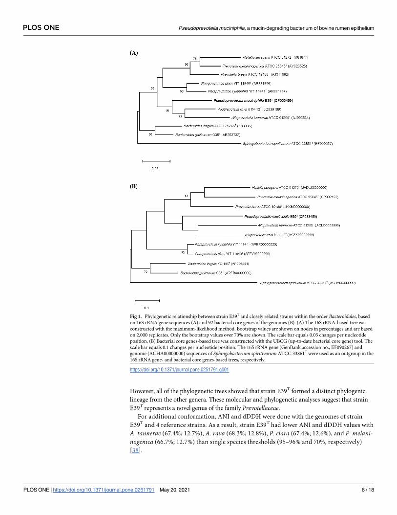
However, all of the phylogenetic trees showed that strain E39T formed a distinct phylogenic
lineage from the other genera. These molecular and phylogenetic analyses suggest that strain
E39T represents a novel genus of the family Prevotellaceae.For additional conformation, ANI and dDDH were done with the genomes of strain
E39T and 4 reference strains. As a result, strain E39T had lower ANI and dDDH values with
A. tannerae (67.4%; 12.7%), A. rava (68.3%; 12.8%), P. clara (67.4%; 12.6%), and P. melani-nogenica (66.7%; 12.7%) than single species thresholds (95–96% and 70%, respectively)
[38].
Fig 1. Phylogenetic relationship between strain E39T and closely related strains within the order Bacteroidales, based
on 16S rRNA gene sequences (A) and 92 bacterial core genes of the genomes (B). (A) The 16S rRNA-based tree was
constructed with the maximum-likelihood method. Bootstrap values are shown on nodes in percentages and are based
on 2,000 replicates. Only the bootstrap values over 70% are shown. The scale bar equals 0.05 changes per nucleotide
position. (B) Bacterial core genes-based tree was constructed with the UBCG (up-to-date bacterial core gene) tool. The
scale bar equals 0.1 changes per nucleotide position. The 16S rRNA gene (GenBank accession no., EF090267) and
genome (ACHA00000000) sequences of Sphingobacterium spiritivorum ATCC 33861T were used as an outgroup in the
16S rRNA gene- and bacterial core genes-based trees, respectively.
https://doi.org/10.1371/journal.pone.0251791.g001
PLOS ONE Pseudoprevotella muciniphila, a mucin-degrading bacterium of bovine rumen epithelium
PLOS ONE | https://doi.org/10.1371/journal.pone.0251791 May 20, 2021 6 / 18

Phenotypic and chemotaxonomic characterization
The transmission electron microscopic analyses showed that the cells of strain E39T were coc-
cus in morphology (680–820 nm in diameter), and lacking in flagella (Fig 2). In addition, fila-
mentous structures were observed from the cell surface. Among the various types of media,
including BHI broth, TSB, Columbia broth, and anaerobe basal broth, strain E39T could only
grow on basal mucin medium. Cells grew at temperatures between 30 and 45˚C, pH between
6.5 and 8.5, and in NaCl concentration between 0.0 and 1.0%. When the headspaces were filled
with anaerobic gas, growth was observed in the absence of a reducing agent, but at a lower rate
than in its presence. Cells did not grow in an aerobic atmosphere regardless of whether a
reducing agent was present or not. These results demonstrate that strain E39T prefers obligate
anaerobic conditions but tolerates a trace amount of oxygen (S3 Fig).
In the API Rapid ID 32A panel, strain E39T had positive activities of mucin-degrading
enzymes including β-galactosidase, N-acetyl-β-glucosaminidase, and α-fucosidase [8]. Lack of
urease activity suggest that strain E39T may not attend to the digestion of urea, which is one of
the roles of epimural bacteria. In particular, strain E39T was distinguished from the reference
taxa by a positive activity of arginine dihydrolase (Table 1). The major cellular fatty acids
Fig 2. A transmission electron micrograph showing the general morphology of negatively stained cells of strain
E39T. Cells were grown on basal mucin agar medium at 39˚C for 3 days. Bar, 200 nm. Filamentous structures were
observed from the cell surface.
https://doi.org/10.1371/journal.pone.0251791.g002
PLOS ONE Pseudoprevotella muciniphila, a mucin-degrading bacterium of bovine rumen epithelium
PLOS ONE | https://doi.org/10.1371/journal.pone.0251791 May 20, 2021 7 / 18
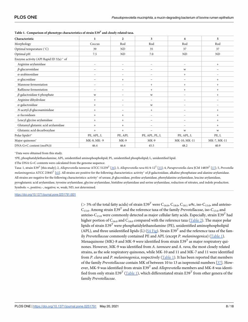
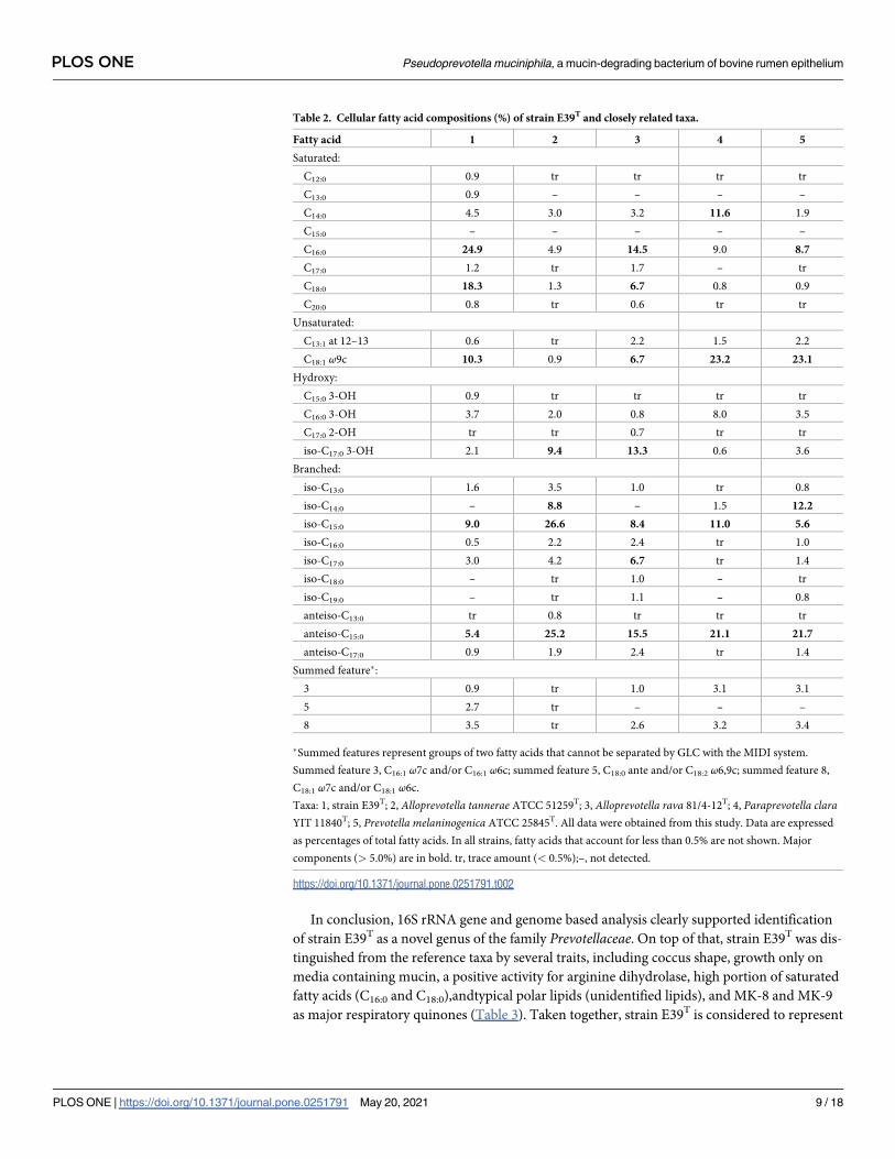
(> 5% of the total fatty acids) of strain E39T were C16:0, C18:0, C18:1 ω9c, iso-C15:0, and anteiso-
C15:0. Among strain E39T and the reference taxa of the family Prevotellaceae, iso-C15:0 and
anteiso-C15:0 were commonly detected as major cellular fatty acids. Especially, strain E39T had
higher portion of C16:0 and C18:0 compared with the reference taxa (Table 2). The major polar
lipids of strain E39T were phosphatidylethanolamine (PE), unidentified aminophospholipid
(APL), and three unidentified lipids (L) (S4 Fig). Strain E39T and the reference taxa of the fam-
ily Prevotellaceae commonly contained PE and APL (except P. melaninogenica) (Table 1).
Menaquinone (MK)-8 and MK-9 were identified from strain E39T as major respiratory qui-
nones. However, MK-9 was identified from A. tanneare and A. rava, the most closely related
strains, as the sole respiratory quinones, while MK-10 and 11 and MK-7 and 11 were identified
from P. clara and P. melaninogenica, respectively (Table 1). It has been reported that members
of the family Prevotellaceae contain MK of between 10 to 13 as isoprenoid numbers [37]. How-
ever, MK-9 was identified from strain E39T and Alloprevotella members and MK-8 was identi-
fied from only strain E39T (Table 1), which differentiated strain E39T from other genera of the
family Prevotellaceae.
Table 1. Comparison of phenotype characteristics of strain E39T and closely related taxa.
Characteristic 1 2 3 4 5
Morphology Coccus Rod Rod Rod Rod
Optimal temperature (˚C) 39 ND 35 37 37
Optimal pH 7.5 ND 7.0 ND ND
Enzyme activity (API Rapid ID 32a) � of
Arginine arylamidase – – – – +
β-glucuronidase – – – w –
α-arabinosidase – – – + –
α-glucosidase – + – – +
Mannose fermentation – – + + +
Raffinose fermentation – – + + +
β-galactosidase 6 phosphate w – w – +
Arginine dihydrolase + – – – –
α-galactosidase + – w – –
N-acetyl-β-glucosaminidase + – + – +
α-fucosidases + + – – +
Leucyl glycine arylamidase + + – – +
Glutamyl glutamic acid arylamidase + + – – +
Glutamic acid decarboxylase + + – w w
Polar lipids†� PE, APL, L PE, APL PE, APL, PL, L PE, APL, L PE, L
Major quinones� MK-8, MK- 9 MK-9 MK-9 MK-10, MK-11 MK-7, MK-11
DNA G+C content (mol%)‡ 46.4 46.6 45.5 48.2 40.9
�Data were obtained from this study.
†PE, phosphatidylethanolamine; APL, unidentified aminophospholipid; PL, unidentified phospholipid; L, unidentified lipid.
‡The DNA G+C contents were calculated from the genome sequence.
Taxa: 1, strain E39T [this study]; 2, Alloprevotella tannerae ATCC 51259T [36]; 3, Alloprevotella rava 81/4-12T [35]; 4, Paraprevotella clara JCM 14859T [37]; 5, Prevotellamelaninogenica ATCC 25845T [45]. All strains are positive for the following characteristics: activity� of β-galactosidase, alkaline phosphatase and alanine arylamidase.
All strains are negative for the following characteristics: activity� of urease, β-glucosidase, proline arylamidase, phenylalanine arylamidase, leucine arylamidase,
pyroglutamic acid arylamidase, tyrosine arylamidase, glycine arylamidase, histidine arylamidase and serine arylamidase, reduction of nitrates, and indole production.
Symbols: +, positive;–, negative; w, weak; ND, not determined.
https://doi.org/10.1371/journal.pone.0251791.t001
PLOS ONE Pseudoprevotella muciniphila, a mucin-degrading bacterium of bovine rumen epithelium
PLOS ONE | https://doi.org/10.1371/journal.pone.0251791 May 20, 2021 8 / 18
In conclusion, 16S rRNA gene and genome based analysis clearly supported identification
of strain E39T as a novel genus of the family Prevotellaceae. On top of that, strain E39T was dis-
tinguished from the reference taxa by several traits, including coccus shape, growth only on
media containing mucin, a positive activity for arginine dihydrolase, high portion of saturated
fatty acids (C16:0 and C18:0),andtypical polar lipids (unidentified lipids), and MK-8 and MK-9
as major respiratory quinones (Table 3). Taken together, strain E39T is considered to represent
Table 2. Cellular fatty acid compositions (%) of strain E39T and closely related taxa.
Fatty acid 1 2 3 4 5
Saturated:
C12:0 0.9 tr tr tr tr
C13:0 0.9 – – – –
C14:0 4.5 3.0 3.2 11.6 1.9
C15:0 – – – – –
C16:0 24.9 4.9 14.5 9.0 8.7
C17:0 1.2 tr 1.7 – tr
C18:0 18.3 1.3 6.7 0.8 0.9
C20:0 0.8 tr 0.6 tr tr
Unsaturated:
C13:1 at 12–13 0.6 tr 2.2 1.5 2.2
C18:1 ω9c 10.3 0.9 6.7 23.2 23.1
Hydroxy:
C15:0 3-OH 0.9 tr tr tr tr
C16:0 3-OH 3.7 2.0 0.8 8.0 3.5
C17:0 2-OH tr tr 0.7 tr tr
iso-C17:0 3-OH 2.1 9.4 13.3 0.6 3.6
Branched:
iso-C13:0 1.6 3.5 1.0 tr 0.8
iso-C14:0 – 8.8 – 1.5 12.2
iso-C15:0 9.0 26.6 8.4 11.0 5.6
iso-C16:0 0.5 2.2 2.4 tr 1.0
iso-C17:0 3.0 4.2 6.7 tr 1.4
iso-C18:0 – tr 1.0 – tr
iso-C19:0 – tr 1.1 – 0.8
anteiso-C13:0 tr 0.8 tr tr tr
anteiso-C15:0 5.4 25.2 15.5 21.1 21.7
anteiso-C17:0 0.9 1.9 2.4 tr 1.4
Summed feature�:
3 0.9 tr 1.0 3.1 3.1
5 2.7 tr – – –
8 3.5 tr 2.6 3.2 3.4
�Summed features represent groups of two fatty acids that cannot be separated by GLC with the MIDI system.
Summed feature 3, C16:1 ω7c and/or C16:1 ω6c; summed feature 5, C18:0 ante and/or C18:2 ω6,9c; summed feature 8,
C18:1 ω7c and/or C18:1 ω6c.
Taxa: 1, strain E39T; 2, Alloprevotella tannerae ATCC 51259T; 3, Alloprevotella rava 81/4-12T; 4, Paraprevotella claraYIT 11840T; 5, Prevotella melaninogenica ATCC 25845T. All data were obtained from this study. Data are expressed
as percentages of total fatty acids. In all strains, fatty acids that account for less than 0.5% are not shown. Major
components (> 5.0%) are in bold. tr, trace amount (< 0.5%);–, not detected.
https://doi.org/10.1371/journal.pone.0251791.t002
PLOS ONE Pseudoprevotella muciniphila, a mucin-degrading bacterium of bovine rumen epithelium
PLOS ONE | https://doi.org/10.1371/journal.pone.0251791 May 20, 2021 9 / 18

a new genus within the family Prevotellaceae, for which the name Pseudoprevotella muciniphilagen. nov., sp. nov. is proposed.
Metabolite changes during mucin and glucose fermentation
The growth of strain E39T was observed in the basal mucin medium and mucin-glucose
medium, but not in glucose medium (Fig 3A). Strain E39T grew in the mucin-glucose medium
better than in the basal mucin medium. Metabolites including free sugars, organic acids, and
amino acids were analyzed by 1H NMR spectroscopy. The concentration of acetate and succi-
nate increased continuously in the both media (Fig 3B). The concentration of mannose and N-
acetylglucosamine which are sugar residues of mucin structure, increased in both media dur-
ing the early fermentation (0–9 h), and then decreased after 9 h (S5 Fig). Alanine, glycine, and
valine were detected as the major amino acids during the fermentation in the both media (S6
Table 3. Comparison of characteristics between Pseudoprevotella gen. nov. and closely related genera within the family Prevotellaceae.
Characteristic 1 2 3 4
Habitat Rumen epithelium Human oral cavity Gut intestinal tract of
mammals
Rumen, gut intestinal tract, and vagina of
mammals
Metabolic end products from
glucose
Acetate,
succinate�†
Acetate, succinate Acetate, succinate Acetate, succinate
Major cellular fatty acids C16:0, C18:0� anteiso-C15:0, iso-
C15:0�
anteiso-C15:0, iso-C15:0, C18:1
ω9c
anteiso-C15:0
Major quinones MK-8, MK-9� MK-9� MK-10, MK-11 MK-10~MK-13
DNA G+C content (mol%) 46.4 45–47 48–49 40–52
�Data were obtained from this study.
† Metabolic end-products from mucin or mucin plus glucose.
Genera: 1, Pseudoprevotella gen. nov.; 2, Alloprevotella [35]; 3, Paraprevotella [37]; 4, Prevotella [45].
https://doi.org/10.1371/journal.pone.0251791.t003
Fig 3. Growth of strain E39T in the basal mucin (M), glucose (G), and mucin-glucose (M+G) media (A) and profiles of glucose, acetate, and succinate in the basal (M) and
mucin-glucose (M+G) media during fermentation (B). Data are presented as mean ± standard error from triplicates.
https://doi.org/10.1371/journal.pone.0251791.g003
PLOS ONE Pseudoprevotella muciniphila, a mucin-degrading bacterium of bovine rumen epithelium
PLOS ONE | https://doi.org/10.1371/journal.pone.0251791 May 20, 2021 10 / 18

Fig). Strain E39T was not able to grow in media without mucin, and the growth of strain E39T
was promoted by the addition of glucose. These results suggest that mucin is an essential
growth nutrient for strain E39T and it can utilize glucose as an energy source only under the
presence of mucin. Furthermore, it was shown that strain E39T might utilize mucin as an
energy source during the early fermentation and produce acetate, succinate, and several amino
acids as major fermented end-products.
Genomic features of the complete genome of Pseudoprevotella muciniphilaE39T
Annotation analysis revealed that the genome of strain E39T comprised 2,920,169 bases, 2,458
genes, 2,396 coding sequences (CDSs), 52 tRNA, 1 transfer-messenger RNA (tmRNA), and 9
rRNA (Table 4).
(1) Mucin degradation and utilization. Mucin is host-derived glycoprotein composed of
a protein backbone, lots of O-glycan, and a small number of N-glycan. N-acetlygalactosamine
(GalNAc) is O-glycosylated to proline-threonine-serine (PTS) domain of a protein backbone
and addition of galactose or N-acetlyglucosamine (GlcNAc) forms 8 types of O-glycan core
structures. Addition of extended core (galactose and GlcNAc) and terminal residues (sialic
acid and fucose) makes mucin structure more complex [39]. To utilize mucin as an energy
source, bacteria need to have series of enzymes to degrade complex mucin structure. Several
glycoside hydrolases (GHs) were known as enzymes involved in mucin degradation such as
sialidases (GH33), α-fucosidases (GH29, GH95), exo- and endo-β-N-acetylglucosaminidases
(GH84, GH85), β-galactosidases (GH2, GH20, GH42), α-N-acetylglucosaminidases (GH89),
endo-β1,4-galactosidases (GH98), and α-N-acetylgalactosaminidases (GH101, GH129) [39,
40]. In this study, the result of CAZyme annotation using dbCAN2 tool showed that P. mucini-phila E39T had 64 GHs, 5 carbohyrate-binding modules, 49 glycosyltransferases, 10 carbohy-
drate esterases, and no polysaccharide lyases (Table 4, S1 Dataset). Among 64 GHs, 28 GHs
were putative enzymes involved in mucin degradation (Table 5). Compared to a Gram-nega-
tive mucin-degrading bacterium, B. thetaiotaomicron, P. muciniphila E39T had fewer but simi-
lar kinds of mucin-degrading GHs. Bacteroides thetaiotaomicron has Sus-like systems for
Table 4. Genomic features of Pseudoprevotella muciniphila E39T.
Number of Contigs 1
Genome Size (bp) 2,920,169
G+C Content (mol%) 46.4
Number of Genes 2,458 Protein encoding genes 2,396
tRNA 52
tmRNA 1
rRNA 9
Proteins with Predicted Functions 1,161
Hypothetical or Uncharacterized Proteins 1,235
Proteins with KEGG Annotations 971
Carbohydrate-Active Enzymes with CAZyme Annotations 128 Glycoside Hydrolase 64
Carbohydrate-Binding Module 5
Glycosyl Transferase 49
Polysaccharide Lyase 0
Carbohydrate Esterase 10
Virulence Factors 17
Antibiotic Resistant Genes 4
https://doi.org/10.1371/journal.pone.0251791.t004
PLOS ONE Pseudoprevotella muciniphila, a mucin-degrading bacterium of bovine rumen epithelium
PLOS ONE | https://doi.org/10.1371/journal.pone.0251791 May 20, 2021 11 / 18

utilization of mucin glycan [41]. Glycan binding proteins on the cell surface bind polysaccha-
ride and GHs partially degrade glycan. And then, two outer membrane proteins, homologs of
SusD and SusC, import oligosaccharide into periplasm. After transportation of glycan from
extracellular place to periplasm, glycan is degraded into monosaccharides by GHs and trans-
ferred to cytoplasm through inner membrane transporters [40, 41]. We confirmed that P.
muciniphila E39T also had homologs of SusC and SusD through a BlastP search against the
genome of P. muciniphila E39T (S1 Dataset). Based on a KEGG pathway analysis and BlastP
analysis, we constructed putative mucin degrading pathway of P. muciniphila E39T. There
were genes associated with metabolism of carbon sources including galactose, sialic acid
(Neu5Ac), fucose, GlcNAc, and mannose on the results of KEGG pathway analysis (Fig 4).
However, the proposed metabolic pathway was incomplete because of the absence of two
genes (galactose 1-phosphate uridyltransferase, EC 2.7.7.12; N-acetylglucosamine kinase, EC
2.7.1.59) involved in galactose and GlcNAc metabolisms, respectively. In addition, P. mucini-phila E39T was negative in the mannose fermentation activity despite it harbored a mannose
metabolic pathway. Further studies on mucin degrading pathway like monosaccharide trans-
porting systems are needed to explain and understand mucin metabolic features of the P.
muciniphila E39T.
(2)Extracellular polymeric substances biosynthesis. Bacteria produce biofilms for vari-
ous purposes. Biofilms are composed of extracellular polymeric substances including nucleic
acids, lipids, proteins, and exopolysaccharides and this complex compounds have a wide range
of roles in adhesion to other bacterial cells or host cells, protection from stresses such as antibi-
otic substances or harmful chemicals, and provision of structure for stratification against rapid
environmental changes [42]. In the Wzx/Wzy-dependent pathway, one of exopolysaccharides
biosynthesis pathways, glycosyltransferases assemble repeating units and a flippase (Wzx pro-
tein) translocates the units into the periplasmic place. The repeating units are elongated by a
polymerase (Wzy protein) and transported across the outer membrane through a polysaccha-
ride export protein [43]. P. muciniphila E39T also produced branch-shaped extracellular struc-
tures (Fig 2) and these structures are predicted to contribute to adhesion to mucin or host
cells. We identified that P. muciniphila E39T had 1 putative extracellular polysaccharide bio-
synthesis locus (L_02166 –L_02195) through a BlastP search against the genome of P. mucini-phila E39T (S1 Dataset). There were 8 putative glycosyltransferases (L_02166, L_02169,
L_02175, L_02179, L_02180, L_02181, L_02183, L_02184), 1 polymerase (L_02174), 1 flippase
(L_02189), 1 serine acetyltransferase (L_02182), 1 N-acetyltransferase (L_02182), 1 amino-
transferase (L_02188), 1 polysaccharide pyruvyl transferase (L_02191), and 1 polysaccharide
export protein (L_02193).
(3)Virulence factors. Bacterial virulence factors allow bacteria to survive in the host, par-
ticipating in adhesion, colonization, invasion, evasion or inhibition of immune responses, etc
[44]. P. muciniphila E39T had 17 putative virulence factors and they involved in adherence
(KpsF, htpB, glf, hasB), antiphagocytosis (cps4J, cps4K, cps4L, hasB), immune evasion (tviC),
stress reaction (clpC, clpP), O-antigen (galE, fcl), lipopolysaccharide (gmd), and metabolic
adaptation (panD) (S1 Dataset). The virulence factors encoded in these genes may enable P.
muciniphila E39T to survive on the rumen wall, a place which host’s defense mechanism is
most active in the rumen, by attaching to epithelial cells and avoiding the host’s immune
responses. The absence of genes encoding exotoxin and involved in invasion suggests that P.
muciniphila E39T may have low pathogenicity. However, we cannot ignore the possibility that
P. muciniphila E39T is a potential pathogen because of its endotoxin (lipopolysaccharide) and
mucinolytic ability. Further researches at molecular level or in vivo studies are required to
determine the pathogenicity of P. muciniphila E39T.
PLOS ONE Pseudoprevotella muciniphila, a mucin-degrading bacterium of bovine rumen epithelium
PLOS ONE | https://doi.org/10.1371/journal.pone.0251791 May 20, 2021 12 / 18

Conclusions
The genetic, physiological, and chemotaxonomic features support that strain E39T represents a
novel genus of the family Prevotellaceae. As such, the name Pseudoprevotella muciniphila gen.
nov., sp. nov. is proposed. Pseudoprevotella muciniphila E39T was isolated from the bovine
rumen epithelium and this bacterium utilized mucin as a sole carbon source. The functional
annotation of the complete genome of P. muciniphila E39T supported that P. muciniphila E39T
possess a series of mucin degrading enzymes and putative mucin-degrading pathway. In addi-
tion, P. muciniphila E39T is predicted to have putative metabolisms to synthesize extracellular
polymeric substances and virulence factors for adhering to rumen epithelial cells and evading
the host’s immune responses. In short, this study contributes to discovery of a novel mucin-
degrading bacterium which has a potential ability to significantly affect host’s physiology and
Table 5. List of predicted mucin degrading enzymes in the complete genome of strain E39T.
Mucin degrading enzymes Glycoside hydrolases (GHs) Locus IDa KEGG ID Prokka annotation
β-galactosidases GH2 L_00290 K01190 (β-galactosidase) β-galactosidase
L_01043 K01190 (β-galactosidase) β-galactosidase
L_01673 K01190 (β-galactosidase) Evolved β-galactosidase subunit α
GH20 L_00323 K12373 (hexosaminidase) β-hexosaminidase
L_00864 – β-N-acetylhexosaminidase
L_00865 – β-N-acetylhexosaminidase
L_01112 K12373 (hexosaminidase) β-hexosaminidase
L_01187 K12373 (hexosaminidase) β-hexosaminidase
L_01443 K12373 (hexosaminidase) β-hexosaminidase
L_01556 K12373 (hexosaminidase) β-hexosaminidase
L_01824 K12373 (hexosaminidase) β-hexosaminidase
GH42 – – –
Endo-β1,4-galactosidases GH98 – – –
α-N-acetylgalactosaminidases GH101 – – –
GH129 – – –
Exo- and endo-β-N-acetylglucosaminidases GH84 L_00360 K01197 (hyaluronoglucosaminidase) Hyaluronoglucosaminidase
L_01917 K01197 (hyaluronoglucosaminidase) O-GlcNAcase
GH85 L_00833 – Hypothetical protein
L_01606 – Hypothetical protein
α-N-acetylglucosaminidases GH89 L_01212 K01205 (α-N-acetylglucosaminidase) Hypothetical protein
L_01965 K01205 (α-N-acetylglucosaminidase) Hypothetical protein
Sialidases GH33 L_00875 – Hypothetical protein
L_01672 K01186 (sialidase-1) Sialidase
L_02283 – Sialidase
Fucosidases GH29 L_00284 K01206 (α-L-fucosidase) Hypothetical protein
L_00599 K01206 (α-L-fucosidase) Hypothetical protein
L_00887 K01206 (α-L-fucosidase) Hypothetical protein
L_00979 K01206 (α-L-fucosidase) Hypothetical protein
L_01701 K01206 (α-L-fucosidase) Hypothetical protein
GH95 L_00982 K15923 (α-L-fucosidase 2) Hypothetical protein
L_01250 K15923 (α-L-fucosidase 2) Hypothetical protein
L_02348 K15923 (α-L-fucosidase 2) Hypothetical protein
aLocus IDs are the results of CAZyme annotation using dbCAN2 tool.
https://doi.org/10.1371/journal.pone.0251791.t005
PLOS ONE Pseudoprevotella muciniphila, a mucin-degrading bacterium of bovine rumen epithelium
PLOS ONE | https://doi.org/10.1371/journal.pone.0251791 May 20, 2021 13 / 18

its putative metabolic pathways which can assist to predict its function in epimural
community.
Description of Pseudoprevotella muciniphila gen. nov., sp. nov.
Pseudoprevotella (Pseu.do.pre.vo.tel0la. Gr. adj. pseudês false; N.L. n. fem. n. Prevotella a bacte-
rial generic name; N.L. fem. n. Pseudoprevotella false Prevotella).
The cells are strictly anaerobic, non-motile, Gram-negative and coccus in shape. The major
cellular fatty acids are C16:0, C18:0, C18:1 ω9c, iso-C15:0, and anteiso-C15:0. The main polar lipids
Fig 4. Proposed mucin degrading and utilizing pathways of the P. muciniphila E39T. These putative pathways were constructed based on
CAZyme annotation, KEGG pathway analysis and BlastP analysis. Mucin is initially degraded into oligo- or monosaccharides by mucin degrading
glycoside hydrolases (GHs) followed by transportation into periplasm by Sus-like outer membrane proteins. Additional degradation is occurred by
periplasmic glycoside hydrolases and mucin-derived monosaccharides are imported through unidentified transporters and utilized as carbon
sources. Metabolic pathways that are present in the P. muciniphila E39T are depicted in gray, and metabolic pathways that are not present in the P.
muciniphila E39T are depicted in red. GlcNAc: N-acetlyglucosamine, GalNAc: N-acetylgalactosamine, Neu5Ac: sialic acid, ManNAc: N-
acetylmannosamine.
https://doi.org/10.1371/journal.pone.0251791.g004
PLOS ONE Pseudoprevotella muciniphila, a mucin-degrading bacterium of bovine rumen epithelium
PLOS ONE | https://doi.org/10.1371/journal.pone.0251791 May 20, 2021 14 / 18

are phosphatidylethanolamine (PE), unidentified aminophospholipid (APL), and three
unidentified lipids. The major respiratory quinones are MK-8 and MK-9. The major fer-
mented end-products of mucin are acetate and succinate. The genus is a member of the family
Prevotellaceae of the phylum Bacteroidetes. The type species is Pseudoprevotella muciniphila.
Description of Pseudoprevotella muciniphila sp. nov.
Pseudoprevotella muciniphila (mu.ci.ni0phi.la. N.L. neut. n. mucinum mucin; Gr. adj. philosloving; N.L. fem. adj. muciniphila mucin-loving).
In addition to the characteristics provided in the genus description above, this species
grows at 30–45˚C (optimum, 39˚C), at pH 6.5–8.5 (optimum, pH 7.5), and in the presence of
0.0–1.0% (w/v) NaCl (optimum, 0.0–0.5%). This species had arginine dihydrolase, α-galactosi-
dase, β-galactosidase, β-galactosidase-6-phosphate, N-acetyl-β-glucosaminidase, glutamic acid
decarboxylase, α-fucosidase, alkaline phosphatase, leucyl glycine arylamidase, alanine arylami-
dase, and glutamyl glutamic acid arylamidase activity, but lacked urease, α-glucosidase, β-glu-
cosidase, α-arabinosidase, β-glucuronidase, mannose fermentation, raffinose fermentation,
reduction of nitrates, indole production, arginine arylamidase, proline arylamidase, phenylala-
nine arylamidase, leucine arylamidase, pyroglutamic acid arylamidase, tyrosine arylamidase,
glycine arylamidase, histidine arylamidase, and serine arylamidase activity. This species has a
DNA G+C content of 46.4 mol%. The type strain is E39T (KCTC 15717T = JCM 32621T), and
it was isolated from the rumen epithelium of Korean cattle.
Supporting information
S1 Fig. Neighbor-joining tree showing the phylogenetic relationship between strain E39T
and closely related strains within the order Bacteroidales, based on 16S rRNA gene
sequences. Bootstrap values over 70% are shown on the nodes as percentages of 2,000 repli-
cates. Sphingobacterium spiritivorum ATCC 33861T (EF090267) was used as an outgroup. Bar
indicates 0.02 changes per nucleotide position.
(DOCX)
S2 Fig. Maximum-parsimony tree showing the phylogenetic relationship between strain
E39T and closely related strains within the order Bacteroidales, based on 16S rRNA gene
sequences. Bootstrap values over 70% are shown on the nodes as percentages of 2,000 repli-
cates. Sphingobacterium spiritivorum ATCC 33861T (EF090267) was used as an outgroup. Bar
indicates 50 changes per nucleotide position.
(DOCX)
S3 Fig. Growth of strain E39T in the absence of a reducing agent or in the aerobic condi-
tion. NROX: No reducing agent and aerobic headspace, NRAN: No reducing agent and anaer-
obic headspace, PROX: presence of reducing agent and aerobic headspace, PRAN: presence of
reducing agent and anaerobic headspace. Data are presented as mean ± standard error from
triplicates.
(DOCX)
S4 Fig. Total polar lipid profiles of strain E39T and closely related strains within the family
Prevotellaceae. Solvent systems: (I) chloroform-methanol-water (65:25:4, v/v/v); (II) chloro-
form-acetic acid-methanol-water (80:15:12:4, v/v/v/v). The TLC plates were sprayed with 10%
ethanolic molybdatophosphoric acid. (A) strain E39T, (B) Alloprevotella tannerae, (C) Allopre-votella rava, (D) Paraprevotella clara, (E) Prevotella melaninogenica. PE, phosphatidylethanol-
amine; APL, unidentified aminophospholipids; PL, unidentified phospholipids; L,
PLOS ONE Pseudoprevotella muciniphila, a mucin-degrading bacterium of bovine rumen epithelium
PLOS ONE | https://doi.org/10.1371/journal.pone.0251791 May 20, 2021 15 / 18

unidentified polar lipids.
(DOCX)
S5 Fig. Profiles of galactose, N-acetylglucosamine, and mannose in the mucin-glucose (A)
and basal mucin (B) media during fermentation. Data are presented as mean ± standard error
from triplicates.
(DOCX)
S6 Fig. Profile of major amino acids in the mucin-glucose (A) and basal mucin (B) media dur-
ing fermentation. Data are presented as mean ± standard error from triplicates.
(DOCX)
S1 Dataset. Genome features of Pseudoprevotella muciniphila E39T.
(XLSX)
Author Contributions
Conceptualization: Sang Weon Na, Che Ok Jeon, Myunggi Baik.
Data curation: Sang Weon Na, Myunggi Baik.
Formal analysis: Sang Weon Na.
Funding acquisition: Sang Weon Na, Myunggi Baik.
Investigation: Sang Weon Na, Byung Hee Chun, Seok-Hyeon Beak, Shehzad Abid Khan, Md.
Najmul Haque, Jae Sung Lee.
Methodology: Che Ok Jeon.
Project administration: Myunggi Baik.
Resources: Che Ok Jeon, Myunggi Baik.
Supervision: Che Ok Jeon, Myunggi Baik.
Validation: Che Ok Jeon, Myunggi Baik.
Writing – original draft: Sang Weon Na, Myunggi Baik.
Writing – review & editing: Sang Weon Na, Che Ok Jeon, Sang-Suk Lee, Myunggi Baik.
References1. Puniya AK, Singh R, Kamra DN. Rumen microbiology: an overview. In: Choudhury PK, Salem AZM,
Jena R, Kumar S, Singh R, Puniya AK, editors. Rumen Microbiology: From Evolution to Revolution.
New Delhi: Springer; 2015. pp. 3–47.
2. Remond D, Meschy F, Boivin R. Metabolites, water and mineral exchanges across the rumen wall:
mechanisms and regulation. Ann Zootechn. 1996; 45: 97–119.
3. Sadet S, Martin C, Meunier B, Morgavi DP. PCR-DGGE analysis reveals a distinct diversity in the bacte-
rial population attached to the rumen epithelium. Animal. 2007; 1: 939–944. https://doi.org/10.1017/
S1751731107000304 PMID: 22444795
4. Cheng KJ, McCowan RP, Costerton JW. Adherent epithelial bacteria in ruminants and their roles in
digestive tract function. Am J Clin Nutr. 1979; 32: 139–148. https://doi.org/10.1093/ajcn/32.1.139
PMID: 367141
5. Wetzels SU, Mann E, Pourazad P, Qumar M, Pinior B, Metzler-Zebeli BU, et al. Epimural bacterial com-
munity structure in the rumen of Holstein cows with different responses to a long-term subacute ruminal
acidosis diet challenge. J Dairy Sci. 2017; 100: 1829–1844. https://doi.org/10.3168/jds.2016-11620
PMID: 28041738
PLOS ONE Pseudoprevotella muciniphila, a mucin-degrading bacterium of bovine rumen epithelium
PLOS ONE | https://doi.org/10.1371/journal.pone.0251791 May 20, 2021 16 / 18

6. Mann E, Wetzels SU, Wagner M, Zebeli Q, Schmitz-Esser S. Metatranscriptome sequencing reveals
insights into the gene expression and functional potential of rumen wall bacteria. Front Microbiol. 2018;
9: 43. https://doi.org/10.3389/fmicb.2018.00043 PMID: 29410661
7. McGuckin MA, Linden SK, Sutton P, Florin TH. Mucin dynamics and enteric pathogens. Nat Rev Mico-
biol. 2011; 9: 265–278. https://doi.org/10.1038/nrmicro2538 PMID: 21407243
8. Derrien M, van Passel MW, van de Bovenkamp JH, Schipper R, de Vos W, Dekker J. Mucin-bacterial
interactions in the human oral cavity and digestive tract. Gut Microbes. 2010; 1: 254–268. https://doi.
org/10.4161/gmic.1.4.12778 PMID: 21327032
9. Derrien M, Vaughan EE, Plugge CM, de Vos WM. Akkermansia muciniphila gen. nov., sp. nov., a
human intestinal mucin-degrading bacterium. Int J Syst Evol Microbiol. 2004; 54: 1469–1476. https://
doi.org/10.1099/ijs.0.02873-0 PMID: 15388697
10. Everard A, Belzer C, Geurts L, Ouwerkerk JP, Druart C, Bindels LB, et al. Cross-talk between Akker-
mansia muciniphila and intestinal epithelium controls diet-induced obesity. Proc Natl Acad Sci. 2013;
110: 9066–9071. https://doi.org/10.1073/pnas.1219451110 PMID: 23671105
11. Hoorens PR, Rinaldi M, Li RW, Goddeeris B, Claerebout E, Vercruysse J, et al. Genome wide analysis
of the bovine mucin genes and their gastrointestinal transcription profile. BMC Genomics. 2011; 12:
140. https://doi.org/10.1186/1471-2164-12-140 PMID: 21385362
12. Fina LR, Hay CA, Bartley EE, Mishra B. Bloat in cattle. V. The role of rumen mucinolytic bacteria. J
Anim Sci. 1961; 20: 654–658.
13. Mishra BD, Fina LR, Bartley EE, Claydon TJ. Bloat in cattle. XI. The role of rumen aerobic (facultative)
mucinolytic bacteria. J Anim Sci. 1967; 26: 606–612. https://doi.org/10.2527/jas1967.263606x PMID:
6039802
14. Mishra BD, Bartley EE, Fina LR, Bryant MP. Bloat in cattle. XIV. Mucinolytic activity of several anaerobic
rumen bacteria. J Anim Sci. 1968; 27: 1651–1656. https://doi.org/10.2527/jas1968.2761651x PMID:
5754400
15. Mead LJ, Jones GA. Isolation and presumptive identification of adherent epithelial bacteria (“Epimural”
bacteria) from the ovine rumen wall. Appl Environ Microbiol. 1981; 41: 1020–1028. https://doi.org/10.
1128/AEM.41.4.1020-1028.1981 PMID: 7195191
16. Muller RE, Iannotti EL, Asplund JM. Isolation and identification of adherent epimural bacteria during
succession in young lambs. Appl Environ Microbiol. 1984; 47: 724–730. https://doi.org/10.1128/AEM.
47.4.724-730.1984 PMID: 6721489
17. Caldwell DR, Bryant MP. Medium without rumen fluid for nonselective enumeration and isolation of
rumen bacteria. Appl Microbiol. 1966; 14: 794–801. PMID: 5970467
18. Sambrook J, Russell DW. Molecular cloning: A laboratory manual. 3rd ed. New York: Cold Spring Har-
bor Laboratory Press; 2001.
19. Lane DJ. 16S/23S rRNA sequencing. In: Stackebrandt E, Goodfellow M, editors. Nucleic Acid Tech-
niques in Bacterial Systematics. New York: John Wiley & Sons; 1991. pp. 115–175.
20. Yoon SH, Ha SM, Kwon S, Lim J, Kim Y, Seo H, et al. Introducing EzBioCloud: a taxonomically united
database of 16S rRNA and whole genome assemblies. Int J Syst Evol Microbiol. 2017; 67: 1613–1617.
https://doi.org/10.1099/ijsem.0.001755 PMID: 28005526
21. Kumar S, Stecher G, Tamura K. MEGA7: Molecular evolutionary genetics analysis version 7.0 for big-
ger datasets. Mol Biol Evol. 2016; 33: 1870–1874. https://doi.org/10.1093/molbev/msw054 PMID:
27004904
22. Gomori G. Preparation of buffers for use in enzyme studies. Methods Enzymol. 1955; 1: 138–146.
23. Sasser M. Identification of bacteria by gas chromatography of cellular fatty acids, MIDI Technical Note
101. Newark: MIDI Inc; 1990.
24. Minnikin DE, Patel PV, Alshamaony L, Goodfellow M. Polar lipid composition in the classification of
Nocardia and related bacteria. Int J Syst Bacteriol. 1977; 27:104–107.
25. Jeon CO, Lee DS, Park JM. Microbial communities in activated sludge performing enhanced biological
phosphorus removal in a sequencing batch reactor. Water Res. 2003; 37: 2195–2205. https://doi.org/
10.1016/S0043-1354(02)00587-0 PMID: 12691905
26. Han DM, Chun BH, Feng T, Kim HM, Jeon CO. Dynamics of microbial communities and metabolites in
ganjang, a traditional Korean fermented soy sauce, during fermentation. Food Microbiol. 2020; 92:
103591. https://doi.org/10.1016/j.fm.2020.103591 PMID: 32950133
27. Chin CS, Alexander DH, Marks P, Klammer AA, Drake J, Heiner C, et al. Nonhybrid, finished microbial
genome assemblies from long-read SMRT sequencing data. Nat Methods. 2013; 10: 563–569. https://
doi.org/10.1038/nmeth.2474 PMID: 23644548
PLOS ONE Pseudoprevotella muciniphila, a mucin-degrading bacterium of bovine rumen epithelium
PLOS ONE | https://doi.org/10.1371/journal.pone.0251791 May 20, 2021 17 / 18

28. Seemann T. Prokka: rapid prokaryotic genome annotation. Bioinformatics. 2014; 30: 2068–2069.
https://doi.org/10.1093/bioinformatics/btu153 PMID: 24642063
29. Na S, Kim YO, Yoon SH, Ha SM, Baek I, Chun J. UBCG: Up-to-date bacterial core gene set and pipe-
line for phylogenomic tree reconstruction. J Microbiol. 2018; 56: 280–285. https://doi.org/10.1007/
s12275-018-8014-6 PMID: 29492869
30. Lee I, Kim YO, Park SC, Chun J. OrthoANI: an improved algorithm and software for calculating average
nucleotide identity. Int J Syst Evol Microbiol. 2016; 66: 1100–1103. https://doi.org/10.1099/ijsem.0.
000760 PMID: 26585518
31. Meier-Kolthoff JP, Auch AF, Klenk HP, Goker M. Genome sequence-based species delimitation with
confidence intervals and improved distance functions. BMC Bioinf. 2013; 14: 60. https://doi.org/10.
1186/1471-2105-14-60 PMID: 23432962
32. Kanehisa M, Sato Y, Morishima K. BlastKOALA and GhostKOALA: KEGG tools for functional charac-
terization of genome and metagenome sequences. J Mol Biol. 2016; 428: 726–731. https://doi.org/10.
1016/j.jmb.2015.11.006 PMID: 26585406
33. Zhang H, Yohe T, Huang L, Entwistle S, Wu P, Yang Z, et al. dbCAN2: a meta server for automated car-
bohydrate-active enzyme annotation. Nucleic Acids Res. 2018; 46: W95–W101. https://doi.org/10.
1093/nar/gky418 PMID: 29771380
34. Li J, Tai C, Deng Z, Zhong W, He Y, Ou HY. VRprofile: gene-cluster-detection-based profiling of viru-
lence and antibiotic resistance traits encoded within genome sequences of pathogenic bacteria. Brief
Bioinform. 2018; 19: 566–574. https://doi.org/10.1093/bib/bbw141 PMID: 28077405
35. Downes J, Dewhirst FE, Tanner ACR, Wade WG. Description of Alloprevotella rava gen. nov., sp. nov.,
isolated from the human oral cavity, and reclassification of Prevotella tannerae gen. nov., comb. nov. Int
J Syst Evol Microbiol. 2013; 63: 1214–1218. https://doi.org/10.1099/ijs.0.041376-0 PMID: 22753527
36. Moore LV, Johnson JL, Moore WE. Descriptions of Prevotella tannerae sp. nov. and Prevotella enoeca
sp. nov. from the human gingival crevice and emendation of the description of Prevotella zoogleofor-
mans. Int J Syst Bacteriol. 1994; 44: 599–602. https://doi.org/10.1099/00207713-44-4-599 PMID:
7981091
37. Morotomi M, Nagai F, Sakon H, Tanaka R. Paraprevotella clara gen. nov., sp. nov. and Paraprevotella
xylaniphila sp. nov., members of the family ’Prevotellaceae’ isolated from human faeces. Int J Syst Bac-
teriol. 2009; 59: 1895–1900. https://doi.org/10.1099/ijs.0.008169-0 PMID: 19567577
38. Richter M, Rossello-Mora R. Shifting the genomic gold standard for the prokaryotic species definition.
Proc Natl Acad Sci USA. 2009; 106: 19126–19131. https://doi.org/10.1073/pnas.0906412106 PMID:
19855009
39. Tailford LE, Crost EH, Kavanaugh D, Juge N. Mucin glycan foraging in the human gut microbiome.
Front Genet. 2015; 6: 81–98. https://doi.org/10.3389/fgene.2015.00081 PMID: 25852737
40. Ndeh D, Gilbert HJ. Biochemistry of complex glycan depolymerisation by the human gut microbiota.
FEMS Microbiol Rev. 2018; 42: 146–164. https://doi.org/10.1093/femsre/fuy002 PMID: 29325042
41. Martens EC, Chiang HC, Gordon JI. Mucosal glycan foraging enhances fitness and transmission of a
saccharolytic human gut bacterial symbiont. Cell Host Microbe. 2008; 4: 447–457. https://doi.org/10.
1016/j.chom.2008.09.007 PMID: 18996345
42. Limoli DH, Jones CJ, Wozniak DJ. Bacterial extracellular polysaccharides in biofilm formation and func-
tion. Microbiol Spectr. 2015; 3. https://doi.org/10.1128/microbiolspec.MB-0011-2014 PMID: 26185074
43. Schmid J, Sieber V, Rehm B. Bacterial exopolysaccharides: biosynthesis pathways and engineering
strategies. Front Microbiol. 2015; 6: 496. https://doi.org/10.3389/fmicb.2015.00496 PMID: 26074894
44. Bakour S, Sankar SA, Rathored J, Biagini P, Raoult D, Fournier PE. Identification of virulence factors
and antibiotic resistance markers using bacterial genomics. Future Microbiol. 2016; 11: 455–466.
https://doi.org/10.2217/fmb.15.149 PMID: 26974504
45. Shah HN, Collins DM. Prevotella, a new genus to include Bacteroides melaninogenicus and related
species formerly classified in the genus Bacteroides. Int J Syst Evol Microbiol. 1990; 40: 205–208.
https://doi.org/10.1099/00207713-40-2-205 PMID: 2223612
PLOS ONE Pseudoprevotella muciniphila, a mucin-degrading bacterium of bovine rumen epithelium
PLOS ONE | https://doi.org/10.1371/journal.pone.0251791 May 20, 2021 18 / 18