An Antarctic hypotrichous ciliate, Parasterkiella thompsoni (Foissner) nov. gen., nov. comb.,...
21
European Journal of Protistology 47 (2011) 103–123 An Antarctic hypotrichous ciliate, Parasterkiella thompsoni (Foissner) nov. gen., nov. comb., recorded in Argentinean peat-bogs: Morphology, morphogenesis, and molecular phylogeny Gabriela Cristina Küppers a,∗ , Thiago da Silva Paiva b , Bárbara do Nascimento Borges c , Maria Lúcia Harada d , Gabriela González Garraza e , Gabriela Mataloni e a Instituto de Limnología Dr. R. A. Ringuelet (CCT LA PLATA CONICET), Av. Calchaquí km 23,5, (1888) Florencio Varela, Buenos Aires Province, Argentina b Laboratório de Protistologia, Departamento de Zoologia, Instituto de Biologia, CCS, Universidade Federal do Rio de Janeiro, Rio de Janeiro, RJ, Brazil c Instituto Socioambiental e dos Recursos Hidricos – Universidade Federal Rural da Amazônia, Belém, PA, Brazil d Laboratório de Biologia Molecular, Instituto de Ciências Biológicas, Cidade Universitária Prof. José Silveira Netto, Universidade Federal do Pará, Belém, PA, Brazil e Grupo de Biodiversidad, Limnología y Biología de la Conservación, 3iA - Instituto de Investigación e Ingeniería Ambiental, Universidad Nacional de San Martín, Buenos Aires Province, Argentina Received 19 August 2010; received in revised form 12 January 2011; accepted 19 January 2011 Available online 3 April 2011 Abstract The ciliate Parasterkiella thompsoni (Foissner, 1996) nov. gen., nov. comb. was originally described from Antarctica. In the present study, we report the morphology, morphogenesis during cell division, and molecular phylogeny inferred from the 18S-rDNA sequence of a population isolated from the Rancho Hambre peat bog, Tierra del Fuego Province (Argentina). The study is based on live and protargol-impregnated specimens. Molecular phylogeny was inferred from trees constructed by means of the maximum parsimony, neighbor joining, and Bayesian analyses. The interphase morphology matches the original description of the species. During the cell division, stomatogenesis begins with the de novo proliferation of two fields of basal bodies, each one left of the postoral ventral cirri and of transverse cirri, which later unify. Primordia IV–VI of the proter develop from disaggregation of cirrus IV/3, while primordium IV of the opisthe develops from cirrus IV/2 and primordia V and VI from cirrus V/4. Dorsal morphogenesis occurs in the Urosomoida pattern—that is, the fragmentation of kinety 3 is lacking. Three macronuclear nodules are generated before cytokinesis. Phylogenetic analyses consistently placed P. thompsoni within the stylonychines. New data on the morphogenesis of the dorsal ciliature justifies the transference of Sterkiella thompsoni to a new genus Parasterkiella. © 2011 Elsevier GmbH. All rights reserved. Keywords: Parasterkiella thompsoni nov. gen., nov. comb.; Stylonychinae; Ontogeny; 18S-rDNA gene; Peat bogs; Argentina ∗ Corresponding author. Fax: +54 11 42758564. E-mail address: [email protected] (G.C. Küppers). Introduction The hypotrichous ciliate Sterkiella thompsoni Foissner, 1996 was originally described from samples of the moss 0932-4739/$ – see front matter © 2011 Elsevier GmbH. All rights reserved. doi:10.1016/j.ejop.2011.01.002
-
Upload
independent -
Category
Documents
-
view
1 -
download
0
Transcript of An Antarctic hypotrichous ciliate, Parasterkiella thompsoni (Foissner) nov. gen., nov. comb.,...

Agm
GM
a
Pb
Rc
d
de
N
RA
A
t1smdbffTtn©
K
0d
European Journal of Protistology 47 (2011) 103–123
n Antarctic hypotrichous ciliate, Parasterkiella thompsoni (Foissner) nov.en., nov. comb., recorded in Argentinean peat-bogs: Morphology,orphogenesis, and molecular phylogeny
abriela Cristina Küppersa,∗, Thiago da Silva Paivab, Bárbara do Nascimento Borgesc,aria Lúcia Haradad, Gabriela González Garrazae, Gabriela Matalonie
Instituto de Limnología Dr. R. A. Ringuelet (CCT LA PLATA CONICET), Av. Calchaquí km 23,5, (1888) Florencio Varela, Buenos Airesrovince, ArgentinaLaboratório de Protistologia, Departamento de Zoologia, Instituto de Biologia, CCS, Universidade Federal do Rio de Janeiro, Rio de Janeiro,J, Brazil
Instituto Socioambiental e dos Recursos Hidricos – Universidade Federal Rural da Amazônia, Belém, PA, BrazilLaboratório de Biologia Molecular, Instituto de Ciências Biológicas, Cidade Universitária Prof. José Silveira Netto, Universidade Federalo Pará, Belém, PA, BrazilGrupo de Biodiversidad, Limnología y Biología de la Conservación, 3iA - Instituto de Investigación e Ingeniería Ambiental, Universidadacional de San Martín, Buenos Aires Province, Argentina
eceived 19 August 2010; received in revised form 12 January 2011; accepted 19 January 2011vailable online 3 April 2011
bstract
The ciliate Parasterkiella thompsoni (Foissner, 1996) nov. gen., nov. comb. was originally described from Antarctica. Inhe present study, we report the morphology, morphogenesis during cell division, and molecular phylogeny inferred from the8S-rDNA sequence of a population isolated from the Rancho Hambre peat bog, Tierra del Fuego Province (Argentina). Thetudy is based on live and protargol-impregnated specimens. Molecular phylogeny was inferred from trees constructed byeans of the maximum parsimony, neighbor joining, and Bayesian analyses. The interphase morphology matches the original
escription of the species. During the cell division, stomatogenesis begins with the de novo proliferation of two fields of basalodies, each one left of the postoral ventral cirri and of transverse cirri, which later unify. Primordia IV–VI of the proter developrom disaggregation of cirrus IV/3, while primordium IV of the opisthe develops from cirrus IV/2 and primordia V and VIrom cirrus V/4. Dorsal morphogenesis occurs in the Urosomoida pattern—that is, the fragmentation of kinety 3 is lacking.hree macronuclear nodules are generated before cytokinesis. Phylogenetic analyses consistently placed P. thompsoni within
he stylonychines. New data on the morphogenesis of the dorsal ciliature justifies the transference of Sterkiella thompsoni to aew genus Parasterkiella.
2011 Elsevier GmbH. All rights reserved.
eywords: Parasterkiella thompsoni nov. gen., nov. comb.; Stylonychinae; Ontogeny; 18S-rDNA gene; Peat bogs; Argentina
∗Corresponding author. Fax: +54 11 42758564.E-mail address: [email protected] (G.C. Küppers).
I
1
932-4739/$ – see front matter © 2011 Elsevier GmbH. All rights reserved.oi:10.1016/j.ejop.2011.01.002
ntroduction
The hypotrichous ciliate Sterkiella thompsoni Foissner,996 was originally described from samples of the moss
104 G.C. Küppers et al. / European Journal of Protistology 47 (2011) 103–123
ho Ham
SDISstAimad(cLtPbtrbTapmrlclbtpa1
sg
M
S
M6Soc
dab(OicsacqpAt
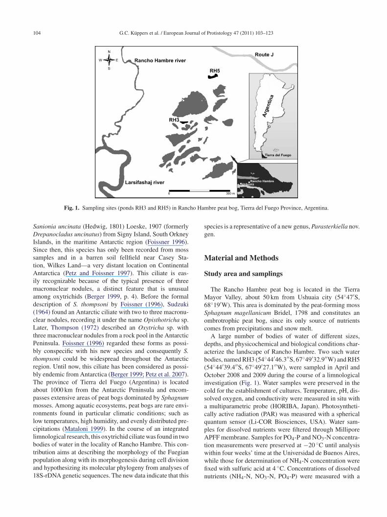
Fig. 1. Sampling sites (ponds RH3 and RH5) in Ranc
anionia uncinata (Hedwig, 1801) Loeske, 1907 (formerlyrepanocladus uncinatus) from Signy Island, South Orkney
slands, in the maritime Antarctic region (Foissner 1996).ince then, this species has only been recorded from mossamples and in a barren soil fellfield near Casey Sta-ion, Wilkes Land—a very distant location on Continentalntarctica (Petz and Foissner 1997). This ciliate is eas-
ly recognizable because of the typical presence of threeacronuclear nodules, a distinct feature that is unusual
mong oxytrichids (Berger 1999, p. 4). Before the formalescription of S. thompsoni by Foissner (1996), Sudzuki1964) found an Antarctic ciliate with two to three macronu-lear nodules, recording it under the name Opisthotricha sp.ater, Thompson (1972) described an Oxytricha sp. with
hree macronuclear nodules from a rock pool in the Antarcticeninsula. Foissner (1996) regarded these forms as possi-ly conspecific with his new species and consequently S.hompsoni could be widespread throughout the Antarcticegion. Until now, this ciliate has been considered as possi-ly endemic from Antarctica (Berger 1999; Petz et al. 2007).he province of Tierra del Fuego (Argentina) is locatedbout 1000 km from the Antarctic Peninsula and encom-asses extensive areas of peat bogs dominated by Sphagnumosses. Among aquatic ecosystems, peat bogs are rare envi-
onments found in particular climatic conditions; such asow temperatures, high humidity, and evenly distributed pre-ipitations (Mataloni 1999). In the course of an integratedimnological research, this oxytrichid ciliate was found in twoodies of water in the locality of Rancho Hambre. This con-
ribution aims at describing the morphology of the Fuegianopulation along with its morphogenesis during cell divisionnd hypothesizing its molecular phylogeny from analyses of8S-rDNA genetic sequences. The new data indicate that thiswwfin
bre peat bog, Tierra del Fuego Province, Argentina.
pecies is a representative of a new genus, Parasterkiella nov.en.
aterial and Methods
tudy area and samplings
The Rancho Hambre peat bog is located in the Tierraayor Valley, about 50 km from Ushuaia city (54◦47′S,
8◦19′W). This area is dominated by the peat-forming mossphagnum magellanicum Bridel, 1798 and constitutes anmbrotrophic peat bog, since its only source of nutrientsomes from precipitations and snow melt.
A large number of bodies of water of different sizes,epths, and physicochemical and biological conditions char-cterize the landscape of Rancho Hambre. Two such waterodies, named RH3 (54◦44′46.3′′S, 67◦49′32.9′′W) and RH554◦44′39.4′′S, 67◦49′27.1′′W), were sampled in April andctober 2008 and 2009 during the course of a limnological
nvestigation (Fig. 1). Water samples were preserved in theold for the establishment of cultures. Temperature, pH, dis-olved oxygen, and conductivity were measured in situ withmultiparametric probe (HORIBA, Japan). Photosyntheti-
ally active radiation (PAR) was measured with a sphericaluantum sensor (Li-COR Biosciences, USA). Water sam-les for dissolved nutrients were filtered through MilliporePFF membrane. Samples for PO4-P and NO3-N concentra-
ion measurements were preserved at −20 ◦C until analysis
ithin four weeks’ time at the Universidad de Buenos Aires,hile those for determination of NH4-N concentration werexed with sulfuric acid at 4 ◦C. Concentrations of dissolvedutrients (NH4-N, NO3-N, PO4-P) were measured with a
urnal of
sp
M
b(Psimafi(ficmowta
fdb(
FdL
2fltcWW
D
cwdTaapma1mNH
N
traatsa
gDsgesa
P
Bpa
iRMcefiack
(Blb(a1
((“B(
f
G.C. Küppers et al. / European Jo
pectrophotometer (Hach Company, USA), with the appro-riate reactants for each analysis.
orphology
In the laboratory, the samples were kept in a culture cham-er, under controlled temperature (4–10 ◦C) and photoperiod12:12 light/dark cycles). Raw cultures were established inetri dishes by enriching samples with wheat kernels totimulate growth of bacteria as a food source for the cil-ates. The ciliates were observed in vivo under the stereo
icroscope and bright-field microscope. In order to visu-lize the infraciliature and the nuclear apparatus, cells werexed in Bouin solution and silver-impregnated with protargolWilbert 1975). Measurements were taken under the bright-eld microscope with a calibrated ocular micrometer. Livingells were free-hand sketched, based on live observations andicrographs. Impregnated cells were illustrated with the aid
f a drawing tube attached to the microscope. Observationsere performed under the stereo microscope at magnifica-
ions of 20× and 40×, and under the bright-field microscopet 40×, 100×, 400×, and 1000×.
Resting cysts of Parasterkiella thompsoni were obtainedrom old cultures, by scratching the bottom of the Petriish with a micropipette. Cysts were observed in vivo byright-field microscopy, stained with methyl-green pyroninFoissner 1991), and impregnated with protargol.
Voucher slides of Parasterkiella thompsoni from Tierra deluego (accession numbers MLP66, MLP67, MLP68) wereeposited in the Colección de Invertebrados of the Museo dea Plata, Argentina, with relevant cells marked.The general terminology used is after Berger (1999, 2006,
008) and Lynn (2008). The terminology concerning bodyexibility is according to Foissner and Stoeck (2008) and for
he oral apparatus after Foissner and Al-Rasheid (2006). Theirri and developing primordia were numbered according toallengren (1900). Resting cysts were described followingalker and Maugel (1980) and Berger (1999).
NA extraction, amplification, and sequencing
Specimens of Parasterkiella thompsoni were picked fromultures from pool RH3 and transferred to embryo dishes,here the isolates were thoroughly washed with mineral andistilled water, then fixed with 70% (v/v) aqueous ethanol.he individuals were then submitted to DNA extractionccording to Sambrook et al. (1989). The 18S-rDNA wasmplified via PCR, through the use of the Hypotricha-specificrimers designed by Paiva et al. (2009). Amplified frag-ents were purified according to Sambrook et al. (1989)
nd sequenced by the dideoxyterminal method (Sanger et al.
977) by means of an ABI 3130 (Applied Biosystems) auto-atic sequencer. The resulting sequence was uploaded to theCBI/GenBank and is available under the accession codeM569264.ta1a
Protistology 47 (2011) 103–123 105
ucleotide matrix assembly and alignment
The nucleotide matrix used in the present study con-ained 29 representatives of supposedly related taxa (notestricted to species with 18 frontal–ventral–transverse cirri)s ingroup, plus nine urostylid sequences as outgroup. Thispproach allows a proper accessing of the monophyly ofhe stylonychines to the inclusion of P. thompsoni in uncon-trained phylogenetic analyses (Kitching et al. 1998; Nixonnd Carpenter 1993).
The nucleotides were aligned through the so-called pro-ressive approach of multiple-sequence alignment (Feng andoolittle 1987) implemented in the Clustal X 1.81 computer
oftware (Thompson et al. 1997), with the adoption of theap-opening and -extension parameters determined in Paivat al. (2009). The resulting matrix was further modified in theoftware BioEdit v7.0.5 (Hall 1999) for manual refinementnd trimming.
hylogenetic analyses
Phylogenetic hypotheses were constructed underayesian, likelihood, and parsimony frameworks, thusermitting the assessment of data sensitivity to differentnalytic criteria.
A Bayesian phylogenetic inference (BI) was performedn the computer software MrBayes 3.1.2 (Huelsenbeck andonquist 2001). The BI was based on two Markov chainonte Carlo (MCMC) simulations that were run with four
hains of 1,000,000 generations, of which trees were sampledach 200 generations (temperature of heat chains = 0.2). Therst 100,000 generations were then discarded as burn-in, andmajority-rule consensus of the remaining trees was used toalculate the Bayesian posterior probabilities of the recoveredinships (Schneider 2007).
Under the likelihood framework, the software PhyML 3.0Guindon and Gascuel 2003) was used to build an initialioNJ (Gascuel 1997) pairwise-distance tree, which had its
ikelihood improved via “subtree pruning and regrafting”ranch-swapping moves to generate a maximum-likelihoodML) tree. To evaluate the stability of the nodes obtained,nonparametric bootstrap analysis was conduced based on
000 replicates.Both BI and ML analyses made use of the GTR + I
=0.6671) + G (=0.5653) nucleotide-substitution modelRodriguez et al. 1990), which was chosen according to theminimum theoretical information criterion” (Akaike 1974;os and Posada 2005), via the software MODELTEST 3.7
Posada and Crandall 1998).Finally, a maximum parsimony (MP) analysis was per-
ormed in the software PAUP* 4b10 (Swofford 2003),
hrough the use of only parsimony-informative charactersnd gaps scored as a “fifth base” (Giribet and Wheeler999; Schneider 2007) along with the successive-weightingpproach (Farris 1969). Initially, the “parsimony ratchet”
1 urnal of
(sttproottpJra
r
ap
R
P
Oo
Fasrv5
06 G.C. Küppers et al. / European Jo
Nixon 1999) strategy implemented in PAUP* via the acces-ory software PaupRat (Sikes and Lewis 2001) was adoptedo speed up the search for fundamental most-parsimoniousrees, based on 250 iterations with 15% of the characterserturbed. The characters were then reweighted after theirescaled consistency index (base weight = 10), and a furtherrdinary heuristic search was conduced with 300 replicatesf random sequence addition. This reweighting and heuris-ic search cycle was successively applied until the length ofhe optimal trees stabilized (Kitching et al. 1998). Node sup-ort in MP was assessed via 1000 jackknife replicates (withAC emulation enabled in PAUP*). The “tree bisection andeconnection” branch swapping algorithm was employed in
ll MP-tree searches.The optimal trees obtained under each framework wereooted a posteriori according to the outgroup position (Nixon
rk
g
igs 2–7. Morphostatic specimen (2–4) and resting cysts (5–7) of Parastnd 5) and protargol impregnation (3, 4, 6, and 7). (2) Ventral view of a repide and nuclear apparatus of the same specimen. Arrowheads mark micidges on the surface of the resting cyst. (7) Fused macronucleus and micacuole; Ma, macronucleus; Mi, micronuclei; 1–4, dorsal kineties 1–4 (k0 �m (3 and 4).
Protistology 47 (2011) 103–123
nd Carpenter 1993), visualized, and edited for aesthetics andublishing in the software MEGA 4 (Tamura et al. 2007).
esults
arasterkiella nov. gen.
Diagnosis. Stylonychinae with undulating membranes inxytricha pattern. With 18 frontal–ventral–transverse cirri,ne left and one right marginal rows of cirri separated poste-
iorly, and four dorsal kineties. Caudal cirri present. Dorsalineties develop without fragmentation.Etymology. Composite of para (Greek, beside) and theeneric name Sterkiella. Feminine gender.
erkiella thompsoni from Tierra del Fuego after live observation (2resentative specimen. (3 and 4) Infraciliature of ventral and dorsal
ronuclei. (5) Cross section of a mature resting cyst. (6) Polygonalronuclei of the kinetosome-resorbing resting cyst. CV, contractileinety 4 is a dorsomarginal row). Scale bars = 30 �m (2 and 5–7),
urnal of
n
a
Pc
(Tomfic
opaV
MdT
1a
T
C
BBBBAAPPAAAMMPPMMMMMAFBFPPTRLCDDDDDDR
M%
G.C. Küppers et al. / European Jo
Type species. Parasterkiella thompsoni (Foissner, 1996)ov. comb. (basionym: Sterkiella thompsoni Foissner, 1996).Species assignable. Presently only the type species is
ssigned to Parasterkiella.
arasterkiella thompsoni (Foissner, 1996) nov.omb.
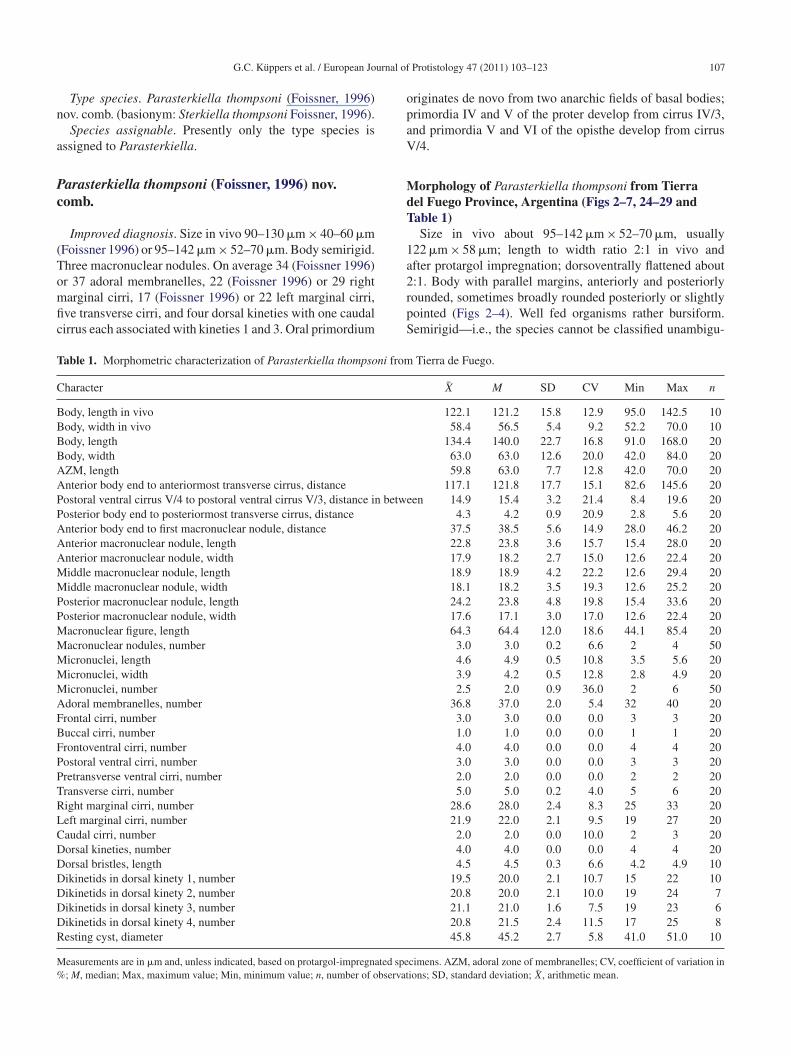
Improved diagnosis. Size in vivo 90–130 �m × 40–60 �mFoissner 1996) or 95–142 �m × 52–70 �m. Body semirigid.hree macronuclear nodules. On average 34 (Foissner 1996)
r 37 adoral membranelles, 22 (Foissner 1996) or 29 rightarginal cirri, 17 (Foissner 1996) or 22 left marginal cirri,ve transverse cirri, and four dorsal kineties with one caudalirrus each associated with kineties 1 and 3. Oral primordium2rpS
able 1. Morphometric characterization of Parasterkiella thompsoni from
haracter
ody, length in vivoody, width in vivoody, lengthody, widthZM, lengthnterior body end to anteriormost transverse cirrus, distanceostoral ventral cirrus V/4 to postoral ventral cirrus V/3, distance in betweosterior body end to posteriormost transverse cirrus, distancenterior body end to first macronuclear nodule, distancenterior macronuclear nodule, lengthnterior macronuclear nodule, widthiddle macronuclear nodule, lengthiddle macronuclear nodule, width
osterior macronuclear nodule, lengthosterior macronuclear nodule, widthacronuclear figure, lengthacronuclear nodules, numbericronuclei, lengthicronuclei, widthicronuclei, numberdoral membranelles, numberrontal cirri, numberuccal cirri, numberrontoventral cirri, numberostoral ventral cirri, numberretransverse ventral cirri, numberransverse cirri, numberight marginal cirri, numbereft marginal cirri, numberaudal cirri, numberorsal kineties, numberorsal bristles, lengthikinetids in dorsal kinety 1, numberikinetids in dorsal kinety 2, numberikinetids in dorsal kinety 3, numberikinetids in dorsal kinety 4, numberesting cyst, diameter
easurements are in �m and, unless indicated, based on protargol-impregnated spe; M, median; Max, maximum value; Min, minimum value; n, number of observat
Protistology 47 (2011) 103–123 107
riginates de novo from two anarchic fields of basal bodies;rimordia IV and V of the proter develop from cirrus IV/3,nd primordia V and VI of the opisthe develop from cirrus/4.
orphology of Parasterkiella thompsoni from Tierrael Fuego Province, Argentina (Figs 2–7, 24–29 andable 1)Size in vivo about 95–142 �m × 52–70 �m, usually
22 �m × 58 �m; length to width ratio 2:1 in vivo andfter protargol impregnation; dorsoventrally flattened about
:1. Body with parallel margins, anteriorly and posteriorlyounded, sometimes broadly rounded posteriorly or slightlyointed (Figs 2–4). Well fed organisms rather bursiform.emirigid—i.e., the species cannot be classified unambigu-Tierra de Fuego.
X M SD CV Min Max n
122.1 121.2 15.8 12.9 95.0 142.5 1058.4 56.5 5.4 9.2 52.2 70.0 10
134.4 140.0 22.7 16.8 91.0 168.0 2063.0 63.0 12.6 20.0 42.0 84.0 2059.8 63.0 7.7 12.8 42.0 70.0 20
117.1 121.8 17.7 15.1 82.6 145.6 20en 14.9 15.4 3.2 21.4 8.4 19.6 20
4.3 4.2 0.9 20.9 2.8 5.6 2037.5 38.5 5.6 14.9 28.0 46.2 2022.8 23.8 3.6 15.7 15.4 28.0 2017.9 18.2 2.7 15.0 12.6 22.4 2018.9 18.9 4.2 22.2 12.6 29.4 2018.1 18.2 3.5 19.3 12.6 25.2 2024.2 23.8 4.8 19.8 15.4 33.6 2017.6 17.1 3.0 17.0 12.6 22.4 2064.3 64.4 12.0 18.6 44.1 85.4 203.0 3.0 0.2 6.6 2 4 504.6 4.9 0.5 10.8 3.5 5.6 203.9 4.2 0.5 12.8 2.8 4.9 202.5 2.0 0.9 36.0 2 6 50
36.8 37.0 2.0 5.4 32 40 203.0 3.0 0.0 0.0 3 3 201.0 1.0 0.0 0.0 1 1 204.0 4.0 0.0 0.0 4 4 203.0 3.0 0.0 0.0 3 3 202.0 2.0 0.0 0.0 2 2 205.0 5.0 0.2 4.0 5 6 20
28.6 28.0 2.4 8.3 25 33 2021.9 22.0 2.1 9.5 19 27 20
2.0 2.0 0.0 10.0 2 3 204.0 4.0 0.0 0.0 4 4 204.5 4.5 0.3 6.6 4.2 4.9 10
19.5 20.0 2.1 10.7 15 22 1020.8 20.0 2.1 10.0 19 24 721.1 21.0 1.6 7.5 19 23 620.8 21.5 2.4 11.5 17 25 845.8 45.2 2.7 5.8 41.0 51.0 10
cimens. AZM, adoral zone of membranelles; CV, coefficient of variation inions; SD, standard deviation; X, arithmetic mean.

108 G.C. Küppers et al. / European Journal of Protistology 47 (2011) 103–123
Figs 8–12. Morphogenesis of ventral side of Parasterkiella thompsoni after protargol impregnation. (8) Oral primordium development fromtwo fields of basal bodies. (9) Enlargement of both basal body fields. Replication bands in macronuclear nodules (arrowhead). (10) Oralprimordium as a unique field of basal bodies. (11) Membranelles differentiation and formation of the primordia I–III of the opisthe andthe primordia IV, V of the proter. (12) Development of the primordia IV, V of the opisthe and the primordia III, VI of the proter. OP,oral primordium; IV/3, IV/2, V/4, ontogenetically active parental cirri; V/3, ontogenetically inactive parental postoral ventral cirrus; I–VI,frontal–ventral–transverse cirri primordia. Scale bars = 50 �m.
Table 2. Origin of the frontal–ventral–transverse cirri primordia during morphogenesis of Parasterkiella thompsoni.
Primordium
I II III IV V VI
Proter UM(I/1) II/2(II/3, II/2, II/1) III/2(III/3, III/2, III/1) IV/3(IV/3, IV/2, IV/1) IV/3(V/4, V/3, V/2, V/1) IV/3(VI/4, VI/3, VI/2, VI/1)Opisthe OP (I/1) OP(II/3, II/2, II/1) OP (III/3, III/2, III/1) IV/2(IV/3, IV/2, IV/1) V/4 (V/4, V/3, V/2, V/1) V/4 (VI/4, VI/3, VI/2, VI/1)
Cirri formed per primordium are in parenthesis. OP, oral primordium; UM, undulating membrane.

urnal of
oOwwAmTm(b(e2mbamdtawcMatDn1lsds(tracl(ecacp
44wwl(lMba
Ma
tcIklrbfbbkr
otorpItTcpiobzfmcsifb
mbmdgo
tdodbtf
G.C. Küppers et al. / European Jo
usly in the rigid (e.g., Stylonychia) or the flexible (e.g.,xytricha) group (see Foissner and Stoeck 2008). Usuallyith three macronuclear nodules (one individual out of 50ith two, and one with four nodules) in central body portion.nterior and posterior macronuclear nodules ellipsoidal;iddle macronuclear nodule almost spherical or ellipsoidal.wo globular micronuclei (one individual out of 50 with sixicronuclei), sometimes faintly impregnated with protargol
Figs 4, 28). Contractile vacuole in midbody near to the leftody margin, with an anterior and a posterior collecting canalFigs 2, 24). Cortical granules absent. Cytoplasm transpar-nt, with several refractive cytoplasmic inclusions, such as.0–3.5 �m long crystals and 5–7 �m-sized lipid globules,ainly along the margins of the cell. Food vacuoles with
acteria, Chlorella-like algae, flagellates, small ciliates, andlso wheat starch in the individuals from cultures. Movesoderately to very fast, crawling on the bottom of the Petri
ish, sometimes remaining still for a while. Somatic ven-ral ciliature composed of 18 frontal–ventral–transverse cirri,rranged in the typical oxytrichid pattern (Figs 3, 28). Rarelyith six transverse cirri. Thick fibres associated with ventral
irri, mainly with the enlarged frontal and transverse cirri.arginal cirri about 12 �m in length in vivo; transverse cirri
bout 17 �m in length in vivo and extending beyond pos-erior end of cell; caudal cirri about 14 �m long in vivo.orsal bristles about 4–5 �m long after protargol impreg-ation, invariably arranged in four kineties. Dorsal kinetyslightly shortened anteriorly, kineties 2 and 3 almost as
ong as body, and kinety 4 posteriorly shortened. Dikinetidsurrounded by oblique fibres, distinctly impregnated aroundikinetids of rows 2 and 3. Usually two delicate and incon-picuous caudal cirri at the rear ends of dorsal kineties 1 and 3Figs 4, 29). One specimen out of 20 with three caudal cirri athe ends of dorsal kineties 1–3. Adoral zone of membranellesepresenting 44.5% of total body length (calculated on theverage values measured on silver-impregnated cells). Buc-al cavity rather large and deep, with a conspicuous hyalineip covering proximal part of adoral zone of membranellesFigs 24, 25). On average 37 adoral membranelles, with lat-ral membranellar cilia extending to the right of the buccalavity; distal membranelles about 18 �m long in vivo. Paroralnd endoral intersect each other optically behind the buc-al cirrus; both membranes composed of dikinetids. Shortharyngeal fibres extend obliquely backwards.
Resting cysts of kinetosome-resorbing type, on average5.8 �m in diameter, golden to light brown colored at0–100× (Figs 5–7, 26, 27 and Table 1). Surface slightlyrinkled, formed by 0.5–1.0 �m high polygonal ridges. Cystall 1.5–2.0 �m thick, with a 0.5–1.0 �m thin outer hya-
ine (mucous?) layer; stains red with methyl-green pyroninwith its ridges intensely stained). Cytoplasm with 3–5 �mipid droplets and 1.0–1.5 �m refractive globular inclusions.
acronucleus organized in a single reniform or rather dumbell-shaped mass. Resting cysts were viable even 14 monthsfter encystment.
ima
Protistology 47 (2011) 103–123 109
orphogenesis during cell division (Figs 8–23, 30–48nd Table 2)Stomatogenesis of the opisthe begins with the prolifera-
ion of basal bodies near the anteriormost postoral ventralirrus IV/2, and near the leftmost transverse cirri II/1 andII/1, resulting in the formation of two anarchic fields ofinetosomes (Figs 8, 9, 30–33). As new basal bodies pro-iferate, these two fields enlarge posteriad and anteriad,espectively, and become a unified anarchic field of basalodies (Figs 10, 34). Primordium I of the opisthe developsrom the oral primordium, and detaches as a streak of basalodies which proliferates anteriad. Some presumptive mem-ranelles develop anteriorly, through the alignment of pairedinetosomes in two rows. The macronuclear nodules showeplication bands at this stage.
Primordia II and III of the opisthe are also formed from theral primordium (Figs 11, 35, 37). The posteriormost fron-oventral cirrus IV/3 disaggregates to form primordia IV-VIf the proter (Figs 11, 12, 36, 38, 40). The frontoventral cir-us III/2 dedifferentiates and generates primordium III of theroter (Figs 12, 39). The buccal cirrus produces primordiumI of the proter, while the paroral dedifferentiates distallyo become primordium I of the proter (Figs 13, 14, 41).he anterior part of the oral primordium of the opistheontinues with the differentiation of membranelles in aosteriad direction. A third row of kinetosomes is addedn the anterior two-rowed membranelles. Primordia I–IIIf the opisthe lengthen and presumptive undulating mem-ranes align longitudinally and parallel to the forming adoralone of membranelles. Primordium IV of the opisthe arisesrom the disaggregated postoral ventral cirrus IV/2 and pri-ordia V and VI from the disaggregated postoral ventral
irrus V/4 (Figs 12–14, 42, 43). Consequently, a total ofix frontal–ventral–transverse cirri primordia are producedn each divider. Cirri II/1, III/1, IV/1, V/1–3, VI/1–4, and therontal cirri are not involved in primordia formation and wille resorbed in later stages of division or in the postdivider.
Two marginal rows primordia develop within each parentalarginal row; the anterior right primordium is generated
y the third or fourth cirrus, while the posterior right pri-ordium is generated from the cirrus behind the presumptive
ivisional furrow. The two left marginal rows primordia areenerated somewhat later than the right ones, and the anteriorne develops from the first left cirrus (Figs 15, 44).
The dorsal kineties primordia develop intrakinetally withinhe dorsal rows 1–3 at the level of the marginal row primor-ia (Fig. 16). In the following stage of division, the endoralf the proter dedifferentiates distally. Whether the endoraledifferentiates completely could not be observed. New cirriegin to segregate within the streaks of both proter and opis-he. The membranelles of the opisthe are almost completelyormed. The undulating membrane primordium of the opisthe
s parallel to the adoral zone of membranelles and the left-ost frontal cirrus is segregated (Figs 15, 44). The parentaldoral zone of membranelles is fully retained in the proter
110 G.C. Küppers et al. / European Journal of Protistology 47 (2011) 103–123
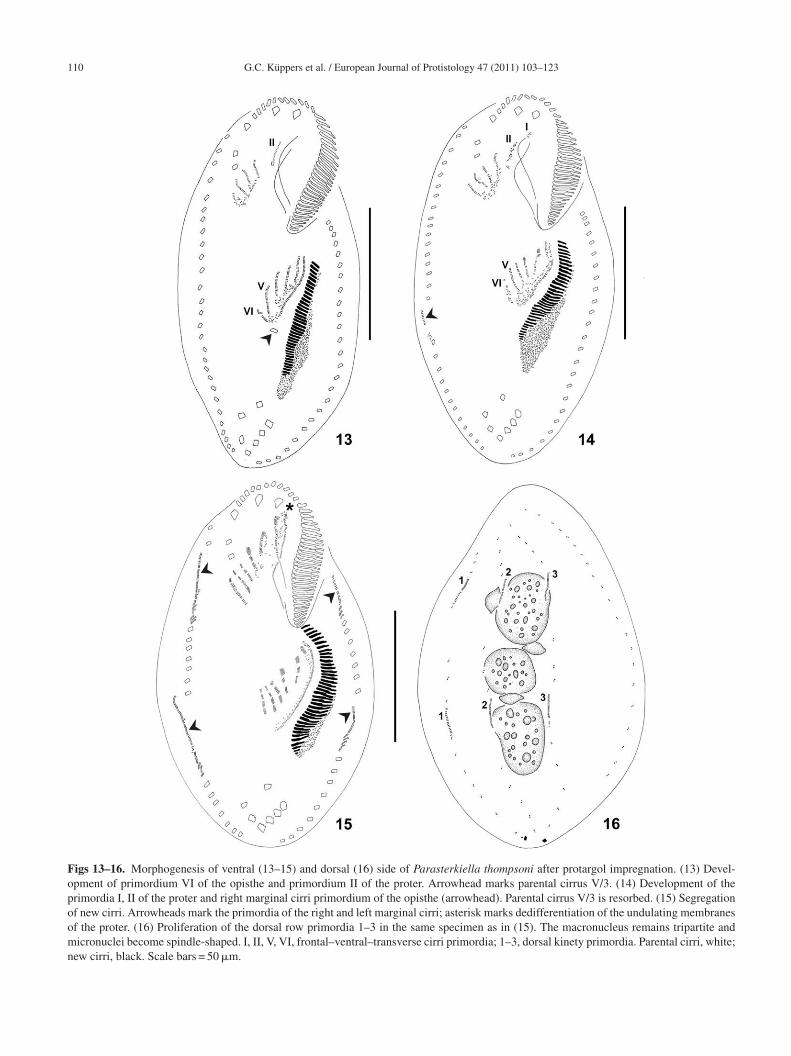
Figs 13–16. Morphogenesis of ventral (13–15) and dorsal (16) side of Parasterkiella thompsoni after protargol impregnation. (13) Devel-opment of primordium VI of the opisthe and primordium II of the proter. Arrowhead marks parental cirrus V/3. (14) Development of theprimordia I, II of the proter and right marginal cirri primordium of the opisthe (arrowhead). Parental cirrus V/3 is resorbed. (15) Segregationof new cirri. Arrowheads mark the primordia of the right and left marginal cirri; asterisk marks dedifferentiation of the undulating membranesof the proter. (16) Proliferation of the dorsal row primordia 1–3 in the same specimen as in (15). The macronucleus remains tripartite andmicronuclei become spindle-shaped. I, II, V, VI, frontal–ventral–transverse cirri primordia; 1–3, dorsal kinety primordia. Parental cirri, white;new cirri, black. Scale bars = 50 �m.

G.C. Küppers et al. / European Journal of Protistology 47 (2011) 103–123 111
Figs 17, 18. Morphogenesis of ventral (17) and dorsal (18) side of Parasterkiella thompsoni after protargol impregnation. (17) Late dividers irri cod specim( white;
tVtdTotoaTbb
c(tdbp
a
etplotccmoaIaonm(
howing the opisthe membranelles and frontal–ventral–transverse corsal row primordia and condensed macronucleus of the samearrowheads). Ma, macronucleus; Mi, micronucleus. Parental cirri,
hough a cryptic reorganization could not be excluded (seeoss and Foissner 1996). Some extra cirri were observed next
o the right anterior and/or posterior marginal row primor-ia, although these cirri could also be parental (Figs 19–21).hese extra cirri seem to be resorbed later since they were notbserved in interphase specimens. In late divisional stages,he streaks differentiate into cirri (Table 2). The adoral zonef the opisthe is completely formed and curves to the rightnteriorly, typically adopting the shape of a question mark.he undulating membranes have already split longitudinally,ut are still parallel to each other (Figs 17, 45). The new cirriegin to migrate to form the characteristic 18-cirri pattern.
Dorsal kineties primordia are lengthened, and one caudalirrus is formed at the end of dorsal kineties 1 and 3Figs 18, 46). One dorsomarginal row differentiates fromhe right marginal row primordium (Figs 17, 44). Parentalikinetids remain among the new developing dorsal kineties,ut these parental kineties are resorbed later or in the
ostdivider.The three macronuclear nodules condense in a single massnd the micronuclei become elliptic. The macronuclear mass
O
l
mpletely formed. Arrowheads mark dorsomarginal rows. (18) Theen as in (17). Caudal cirri are formed only on kineties 1 and 3new cirri, black. Scale bar = 50 �m.
longates and divides into two pieces, one for the proter andhe other for the opisthe. Later, the macronuclear mass of theroter divides again into two nodules, with the anterior beingarger than the posterior; while the macronuclear mass of thepisthe divides into an anterior rounded nodule and a pos-erior enlarged nodule. Both enlarged macronuclear nodulesonstrict again, resulting in the typical tripartite macronu-leus for each divider. Meanwhile, the micronuclei divideitotically (Figs 18–21, 45–48). The undulating membranes
f the proter reorganize and the membranes of both the proternd the opisthe intersect each other optically (Figs 21, 48).n the postdivider, the cirri have already achieved theirppropriate positions but some parental marginal (proter)r transverse cirri (opisthe) are still not resorbed. Theuclear apparatus already consists of the typical threeacronuclear nodules and the two or three micronuclei
Figs 22, 23).
ccurrence and ecologyParasterkiella thompsoni was found in two small, shal-
ow water pools (about 500 m2, 0.33 m deep) from a Fuegian
112 G.C. Küppers et al. / European Journal of Protistology 47 (2011) 103–123
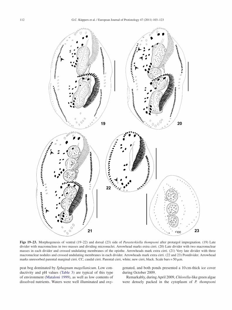
Figs 19–23. Morphogenesis of ventral (19–22) and dorsal (23) side of Parasterkiella thompsoni after protargol impregnation. (19) Latedivider with macronucleus in two masses and dividing micronuclei. Arrowhead marks extra cirri. (20) Late divider with two macronuclearm opisthm ivider.m cirri, w
pdod
g
asses in each divider and crossed undulating membranes of theacronuclear nodules and crossed undulating membranes in each darks unresorbed parental marginal cirri. CC, caudal cirri. Parental
eat bog dominated by Sphagnum magellanicum. Low con-
uctivity and pH values (Table 3) are typical of this typef environment (Mataloni 1999), as well as low contents ofissolved nutrients. Waters were well illuminated and oxy-d
w
e. Arrowheads mark extra cirri. (21) Very late divider with threeArrowheads mark extra cirri. (22 and 23) Postdivider. Arrowheadhite; new cirri, black. Scale bars = 50 �m.
enated, and both ponds presented a 10 cm-thick ice cover
uring October 2009.Remarkably, during April 2009, Chlorella-like green algaeere densely packed in the cytoplasm of P. thompsoni

G.C. Küppers et al. / European Journal of Protistology 47 (2011) 103–123 113
Figs 24–27. Interphase morphology (24 and 25) and resting cyst (26 and 27) of Parasterkiella thompsoni in vivo (24–26) and after protargolimpregnation (27). (24) Ventral view of a specimen with food. (25) Ventral view of a slender specimen. Arrowhead marks the conspicuousbuccal lip. (26) Optical cross section of a mature resting cyst. Arrowhead marks a ridge on the cyst wall. (27) View of polygonal ridges onthe surface of the resting cyst. CV, contractile vacuole; Ma, macronucleus. Scale bars = 30 �m.
Table 3. Physicochemical variables in sampling sites RH3 and RH5, where Parasterkiella thompsoni was found.
Environmental variable RH3 RH5
Temperature (◦C) 7.1 (3.2–11.4) 5.4 (1.7–7.5)pH 4.5 (4.4–4.7) 4.5 (4.4–4.8)Conductivity (�S cm−1) 14 (10–17) 11 (5.5–18)Dissolved oxygen (mg L−1) 8.8 (8.7–8.9) 8.7 (8.7–8.8)PAR (� photons m−2 s−1) 707.4 (410.3–1,184) 635.5 (244.8–1026)DIN (�g L−1) 30.4 (10–51) 42.9 (31.3–54.4)PO4-P (�g L−1) 80 (30–130) 30 (20–40)Total N (�g L−1) 6800 (5500–9400) 6033 (4900–8300)Total P (�g L−1) 143 (90–170) 307 (80–420)
T n parena sphoru
ocaoDkb
T(
he arithmetic mean is followed by minimum and maximum observations ictive radiation; PO4-P, phosphate; Total N, total nitrogen; Total P, total pho
btained from the pool RH5. Protargol impregnation of theseells showed no difference from the specimens lacking thelgae (i.e., cells from RH5 obtained during other samplingccasions and from RH3). Unfortunately, we could not obtain
NA from the specimens with algae, thus we assume bothinds of specimens to be conspecific until additional dataecome available.tAa
theses (n = 3). DIN, dissolved inorganic nitrogen; PAR, photosyntheticallys.
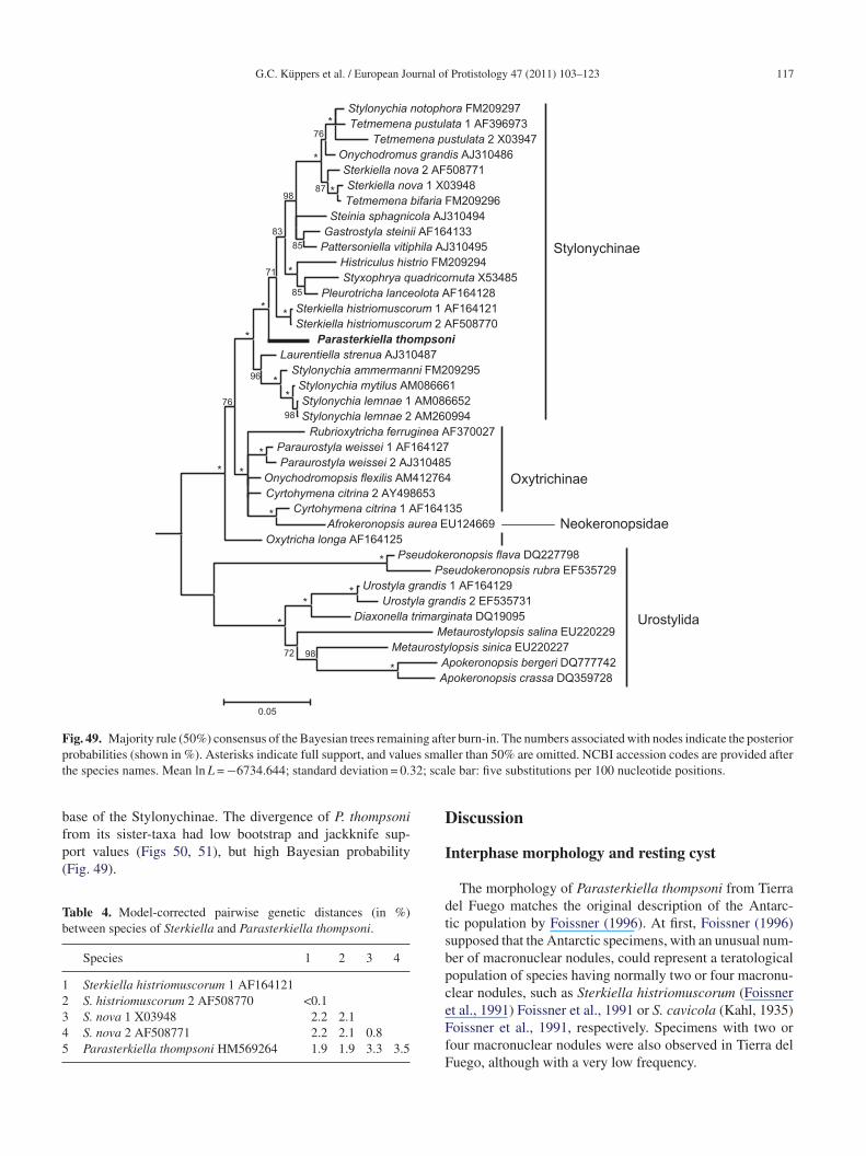
he 18S-rDNA data and phylogenetic analysesFigs 49–51 and Table 4)
The obtained 18S-rDNA fragment of Parasterkiella
hompsoni had 1621 nucleotides and a CG content of 44.81%.fter aligning and trimming, the nucleotide matrix yieldedtotal of 1671 characters, of which 1332 were constant and
114 G.C. Küppers et al. / European Journal of Protistology 47 (2011) 103–123
Figs 28–35. Interphase morphology (28 and 29) and morphogenesis (30–35) of Parasterkiella thompsoni after protargol impregnation. (28)Ventral infraciliature. Arrowhead marks micronucleus. (29) Dorsal kineties. Arrowhead marks an extra dikinetid, possibly a still unresorbedparental dikinetid. (30 and 31) Very early divider. Arrowhead marks the beginning of stomatogenesis, with proliferation of basal bodies nearthe postoral ventral cirri (30); detail of the proliferation of basal bodies near postoral ventral cirri (31, arrowhead). (32) Very early dividershowing proliferation of a second field of basal bodies near the transverse cirri. Arrowheads mark both fields of basal bodies. (33) Detail ofenlarged fields of basal bodies. Arrowhead marks replication band in macronucleus. (34) Detail of unique field of basal bodies of an earlydivider. (35) Early to middle divider showing the oral primordium. Arrowhead marks disaggregated cirrus IV/2. PO, postoral ventral cirri.Scale bars = 20 �m (31 and 34), 30 �m (28–30, 32, 33, and 35).

G.C. Küppers et al. / European Journal of Protistology 47 (2011) 103–123 115
Figs 36–42. Middle dividers of Parasterkiella thompsoni after protargol impregnation. (36) Disaggregating cirrus IV/3 and primordium IVof the proter (arrowhead). (37) Oral primordium and primordia I–III of the opisthe. (38) Primordia IV, V of the proter (arrowhead). (39)P (41) PrI I, fron3
2os
cnwttom
AnRPtEpa
rimordium III of the proter. (40) Primordia IV–VI of the proter.–VI of the opisthe; postoral ventral cirrus V/3 still unresorbed. I–V9–42), 30 �m (37 and 38).
22 were parsimony-informative. The 18S-rDNA sequencef P. thompsoni differs by up to 3.5% from the analogousequences of Sterkiella species (Table 4).
The phylogenetic patterns obtained from BI, MP, and MLonsistently hypothesized a monophyly of the Stylonychi-ae (Figs 49–51). The internal kinships of the ingroupere slightly variable as a result of different analytic cri-
eria. The BI and ML results yielded almost identical
opologies, differing because of a consensus compromisef the relationships within the group formed by Cyrtohy-ena citrina (Berger and Foissner, 1987) Foissner, 1989,bls
imordia II–VI of the proter. (42) Oral primordium and primordiatal–ventral–transverse cirri primordia. Scale bars = 20 �m (36 and
frokeronopsis aurea (Foissner and Stoeck, 2008) Foiss-er et al., 2010, Onychodromopsis flexilis Stokes, 1887,ubrioxytricha ferruginea (Stein, 1859) Berger, 1999, andaraurostyla weissei (Stein, 1859) Borror, 1972 in the BI
ree. The genera Sterkiella Foissner et al., 1991, Stylonychiahrenberg, 1830, and Tetmemena Eigner, 1999 are all poly-hyletic. Parasterkiella thompsoni was consistently placedmong the stylonychines. In BI and ML trees, P. thompsoni
ranched after the Laurentiella + Stylonychia ammermanni-emnae-mytilus group; while in the MP trees both groupswitched their positions, so that P. thompsoni split at the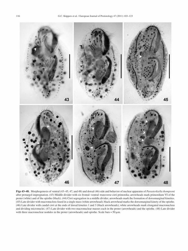
116 G.C. Küppers et al. / European Journal of Protistology 47 (2011) 103–123
Figs 43–48. Morphogenesis of ventral (43–45, 47, and 48) and dorsal (46) side and behavior of nuclear apparatus of Parasterkiella thompsoniafter protargol impregnation. (43) Middle divider with six frontal–ventral–transverse cirri primordia; arrowheads mark primordium VI of theproter (white) and of the opisthe (black). (44) Cirri segregation in a middle divider; arrowheads mark the formation of dorsomarginal kineties.(45) Late divider with macronucleus fused in a single mass (white arrowhead); black arrowhead marks the dorsomarginal kinety of the opisthe.(46) Late divider with caudal cirri at the ends of dorsal kineties 1 and 3 (black arrowheads); white arrowheads mark elongated macronucleusand dividing micronuclei. (47) Late divider with two macronuclear masses each in the proter (arrowheads) and the opisthe. (48) Late dividerwith three macronuclear nodules in the proter (arrowheads) and opisthe. Scale bars = 50 �m.
G.C. Küppers et al. / European Journal of Protistology 47 (2011) 103–123 117
Stylonychia notophora FM209297Tetmemena pustulata 1 AF396973
Tetmemena pustulata 2 X03947Onychodromus grandis AJ310486Sterkiella nova 2 AF508771Sterkiella nova 1 X03948Tetmemena bifaria FM209296
Steinia sphagnicola AJ310494Gastrostyla steinii AF164133
Pattersoniella vitiphila AJ310495Histriculus histrio FM209294Styxophrya quadricornuta X53485
Pleurotricha lanceolota AF164128Sterkiella histriomuscorum 1 AF164121Sterkiella histriomuscorum 2 AF508770
Parasterkiella thompsoniLaurentiella strenua AJ310487
Stylonychia ammermanni FM209295Stylonychia mytilus AM086661Stylonychia lemnae 1 AM086652Stylonychia lemnae 2 AM260994
Rubrioxytricha ferruginea AF370027Paraurostyla weissei 1 AF164127Paraurostyla weissei 2 AJ310485
Onychodromopsis flexilis AM412764Cyrtohymena citrina 2 AY498653
Cyrtohymena citrina 1 AF164135Afrokeronopsis aurea EU124669
Oxytricha longa AF164125Pseudokeronopsis flava DQ227798
Pseudokeronopsis rubra EF535729Urostyla grandis 1 AF164129
Urostyla grandis 2 EF535731Diaxonella trimarginata DQ19095
Metaurostylopsis salina EU220229Metaurostylopsis sinica EU220227
Apokeronopsis bergeri DQ777742Apokeronopsis crassa DQ359728
0.05
85
98
76
*
*
*87
83
85
*71
**
*
**
98
76
*
*
*
*
*
*
**
*9872
96
Stylonychinae
Urostylida
Oxytrichinae
Neokeronopsidae
Fig. 49. Majority rule (50%) consensus of the Bayesian trees remaining after burn-in. The numbers associated with nodes indicate the posteriorp es smalt 2; sca
bfp(
Tb
12345
D
robabilities (shown in %). Asterisks indicate full support, and valuhe species names. Mean ln L = −6734.644; standard deviation = 0.3
ase of the Stylonychinae. The divergence of P. thompsoni
rom its sister-taxa had low bootstrap and jackknife sup-ort values (Figs 50, 51), but high Bayesian probabilityFig. 49).able 4. Model-corrected pairwise genetic distances (in %)etween species of Sterkiella and Parasterkiella thompsoni.
Species 1 2 3 4
Sterkiella histriomuscorum 1 AF164121S. histriomuscorum 2 AF508770 <0.1S. nova 1 X03948 2.2 2.1S. nova 2 AF508771 2.2 2.1 0.8Parasterkiella thompsoni HM569264 1.9 1.9 3.3 3.5
I
dtsbpceFfF
ler than 50% are omitted. NCBI accession codes are provided afterle bar: five substitutions per 100 nucleotide positions.
iscussion
nterphase morphology and resting cyst
The morphology of Parasterkiella thompsoni from Tierrael Fuego matches the original description of the Antarc-ic population by Foissner (1996). At first, Foissner (1996)upposed that the Antarctic specimens, with an unusual num-er of macronuclear nodules, could represent a teratologicalopulation of species having normally two or four macronu-lear nodules, such as Sterkiella histriomuscorum (Foissnert al., 1991) Foissner et al., 1991 or S. cavicola (Kahl, 1935)
oissner et al., 1991, respectively. Specimens with two orour macronuclear nodules were also observed in Tierra deluego, although with a very low frequency.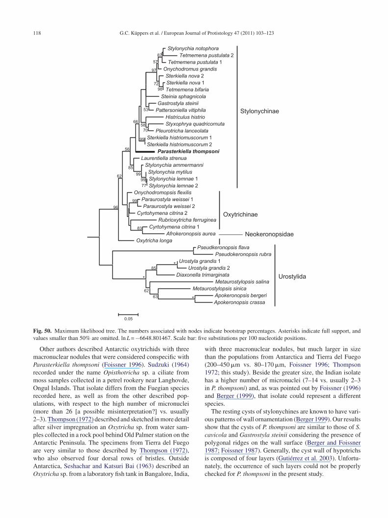
118 G.C. Küppers et al. / European Journal of Protistology 47 (2011) 103–123
Stylonychia notophoraTetmemena pustulata 2
Tetmemena pustulata 1Onychodromus grandisSterkiella nova 2Sterkiella nova 1Tetmemena bifaria
Steinia sphagnicolaGastrostyla steinii
Pattersoniella vitiphilaHistriculus histrioStyxophrya quadricornuta
Pleurotricha lanceolataSterkiella histriomuscorum 1Sterkiella histriomuscorum 2
Parasterkiella thompsoniLaurentiella strenua
Stylonychia ammermanniStylonychia mytilusStylonychia lemnae 1Stylonychia lemnae 2
Onychodromopsis flexilisParaurostyla weissei 1Paraurostyla weissei 2
Cyrtohymena citrina 2Rubrioxytricha ferruginea
Cyrtohymena citrina 1Afrokeronopsis aurea
Oxytricha longaPseudkeronopsis flava
Pseudokeronopsis rubraUrostyla grandis 1
Urostyla grandis 2Diaxonella trimarginata
Metaurostylopsis salinaMetaurostylopsis sinica
Apokeronopsis bergeriApokeronopsis crassa
85
63
62
99
83
7799
99
85
99
7058
53
9872
87
57
97
66
56
62
96
0.05
*
*
*
Stylonychinae
Oxytrichinae
Urostylida
Neokeronopsidae
F odes iv ar: five
mPrmOru(2apAawAO
wt(1hias
oscp1
ig. 50. Maximum likelihood tree. The numbers associated with nalues smaller than 50% are omitted. ln L = −6648.801467. Scale b
Other authors described Antarctic oxytrichids with threeacronuclear nodules that were considered conspecific witharasterkiella thompsoni (Foissner 1996). Sudzuki (1964)ecorded under the name Opisthotricha sp. a ciliate fromoss samples collected in a petrel rookery near Langhovde,ngul Islands. That isolate differs from the Fuegian species
ecorded here, as well as from the other described pop-lations, with respect to the high number of micronucleimore than 26 [a possible misinterpretation?] vs. usually–3). Thompson (1972) described and sketched in more detailfter silver impregnation an Oxytricha sp. from water sam-les collected in a rock pool behind Old Palmer station on thentarctic Peninsula. The specimens from Tierra del Fuego
re very similar to those described by Thompson (1972),
ho also observed four dorsal rows of bristles. Outsidentarctica, Seshachar and Katsuri Bai (1963) described anxytricha sp. from a laboratory fish tank in Bangalore, India,inc
ndicate bootstrap percentages. Asterisks indicate full support, andsubstitutions per 100 nucleotide positions.
ith three macronuclear nodules, but much larger in sizehan the populations from Antarctica and Tierra del Fuego200–450 �m vs. 80–170 �m, Foissner 1996; Thompson972; this study). Beside the greater size, the Indian isolateas a higher number of micronuclei (7–14 vs. usually 2–3n P. thompsoni) and, as was pointed out by Foissner (1996)nd Berger (1999), that isolate could represent a differentpecies.
The resting cysts of stylonychines are known to have vari-us patterns of wall ornamentation (Berger 1999). Our resultshow that the cysts of P. thompsoni are similar to those of S.avicola and Gastrostyla steinii considering the presence ofolygonal ridges on the wall surface (Berger and Foissner987; Foissner 1987). Generally, the cyst wall of hypotrichs
s composed of four layers (Gutiérrez et al. 2003). Unfortu-ately, the occurrence of such layers could not be properlyhecked for P. thompsoni in the present study.
G.C. Küppers et al. / European Journal of Protistology 47 (2011) 103–123 119
Tetmemena pustulata 1Tetmemena pustulata 2Stylonychia notophoraOnychodromus grandisSterkiella nova 1Tetmemena bifariaSterkiella nova 2Steinia sphagnicolaGastrostyla steiniiPattersoniella vitiphilaHistriculus histrioStyxophrya quadricornutaPleurotricha lanceolotaSterkiella histriomuscorum 1Sterkiella histriomuscorum 2Laurentiella strenuaStylonychia ammermanniStylonychia mytilusStylonychia lemnae 1Stylonychia lemnae 2Parasterkiella thompsoniAfrokeronopsis aureaOnychodromopsis flexilisRubrioxytricha ferrugineaCyrtohymena citrina 1Cyrtohymena citrina 2Paraurostyla weissei 1Paraurostyla weissei 2Oxytricha longaPseudokeronopsis flavaPseudokeronopsis rubraApokeronopsis bergeriApokeronopsis crassaMetaurostylopsis sinicaMetaurostylopsis salinaDiaxonella trimarginataUrostyla grandis 1Urostyla grandis 2
*
64
99 *
85
*
*
90
95
53
61
57
67
66
85
*
96
91
94
74
72
84
79
69
9992
63
73
Stylonychinae
Oxytrichinae
Urostylida
Neokeronopsidae
F umberi ed. Tri
D
e(2Tt(dimo1
g
oloIIdoahcS1b
ig. 51. Strict consensus of four maximum parsimony trees. The nndicate full support, and values smaller than 50% are not displayndex = 0.8631; rescaled consistency index = 0.6077.
ivisional morphogenesis
The morphogenesis of P. thompsoni shows some differ-nces compared to the morphogenesis of Sterkiella speciesBerger et al. 1985; Foissner and Berger 1999; Foissner et al.002; Fryd-Versavel et al. 2010; Petz and Foissner 1997).he most noticeable difference lies in the development of
he dorsal ciliature, which occurs in the Urosomoida patternsee Berger 1999). In P. thompsoni the presence of only fourorsal rows of bristles results from the lack of fragmentationn dorsal kinety 3 and the development of only one dorso-
arginal kinety from the right marginal row primordium, asccurs in the Oxytrichinae genera Urosoma Kowalewskiego,
882 and Urosomoida Hemberger in Foissner, 1982.Further differences were observed with respect to the ori-in of the oral primordium of the opisthe. The development
e2f
s associated to branches indicate jackknife percentages. Asterisksee length (in steps) = 2514; consistency index = 0.7041; retention
f this primordium in P. thompsoni begins with the pro-iferation of two separated anarchic fields of basal bodies,ne generated near the anteriormost postoral ventral cirrusV/2 and the other near the leftmost transverse cirri II/1 andII/1. In S. cavicola, a small patch of basal bodies, in fact,evelops close to cirrus IV/2 and probably contributes to theral primordium. This contribution, however, would occurfter the basal bodies originating near the transverse cirriave proliferated and reached the level of the postoral ventralirri (Foissner et al. 2002). Such pattern differs from that ofterkiella histriomuscorum and S. nova Foissner and Berger,999, where the oral primordium develops solely from basalodies that originate near the leftmost transverse cirri (Berger
t al. 1985; Foissner and Berger 1999; Fryd-Versavel et al.010; Petz and Foissner 1997). Indeed, the pattern of earlyormation of the oral primordium observed in P. thompsoni
1 urnal of
rvagN(H
sBpuc
F
w1iobaostFcp
stmt1celofic1a
atsb1cnp
mU
sOSSehtaoF
P
Siwecefttscidr1bits2
Mtmv2aemtohbewrl
20 G.C. Küppers et al. / European Jo
arely occurs among the Stylonychinae – e.g., Pattersoniellaitiphila Foissner, 1987 – and is apparently more commonmong the flexible-body oxytrichids—e.g., Oxytricha longi-ranulosa Berger and Foissner, 1989, Rubrioxytricha indicaaqvi et al., 2006, Tachysoma terricola Hemberger, 1985
Berger 1999; Berger and Foissner 1989; Foissner 1987;emberger 1985; Naqvi et al. 2006).With respect to macronucleus development in Oxytricha
p. with three macronuclear nodules (Seshachar and Katsuriai 1963), the typical number of nodules is achieved in theostdivider about 24 h after fission, whereas in P. thompsonisually three nodules are generated in each divider beforeytokinesis.
oundation for the creation of the new genus
The genus Sterkiella was created to include those speciesith caudal cirri previously assigned to Histriculus Corliss,960 (Foissner et al. 1991). At the present time, this genuss defined by a combination of characters—e.g., the rigidr semirigid body, curved and intersecting undulating mem-ranes, separated marginal rows of cirri at the posterior end,nd the presence of caudal cirri. All these features, however,ccur in other genera and thus are not unique. The supposedole autapomorphy of the genus was the de novo formation ofhe primordia V and VI of the opisthe (Berger 1999). Later,oissner et al. (2002) recognized this origin as a mistake andonfirmed that both primordia V and VI develop from theostoral ventral cirrus V/4.
Foissner (1996) and Berger (1999) considered the clas-ification of the Antarctic ciliate in the genus Sterkiella asentative, since this ciliate possesses characteristics com-
on to several oxytrichids. The dorsal ciliature resembleshe Oxytrichinae genera Urosoma and Urosomoida (Berger999) and, as confirmed in the present study, the dorsaliliature actually develops in the Urosomoida pattern. Nev-rtheless, with respect to features like the semirigid body, theack of cortical granules, the adoral zone of membranellesccupying about 50% of body length, the presence of obliquebres around dorsal cilia, and the morphogenetic inactivity ofirrus V/3, this species belongs to the Stylonychinae (Berger999). This classification is confirmed by our phylogeneticnalyses (see below).
In Sterkiella the rightmost dorsal primordium fragmentsnd forms two dorsal kineties (Foissner et al. 2002). Hence,he lack of fragmentation in the dorsal primordia of P. thomp-oni clearly separates this genus not only from Sterkiella,ut also from all other 18-cirri stylonychine genera (Berger999). This difference in the morphogenesis of the dorsaliliature is herein regarded as justifiable for the creation of aew genus, namely Parasterkiella, for S. thompsoni. For the
resent, though, P. thompsoni remains monotypic.Among the non-stylonychine 18-cirri genera in whichorphogenetic data also confirmed the occurrence of therosomoida pattern, Parasterkiella is comparable to Uro-
acat
Protistology 47 (2011) 103–123
oma, Urosomoida, and some species currently assigned toxytricha Bory De Saint-Vincent in Lamouroux, Bory Deaint-Vincent and Deslongchamps (e.g., O. longa Gelei andzabados, 1950 and O. setigera Stokes, 1891). These gen-ra all differ in a fundamental way from Parasterkiella byaving flexible bodies, by exhibiting participation of the pos-oral ventral cirrus V/3 in primordia formation, and by havingn adoral zone of membranelles that occupies less than 50%f the body length (Berger 1999; Berger and Foissner 1988;oissner 1983; Ganner et al. 1987).
hylogenetic position
The pairwise genetic distances between populations ofterkiella histriomuscorum and of S. nova were smaller thannterspecific distances, including P. thompsoni. Nevertheless,e found, in accordance with others (Foissner 2007; Foissner
t al. 2003; Schmidt et al. 2007), that such comparisonsould be misleading as an objective criterion for species orven genus discrimination among stylonychines when per-ormed independently of a morphological study. Althoughhe 18S-rDNA of P. thompsoni differs by up to 3.5% fromhe analogous sequence from Sterkiella spp., the P. thompsoniequence does so less when compared with the 18S-rDNAs ofertain stylonychines that are morphologically more dissim-lar. For instance, the 18S-rDNA sequence of P. thompsoniiffered by 2.7% from those of Gastrostyla steinii and Lau-entiella strenua (Dingfelder, 1962) Berger and Foissner,989 – which have conspicuous ventral row[s] of cirri formedy the ventral primordia – and by 2.6% from the correspond-ng nucleotide sequences of Pattersoniella vitiphila; in whichhe ventral cirri form a midventral complex and multiple dor-al kinety fragmentation occurs (Berger 1999; Foissner et al.002; Martin et al. 1983).
The phylogenetic patterns obtained from BI, MP, andL did not reject the ingroup monophyly with respect to
he urostylid outgroup and consistently hypothesized theonophyly of the Stylonychinae, thus corroborating pre-
ious analyses along the same lines (e.g., Bernhard et al.001; Foissner et al., 2004; Hewitt et al. 2003; Paiva etl., 2009; Schmidt et al. 2007, 2008; Shao et al. 2007; Yit al. 2008, 2009). The MP trees were generally in agree-ent with those obtained by Paiva et al. (2009) concerning
he Stylonychinae kinships and the pectinate distributionf its clades. Paraurostyla weissei and Oxytricha longa,owever, occupied slightly different positions, branchingefore the cyrtohymenids. The morphologically similar gen-ra Sterkiella, Stylonychia, and Tetmemena (Berger 1999)ere all polyphyletic within the present analyses, thus cor-
oborating the results from Schmidt et al. (2008). We foundittle inconsistency regarding the placement of P. thompsoni
mong stylonychines. In the BI and ML trees, P. thompsonionsistently branched after the Laurentiella + Stylonychiammermanni-lemnae-mytilus group, while in the MP trees P.hompsoni split at the base of the Stylonychinae. Although the
urnal of
dswost2e
tadgsPmTPsPshB
A
PDdTaamdA8
R
A
B
B
B
B
B
B
B
B
B
C
F
F
F
F
F
F
F
F
F
F
F
F
G.C. Küppers et al. / European Jo
ivergence of P. thompsoni from its sister-taxa had low boot-trap and jackknife support values, the Bayesian probabilityas high. Worthy of note is that the Bayesian probabilities areften inflated and thus their reliability as a measure of supporthould be interpreted with extreme caution, especially whenheir conclusions contradict other supports (Cummings et al.003; Randle and Prickett 2010; Schneider 2007; Simmonst al. 2004).
Berger (2006, 2008) established the Dorsomarginalia forhe Hypotricha Stein, 1859 with dorsomarginal kinetiess supposed morphological synapomorphy. The Oxytrichi-ae (including the Stylonychinae) belong to the formerroup, with the fragmentation of dorsal kinety 3 as a robustynapomorphy (Berger 1999, 2006, 2008). Accordingly,arasterkiella would represent a non-oxytrichid Dorso-arginalia, since fragmentation of dorsal kinety 3 is lacking.he phylogeny of the 18S-rDNA marker, however, places. thompsoni as either a basal (MP) or internal (BI, ML)tylonychine branch. In conclusion, if the classification of. thompsoni in the stylonychines is correct, then the Uro-omoida pattern likely evolved convergently within otherypotrich lineages, as has been previously conjectured byerger (1999).
cknowledgements
We are grateful to our research colleagues in the projectICT 2006-01697 for their help with the samplings and tors. Lía Lunaschi and Fabiana Drago for the use of therawing tube in their laboratory at the Museo de La Plata.he editor and the reviewers are also acknowledged for valu-ble suggestions. Dr. Donald Haggerty, a career investigatornd native English speaker, edited the final version of theanuscript. Financial support was from the Consejo Nacional
e Investigaciones Científicas y Técnicas (CONICET) andNPCyT, PICT 2006-01697. This is contribution number99 from the Instituto de Limnología Dr. R.A. Ringuelet.
eferences
kaike, H., 1974. A new look at the statistical model identification.IEEE Trans. Automat. Control 19, 716–723.
erger, H., 1999. Monograph of the Oxytrichidae (Ciliophora,Hypotrichia). Monogr. Biol. 78, 1–1080.
erger, H., 2006. Monograph of the Urostyloidea (Ciliophora,Hypotricha). Monogr. Biol. 85, 1–1304.
erger, H., 2008. Monograph of the Amphisiellidae and Trache-lostylidae (Ciliophora, Hypotricha). Monogr. Biol. 88, 1–737.
erger, H., Foissner, W., 1987. Morphology and biometry of somesoil hypotrichs (Protozoa: Ciliophora). Zool. Jb. Syst. 114,193–239.
erger, H., Foissner, W., 1988. The morphogenesis of Kahliellafranzi (Foissner, 1982) nov. comb. and Oxytricha giganteaHorváth, 1933 (Ciliophora, Hypotrichida). Arch. Protistenk.136, 65–77.
F
Protistology 47 (2011) 103–123 121
erger, H., Foissner, W., 1989. Morphology and biometry of somesoil hypotrichs (Protozoa, Ciliophora) from Europe and Japan.Bull. Br. Mus. Nat. Hist. (Zool.) 55, 19–46.
erger, H., Foissner, W., Adam, H., 1985. Morphological varia-tion and comparative analysis of morphogenesis in Parakahliellamacrostoma (Foissner, 1982) nov. gen. and Histriculus musco-rum (Kahl, 1932), (Ciliophora, Hypotrichida). Protistologica 21,295–311.
ernhard, D., Stechmann, A., Foissner, W., Ammermann, D.,Hehn, M., Schlegel, M., 2001. Phylogenetic relationships withinthe class Spirotrichea (Ciliophora) inferred from small subunitrRNA gene sequences. Mol. Phylogenet. Evol. 21, 86–92.
os, D.H., Posada, D., 2005. Using models of nucleotide evolutionto build phylogenetic trees. Dev. Comp. Immunol. 29, 211–227.
ummings, M.P., Handley, S.A., Myers, D.S., Reed, D.L., Rokas,A., Winka, K., 2003. Comparing bootstrap and posterior proba-bility values in the four-taxon case. Syst. Biol. 52, 477–487.
arris, J.S., 1969. A successive approximations approach to charac-ter weighting. Syst. Zool. 18, 374–385.
eng, D., Doolittle, R.F., 1987. Progressive sequence alignment asa prerequisite to correct phylogenetic trees. J. Mol. Evol. 60,351–360.
oissner, W., 1983. Die Morphogenese von Urosoma macrostyla(Wrzesniowski, 1870) (Ciliophora: Oxytrichidae). Arch. Protis-tenk. 127, 413–428.
oissner, W., 1987. Neue und wenig bekannte hypotriche und colpo-dide Ciliaten (Protozoa: Ciliophora) aus Böden und Moosen.Zool. Beitr. 31, 187–282.
oissner, W., 1991. Basic light and scanning electron microscopicmethod for taxonomic studies of ciliated protozoa. Eur. J. Pro-tistol. 27, 313–330.
oissner, W., 1996. Faunistics, taxonomy and ecology of mossand soil ciliates (Protozoa, Ciliophora) from Antarctica, withdescription of new species, including Pleuroplitoides smithi gen.n., sp. n. Acta Protozool. 35, 95–123.
oissner, W., 2007. Dispersal and biogeography of protists: recentadvances. Jpn. J. Protozool. 40, 1–16.
oissner, W., Agatha, S., Berger, H., 2002. Soil ciliates (Protozoa,Ciliophora) from Namibia (Southwest Africa), with emphasis ontwo contrasting environments, the Etosha region and the Namibdesert. Denisia 5, 1–1459.
oissner, W., Al-Rasheid, K., 2006. A unified organization of the sti-chotrichine oral apparatus, including a description of the buccalseal (Ciliophora: Sprirotrichea). Acta Protozool. 45, 1–16.
oissner, W., Berger, H., 1999. Identification and ontogenesis of thenomen nudum hypotrichs (Protozoa: Ciliophora) Oxytricha nova(=Sterkiella nova sp. n.) and O. trifallax (=S. histriomuscorum).Acta Protozool. 38, 215–248.
oissner, W., Blatterer, H., Berger, H., Kohmann, F., 1991.Taxonomische und ökologische Revision der Ciliaten desSaprobiensystems—Band I: Cyrtophorida, Oligotrichida,Hypotrichia, Colpodea. Informationsberichte des Bayer.Landesamtes für Wasserwirtschaft 1/91, 1–478.
oissner, W., Moon-van der Staay, S.Y., van der Staay, G.W., Hack-stein, J.H.P., Krautgartner, W.-D., Berger, H., 2004. Reconcilingmorphological and 18S rRNA phylogenies in the stichotrichines(Ciliophora, Spirotrichea), including new sequences from some
rare species. Eur. J. Protistol. 40, 265–281.oissner, W., Stoeck, T., 2008. Morphology, ontogenesis and molec-ular phylogeny of Neokeronopsis (Afrokeronopsis) aurea nov.subgen., nov. spec. (Ciliophora: Hypotricha), a new African flag-

1 urnal of
F
F
G
G
G
G
G
H
H
H
H
K
L
M
M
N
N
N
P
P
P
P
R
R
S
S
S
S
S
S
S
S
S
S
S
T
T
22 G.C. Küppers et al. / European Jo
ship ciliate confirms the CEUU hypothesis. Acta Protozool. 47,1–33.
oissner, W., Strüder-Kypke, M., van der Staay, G.W.M., Hackstein,J.H.P., 2003. Endemic ciliates (Protozoa, Ciliophora) from tankbromeliads: a combined morphological, molecular, and ecolog-ical study. Eur. J. Protistol. 39, 365–372.
ryd-Versavel, G., Lemullois, M., Aubusson-Fleury, A., 2010.Maintaining cell polarity through vegetative cell pattern ded-ifferentiation: cytoskeleton and morphogenesis in the hypotrichciliate Sterkiella histriomuscorum. Protist 161, 222–236.
anner, B., Foissner, W., Adam, H., 1987. Morphogenetic andbiometric comparison of four populations of Urosomoida agili-formis (Ciliophora, Hypotrichida). Ann. Sci. Nat. (Zool.) 8,199–207.
ascuel, O., 1997. An improved version of the NJ algorithm basedon a simple model of sequence data. Mol. Biol. Evol. 14,685–695.
iribet, G., Wheeler, W., 1999. On gaps. Mol. Phylogenet. Evol.13, 132–143.
uindon, S., Gascuel, O., 2003. A simple, fast, and accurate algo-rithm to estimate large phylogenies by maximum likelihood.Syst. Biol. 52, 696–704.
utiérrez, J.C., Díaz, S., Ortega, R., Martín-González, A., 2003.Ciliate resting cyst walls: a comparative review. Recent Res.Dev. Microbiol. 7, 361–379.
all, T.A., 1999. BioEdit: a user-friendly biological sequencealignment editor and analysis program for Windows 95/98/NT.Nucleic Acids Symp. Ser. 41, 95–98.
emberger, H., 1985. Neue Gattungen und Arten hypotricher Cili-aten. Arch. Protistenk. 130, 397–417.
ewitt, E.A., Müller, K.M., Cannone, J., Hogan, D.J., Gutell,R., Prescott, D.M., 2003. Phylogenetic relationships among 28spirotrichous ciliates documented by rDNA. Mol. Phylogenet.Evol. 29, 258–267.
uelsenbeck, J.P., Ronquist, F., 2001. MrBayes: Bayesian inferenceof phylogenetic trees. Bioinformatics 17, 754–755.
itching, I.J., Forey, P.L., Humphries, C.J., Williams, D.M., 1998.Cladistics. The Theory and Practice of Parsimony Analysis.Oxford University Press, Oxford.
ynn, D.H., 2008. The Ciliated Protozoa. Characterization, Classifi-cation, and Guide to the Literature, 3rd ed. Springer, Dordrecht,The Netherlands.
artin, J., Concepcion, F., Perez-Silva, J., 1983. Morphogeneticpattern in Laurentiella acuminata (Ciliophora, Hypotrichida): itssignificance for the comprehension of ontogeny and phylogenyof hypotrichous ciliates. J. Protozool. 30, 519–529.
ataloni, G., 1999. Ecological studies on algal communities fromTierra del Fuego peat bogs. Hydrobiologia 391, 157–171.
aqvi, I., Gupta, R., Borgohain, P., Sapra, G.R., 2006. Morphologyand morphogenesis of Rubrioxytricha indica n. sp. (Ciliophora:Hypotrichida). Acta Protozool. 45, 53–64.
ixon, K.C., 1999. The Parsimony Ratchet, a new method for rapidparsimony analysis. Cladistics 15, 407–414.
ixon, K.C., Carpenter, J.M., 1993. On outgroups. Cladistics 9,413–426.
aiva, T.S., Borges, B.N., Harada, M.L., Silva-Neto, I.D., 2009.Comparative phylogenetic study of Stichotrichia (Alveolata: Cil-
iophora: Spirotrichea) based on 18S-rDNA sequences. Genet.Mol. Res. 8, 223–246.etz, W., Foissner, W., 1997. Morphology and infraciliature of somesoil ciliates (Protozoa, Ciliophora) from continental Antarctica,
T
Protistology 47 (2011) 103–123
with notes on the morphogenesis of Sterkiella histriomuscorum.Polar Rec. 33, 307–326.
etz, W., Valbonessi, A., Schiftner, U., Quesada, A., Ellis-Evans,J.C., 2007. Ciliate biogeography in Antarctic and Arctic fresh-water ecosystems: endemism or global distribution of species?FEMS Microbiol. Ecol. 59, 396–408.
osada, D., Crandall, K.A., 1998. Modeltest: testing the model ofDNA substitution. Bioinformatics 14, 817–818.
andle, C.P., Prickett, K.M., 2010. The conflation of ignorance andknowledge in the inference of clade posteriors. Cladistics 26,1–10.
odriguez, F.J.L.O.J., Martin, A., Medina, J.R., 1990. The generalstochastic model of nucleotide substitution. J. Theor. Biol. 124,485–501.
ambrook, J., Fritsch, E.F., Maniatis, T., 1989. Molecular Cloning,A Laboratory Manual, 2nd ed. Cold Spring Harbor LaboratoryPress, Cold Spring Harbor, New York.
anger, F., Nicklen, S., Coulson, A.R., 1977. DNA sequencing withchain-terminating inhibitors. Proc. Natl. Acad. Sci. U.S.A. 74,5463–5467.
chmidt, S.L., Bernhard, D., Schlegel, M., 2008. Die Ver-wandtschaftsbeziehungen innerhalb der Stylonychinae Berger &Foissner, 1997 (Spirotrichea, Stichotrichia, Oxytrichidae) rekon-struiert mit Sequenzen der 18S rDNA. Denisia 23, 403–409.
chmidt, S.L., Bernhard, D., Schlegel, M., Foissner, W., 2007.Phylogeny of the Stichotrichia (Ciliophora; Spirotrichea) recon-structed with nuclear small subunit rRNA gene sequences:discrepancies and accordances with morphological data. J.Eukaryot. Microbiol. 54, 201–209.
chneider, H., 2007. Métodos de análise filogenética. Um guiaprático , 3a ed. Holos Editora, Ribeirão Preto, SP, Brazil.
eshachar, B.R., Katsuri Bai, A.R., 1963. Cytology of a new speciesof Oxytricha. Arch. Protistenkd. 106, 456–464.
hao, C., Song, W., Yi, Z., Gong, J., Li, J., Lin, X., 2007. Mor-phogenesis of the marine spirotrichous ciliate, Trachelostylapediculiformis (Cohn, 1866) Borror, 1972 (Ciliophora, Sti-chotrichia), with consideration of its phylogenetic position. Eur.J. Protistol. 43, 255–264.
ikes, D.S., Lewis, P.O., 2001. Beta Software, Version 1. PAUPRat:PAUP Implementation of the Parsimony Ratchet. Distributed bythe authors, Department of Ecology and Evolutionary Biology,University of Connecticut, Storrs.
immons, M.P., Pickett, K.M., Miya, M., 2004. How meaningfulare bayesian support values? Mol. Biol. Evol. 21, 188–199.
udzuki, M., 1964. On the microfauna of the Antarctic Region. I.Moss-water community at Langhovde. Sci. Rep. Jap. AntarcticRes. Expedition, Series E 19 (1–41), plates I–XI.
wofford, D.L., 2003. PAUP*. Phylogenetic Analysis Using Par-simony (* and Other Methods), Version 4. Sinauer Associates,Sunderland, MA, USA.
amura, K., Dudley, J., Nei, M., Kumar, S., 2007. MEGA4: Molec-ular Evolutionary Genetics Analysis (MEGA) software version4.0. Mol. Biol. Evol. 24, 1596–1599.
hompson Jr., J.C., 1972. Ciliated protozoa of the AntarcticPeninsula. In: Llano, G.A. (Ed.), Antarctic Terrestrial Biology.Antarctic Research Series vol. 20. American Geophysical Union,Washington, pp. 261–288.
hompson, J.D., Gibson, T.J., Plewniak, F., Jeanmougin, F., Hig-gins, D.G., 1997. The CLUSTAL X windows interface: flexiblestrategies for multiple sequence alignment aided by quality anal-ysis tools. Nucleic Acids Res. 25, 4876–4882.

urnal of
V
W
W
W
Y
Y
suggest that Psammomitra (Ciliophora, Urostylida) should rep-
G.C. Küppers et al. / European Jo
oss, H.-J., Foissner, W., 1996. Divisional morphogenesis in Steiniasphagnicola (Ciliophora, Hypotrichida): a comparative light andscanning electron microscopic study. Eur. J. Protistol. 32, 31–46.
alker, G.K., Maugel, T.K., 1980. Encystment and excystment inhypotrich ciliates. 11. Diophrys scutum and remarks on compar-ative features. Protistologica 16, 525–531.
allengren, H., 1900. Studier öfver ciliata infusorier. IV. Acta Univ.Lund. 36, 1–54, Platta I, II.
ilbert, N., 1975. Eine verbesserte Technik der Protargolimpräg-nation für Ciliaten. Mikrokosmos 64, 171–179.
i, Z., Chen, Z., Warren, A., Roberts, D., Al-Rasheid, K.A.S., Miao,M., Gao, S., Shao, S., Song, W., 2008. Molecular phylogeny
Protistology 47 (2011) 103–123 123
of Pseudokeronopsis (Protozoa, Ciliophora, Urostylida), withreconsideration of three closely related species at inter- and intra-specific levels inferred from the small subunit ribosomal RNAgene and the ITS1-5. 8S-ITS2 region sequences. J. Zool. 275,268–275.
i, Z., Song, W., Stoeck, T., Al-Rasheid, K.A.S., Al-Khedhairy,A.A., Gong, J., Ma, H., Chen, Z., 2009. Phylogenetic analyses
resent an urostylid family, based on small subunit rRNA andalpha-tubulin gene sequence information. Zool. J. Linn. Soc.157, 227–236.