Analysis of a cholera toxin B subunit (CTB) and human mucin 1 (MUC1) conjugate protein in a...
18
Analysis of a Cholera Toxin B Subunit (CTB) and Human Mucin 1 (MUC1) Conjugate Protein in a MUC1 Tolerant Mouse Model Julia Pinkhasov a , M. Lucrecia Alvarez a , Latha B. Pathangey b , Teresa L. Tinder b , Hugh S. Mason a , Amanda M. Walmsley a , Sandra J. Gendler b , and Pinku Mukherjee b a Center for Infectious Diseases and Vaccinology (CIDV), The Biodesign Institute at Arizona State University, 1001 South McAllister Avenue, Tempe, AZ 85287-4501, USA b Mayo Clinic College of Medicine, Department of Biochemistry and Molecular Biology, Mayo Clinic, Scottsdale, AZ, USA Abstract Since epithelial mucin 1 (MUC1) is associated with several adenocarcinomas at mucosal sites, it is pertinent to test the efficacy of a mucosally targeted vaccine formulation. The B subunit of the Vibrio cholerae cholera toxin (CTB) has great potential to act as a mucosal carrier for subunit vaccines. In the present study we evaluated whether a MUC1 tandem repeat (TR) peptide chemically linked to CTB would break self-antigen tolerance in the transgenic MUC1 tolerant mouse model (MUC1.Tg) through oral or parenteral immunizations. We report that oral immunization with the CTB-MUC1 conjugate along with mucosal adjuvant, unmethylated CpG oligodeoxynucleotide (ODN) and interleukin-12 (IL-12), did not break self-antigen tolerance in MUC1.Tg mice, but induced a strong humoral response in wild-type C57BL/6 mice. However, self-antigen tolerance in the MUC1.Tg mouse model was broken after parenteral immunizations with different doses of the CTB-MUC1 conjugate protein and with the adjuvant CpG ODN co- delivered with CTB-MUC1. Importantly, mice immunized systemically with CpG ODN alone and with CTB-MUC1 exhibited decreased tumor burden when challenged with a mammary gland tumor cell line that expresses human MUC1. Keywords Epithelial mucin 1; Cholera toxin B subunit; MUC1 transgenic mouse model; tumor immunotherapy; oral immunization Introduction The identification of tumor-associated antigens (TAAs) and their subsequent isolation has revolutionized tumor immunology. TAAs are abnormal or altered self-proteins that are potentially immunogenic. Epithelial mucin 1 (MUC1) is the test antigen of choice in this study because of its prevalence on most solid adenocarcinomas, including breast, lung, pancreas, prostate, stomach, colon and ovary (1–4). MUC1 is a member of a group of heavily glycosylated proteins known as mucins that are localized to the apical surface of healthy epithelial cells. Aberrant glycosylation and re-orientation of MUC1 on the cell Corresponding Author: Previous address: Pinku Mukherjee, Ph.D., Associate Professor, Department of Immunology, Director, Cellular Immunology Laboratory, Mayo Clinic Scottsdale, 13400 E. Shea Blvd., Scottsdale, AZ 85255, Phone: 480-301-5168, Fax: 480-301-7017, [email protected]. Present address: Irwin Belk Endowed Professor of Cancer Research, Department of Biology, University of North Carolina Charlotte, 9201 University City Blvd., Charlotte, NC 28223 NIH Public Access Author Manuscript Cancer Immunol Immunother. Author manuscript; available in PMC 2012 January 12. Published in final edited form as: Cancer Immunol Immunother. 2010 December ; 59(12): 1801–1811. doi:10.1007/s00262-010-0906-1. NIH-PA Author Manuscript NIH-PA Author Manuscript NIH-PA Author Manuscript
-
Upload
independent -
Category
Documents
-
view
1 -
download
0
Transcript of Analysis of a cholera toxin B subunit (CTB) and human mucin 1 (MUC1) conjugate protein in a...

Analysis of a Cholera Toxin B Subunit (CTB) and Human Mucin 1(MUC1) Conjugate Protein in a MUC1 Tolerant Mouse Model
Julia Pinkhasova, M. Lucrecia Alvareza, Latha B. Pathangeyb, Teresa L. Tinderb, Hugh S.Masona, Amanda M. Walmsleya, Sandra J. Gendlerb, and Pinku Mukherjeeb
a Center for Infectious Diseases and Vaccinology (CIDV), The Biodesign Institute at Arizona StateUniversity, 1001 South McAllister Avenue, Tempe, AZ 85287-4501, USAb Mayo Clinic College of Medicine, Department of Biochemistry and Molecular Biology, MayoClinic, Scottsdale, AZ, USA
AbstractSince epithelial mucin 1 (MUC1) is associated with several adenocarcinomas at mucosal sites, it ispertinent to test the efficacy of a mucosally targeted vaccine formulation. The B subunit of theVibrio cholerae cholera toxin (CTB) has great potential to act as a mucosal carrier for subunitvaccines. In the present study we evaluated whether a MUC1 tandem repeat (TR) peptidechemically linked to CTB would break self-antigen tolerance in the transgenic MUC1 tolerantmouse model (MUC1.Tg) through oral or parenteral immunizations. We report that oralimmunization with the CTB-MUC1 conjugate along with mucosal adjuvant, unmethylated CpGoligodeoxynucleotide (ODN) and interleukin-12 (IL-12), did not break self-antigen tolerance inMUC1.Tg mice, but induced a strong humoral response in wild-type C57BL/6 mice. However,self-antigen tolerance in the MUC1.Tg mouse model was broken after parenteral immunizationswith different doses of the CTB-MUC1 conjugate protein and with the adjuvant CpG ODN co-delivered with CTB-MUC1. Importantly, mice immunized systemically with CpG ODN alone andwith CTB-MUC1 exhibited decreased tumor burden when challenged with a mammary glandtumor cell line that expresses human MUC1.
KeywordsEpithelial mucin 1; Cholera toxin B subunit; MUC1 transgenic mouse model; tumorimmunotherapy; oral immunization
IntroductionThe identification of tumor-associated antigens (TAAs) and their subsequent isolation hasrevolutionized tumor immunology. TAAs are abnormal or altered self-proteins that arepotentially immunogenic. Epithelial mucin 1 (MUC1) is the test antigen of choice in thisstudy because of its prevalence on most solid adenocarcinomas, including breast, lung,pancreas, prostate, stomach, colon and ovary (1–4). MUC1 is a member of a group ofheavily glycosylated proteins known as mucins that are localized to the apical surface ofhealthy epithelial cells. Aberrant glycosylation and re-orientation of MUC1 on the cell
Corresponding Author: Previous address: Pinku Mukherjee, Ph.D., Associate Professor, Department of Immunology, Director,Cellular Immunology Laboratory, Mayo Clinic Scottsdale, 13400 E. Shea Blvd., Scottsdale, AZ 85255, Phone: 480-301-5168, Fax:480-301-7017, [email protected] address: Irwin Belk Endowed Professor of Cancer Research, Department of Biology, University of North Carolina Charlotte,9201 University City Blvd., Charlotte, NC 28223
NIH Public AccessAuthor ManuscriptCancer Immunol Immunother. Author manuscript; available in PMC 2012 January 12.
Published in final edited form as:Cancer Immunol Immunother. 2010 December ; 59(12): 1801–1811. doi:10.1007/s00262-010-0906-1.
NIH
-PA Author Manuscript
NIH
-PA Author Manuscript
NIH
-PA Author Manuscript

surface allows access of anti-MUC1 immune effector cells to tumor sites that are mostlyexcluded from normal epithelial tissues. MUC1 has been used in several clinical andpreclinical studies and has shown promising results (5–18). Since MUC1 is associated withseveral adenocarcinomas at mucosal sites, we considered it pertinent to test the efficacy of amucosally targeted vaccine formulation.
One of the most notable benefits of mucosal immunization is the fact that both systemic andmucosal immunity are triggered, which is particularly advantageous in the cases where theinitial site of tumor formation is along the mucosa. Another benefit is the ease ofadministration of an oral vaccine, which may be used in a prophylactic setting for high riskpatients. Animal studies using TAA targeted therapies have been successful in preventingtumor formation or reducing tumor burden when used as prophylactic agents (13, 17, 19).Disappointingly, this has not been recreated in human clinical trials since the antigenvaccines are being used as alternative therapies in advanced disease cases and not in aprophylactic setting that would allow the immune system to reach the robustness needed totarget and kill the tumor cells before they acquired the mechanisms to escape immunerecognition and/or killing. Therefore, targeting tumors at their primary site is pertinent.Many cancers, such as pancreatic, colorectal, cervical, lung and bladder, have primarytumors that originate at mucosal sites, which are first encountered by the mucosal immunesystem.
Unfortunately, most clinically relevant vaccine candidates show weak immunogenicitywhen delivered mucosally and poor transport across biological barriers. In order to bypassthis weakness we conjugated the MUC1 tandem repeat peptide to the B subunit of the Vibriocholerae cholera toxin (CTB), which has great potential to act as a mucosal carrier forsubunit vaccines (20–21). The efficacy of CTB as a mucosal carrier molecule relies on itsstrong affinity for the GM1 ganglioside receptor that is present on most cells in the body,including epithelial cells and leukocytes. Binding to the GM1 receptors on mucosalepithelial cells, specifically microfold cells (M-cells) located above the Peyer’s patch in theintestine, is thought to increase the uptake of the antigen across the mucosa and lead to anenhanced presentation of the antigen to the immune system (22–24). CTB hasimmunomodulating effects by lowering the threshold concentration of the conjugatedantigen required for immune cell activation and by increasing CD40 and CD86 expressionon antigen presenting cells (APCs) (25).
Our goal was to evaluate if a MUC1-specific mucosally targeted vaccine could break self-antigen tolerance and translate to a systemic response that would lead to anti-tumorimmunity. The MUC1.Tg mouse model is an inbred C57BL/6 mouse strain that expresseshuman MUC1 in a tissue specific fashion and is driven by its own promoter (26). Thismodel system allowed us to test our anti-MUC1 vaccine formulation in an appropriateimmunologically tolerant host.
In the present study we evaluated whether MUC1 tandem repeat (TR) peptide chemicallylinked to CTB (CTB-MUC1) would break self-antigen tolerance in MUC1.Tg mice throughoral or parenteral immunizations. Oral immunizations included strong mucosal adjuvants,specifically unmethylated CpG oligodeoxynucleotides (ODNs) and recombinant mouseinterleukin 12 (IL-12). We report that oral immunization with the CTB-MUC1 conjugatealong with the adjuvants did not break self-antigen tolerance in MUC1.Tg mice. However,self-antigen tolerance was broken after parenteral immunizations with different dosages (10and 30 g) of CTB-MUC1. Importantly, mice immunized with the higher dose (30 g) ofCTB-MUC1 showed decreased tumor burden after challenge with a mammary gland tumorcell line that expresses human MUC1 (C57mg.MUC1). The addition of the immunological
Pinkhasov et al. Page 2
Cancer Immunol Immunother. Author manuscript; available in PMC 2012 January 12.
NIH
-PA Author Manuscript
NIH
-PA Author Manuscript
NIH
-PA Author Manuscript
adjuvant CpG ODN to 10 g CTB-MUC1 augmented vaccine efficacy as demonstrated byantibody production and protection from tumor challenge.
Materials and MethodsSynthesis and conjugation of CTB-MUC1
The MUC1 tandem repeat peptide with a cysteine attached to the N terminus of the peptidewas synthesized at Arizona State University Department of Biochemistry (Tempe, AZ). Thesynthetic MUC1 peptide (C-STAPPAHGVTSAPDTRPAPGSTAPPA) was then conjugatedto CTB (C9903, Sigma, St. Louis, MO) by Biosynthesis Incorporated (Lewisville, Texas).The conjugation method used m-maleimidobenzoyl-N-hydroxysuccinimide ester (MBS).For peptide conjugation, the peptide usually provides the thiol group in the form of acysteine residue (provided by the added Cys on the MUC1 peptide), whereas the carrierprovides amino groups in the form of lysine residues (CTB monomer contains 11 Lysresidues per monomer). Due to the pentamer nature of CTB and the inexactness of theconjugation method, we were unable to quantify the amount of MUC1, although weattempted with a semi-quantitative anti-MUC1 ELISA. Therefore, we used a one to one ratioof CTB to MUC1 peptide for conjugation and determined the quantity of vaccine dose bythe amount of CTB used for conjugation.
MiceWild-type (w.t.) and MUC1.Tg mice (both are C57BL/6 background) (26) were bred in-house at the Mayo Clinic Scottsdale Natalie Schafer Transgenic Animal Facility inpathogen-free conditions. All experimental procedures were conducted according to IACUCguidelines.
Cell LineC57 mammary gland cancer cells derived from spontaneous mammary gland tumors fromthe C57BL/6 mice transfected with full-length human MUC1 gene (C57mg.MUC1 cells)(12) were maintained in DMEM with 10% fetal bovine serum (FBS), penicillin (50 U/ml)and streptomycin (50 μg/ml), supplemented with 150 μg/ml G418 and 20 ng/ml insulin(Sigma, St. Louis, MO, USA). In all experiments, 1 × 10⁶ C57mg.MUC1 cells were injectedsubcutaneously.
Oral immunizationsWe conducted three separate oral immunizations using MUC1.Tg and C57BL/6 mice. In thefirst animal trial, we tested three doses of the CTB-MUC1 conjugate protein (5, 10 and 30μg) without any adjuvant. In the second animal trial, 10 μg of CTB-MUC1 was co-deliveredwith the adjuvant CpG ODN (Coley Pharmaceutical Group, Canada). In the third animaltrial, 10 μg of CTB-MUC1 was co-delivered with the adjuvant recombinant mouse IL-12(rmIL-12) (PeproTech, Inc., Rocky Hill, NJ). Mice were immunized by gavage (100 μl vol.)on days 0, 7, 14, 21, 28 and 35 and challenged on day 14 by subcutaneous injection ofC57mg.MUC1 (1 × 10⁶) tumor cells in the right flank region.
For the IL-12 adjuvant study, rmIL-12 was combined with DOTAP liposomes (RocheApplied Science, Indianapolis, IN) to protect the cytokine from the gastric environment asreported previously (27). Prior to immunization, mice were deprived of food for 2 hours,followed by intragastric administration (100 μl) of an isotonic bicarbonate solution [8 partsHBSS and 2 parts 7.5% sodium bicarbonate]. Thirty minutes later, PBS and 10 μg CTB-MUC1 were administered by gavage. After an additional 30 minutes, the liposomal rmIL-12(1 μg) was administered by gavage. The rmIL-12 + 10 μg CTB-MUC1 group also received 1μg of rmIL-12 on days 4, 11 and 17.
Pinkhasov et al. Page 3
Cancer Immunol Immunother. Author manuscript; available in PMC 2012 January 12.
NIH
-PA Author Manuscript
NIH
-PA Author Manuscript
NIH
-PA Author Manuscript

Parenteral immunizationsVaccine fomulations were initially tested in C57BL/6 mice using subcutaneous (s.c.) andintraperitoneal (i.p.) routes of injection and found that both induced similar immuneresponses. Since this study is a proof of principle, rather than the final design of a clinicaltrial and due to ease of administration, we used the intraperitonal route for all further studies.Two animal studies were conducted for the i.p. immunizations. The aim of the first trial wasto test if i.p. delivery can break MUC1 tolerance in MUC1.Tg mice. The second study wasto test the efficacy of the CpG ODN adjuvant when combined with the CTB-MUC1conjugate protein. Vaccine regimen included weekly injections (100 μl vol.) on days 0, 7,14, 21, 28 and 35. Mice were challenged on day 14 with the tumor cell line C57mg.MUC1(1×10⁶ cells).
Endpoint analysis consisted of serum analysis for anti-MUC1 IgG, IgG2a, and IgG1, 3H-thymidine proliferation assays, and Mouse Th1/Th2 Cytometric Bead Array (BDBiosciences Pharmingen, San Diego, CA) analysis.
Antibody responsesTo detect anti-MUC1 antibodies, 96-well plates (Falcon 353911, BD Biosciences DiscoveryLabware, Bedford, MA) were coated with 5 μg of the chemically synthesized MUC1peptide. Serum from vaccinated animals were then serially diluted in the plates followed bysecondary HRP conjugated goat anti-mouse-IgG (Pierce Biotechnology, Rockford, IL),IgG1, or IgG2a (Southern Biotech, Birmingham, AL), Used TMB peroxidase (Bio-Rad,Hercules, CA) for detection. Concentrations of serum anti-MUC1 were determined by linearregression from a standard curve of BC2, a mouse monoclonal antibody with epitopes in theTR domain on MUC1 (28–29). To confirm that antibody detection is on the MUC1 peptideand not the cysteine within the synthesized MUC1 peptide, parallel experiments wereconducted using plates coated with a MUC1 peptide without a cysteine (TAPPA).
The anti-CTB antibody ELISA was performed as the anti-MUC1 ELISA except plates werecoated with ganglioside 0.5 μg/50 μl per well (Sigma-Aldrich G3018, St. Louis, MO, USA)and incubated overnight at room temperature, before adding 125 ng of CTB in 50 μl per well(Sigma-Aldrich C9903, St. Louis, MO, USA). Concentrations of serum anti-CTB weredetermined by linear regression from a standard curve of rabbit anti-CTB, a rabbitpolyclonal antibody purified using the HiTrap Protein G HP affinity column as permanufacturer’s instructions (Amersham Biosciences, Piscataway, NJ) and then quantifiedusing the Beckman DU-600 Spectrophotometer (Beckman Instruments, Inc., Fullerton, CA).
T cell proliferation assayT cell proliferation assays were conducted to test if T cell tolerance was broken in theimmunized MUC1.Tg mice. DCs were made from bone marrow as described previously(13) and pulsed with 10 μg/ml MUC1 TR peptide. Irradiated DCs (3000R), were then co-incubated with splenocytes at a 1:20 ratio for 5 days. The cells were then pulsed with 3H-Thymidine to measure the proliferative activity using a topcount liquid scintillation counter(PerkinElmer Life and Analytical Sciences, Inc., Wellesley, MA).
Tumor protective immunityMice were challenged with C57mg.MUC1 tumor cells (1 × 10⁶ cells) by subcutaneousinjection in the right flank region. Cells were obtained from cultures, washed extensivelywith PBS, and viability determined by trypan blue dye exclusion. Tumor burden wasmonitored by daily palpations. Palpable tumors were measured in cm by calipers and tumorvolume was calculated according to the formula: mm3 = (length × width × height) × 10.
Pinkhasov et al. Page 4
Cancer Immunol Immunother. Author manuscript; available in PMC 2012 January 12.
NIH
-PA Author Manuscript
NIH
-PA Author Manuscript
NIH
-PA Author Manuscript

Immunohistochemistry and HistologyTo evaluate MUC1 expression, tumors were harvested from control and immunizedMUC1.Tg mice at time of sacrifice. Tumors were fixed in methacarn (60% methanol, 30%chloroform, 10% glacial acetic acid) followed by 70% ethanol, paraffin embedded, and step-sectioned for immunohistochemical analysis. B27.29 antibody that recognizes the humanMUC1 tandem repeat sequences but is not reactive with the mouse Muc1 protein was usedto detect MUC1 on tumor sections. Secondary antibody was a goat anti-mouse IgGconjugated to HRP (Dako, Carpinteria, CA, U.S.A.). Antibody staining was blocked withthe appropriate peptides.
Flow CytometryPooled serum from immunized mice (n=5 per group) were incubated with 1×10⁶
C57mg.MUC1 or C57mg wild type tumor cells for one hour at 4°C. To detect specificallybound antibodies from the serum on the tumor cells, a PE conjugated goat anti-mouse IgGsecondary antibody (BD Pharmingen Franklin Lakes, NJ) was used. As a positive control,the C57mg.MUC1 cell line was stained with a FITC anti-MUC1 antibody, to confirm thepresence of MUC1 on the cell line. The cells were then analyzed on a flow cytometer.
In vitro stimulation (IVS)In order to examine if the CTB-MUC1 conjugate can stimulate MUC1.Tg and/or wild-typeC57BL/6 splenocytes, two rounds of in vitro stimulation (IVS) were employed. MUC1.TgDCs were pulsed with: 1) 50 μg MUC1 TR peptide; 2) 10 μg CTB-MUC1; 3) 25 μg CTB-MUC1; 4) 50 μg CTB-MUC1 and 5) no peptide. Splenocytes from non-treated MUC1.Tgand C57BL/6 mice were co-incubated with DCs for 5 days per round of IVS. DCs weremade from bone marrow and pulsed with the specified peptides on day 5 and then 1μg/mlLPS on day 6. On day 7, the DCs were irradiated (3000R), scraped and then co-incubatedwith splenocytes at a 1:20 ratio (1 × 10⁴ cells/well DC: 2 × 10⁵ cells/well splenocytes). Forthe first round of IVS, the cells were allowed to co-incubate in a 24-well flat-bottom platefor 5 days in 37°C in a 5% CO2 incubator. On day 5, the floating splenocytes were collectedand re-plated on a 96-well plate with freshly prepared DCs (pulsed and irradiated in thesame manner as the previous DCs) and allowed to co-incubate for an additional 5 days (day10). On day 11, we performed a T cell proliferation assay (as described above).
Cytokine analysisThe Mouse Th1/Th2 Cytokine Bead Array kit (BD Biosciences, Franklin Lakes, NJ) wasused to detect IL-2, IL-4, IL-5, IFN-γ and TNF-α in culture supernatant of spleen fromimmunized mice. Splenocytes from immunized and non-immunized mice were co-incubatedwith DCs pulsed with 10 g/ml MUC1 TR peptide for 5 days, as described in T cellproliferation assay. The supernatant was then collected and placed at −80°C until furtheranalysis. The assay was performed as directed by manufacturer.
Statistical AnalysisStatistics were analyzed with JMP 5.1.2 software (SAS Institute, Inc.). P values weregenerated using the two-tailed Student’s t test. Values were considered significant when p <0.05.
ResultsOral immunizations with the CTB-MUC1 conjugate protein
MUC1.Tg and C57BL/6 mice were orally immunized with CTB-MUC1 on a weekly basisfor six weeks. MUC1-specific antibodies were not detected in the serum of immunized
Pinkhasov et al. Page 5
Cancer Immunol Immunother. Author manuscript; available in PMC 2012 January 12.
NIH
-PA Author Manuscript
NIH
-PA Author Manuscript
NIH
-PA Author Manuscript

MUC1.Tg mice treated with the CTB-MUC1 conjugate protein (Fig. 1A), whereas C57BL/6mice showed a significant increase in MUC1-specific IgG levels for the 5 and 10 μg CTB-MUC1 treatment group compared to the PBS control group (p < 0.001 and p < 0.01,respectively).
Therefore, orally delivered CTB-MUC1 conjugate protein was not able to break tolerance toMUC1 in the transgenic mice. Nonetheless, in the C57BL/6 mice a strong antibody responseagainst MUC1 was observed, suggesting that the peptide is intact in the gastric environmentand it is immunogenic if it does not have to overcome self-antigen tolerance. All micetreated with CTB and CTB-MUC1 showed a strong anti-CTB IgG response (Fig. 1B). In theorally immunized C57BL/6 mice, we observed an unexpected decrease in anti-MUC1 IgGlevels in the 30 μg CTB-MUC1 treatment group compared to the 5 and 10 μg CTB-MUC1groups. At this time we can’t explain this decrease, but it may be that CTB dominated theimmune response and reduced the MUC1-specific humoral response generated. Anti-CTBIgG levels showed a steady increase in antibody levels with the increased dosage of CTB-MUC1 (Fig. 1B). Again, this difference in anti-MUC1 and anti-CTB IgG levels withincreasing CTB-MUC1 dosage may point to the immunodominant property of CTB inrespect to the humoral immune response, which has been reported in a previous study (30).Although oral tolerance studies have shown that high doses of antigen help induce tolerance,whereas lower doses are less likely, we are not sure if this is the case in our study since weare only using micrograms of fusion protein versus milligrams of protein used in the studiesdescribed in the literature. In addition, the average anti-MUC1 IgG responses between thelow and high CTB-MUC1 doses are comparable in the C57BL/6 mice depicted in Figure 1.
Oral immunizations with strong mucosal adjuvants and CTB-MUC1Since oral immunization with the CTB-MUC1 conjugate protein did not induce a MUC1-specific immune response in the MUC1.Tg mouse model, we decided to co-deliver strongmucosal adjuvants that promote a Th1-type response along with the CTB-MUC1 conjugate.The first adjuvant study included the synthetic CpG ODN. MUC1.Tg mice treated with CpGODN + 10 μg CTB-MUC1 did not stimulate antibodies against MUC1 (tested for IgG andIgA; data not shown).
To investigate the cellular immune response, we examined the spleen and Peyer’s patch oforally immunized MUC1.Tg mice by T cell proliferation assays and cytokine analysis.Splenocytes and Peyer’s patch cells were co-cultivated with DCs pulsed with the MUC1 TRpeptide for 5 days. The supernatant was then collected and analyzed for Th1/Th2 cytokines(IL-2, IL-4, IL-5, IFN-γ and TNF-α) and cell proliferation was examined by 3H-thymidineuptake. Proliferation and cytokine levels obtained during the analysis fell within backgroundlevels (data not shown), thus it is unlikely that T cell tolerance to MUC1 was broken withthis treatment.
Since cytokines are known to play an essential role in stimulating immune responses, thesecond adjuvant used was recombinant mouse IL-12 (rmIL-12) that is known to drive a Th1response by promoting IFN-γ formation by T cells and NK cells. Because of the harshenvironment in the gut, rmIL-12 was encased and protected in DOTAP liposomes.MUC1.Tg mice treated with rmIL-12 + 10 μg CTB-MUC1 did not stimulate antibodies or Tcells against MUC1, thus this treatment failed to break MUC1 self-antigen tolerance (datanot shown).
Intraperitoneal immunizations with the CTB-MUC1 conjugate proteinDue to the ineffectiveness of oral immunization with CTB-MUC1 to produce a MUC1-specific immune response in the MUC1.Tg mouse model, we decided to employ an alternate
Pinkhasov et al. Page 6
Cancer Immunol Immunother. Author manuscript; available in PMC 2012 January 12.
NIH
-PA Author Manuscript
NIH
-PA Author Manuscript
NIH
-PA Author Manuscript
delivery method with the CTB-MUC1 conjugate protein along with the CpG ODN adjuvantand the proper controls. Since we did not observe any difference in the anti-MUC1 IgGlevels in the mice injected with CTB-MUC1 (10 μg) + CpG ODN either i.p. or s.c., weselected i.p. injections for the future experiments. (The values for anti-MUC1 IgG were 72 +42 μg/ml for i.p. versus 78 + 49 μg/ml for s.c. injection in n=5 mice per group.) MUC1.Tgmice were immunized i.p. on a weekly basis for seven weeks. C57BL/6 and MUC1.Tg micetreated with MUC1 peptide alone did not generate MUC1-specific IgG, whereasimmunization with the CTB-MUC1 conjugate induced a strong anti-MUC1 IgG response(Fig. 2). Moreover, the addition of the adjuvant CpG ODN (100 μg) to 10 μg CTB-MUC1led to a significant increase (p < 0.01) in anti-MUC1 IgG levels compared to the 10 μgCTB-MUC1 group (Fig. 2). Furthermore, serum analysis for MUC1-specific IgG1 andIgG2a showed that the response was primarily T-helper type 2 (Th2) with high IgG1 titers inall the CTB-MUC1 treatment groups (Fig. 3A). However, the addition of CpG ODN (100μg) to 10 μg CTB-MUC1 showed a strong anti-MUC1 and anti-CTB IgG2a titer (Fig. 3Band 3D) that was significantly higher than the PBS control group (p < 0.001), suggesting ashift from a Th2 to a Th1-type response.
On day fourteen, immunized mice were challenged with a mammary gland tumor cell linethat expresses human MUC1 (C57mg.MUC1) by subcutaneous injection (1 × 10⁶ cells) intothe right flank region. C57mg.MUC1 cells do not metastasize from s.c. injections and nometastasis was apparent in any treatment group. Tumor burden was determined by dailypalpation after tumor challenge and continued until day of sacrifice (26 days post tumorchallenge). Compared to the MUC1 peptide control group, tumor burden was significantlylower (p < 0.001) in groups immunized with CpG ODN (Fig. 4B) and with higher dose (30μg) of CTB-MUC1 conjugate protein (p < 0.01). All mice were taken down at the same timepoint (day 26) due to the large tumor size observed in the control groups (i.e. PBS),therefore we cannot say for certain whether or not the tumors in the CTB-MUC1 vaccinatedgroup would have continued to grow out with time.
Tumor sections from MUC1.Tg mice immunized with CTB-MUC1 showed a decrease inMUC1 expression with increased CTB-MUC1 dose regimen (Fig. 5). Paraffin embeddedtumor sections were stained with the B27.29 antibody that recognizes the human MUC1tandem repeat sequences but is not reactive with the mouse Muc1 protein. The structuralintegrity of the cellular mass was clearly delineated in the hematoxylin and eosin (H&E)stain. The B27.29 staining visually shows a decrease in MUC1 staining with increased doseof the CTB-MUC1 regimen compared to the CTB alone control, suggesting that thedecrease in tumor burden was a MUC1-specific response. Decreased MUC1 expression maybe attributed to fewer tumor cells, as measured by tumor burden (Fig. 4); yet we can notdiscount the possibility that the leftover tumor cells become resistant by decreasing MUC1expression on their surface.
To demonstrate that MUC1.Tg mice immunized with the CTB-MUC1 conjugate proteininduced antibodies that react with MUC1 as it is naturally expressed on tumor cells, we co-incubated C57mg.MUC1 cell line with the serum from immunized animals and analyzed byflow cytometry. To distinguish between MUC1-specific binding and non-specific antibodybinding, we used C57mg wild type tumor cell line that does not express MUC1. As apositive control, the tumor cell lines were stained with a commercially available FITC anti-MUC1 antibody from BD Pharmingen; C57mg.MUC1 cells showed very high MUC1expression (80%, Fig. 6B), whereas virtually no expression was detected in the C57mg cells(1.0%, Fig. 6A). Sera from MUC1.Tg mice immunized with PBS, CTB, and CpG ODNalone, showed low levels of MUC1 specific binding in the C57mg.MUC1 tumor cell lineand low levels of non-specific binding in the C57mg cells. The two doses of CTB-MUC1showed MUC1-specific binding with the highest level of binding in the 10 μg dose (77.3%),
Pinkhasov et al. Page 7
Cancer Immunol Immunother. Author manuscript; available in PMC 2012 January 12.
NIH
-PA Author Manuscript
NIH
-PA Author Manuscript
NIH
-PA Author Manuscript

followed by the 30 μg dose (69.3%). In the C57mg cells, both CTB-MUC1 dosages showedlow levels of non-specific binding (2–4%). The CpG ODN + 10 μg CTB-MUC1 showedhigh MUC1-specific binding in the C57mg.MUC1 cells (76.4%) and low non-specificbinding in the C57mg cells (4.8%). These results were obtained by combing sera from allmice in each treatment group and due to the high variation of response we observed withineach treatment group, we cannot say for certain that the reactivity of one treatment wasstronger than the other, but we can say that the antibodies were specific to the MUC1expressed on the C57mg.MUC1 tumor cell line. From these results, we can conclude thatour vaccine formulation did in fact induce antibodies that react with MUC1 as it is naturallyexpressed on tumor cells.
DiscussionIn this study we investigated if the tumor associated antigen MUC1 conjugated to the carriermolecule CTB would break self-antigen tolerance in the MUC1.Tg mouse model via oraland i.p. delivery. We found that oral delivery of the CTB-MUC1 conjugate protein did notbreak MUC1 self-antigen tolerance (Fig. 1A). However, i.p. delivery of the CTB-MUC1conjugate protein along with the adjuvant CpG ODN broke MUC1 tolerance (Figs. 2, 3) anddecreased tumor burden on challenge with the C57mg.MUC1 tumor cell line (Fig. 4).
Cholera toxin (CT) and the closely related Escherichia coli heat labile toxin (LT), whichbind to ganglioside receptors on all nucleated cells, have strong immunoenhancing effectson both systemic and mucosal immune responses (31). These toxins are comprised of asingle A-subunit with ADP ribosyltransferase activity and five B subunits responsible forbinding to cell surface gangliosides. After B subunit binding to ganglioside receptors,holotoxins are internalized and cause intracellular increases in cyclic AMP (cAMP) whichresults in classical watery diarrhea. Optimal immunomodulating effect is dependent on bothbinding and enzymatic activities of CT (32).
Recent evidence suggests that CT binds directly to antigen presenting cells (APCs) throughGM1-ganglioside interaction and induced nuclear translocation of nuclear factor (NF)-kB,leading to DC maturation and activation (33). Therefore, CTB may not only be useful as acarrier for mucosal immunizations due to their M-cell interaction in the Peyer’s Patches, butmore generally as an immunomodulating APC targeting molecule. We observed this DCspecific effect of our CTB-MUC1 conjugate protein by an in-vitro stimulation assay, whereDCs pulsed with MUC1 alone or CTB-MUC1 were co-cultured with MUC1.Tg splenocytesand only the CTB-MUC1 treated DCs induced blast formation and proliferation (Supp Fig1).
Since our goal was to increase the pool of effector cells to include Th1 and Th2 cells, we co-delivered CpG ODN with the CTB-MUC1 conjugate. Bacterial DNA and synthetic ODNsexpressing unmethylated CpG motifs trigger an immunostimulatory cascade that culminatesin the maturation, differentiation and proliferation of multiple immune cells, including DCs,macrophages, monocytes, NK cells, B and T lymphocytes (34). Together, these cells secretecytokines and chemokines that create a pro-inflammatory and Th1-biased immune milieu. Inour study, oral delivery of the CTB-MUC1 conjugate along with CpG ODN did not breakMUC1 self-antigen tolerance in the MUC1.Tg mouse model. However, i.p. co-delivery ofCpG ODN augmented the immune response of the CTB-MUC1 conjugate and shifted theresponse towards a Th1-type response, as demonstrated by high anti-MUC1 IgG2a levels(Fig. 3B).
Significant decrease in tumor burden was observed in CpG ODN alone (Fig. 4B; p < 0.001)and CpG ODN + 10 μg CTB-MUC1 (p < 0.001) treatment groups. Cytokine analysis of
Pinkhasov et al. Page 8
Cancer Immunol Immunother. Author manuscript; available in PMC 2012 January 12.
NIH
-PA Author Manuscript
NIH
-PA Author Manuscript
NIH
-PA Author Manuscript
culture supernatant of splenocytes from CpG ODN and CpG ODN + 10 μg CTB-MUC1treatment groups did not show an increase in IFN-γ levels when co-incubated with DCspulsed with the MUC1 TR peptide (Supp Fig. 2). The lack of response may be due to thespecificity of the assay for the MUC1 TR antigen, suggesting that the immune responsestimulated by CpG ODN is not specific to the MUC1 antigen. Therefore, the decrease intumor burden in the CpG ODN alone treatment may be due to NK cell activation and theTh1-biased immune milieu that is induced by CpG ODN stimulation which has beenpreviously reported by Baines et al. (35).
The current goal for cancer vaccines is to promote a CTL response that leads to tumorrejection on challenge. However, it is important to recognize the essential role antibodiesplay in the immune system and, if used in prophylactic settings, how they might aid in theanti-tumor immune response. We showed by flow cytometry that the antibodies induced inour vaccine formulation specifically bind to MUC1 on the C57mg.MUC1 tumor cell line(Fig. 6B). Antibodies act on tumor cells by fixing complement, facilitating antibody-dependant cellular cytotoxicity (ADCC) and/or promoting tumor uptake by APCs viaopsonization. More specifically, MUC1 vaccination trials in breast cancer patients havedemonstrated that the induced MUC1 antibodies can affect tumor cell killing by ADCC andthat NK cells were the essential effector cells (36). Moreover, early breast cancer patientswith a natural antibody response to MUC1 have a higher probability of being free fromdistant disease recurrence (37). Therefore, it may be worthwhile to formulate cancervaccines that stimulate diverse immune effector cells at diverse sites in order to curtail theevasive properties of tumor cells.
Our study is the first published attempt at breaking MUC1 self-antigen tolerance using themucosal carrier peptide CTB via alternate delivery methods. Oral delivery of the CTB-MUC1 conjugate protein induced a MUC1-specific immune response in wild-type C57BL/6mice, but not in MUC1.Tg mice. This suggests a different requirement for induction of amucosal immune response to a foreign antigen compared to a self-antigen. Although it iswell known that oral delivery of peptides can induce immunological tolerance, we know thisis not the case in our study since the wild-type C57BL/6 mice exhibited a strong anti-MUC1humoral response. However, breaking self-antigen tolerance and decreasing tumor burdenwere achieved in MUC1.Tg mice using parenteral delivery. Future studies will examine aprime boost regimen that include parenteral and mucosal delivery of the vaccine formulationto test if breaking tolerance via parenteral delivery would translate into breakingimmunological tolerance in the mucosal immune system. Also, a multivalent vaccineformulation that targets multiple tumor-associated antigens would be the ideal therapy inorder to overcome the evasive properties of tumor cells and to target a greater percentage ofcancerous cells in contrast to traditional vaccines comprised of a single antigen. Using themultivalent vaccine formulation along with alternate routes of administration in conjunctionwith carrier peptides and adjuvants is an approach that may generate a multi-faceted immuneresponse that leads to tumor rejection.
Supplementary MaterialRefer to Web version on PubMed Central for supplementary material.
AcknowledgmentsThe study was supported by CA107959-01A1 and DOD Breast Cancer Research Program (BCRP) BC022017. Wewould like to thank the animal facility and the histology staff at Mayo Clinic Arizona for their help with the animalstudy and with the histology. We would like to thank Ms. Mahnaz Sahraei for her help with the final figures.
Pinkhasov et al. Page 9
Cancer Immunol Immunother. Author manuscript; available in PMC 2012 January 12.
NIH
-PA Author Manuscript
NIH
-PA Author Manuscript
NIH
-PA Author Manuscript

References1. Dahiya R, Kwak KS, Byrd JC, Ho S, Yoon WH, Kim YS. Mucin synthesis and secretion in various
human epithelial cancer cell lines that express the MUC-1 mucin gene. Cancer Res. 1993; 53:1437–43. [PubMed: 8443822]
2. Dong Y, Walsh MD, Cummings MC, Wright RG, Khoo SK, Parsons PG, McGuckin MA.Expression of MUC1 and MUC2 mucins in epithelial ovarian tumours. J Pathol. 1997; 183:311–7.[PubMed: 9422987]
3. Leroy X, Zerimech F, Zini L, Copin MC, Buisine MP, Gosselin B, Aubert JP, Porchet N. MUC1expression is correlated with nuclear grade and tumor progression in pT1 renal clear cell carcinoma.Am J Clin Pathol. 2002; 118:47–51. [PubMed: 12109855]
4. Nakamori S, Ota DM, Cleary KR, Shirotani K, Irimura T. MUC1 mucin expression as a marker ofprogression and metastasis of human colorectal carcinoma. Gastroenterology. 1994; 106:353–61.[PubMed: 7905449]
5. Acres B, Apostolopoulos V, Balloul JM, Wreschner D, Xing PX, Ali-Hadji D, Bizouarne N, KienyMP, McKenzie IF. MUC1-specific immune responses in human MUC1 transgenic mice immunizedwith various human MUC1 vaccines. Cancer Immunol Immunother. 2000; 48:588–94. [PubMed:10630311]
6. Acres RB, Hareuveni M, Balloul JM, Kieny MP. Vaccinia virus MUC1 immunization of mice:immune response and protection against the growth of murine tumors bearing the MUC1 antigen. JImmunother Emphasis Tumor Immunol. 1993; 14:136–43. [PubMed: 8280702]
7. Apostolopoulos V, Xing PX, McKenzie IF. Murine immune response to cells transfected withhuman MUC1: immunization with cellular and synthetic antigens. Cancer Res. 1994; 54:5186–93.[PubMed: 7923138]
8. Gilewski T, Adluri S, Ragupathi G, Zhang S, Yao TJ, Panageas K, Moynahan M, Houghton A,Norton L, Livingston PO. Vaccination of high-risk breast cancer patients with mucin-1 (MUC1)keyhole limpet hemocyanin conjugate plus QS-21. Clin Cancer Res. 2000; 6:1693–701. [PubMed:10815887]
9. Goydos JS, Elder E, Whiteside TL, Finn OJ, Lotze MT. A phase I trial of a synthetic mucin peptidevaccine. Induction of specific immune reactivity in patients with adenocarcinoma. J Surg Res. 1996;63:298–304. [PubMed: 8667619]
10. Karanikas V, Hwang LA, Pearson J, Ong CS, Apostolopoulos V, Vaughan H, Xing PX, JamiesonG, Pietersz G, Tait B, Broadbent R, Thynne G, McKenzie IF. Antibody and T cell responses ofpatients with adenocarcinoma immunized with mannan-MUC1 fusion protein. J Clin Invest. 1997;100:2783–92. [PubMed: 9389743]
11. Loveland BE, Zhao A, White S, Gan H, Hamilton K, Xing PX, Pietersz GA, Apostolopoulos V,Vaughan H, Karanikas V, Kyriakou P, McKenzie IF, Mitchell PL. Mannan-MUC1-pulseddendritic cell immunotherapy: a phase I trial in patients with adenocarcinoma. Clin Cancer Res.2006; 12:869–77. [PubMed: 16467101]
12. Mukherjee P, Ginardi AR, Tinder TL, Sterner CJ, Gendler SJ. MUC1-specific cytotoxic Tlymphocytes eradicate tumors when adoptively transferred in vivo. Clin Cancer Res. 2001;7:848s–55s. [PubMed: 11300482]
13. Mukherjee P, Madsen CS, Ginardi AR, Tinder TL, Jacobs F, Parker J, Agrawal B, LongeneckerBM, Gendler SJ. Mucin 1-specific immunotherapy in a mouse model of spontaneous breastcancer. J Immunother. 2003; 26:47–62. [PubMed: 12514429]
14. Mukherjee P, Tinder TL, Basu GD, Pathangey LB, Chen L, Gendler SJ. Therapeutic efficacy ofMUC1-specific cytotoxic T lymphocytes and CD137 co-stimulation in a spontaneous breastcancer model. Breast Dis. 2004; 20:53–63. [PubMed: 15687707]
15. Taylor-Papadimitriou J, Burchell JM, Plunkett T, Graham R, Correa I, Miles D, Smith M. MUC1and the immunobiology of cancer. J Mammary Gland Biol Neoplasia. 2002; 7:209–21. [PubMed:12463741]
16. Vaughan HA, Ho DW, Karanikas VA, Ong CS, Hwang LA, Pearson JM, McKenzie IF, PieterszGA. Induction of humoral and cellular responses in cynomolgus monkeys immunised withmannan-human MUC1 conjugates. Vaccine. 1999; 17:2740–52. [PubMed: 10418926]
Pinkhasov et al. Page 10
Cancer Immunol Immunother. Author manuscript; available in PMC 2012 January 12.
NIH
-PA Author Manuscript
NIH
-PA Author Manuscript
NIH
-PA Author Manuscript
17. Xia J, Tanaka Y, Koido S, Liu C, Mukherjee P, Gendler SJ, Gong J. Prevention of spontaneousbreast carcinoma by prophylactic vaccination with dendritic/tumor fusion cells. J Immunol. 2003;170:1980–6. [PubMed: 12574367]
18. Zhang S, Graeber LA, Helling F, Ragupathi G, Adluri S, Lloyd KO, Livingston PO. Augmentingthe immunogenicity of synthetic MUC1 peptide vaccines in mice. Cancer Res. 1996; 56:3315–9.[PubMed: 8764127]
19. Pupa SM, Invernizzi AM, Forti S, Di Carlo E, Musiani P, Nanni P, Lollini PL, Meazza R, FerriniS, Menard S. Prevention of spontaneous neu-expressing mammary tumor development in micetransgenic for rat proto-neu by DNA vaccination. Gene Ther. 2001; 8:75–9. [PubMed: 11402305]
20. Holmgren J, Adamsson J, Anjuere F, Clemens J, Czerkinsky C, Eriksson K, Flach CF, George-Chandy A, Harandi AM, Lebens M, Lehner T, Lindblad M, Nygren E, Raghavan S, Sanchez J,Stanford M, Sun JB, Svennerholm AM, Tengvall S. Mucosal adjuvants and anti-infection and anti-immunopathology vaccines based on cholera toxin, cholera toxin B subunit and CpG DNA.Immunol Lett. 2005; 97:181–8. [PubMed: 15752556]
21. Sun JB, Holmgren J, Czerkinsky C. Cholera toxin B subunit: an efficient transmucosal carrier-delivery system for induction of peripheral immunological tolerance. Proc Natl Acad Sci U S A.1994; 91:10795–9. [PubMed: 7526379]
22. Fan E, Merritt EA, Zhang Z, Pickens JC, Roach C, Ahn M, Hol WG. Exploration of the GM1receptor-binding site of heat-labile enterotoxin and cholera toxin by phenyl-ring-containinggalactose derivatives. Acta Crystallogr D Biol Crystallogr. 2001; 57:201–12. [PubMed: 11173465]
23. Merritt EA, Sixma TK, Kalk KH, van Zanten BA, Hol WG. Galactose-binding site in Escherichiacoli heat-labile enterotoxin (LT) and cholera toxin (CT). Mol Microbiol. 1994; 13:745–53.[PubMed: 7997185]
24. Sixma TK, Pronk SE, Kalk KH, Wartna ES, van Zanten BA, Witholt B, Hol WG. Crystal structureof a cholera toxin-related heat-labile enterotoxin from E. coli. Nature. 1991; 351:371–7. [PubMed:2034287]
25. George-Chandy A, Eriksson K, Lebens M, Nordstrom I, Schon E, Holmgren J. Cholera toxin Bsubunit as a carrier molecule promotes antigen presentation and increases CD40 and CD86expression on antigen-presenting cells. Infect Immun. 2001; 69:5716–25. [PubMed: 11500448]
26. Rowse GJ, Tempero RM, VanLith ML, Hollingsworth MA, Gendler SJ. Tolerance and immunityto MUC1 in a human MUC1 transgenic murine model. Cancer Res. 1998; 58:315–21. [PubMed:9443411]
27. Marinaro M, Boyaka PN, Finkelman FD, Kiyono H, Jackson RJ, Jirillo E, McGhee JR. Oral butnot parenteral interleukin (IL)-12 redirects T helper 2 (Th2)-type responses to an oral vaccinewithout altering mucosal IgA responses. J Exp Med. 1997; 185:415–27. [PubMed: 9053442]
28. Price, MR.; Rye, PD.; Petrakou, E.; Murray, A.; Brady, K.; Imai, S.; Haga, S.; Kiyozuka, Y.;Schol, D.; Meulenbroek, MF.; Snijdewint, FG.; von Mensdorff-Pouilly, S.; Verstraeten, RA.;Kenemans, P.; Blockzjil, A.; Nilsson, K.; Nilsson, O.; Reddish, M.; Suresh, MR.; Koganty, RR.;Fortier, S.; Baronic, L.; Berg, A.; Longenecker, MB.; Hilgers, J., et al. Tumour Biol; Summaryreport on the ISOBM TD-4 Workshop: analysis of 56 monoclonal antibodies against the MUC1mucin; San Diego, Calif. November 17–23, 1996; 1998. p. 1-20.
29. Xing PX, Tjandra JJ, Stacker SA, Teh JG, Thompson CH, McLaughlin PJ, McKenzie IF.Monoclonal antibodies reactive with mucin expressed in breast cancer. Immunol Cell Biol. 1989;67 ( Pt 3):183–95. [PubMed: 2477330]
30. Matoba N, Geyer BC, Kilbourne J, Alfsen A, Bomsel M, Mor TS. Humoral immune responses byprime-boost heterologous route immunizations with CTB-MPR(649–684), a mucosal subunit HIV/AIDS vaccine candidate. Vaccine. 2006; 24:5047–55. [PubMed: 16621185]
31. Rappuoli R, Pizza M, Douce G, Dougan G. Structure and mucosal adjuvanticity of cholera andEscherichia coli heat-labile enterotoxins. Immunol Today. 1999; 20:493–500. [PubMed:10529776]
32. Williams NA, Hirst TR, Nashar TO. Immune modulation by the cholera-like enterotoxins: fromadjuvant to therapeutic. Immunol Today. 1999; 20:95–101. [PubMed: 10098329]
33. Kawamura YI, Kawashima R, Shirai Y, Kato R, Hamabata T, Yamamoto M, Furukawa K,Fujihashi K, McGhee JR, Hayashi H, Dohi T. Cholera toxin activates dendritic cells through
Pinkhasov et al. Page 11
Cancer Immunol Immunother. Author manuscript; available in PMC 2012 January 12.
NIH
-PA Author Manuscript
NIH
-PA Author Manuscript
NIH
-PA Author Manuscript

dependence on GM1-ganglioside which is mediated by NF-kappaB translocation. Eur J Immunol.2003; 33:3205–12. [PubMed: 14579289]
34. Klinman DM, Currie D, Gursel I, Verthelyi D. Use of CpG oligodeoxynucleotides as immuneadjuvants. Immunol Rev. 2004; 199:201–16. [PubMed: 15233736]
35. Baines J, Celis E. Immune-mediated tumor regression induced by CpG-containingoligodeoxynucleotides. Clin Cancer Res. 2003; 9:2693–700. [PubMed: 12855649]
36. Snijdewint FG, von Mensdorff-Pouilly S, Karuntu-Wanamarta AH, Verstraeten AA, LivingstonPO, Hilgers J, Kenemans P. Antibody-dependent cell-mediated cytotoxicity can be induced byMUC1 peptide vaccination of breast cancer patients. Int J Cancer. 2001; 93:97–106. [PubMed:11391628]
37. von Mensdorff-Pouilly S, Verstraeten AA, Kenemans P, Snijdewint FG, Kok A, Van Kamp GJ,Paul MA, Van Diest PJ, Meijer S, Hilgers J. Survival in early breast cancer patients is favorablyinfluenced by a natural humoral immune response to polymorphic epithelial mucin. J Clin Oncol.2000; 18:574–83. [PubMed: 10653872]
Pinkhasov et al. Page 12
Cancer Immunol Immunother. Author manuscript; available in PMC 2012 January 12.
NIH
-PA Author Manuscript
NIH
-PA Author Manuscript
NIH
-PA Author Manuscript

Fig. 1.Antibodies to MUC1 were elicited in C57BL/6 mice but not in tolerant MUC1.Tg micefollowing oral administration of CTB-MUC1, yet antibodies to CTB were elicited in allmice. (A) Circulating MUC1 IgG levels. MUC1.Tg mice treated with 5-, 10- and 30 μgCTB-MUC1 (CM1) do not show any circulating MUC1 IgG, whereas C57BL/6 mice showhigh levels of MUC1 IgG. MUC1.Tg mice treated with 10 μg MUC1 peptide alone did notelicit antibodies to MUC1. C57BL/6 show a significant increase in MUC1 IgG levels ascompared to the PBS alone group for the 5 μg CTB-MUC1 treatment group (p < 0.001) andthe 10 μg CTB-MUC1 treatment group (p < 0.01). (B) Circulating CTB IgG levels.MUC1.Tg mice treated with 10 μg CTB-MUC1 (p < 0.01), 30 μg CTB-MUC1 (p < 0.001),and CTB alone (p < 0.01) show a significant increase compared to the PBS control group.C57BL/6 mice also gave significantly higher CTB IgG levels when treated with 10 and 30μg CTB-MUC1 MUC1 (p< 0.001) as compared to PBS control group. Control representsPBS treated wild-type C57BL/6 mice. CM1 represents the CTB-MUC1 conjugate protein.Individual mouse data are shown (n = 5 mice per group).
Pinkhasov et al. Page 13
Cancer Immunol Immunother. Author manuscript; available in PMC 2012 January 12.
NIH
-PA Author Manuscript
NIH
-PA Author Manuscript
NIH
-PA Author Manuscript
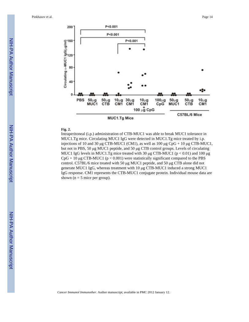
Fig. 2.Intraperitoneal (i.p.) administration of CTB-MUC1 was able to break MUC1 tolerance inMUC1.Tg mice. Circulating MUC1 IgG were detected in MUC1.Tg mice treated by i.p.injections of 10 and 30 μg CTB-MUC1 (CM1), as well as 100 μg CpG + 10 μg CTB-MUC1,but not in PBS, 50 μg MUC1 peptide, and 50 μg CTB control groups. Levels of circulatingMUC1 IgG levels in MUC1.Tg mice treated with 30 μg CTB-MUC1 (p < 0.01) and 100 μgCpG + 10 μg CTB-MUC1 (p < 0.001) were statistically significant compared to the PBScontrol. C57BL/6 mice treated with 50 μg MUC1 peptide, and 50 μg CTB alone did notgenerate MUC1 IgG, whereas treatment with 10 μg CTB-MUC1 induced a strong MUC1IgG response. CM1 represents the CTB-MUC1 conjugate protein. Individual mouse data areshown (n = 5 mice per group).
Pinkhasov et al. Page 14
Cancer Immunol Immunother. Author manuscript; available in PMC 2012 January 12.
NIH
-PA Author Manuscript
NIH
-PA Author Manuscript
NIH
-PA Author Manuscript
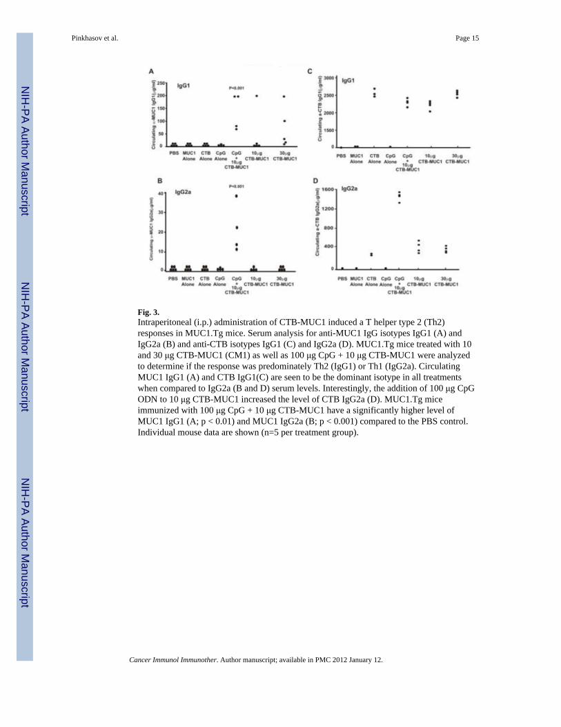
Fig. 3.Intraperitoneal (i.p.) administration of CTB-MUC1 induced a T helper type 2 (Th2)responses in MUC1.Tg mice. Serum analysis for anti-MUC1 IgG isotypes IgG1 (A) andIgG2a (B) and anti-CTB isotypes IgG1 (C) and IgG2a (D). MUC1.Tg mice treated with 10and 30 μg CTB-MUC1 (CM1) as well as 100 μg CpG + 10 μg CTB-MUC1 were analyzedto determine if the response was predominately Th2 (IgG1) or Th1 (IgG2a). CirculatingMUC1 IgG1 (A) and CTB IgG1(C) are seen to be the dominant isotype in all treatmentswhen compared to IgG2a (B and D) serum levels. Interestingly, the addition of 100 μg CpGODN to 10 μg CTB-MUC1 increased the level of CTB IgG2a (D). MUC1.Tg miceimmunized with 100 μg CpG + 10 μg CTB-MUC1 have a significantly higher level ofMUC1 IgG1 (A; p < 0.01) and MUC1 IgG2a (B; p < 0.001) compared to the PBS control.Individual mouse data are shown (n=5 per treatment group).
Pinkhasov et al. Page 15
Cancer Immunol Immunother. Author manuscript; available in PMC 2012 January 12.
NIH
-PA Author Manuscript
NIH
-PA Author Manuscript
NIH
-PA Author Manuscript
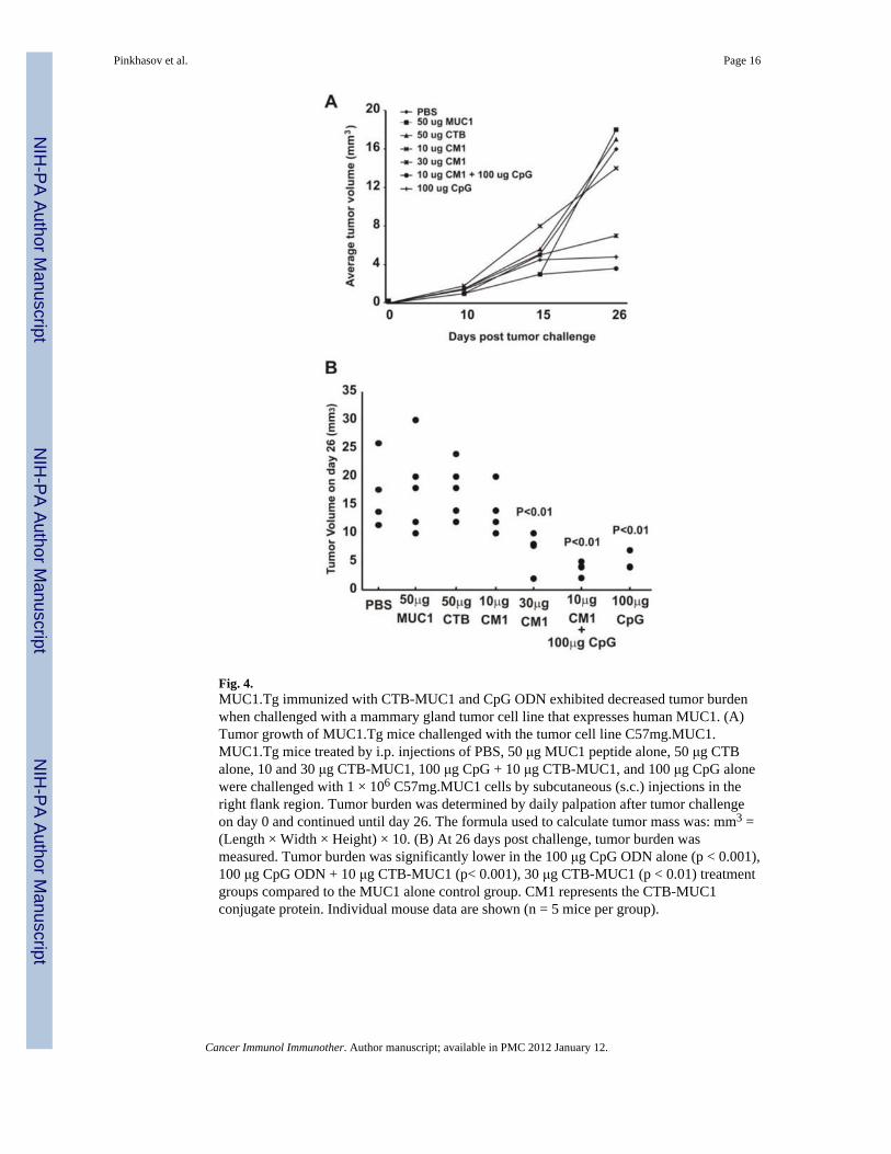
Fig. 4.MUC1.Tg immunized with CTB-MUC1 and CpG ODN exhibited decreased tumor burdenwhen challenged with a mammary gland tumor cell line that expresses human MUC1. (A)Tumor growth of MUC1.Tg mice challenged with the tumor cell line C57mg.MUC1.MUC1.Tg mice treated by i.p. injections of PBS, 50 μg MUC1 peptide alone, 50 μg CTBalone, 10 and 30 μg CTB-MUC1, 100 μg CpG + 10 μg CTB-MUC1, and 100 μg CpG alonewere challenged with 1 × 10⁶ C57mg.MUC1 cells by subcutaneous (s.c.) injections in theright flank region. Tumor burden was determined by daily palpation after tumor challengeon day 0 and continued until day 26. The formula used to calculate tumor mass was: mm3 =(Length × Width × Height) × 10. (B) At 26 days post challenge, tumor burden wasmeasured. Tumor burden was significantly lower in the 100 μg CpG ODN alone (p < 0.001),100 μg CpG ODN + 10 μg CTB-MUC1 (p< 0.001), 30 μg CTB-MUC1 (p < 0.01) treatmentgroups compared to the MUC1 alone control group. CM1 represents the CTB-MUC1conjugate protein. Individual mouse data are shown (n = 5 mice per group).
Pinkhasov et al. Page 16
Cancer Immunol Immunother. Author manuscript; available in PMC 2012 January 12.
NIH
-PA Author Manuscript
NIH
-PA Author Manuscript
NIH
-PA Author Manuscript
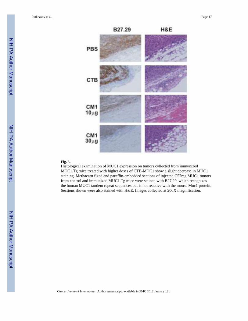
Fig. 5.Histological examination of MUC1 expression on tumors collected from immunizedMUC1.Tg mice treated with higher doses of CTB-MUC1 show a slight decrease in MUC1staining. Methacarn fixed and paraffin-embedded sections of injected C57mg.MUC1 tumorsfrom control and immunized MUC1.Tg mice were stained with B27.29, which recognizesthe human MUC1 tandem repeat sequences but is not reactive with the mouse Muc1 protein.Sections shown were also stained with H&E. Images collected at 200X magnification.
Pinkhasov et al. Page 17
Cancer Immunol Immunother. Author manuscript; available in PMC 2012 January 12.
NIH
-PA Author Manuscript
NIH
-PA Author Manuscript
NIH
-PA Author Manuscript

Fig. 6.The vaccine formulation induced antibodies that react with MUC1 as it is naturallyexpressed on tumor cells. Serum from MUC1.Tg mice immunized with the 10 and 30 μgCTB-MUC1 conjugate protein and 100 μg CpG ODN+ 10 μg CTB-MUC1 react withMUC1 as it is naturally expressed on the tumor cell line C57mg.MUC1 (B) but not on thewild type control cell line C57mg (A). A FITC conjugated anti-MUC1 antibody was used asa positive control to show MUC1 expression on the C57mg.MUC1 cell line but not on thewild type control group. Mice immunized with PBS, 50 μg CTB and 100 μg CpG ODNalone showed low levels of reactivity in C57mg.wild type and C57mg.MUC1 cell line.Percent gated populations are shown in the appropriate quadrants. SA-PE stands forsecondary antibody conjugated to phycoerythrin (PE).
Pinkhasov et al. Page 18
Cancer Immunol Immunother. Author manuscript; available in PMC 2012 January 12.
NIH
-PA Author Manuscript
NIH
-PA Author Manuscript
NIH
-PA Author Manuscript