
N- and O-glycans of recombinant human C1 inhibitor expressed in the milk of transgenic rabbits
Upload
independentCategory
view
6download
0
Schloerer and Horst SchrotenFranz-Georg Hanisch, David Bonar, Nils Helicobacter pyloriwith Antibiotic Activity against
-GlcNAc-capped Mucin GlycansαBinds Human Trefoil Factor 2 Is a Lectin ThatGlycobiology and Extracellular Matrices:
doi: 10.1074/jbc.M114.597757 originally published online August 14, 20142014, 289:27363-27375.J. Biol. Chem.
10.1074/jbc.M114.597757Access the most updated version of this article at doi:
.JBC Affinity SitesFind articles, minireviews, Reflections and Classics on similar topics on the
Alerts:
When a correction for this article is posted•
When this article is cited•
to choose from all of JBC's e-mail alertsClick here
http://www.jbc.org/content/289/40/27363.full.html#ref-list-1
This article cites 37 references, 13 of which can be accessed free at
at DE
UT
SC
HE
ZE
NT
RA
LBIB
LIOT
HE
K F
UE
R M
ED
IZIN
on October 29, 2014
http://ww
w.jbc.org/
Dow
nloaded from
at DE
UT
SC
HE
ZE
NT
RA
LBIB
LIOT
HE
K F
UE
R M
ED
IZIN
on October 29, 2014
http://ww
w.jbc.org/
Dow
nloaded from

Human Trefoil Factor 2 Is a Lectin That Binds�-GlcNAc-capped Mucin Glycans with Antibiotic Activityagainst Helicobacter pylori*
Received for publication, July 17, 2014, and in revised form, August 8, 2014 Published, JBC Papers in Press, August 14, 2014, DOI 10.1074/jbc.M114.597757
Franz-Georg Hanisch‡§1, David Bonar‡, Nils Schloerer¶, and Horst Schroten�
From the ‡Institute of Biochemistry II, Medical Faculty, University of Cologne, Joseph-Stelzmann-Str. 52, 50931 Koln, the §Center forMolecular Medicine Cologne, University of Cologne, Robert-Koch-Str. 21, 50931 Koln, the ¶Institute of Organic Chemistry, Universityof Cologne, Greinstr. 4, 50939 Koln, and the �University Children’s Hospital, Mannheim, Heidelberg University, Theodor-Kutzer-Ufer1–3, 68167 Mannheim, Germany
Background: Trefoil factor 2 (TFF2) forms complexes with MUC6 and is concertedly secreted by deep gastric glands.
Results: TFF2 is a lectin that binds to �-GlcNAc-capped MUC6 O-glycans with antibiotic activity against Helicobacter pylori.
Conclusion: There may be a functional link between mucin glycans and TFF2 in H. pylori defense.
Significance: The findings impact in development of defense strategies against H. pylori and in TFF2 receptor-mediated cell
signaling.
Helicobacter pylori infection is themajor cause of gastric can-
cer and remains an important health care challenge. The trefoil
factor peptides are a family of small highly conserved proteins
that are claimed to play essential roles in cytoprotection and
epithelial repair within the gastrointestinal tract.H. pylori colo-
calizes with MUC5AC at the gastric surface epithelium, but not
with MUC6 secreted in concert with TFF2 by deep gastric
glands. Both components of the gastric gland secretome associ-
ate non-covalently and show increased expression upon H.
pylori infection. Although blood group active O-glycans of the
Lewis-type form the basis of H. pylori adhesion to the surface
mucin layer and to epithelial cells, �1,4-GlcNAc-cappedO-gly-
cans ongastricmucinswereproposed to inhibitH.pylori growth
as a natural antibiotic. We show here that the gastric glycoform
of TFF2 is a calcium-independent lectin, which binds with high
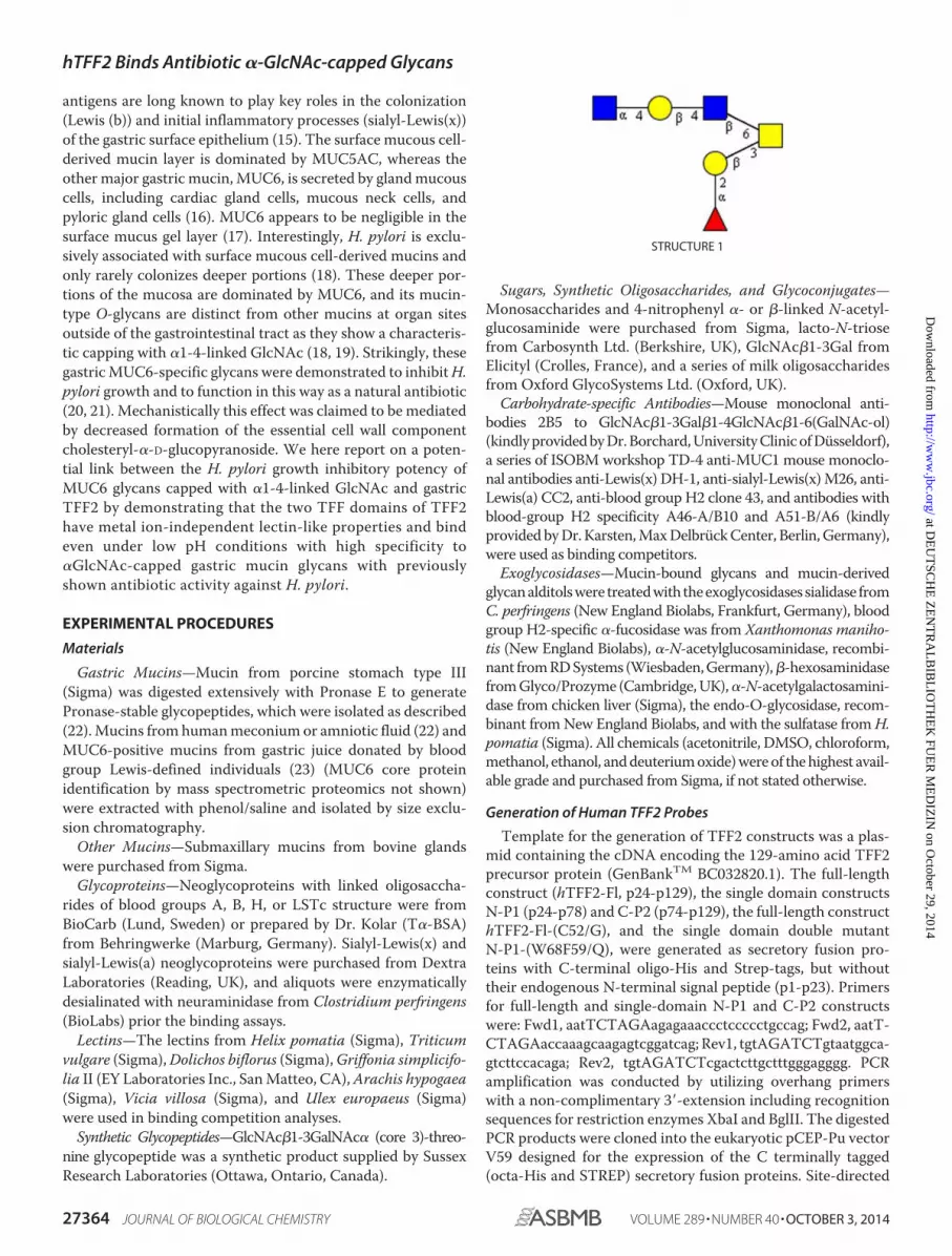
specificity to O-linked �1,4-GlcNAc-capped hexasaccharides
on human and porcine stomach mucin. The structural assign-
ments of twohexasaccharide isomers and thebinding active glyco-
topewerebasedonmassspectrometry, linkageanalysis, 1Hnuclear
magnetic resonance spectroscopy, glycan inhibition, and lectin
competition of TFF2-mucin binding. Neoglycolipids derived from
the C3/C6-linked branches of the two isomers revealed highly
specific TFF2 binding to the 6-linked trisaccharide in
GlcNAc�1-4Gal�1-4GlcNAc�1-6(Fuc�1-2Gal�1-3)GalNAc-ol
(Structure 1). Supposedly, lectin TFF2 is involved in protection of
gastric epithelia via a functional relationship to defense againstH.
pylori launched by antibiotic�1,4-GlcNAc-cappedmucin glycans.
Lectin-carbohydrate interactionmay have also an impact onmore
general functional aspects of TFF members by mediating their
binding to cell signaling receptors.
Human trefoil factor 2 (hTFF2 or TFF2)2 (UniProt Q03403,chromosome21) is a secretory peptidewith amolecularmass of14.284 Da that comprises 106 amino acid residues and belongsto the trefoil factor family peptides characterized by character-istic cysteine patterns (1–4). It was discovered originally in theporcine pancreas where it was termed “pancreatic spasmolyticpolypeptide” (5). However, in humans the major expressionsite of TFF2 is the stomach (1) where it is secreted togetherwith the mucin MUC6 by cardiac glands, mucous neck cells,and antral gland cells (6, 7). Peptides of the trefoil factorfamily are characterized by at least one TFF domain, a40-mer peptide with three conserved disulfide bridges.TFF2, in particular, is composed of two TFF domains (P1,P2), and modified mostly by N-linked glycans within the P1domain. The stomach-associated glycoform was shown tocarry a biantennary complex-type N-glycan with terminalmonofucosylated N,N�-diacetyl-lactosediamine antennae(8). It has been postulated that the hydrophobic grooveformed by loops 2 and 3 of the TFF domains could exertspecific protein and/or carbohydrate binding capacities (9).Different functional aspects of TFF2 have been reported in
the past. On the one hand, gastric TFF2 is strongly associatedwith mucins (7, 10) where it changes the rheological propertiesof mucus (11). It is a characteristic constituent of the alternat-ing laminated structure of the gastric surface mucous gel layerwhere it co-localizes with MUC6 (7, 12).TFF2 has recently been identified as a gastric stem/progeni-
tor cell marker in Helicobacter pylori infection (13). Furtherevidence for a link between trefoil factor peptides andH. pyloriinfection was revealed by demonstrating aberrant increasedepithelial expression of TFF2 and MUC6 in H. pylori-infectedgastric antrum, incisura, and body (14). Mucin-type O-linkedglycans on gastric mucins expressing Lewis-type blood group
* This work was supported by Deutsche Forschungsgemeinschaft Grant HA2092/21-1 (to F. G. H.).
1 To whom correspondence should be addressed: Institute of Biochemistry II,Medical Faculty, University of Cologne, Joseph-Stelzmann-Str. 52, 50931Koln, Germany. Tel.: 49-221-478-4493; Fax: 49-221-478-7788; E-mail:[email protected].
2 The abbreviations used are: hTFF2, human trefoil factor family 2; PSM, por-cine stomach mucin; PMAA, Partially methylated alditol acetates; TOCSY,Two-dimensional total correlation spectroscopy; GSA, isolectin II from G.simplicifolia.
THE JOURNAL OF BIOLOGICAL CHEMISTRY VOL. 289, NO. 40, pp. 27363–27375, October 3, 2014© 2014 by The American Society for Biochemistry and Molecular Biology, Inc. Published in the U.S.A.
OCTOBER 3, 2014 • VOLUME 289 • NUMBER 40 JOURNAL OF BIOLOGICAL CHEMISTRY 27363
at DE
UT
SC
HE
ZE
NT
RA
LBIB
LIOT
HE
K F
UE
R M
ED
IZIN
on October 29, 2014
http://ww
w.jbc.org/
Dow
nloaded from
antigens are long known to play key roles in the colonization
(Lewis (b)) and initial inflammatory processes (sialyl-Lewis(x))
of the gastric surface epithelium (15). The surface mucous cell-
derived mucin layer is dominated by MUC5AC, whereas the
other major gastric mucin, MUC6, is secreted by glandmucous
cells, including cardiac gland cells, mucous neck cells, and
pyloric gland cells (16). MUC6 appears to be negligible in the
surface mucus gel layer (17). Interestingly, H. pylori is exclu-
sively associated with surface mucous cell-derived mucins and
only rarely colonizes deeper portions (18). These deeper por-
tions of the mucosa are dominated by MUC6, and its mucin-
type O-glycans are distinct from other mucins at organ sites
outside of the gastrointestinal tract as they show a characteris-
tic capping with �1-4-linked GlcNAc (18, 19). Strikingly, these
gastricMUC6-specific glycans were demonstrated to inhibitH.
pylori growth and to function in this way as a natural antibiotic
(20, 21). Mechanistically this effect was claimed to be mediated
by decreased formation of the essential cell wall component
cholesteryl-�-D-glucopyranoside. We here report on a poten-
tial link between the H. pylori growth inhibitory potency of
MUC6 glycans capped with �1-4-linked GlcNAc and gastric
TFF2 by demonstrating that the two TFF domains of TFF2
have metal ion-independent lectin-like properties and bind
even under low pH conditions with high specificity to
�GlcNAc-capped gastric mucin glycans with previously
shown antibiotic activity against H. pylori.
EXPERIMENTAL PROCEDURES
Materials
Gastric Mucins—Mucin from porcine stomach type III
(Sigma) was digested extensively with Pronase E to generate
Pronase-stable glycopeptides, which were isolated as described
(22).Mucins from humanmeconium or amniotic fluid (22) and
MUC6-positive mucins from gastric juice donated by blood
group Lewis-defined individuals (23) (MUC6 core protein
identification by mass spectrometric proteomics not shown)
were extracted with phenol/saline and isolated by size exclu-
sion chromatography.
Other Mucins—Submaxillary mucins from bovine glands
were purchased from Sigma.
Glycoproteins—Neoglycoproteins with linked oligosaccha-
rides of blood groups A, B, H, or LSTc structure were from
BioCarb (Lund, Sweden) or prepared by Dr. Kolar (T�-BSA)
from Behringwerke (Marburg, Germany). Sialyl-Lewis(x) and
sialyl-Lewis(a) neoglycoproteins were purchased from Dextra
Laboratories (Reading, UK), and aliquots were enzymatically
desialinated with neuraminidase from Clostridium perfringens
(BioLabs) prior the binding assays.
Lectins—The lectins from Helix pomatia (Sigma), Triticum
vulgare (Sigma),Dolichos biflorus (Sigma),Griffonia simplicifo-
lia II (EY Laboratories Inc., SanMatteo, CA),Arachis hypogaea
(Sigma), Vicia villosa (Sigma), and Ulex europaeus (Sigma)
were used in binding competition analyses.
Synthetic Glycopeptides—GlcNAc�1-3GalNAc� (core 3)-threo-
nine glycopeptide was a synthetic product supplied by Sussex
Research Laboratories (Ottawa, Ontario, Canada).
Sugars, Synthetic Oligosaccharides, and Glycoconjugates—
Monosaccharides and 4-nitrophenyl �- or �-linked N-acetyl-glucosaminide were purchased from Sigma, lacto-N-triosefrom Carbosynth Ltd. (Berkshire, UK), GlcNAc�1-3Gal fromElicityl (Crolles, France), and a series of milk oligosaccharidesfrom Oxford GlycoSystems Ltd. (Oxford, UK).Carbohydrate-specific Antibodies—Mouse monoclonal anti-
bodies 2B5 to GlcNAc�1-3Gal�1-4GlcNAc�1-6(GalNAc-ol)(kindlyprovidedbyDr.Borchard,UniversityClinicofDusseldorf),a series of ISOBM workshop TD-4 anti-MUC1 mouse monoclo-nal antibodies anti-Lewis(x) DH-1, anti-sialyl-Lewis(x)M26, anti-Lewis(a) CC2, anti-blood group H2 clone 43, and antibodies withblood-group H2 specificity A46-A/B10 and A51-B/A6 (kindlyprovidedbyDr.Karsten,MaxDelbruckCenter, Berlin,Germany),were used as binding competitors.Exoglycosidases—Mucin-bound glycans and mucin-derived
glycanalditolswere treatedwith theexoglycosidases sialidase fromC. perfringens (New England Biolabs, Frankfurt, Germany), bloodgroup H2-specific �-fucosidase was from Xanthomonas maniho-
tis (New England Biolabs), �-N-acetylglucosaminidase, recombi-nant fromRDSystems (Wiesbaden,Germany),�-hexosaminidasefromGlyco/Prozyme (Cambridge,UK),�-N-acetylgalactosamini-dase from chicken liver (Sigma), the endo-O-glycosidase, recom-binant fromNew England Biolabs, and with the sulfatase fromH.
pomatia (Sigma). All chemicals (acetonitrile, DMSO, chloroform,methanol, ethanol, anddeuteriumoxide)wereof thehighest avail-able grade and purchased from Sigma, if not stated otherwise.
Generation of Human TFF2 Probes
Template for the generation of TFF2 constructs was a plas-mid containing the cDNA encoding the 129-amino acid TFF2precursor protein (GenBankTM BC032820.1). The full-lengthconstruct (hTFF2-Fl, p24-p129), the single domain constructsN-P1 (p24-p78) and C-P2 (p74-p129), the full-length constructhTFF2-Fl-(C52/G), and the single domain double mutantN-P1-(W68F59/Q), were generated as secretory fusion pro-teins with C-terminal oligo-His and Strep-tags, but withouttheir endogenous N-terminal signal peptide (p1-p23). Primersfor full-length and single-domain N-P1 and C-P2 constructswere: Fwd1, aatTCTAGAagagaaaccctccccctgccag; Fwd2, aatT-CTAGAaccaaagcaagagtcggatcag; Rev1, tgtAGATCTgtaatggca-gtcttccacaga; Rev2, tgtAGATCTcgactcttgctttgggagggg. PCRamplification was conducted by utilizing overhang primerswith a non-complimentary 3�-extension including recognitionsequences for restriction enzymes XbaI and BglII. The digestedPCR products were cloned into the eukaryotic pCEP-Pu vectorV59 designed for the expression of the C terminally tagged(octa-His and STREP) secretory fusion proteins. Site-directed
STRUCTURE 1
hTFF2 Binds Antibiotic �-GlcNAc-capped Glycans
27364 JOURNAL OF BIOLOGICAL CHEMISTRY VOLUME 289 • NUMBER 40 • OCTOBER 3, 2014
at DE
UT
SC
HE
ZE
NT
RA
LBIB
LIOT
HE
K F
UE
R M
ED
IZIN
on October 29, 2014
http://ww
w.jbc.org/
Dow
nloaded from

mutations were introduced with the QuikChange II Site-di-rected Mutagenesis Kit. Cys42 and Cys52, respectively, wereexchanged forGly in hTFF2-Fl, and a novelN-glycosylation sitewas generated in the otherwise N-glycosylation-negative P2domain by mutation of Asp87 to Asn87 and Arg89 to Thr89 inone step using the following primers: Fl-(Cys42/Gly), Fwd,cataacaggacgaacggcggcttccct; Fl(Cys52/Gly), Fwd, atcaccag-tgaccaggtgtttgacaat. C-P2 (Asp87/Asn, Arg89/Thr), Fwd, tgcgt-catggaggtctcaaaccgaacaaactgtggctacc. The construct N-P1-(W68F59/Q) was synthesized by Geneart-Life Technolgies(Regensburg, Germany), and ligated into V59 vector as above.
Cells and Cell Culture
Culture media and cell culture flasks were purchased fromBiochrom (Berlin, Germany). The human embryonic kidneycell line HEK-293 (Invitrogen) was cultivated and transfected,and recombinant TFF2 probes were isolated via their oligo-Histag as described (24).
Isolation of Gastric Mucin-derived O-Glycans
The glycan chains were released from themucin core proteinby reductive �-elimination (24) or by hydrazinolysis to retainthe pyranosidic ring structure of the reducing core-GalNAc(25). After desalting on graphitized carbon, glycan alditols orreducing glycans were chromatographed by polar amino-phaseHPLC (SystemGold, Beckman-Coulter, Germany) as described(22, 25).
ELISA Binding, Competition, and Inhibition Analyses
Mucin ormucin-derived glycopeptides (100 �g or 10 �g/ml)were immobilized on polystyrene microtitration plates (Nunc,Wiesbaden, Germany). After blocking with 200 �l of 5% BSA/PBS (37 °C, 1 h) the Strep-tagged TFF2 probe (1 �g/ml of cal-cium- and magnesium-free phosphate-buffered saline) wasadded in the presence or absence of inhibitors (37 °C, 2 h), andbound TFF2 was detected with Strep-Tactin/alkaline phospha-tase conjugate (IBA GmbH, Goettingen, Germany, 1:1000,37 °C, 1 h) followed by development with p-nitrophenyl phos-phate. Plates were read in a Tecan ELISA reader at 405 nm.Statistical evaluation of triplicate assays was based on inde-pendent sample t tests and calculation of t and p values. The tquantile (degrees of freedom 4, probability level 0.05) was 2.78.
Structural Analyses
Profiling and Oligosaccharide Sequencing of Permethylated
Glycan Alditols by MALDI-MS—Glycan alditols were per-methylated (26) and analyzed by mass spectrometry on anUltrafleXtreme MALDI-TOF-TOF instrument (Bruker Dal-tonics, Bremen, Germany) as described (24). MS/MS was per-formed by analysis of post-source decay fragments in the laser-induced dissociation mode. Fragment annotation of glycanswas assisted by application of the GlycoWorkbench tool (27).T3 sequencing of in-source decay B-type fragment ions wasperformed under the same MS/MS conditions.Oligosaccharide sequencing by LC-ESI-MS/MS—Methylated
glycan alditols were analyzed by LC-MS/MS on an ESI iontrap,the HCT ultra ETDII PTMDiscovery System (Bruker-Dalton-
ics), coupled with an online easy-nano-LC system (Proxeon,Odense, Denmark) as described (26).
GC-MS of Partially Methylated Alditol Acetates for Linkage
Analysis
Partially methylated alditol acetates (PMAA) were preparedfrom permethylated glycan alditols according to Albersheim et
al. (28) and were analyzed by GC-MS on a 15-m RTX5-SILMScapillary column (Restek, Bad Homburg, Germany) (24).
1H Nuclear Resonance Spectroscopy of TFF2 Binding
Hexasaccharide Alditol
All NMR spectra were recorded at 303 K on a Bruker AvanceII� 600 spectrometer (1H frequency 600.2 MHz) using a tripleresonance z gradient probe (H,P,BB) for inverse detection.They were processed using TopSpin 3.1 software (Bruker). Thehexasaccharide analyte (approximately 100 �g) dissolved inwater was repeatedly dried by vacuum rotation and resolubi-lized in deuterated solvent (D2O), which was purchased fromAldrich. The final volume was 450 �l (0.2 mM).For all experiments, suppression of the residual water signal
was achieved by presaturation. One-dimensional high-resolu-tion experiments were recorded with 64k complex data pointsand 64 scans were collected at a spectral width of 12,000 Hz.The original free induction decay was zero filled to 128k andFourier transformation using an exponential window functionwas applied. Two-dimensional total correlation spectroscopy(TOCSY) spectra were recorded using the MLEV-17 pulsesequence with mixing times (spin-lock) in the range of 80 ms.Usually 4k data points were collected for the direct domain (t2)with 32 scans each, and 512 t1 transients were collected.
Preparation and Analysis of Glycan Alditol-derived
Neoglycolipids
Glycan alditols (10 �g) were converted to neoglycolipidsafter mild periodate oxidation by reductive amination to L-1,2-dipalminoyl-sn-glycero-3-phosphoethanolamine according toa previously published protocol for microscale applications(29). Neoglycolipids were eluted from silica gel with chloro-form/methanol/water (10:10:1). Duplicate ELISAs were per-formed on polystyrene microtitration plates (approximately400 pmol of neoglycolipid per well were applied by drying a 50�l solution in methanol). About 100–200 pmol of the neogly-colipids in 0.5 �l of methanol (TLC-fractions 1–4) were mixedwith an equal aliquot of matrix (saturated solution of 4-hy-droxy-�-cyanocinnamic acid in acetonitrile, 0.1% aqueous trif-luoroacetic acid, 1:1) and MALDI mass spectrometry was per-formed in the negative ion mode.
RESULTS
The Human Gastric Glycoform of TFF2 Binds Calcium
Independent to Gastric Mucin
Human TFF2 had long been proposed to exhibit lectin-likeactivities, although strong evidence was never provided. As tre-foil factor peptides in general are found in association withmucins of the gastrointestinal tract we tested first whetherTFF2 (expressed in HEK-293 cells as a strep-tagged TFF2-Fl
hTFF2 Binds Antibiotic �-GlcNAc-capped Glycans
OCTOBER 3, 2014 • VOLUME 289 • NUMBER 40 JOURNAL OF BIOLOGICAL CHEMISTRY 27365
at DE
UT
SC
HE
ZE
NT
RA
LBIB
LIOT
HE
K F
UE
R M
ED
IZIN
on October 29, 2014
http://ww
w.jbc.org/
Dow
nloaded from

probe) was binding active to a series of mucin preparationsfrom human gastric juice. For this purpose we performed bind-ing and binding inhibition studies in a standardized enzyme-linked immunosorbent assay by immobilizing mucins ormucin-derived Pronase-stable glycopeptides onto polystyrenemicroplates. We could demonstrate a high binding activity offull-length TFF2 to these mucin samples, which was indepen-dent of the blood group Lewis status of the gastric juice donors(Figs. 1 and 2A). Similar high binding was found on mucinsfrom human meconium, but no binding above background tobovine submaxillary mucin (Fig. 1A). Further evidence wasrevealed in binding studies on gastric mucins from porcinestomach (PSM) andderivedPronase-stable PSMglycopeptides,which showed the highest TFF2 binding capacities comparedwith all other mucins under study (Fig. 1). Binding of TFF2 tomucins revealed concentration-dependent logarithmic charac-teristics (Fig. 1B). As all binding assays were performed in cal-cium and magnesium-free PBS, the lectin activity of TFF2 wasapparently independent of calciumor other divalentmetal ions.Over 2-h time periods at 37 °C the binding activity showed apartial resistance to low pH conditions (Fig. 1A).
TFF2 Binds Carbohydrate-dependent, but Blood Group AB and
Lewis-independent to Mucin Glycans
Mild selective periodate oxidation of mucin-bound sugarscompletely abrogated TFF2 binding activity to the backgroundlevel (p value 2.6� 10�9), indicating a carbohydrate-dependentinteraction with the immobilizedmucins (Fig. 2A). A contribu-tion of the mucin core proteins to the binding epitope can belargely ruled out, as binding activity was retained on Pronase-sta-ble glycopeptides derived from porcine stomach mucin (Fig. 1B,refer also to neoglycolipid binding reported below). Treatmentswith exoglycosidases (sialidase, �2-fucosidase, �-galactosidase,�-hexosaminidase, �-N-acetylglucosaminidase, and �-N-acetyl-galactosaminidase) and endo-N-acetylgalactosaminidase did notdestroy the binding active epitope (data not shown). More-over, none of the tested neoglycoproteins carrying bloodgroup-related glycans were found binding positive, with theexception of blood group H disaccharide-HSA, whichshowed weak, but significant binding activity (p value 1.34 �
10�6) compared with blood group A active neoglycoprotein(Fig. 2B). These initial findings indicated a carbohydratedependence of TFF2 binding to mucins and demonstratedthat blood group A/B or Lewis-related context of this bind-ing can be largely ruled out.
Structural Requirements of TFF2 Lectin Activity
The lectin-like binding capacity of TFF2 on gastric mucinswas retained using the single trefoil domain constructs N-P1and C-P2* (the latter with a designed N-glycosylation site),however, both were at lower avidity compared with the wildtype full-length construct (Table 1). Mutant forms of TFF2(Cys52/Gly) with distorted disulfide bridge formation lackedany binding activity (Table 1). Strikingly, a mild periodateoxidation of the N-linked glycan that modifies the nativefull-length TFF2 probe and the natural gastric glycoformresulted in strong reduction of lectin activity (Table 1; seealso below). This latter finding indicates a potential contri-
bution of N-glycosylation at Asn38 to the active conforma-tion of TFF2. Artificial amino acid sequences introduced astags (oligo-His and Strep-tag) apparently do not contributeto the binding activity as the double point-mutated con-struct N-P1-(W68F59/Q) with the affected hydrophobicpocket was binding inactive (Table 1).
FIGURE 1. Binding of TFF2 (hTFF2-Fl) to human gastric mucin and por-cine gastric mucin glycopeptides. A, triplicate assay (with S.D.) of TFF2binding to human mucins from gastric juice (HGM, pooled sample), andmeconium (HMM), bovine submaxillary mucin (BSM), 5 �g/well, and por-cine stomach mucin (PSM) glycopeptides (0.5 �g/well) in calcium- andmagnesium-free Dulbecco’s phosphate-buffered saline at pH 7.0 (PBS).Binding to PSM glycopeptides was also tested in acidified PBS at pH 4.0,2.0, and 1.5. Average background binding was at 0.06 (A405 nm). B, triplicateassays (with S.D.) of TFF2 binding to PSM-derived glycopeptides; variationof immobilized glycopeptide amounts tested with hTFF2-Fl at 1 �g/ml(upper part) or variation of hTFF2-Fl concentration tested on 0.5 �g/well ofPSM glycopeptides (lower part). Average background binding was at 0.06(A405 nm).
hTFF2 Binds Antibiotic �-GlcNAc-capped Glycans
27366 JOURNAL OF BIOLOGICAL CHEMISTRY VOLUME 289 • NUMBER 40 • OCTOBER 3, 2014
at DE
UT
SC
HE
ZE
NT
RA
LBIB
LIOT
HE
K F
UE
R M
ED
IZIN
on October 29, 2014
http://ww
w.jbc.org/
Dow
nloaded from

Human TFF2 Carbohydrate Binding Specificity as Revealed by
Inhibition and Competition-based Immunoassays
Lectins—Among a panel of lectins tested as potential com-
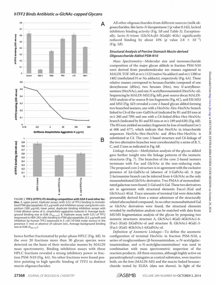
petitors of TFF2 the isolectin II from G. simplicifolia (GSA II)
showed significant strong competitive effects (p value 1.21 �
10�6) and logarithmic binding inhibition curves with ID50 val-
ues in the range of 15 �g/ml (Fig. 3A). Other lectins, likeWGA
andUEA I, showed a less inhibitory potential at concentrations
ranging above 100 �g/ml (Fig. 3A, upper panel). GSA II is
known to bind terminal GlcNAc residues, however, it does not
discriminate between �- and �-anomeric GlcNAc, whereas
wheat germ agglutinin is proposed to bind preferentially to the
�-anomer. In conclusion, a terminal GlcNAc residue can be
assumed to play a role in the TFF2 bindingmucin glycan. Com-
petitive effects were also seen with UEA I (p value 0.18� 10�6),
which might correspond to TFF2 binding activity on blood
group H-disaccharide. A very weak, but still significant inhibi-
tion was found with PNA (p value 0.018).
Control experiments with non-glycosylated human TFF2expressed in E. coli revealed partial competitive inhibition ofthe gastric glycoform of TFF2 expressed in HEK-293 cells (Fig.3B). This findingmay indicate a dependence of lectin activity onparticular TFF2 glycoforms (see above). The latter assumptionwould be in accordance with the observation that porcine pan-creatic TFF2 is not associated with high-molecular massmucins expressing �1,4-GlcNAc-capped GSA II binding gly-cans (30). The presence of calcium ions had no significant effecton binding and binding competition (p value 0.068).Carbohydrate-specific Antibodies—Monoclonal antibodies
with a defined carbohydrate specificity were used to unravel thestructural features of the TFF2 glycotope. The panel covered aseries of carbohydrate blood group specificities, including H2,Lewis(y), Lewis(x), sialyl-Lewis(x), and blood group A/B. Nosignificant binding competition was revealed for any of theseantibodies indicating again that no direct blood group depen-dence of the TFF2 binding to gastric mucins was involved(Table 2).
Mucin Glycans, Oligosaccharides, Glycopeptides, and Other
Glycoconjugates
To define the structural aspects of the TFF2 glycotope wecleaved the O-linked glycans from binding active human andporcine gastric mucins and tested HPLC-purified fractions fortheir inhibitory potency in TFF2/mucin binding assays (Fig. 4).A prior fractionation of the glycan alditols by anion exchangechromatography revealed that all inhibitory potency wasconfined to the neutral fraction representing the quantita-tively dominating portion (Fig. 5A). The inhibitory potentialof glycans with intact reducing termini was similar to that ofthe reductively eliminated glycan alditols in binding inhibi-tion assays (data not shown). Neutral glycan alditols were
FIGURE 2. Evidence for blood group Lewis-independent carbohydratespecificity of TFF2 (hTFF2-Fl) binding to mucins. A, triplicate assay (withS.D.) of TFF2 binding to blood group Lewis-defined human gastric mucin(HGM, 5 �g/well) and to a blood group undefined sample (5 �g/well) testedprior to and after mild periodate oxidation of carbohydrates (10 mM sodiummetaperiodate in 0.1 M sodium acetate, pH 4.0, overnight incubation at 4 °C inthe dark). Average background binding was at 0.06 (A405 nm). B, triplicate assay(with S.D.) of TFF2 binding to neoglycoproteins (0.5 �g/well) carrying bloodgroup-related carbohydrate antigens. Average background binding was at0.06 (A405 nm).
TABLE 1
Survey of hTFF2 probes generated and tested for mucin glycopeptidebindingIn the schematic presentation of constructs generated green circles refer to loops1–3 in theTFFdomain P1, blue circles to the corresponding P2 domain (grey shadedareas indicate mutant versions of loops 2 and/or 3). Quantitative data of the probes’binding activities are given in % relative to the native full-length construct hTFF2(triplicate assays). * Probes were shown to carry N-linked glycans with LacdiNAcmodification (authentic gastric glycoform, Ref. 8). **probes to carry aberrant com-plex-type or high-mannose-type glycans (Bonar, Hanisch; unpublished data, man-uscript in preparation).
hTFF2 Binds Antibiotic �-GlcNAc-capped Glycans
OCTOBER 3, 2014 • VOLUME 289 • NUMBER 40 JOURNAL OF BIOLOGICAL CHEMISTRY 27367
at DE
UT
SC
HE
ZE
NT
RA
LBIB
LIOT
HE
K F
UE
R M
ED
IZIN
on October 29, 2014
http://ww
w.jbc.org/
Dow
nloaded from
hence further fractionated by polar-phase HPLC (Fig. 4B). Inthe over 20 fractions more than 30 glycan species weredetected on the basis of their molecular masses by MALDImass spectrometry. Binding inhibition assays with theseHPLC fractions revealed a strong inhibitory power in frac-tion PSM-N10 (Fig. 4A). No other fractions were found pos-itive pointing to high specific binding of TFF2 to distinctmucin oligosaccharides.
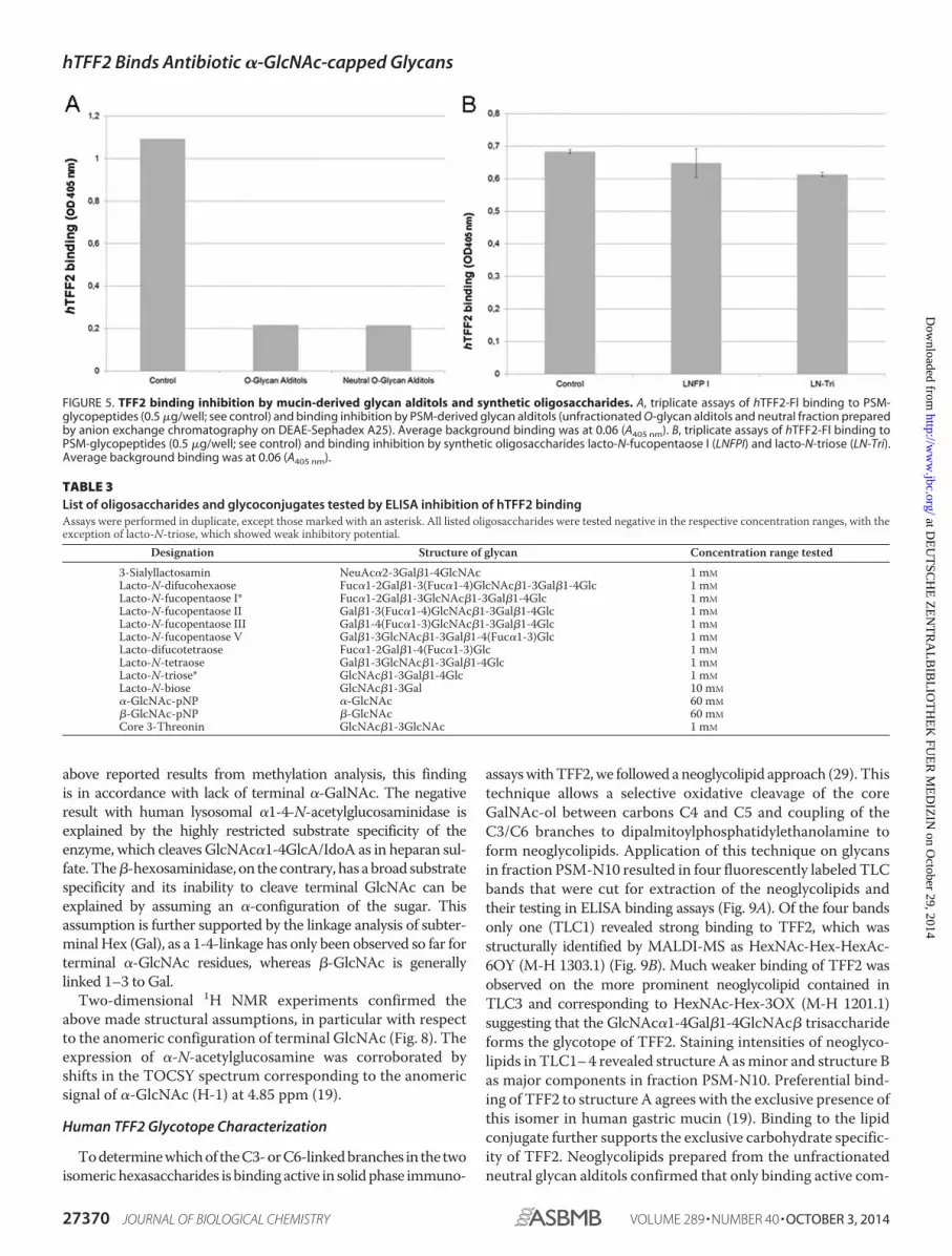
All other oligosaccharides from different sources (milk oli-gosaccharides, like lacto-N-fucopentaose I (p value 0.142), lackedinhibitory binding activity (Fig. 5B and Table 3). Exception-ally, lacto-N-triose (GlcNAc�1-3Gal�1-4Glc) significantlyreduced binding by about 10% (p value 2.8 � 10�5)(Fig. 5B).
Structural Analysis of Porcine Stomach Mucin-derived
Oligosaccharide Alditol PSM-N10
Mass Spectrometry—Molecular size and monosaccharidecomposition of the major glycan alditols in fraction PSM-N10were derived from pseudomolecular ion masses registered inMALDI-TOF-MSatm/z1122 (nativeNaadduct) andm/z1380or1402 (methylated H or Na adducts), respectively (Fig. 6A). Theserelative masses correspond to hexasaccharides composed of onedeoxyhexose (dHex), two hexoses (Hex), two N-acetylhexo-samines (HexNAc), andoneN-acetylhexosaminitol (HexNAc-ol).SequencingbyMALDI-MS2 (Fig. 6B), post-sourcedecayMALDI-MS3 analysis of in-source B-ion fragments (Fig. 6C), and ESIMS2andMS3 (Fig. 6D) revealed a core 2-based glycan alditol formingtwo branched isomers, onewith aHexNAc-Hex-HexNAc branchlinked toC6 of the core-GalNAcol (indicated by B1 and B3 ions atm/z 260 and 709) and one with a C6-linked dHex-Hex-HexNAcbranch (indicated by B1 and B3 ions atm/z 189 and 638) (Fig. 6B).TheB3 ions yielded secondary fragments by loss ofmethanol (m/zat 606 and 677), which indicate that HexNAc in trisaccharidesequences HexNAc-Hex-HexNAc and dHex-Hex-HexNAc issubstituted at C4. The core 2-based structure and C6-linkage ofthe two alternative brancheswere corroborated by a series of B, Y,C, and Z ions as indicated in Fig. 6B.Linkage Analysis—Methylation analysis of the glycan alditol
gave further insight into the linkage patterns of the isomericstructures (Fig. 7). The branches of the core 2-based isomersterminate with Fuc and GlcNAc at the non-reducing ends.The proposed core 2 structure is in agreementwith the exclusivepresence of 3,6-GalNAc-ol (absence of 3-GalNAc-ol). A type2-lactosamine branch can be inferred from 4-GlcNAc as the onlymonosubstitutedGlcNAc derivative. Two PMAAofmonosubsti-tutedgalactosewere found:2-Galand4-Gal.These twoderivativesare in agreement with structural elements Fuc�1-2Gal andGlcNAc�1-4Gal. Trace amounts of terminal Gal were detectablepresumably derived from a minor admixture of the structurallyrelated afucosylated compound.AsnoothermonosubstitutedGalor GlcNAc derivatives were found, the structural elementsrevealed by methylation analysis can be matched with data fromMS/MS fragmentation analysis of the glycan by proposing twoisomeric structures: structure A, GlcNAc1-4Gal1-4GlcNAc1-6-(Fuc1-2Gal1-3)GalNAc-ol and structure B, GlcNAc1-4Gal1-3-(Fuc1-2Gal1-4GlcNAc1-6)GalNAc-ol.Definition of Anomeric Linkages—To define the anomeric
configuration of terminal HexNAc in fraction PSM-N10, aseries of exoglycosidases (�-hexosaminidase, �-N-acetylgalac-tosaminidase, and �-N-acetylglucosaminidase) was used incombination with mass spectrometric inspection of thereaction products. All three enzymes, although highly active onparanitrophenyl conjugates as control substrates, were inactiveboth, on the free (MALDI-MS) and the mucin-linked hexasac-charide tested by ELISA (data not shown). In light of the
FIGURE 3. TFF2 (hTFF2-Fl)-binding competition with GSA II and other lec-tins. A, upper panel, triplicate assays (with S.D.) of TFF2 binding to immobi-lized PSM glycopeptides (0.5 �g/well) and inhibition assays using lectin com-petitors (500 �g/ml); lower panel, duplicate binding inhibition assays with5-fold dilution series of G. simplicifolia agglutinin isolectin II. Average back-ground binding was at 0.06 (A405 nm). B, triplicate assay (with S.D.) of TFF2(expressed in HEK-293 cells) binding to PSM glycopeptides (0.5 �g/well) andinhibition by human TFF2 expressed in E. coli (10-fold molar excess) in thepresence (1 mM) or absence of calcium ions. Average background bindingwas at 0.06 (A405 nm).
hTFF2 Binds Antibiotic �-GlcNAc-capped Glycans
27368 JOURNAL OF BIOLOGICAL CHEMISTRY VOLUME 289 • NUMBER 40 • OCTOBER 3, 2014
at DE
UT
SC
HE
ZE
NT
RA
LBIB
LIOT
HE
K F
UE
R M
ED
IZIN
on October 29, 2014
http://ww
w.jbc.org/
Dow
nloaded from

TABLE 2
List of monoclonal antibodies with carbohydrate specificity tested in ELISA competition of hTFF2 binding to porcine stomach mucins asnegativeAssays were performed in duplicate.
Designation Specificity Concentration range tested
19-OLE Blood group H2 Hybridoma supernatantFW6 Fucosylated polylactosamines Hybridoma supernatantA46-B10 Blood group H2 Hybridoma supernatantA51-B/A6 Blood group Lewis y/H2 Hybridoma supernatant2B5 GlcNAc�1-3Gal�1-4GlcNAc�1-6(GalNAc-ol) Hybridoma supernatantISOBM128 (DH-1) Blood group Lewis x 50 �g/mlISOBM143 (M26) Blood group sialyl-Lewis x 50 �g/mlISOBM164 (CC2) Blood group Lewis a 50 �g/ml
FIGURE 4. Gastric mucin-derived hexasaccharides inhibit TFF2 (hTFF2-Fl) binding. A, duplicate ELISA inhibition assay of hTFF2-Fl binding to PSM-glyco-peptides (0.5 �g/well) by dried aliquots from polar-phase HPLC-separated PSM-derived glycan alditols (fractions 5–19, estimated concentration range ofglycan alditols applied: 50 –500 �M). Average background binding was at 0.06 (A405 nm). B, representative chromatogram of neutral glycan alditols (250 �g)from PSM separated by polar-phase HPLC.
hTFF2 Binds Antibiotic �-GlcNAc-capped Glycans
OCTOBER 3, 2014 • VOLUME 289 • NUMBER 40 JOURNAL OF BIOLOGICAL CHEMISTRY 27369
at DE
UT
SC
HE
ZE
NT
RA
LBIB
LIOT
HE
K F
UE
R M
ED
IZIN
on October 29, 2014
http://ww
w.jbc.org/
Dow
nloaded from
above reported results from methylation analysis, this findingis in accordance with lack of terminal �-GalNAc. The negativeresult with human lysosomal �1-4-N-acetylglucosaminidase isexplained by the highly restricted substrate specificity of theenzyme, which cleavesGlcNAc�1-4GlcA/IdoA as in heparan sul-fate.The�-hexosaminidase, on thecontrary, has abroad substratespecificity and its inability to cleave terminal GlcNAc can beexplained by assuming an �-configuration of the sugar. Thisassumption is further supported by the linkage analysis of subter-minal Hex (Gal), as a 1-4-linkage has only been observed so far forterminal �-GlcNAc residues, whereas �-GlcNAc is generallylinked 1–3 to Gal.Two-dimensional 1H NMR experiments confirmed the
above made structural assumptions, in particular with respectto the anomeric configuration of terminal GlcNAc (Fig. 8). Theexpression of �-N-acetylglucosamine was corroborated byshifts in the TOCSY spectrum corresponding to the anomericsignal of �-GlcNAc (H-1) at 4.85 ppm (19).
Human TFF2 Glycotope Characterization
Todeterminewhichof theC3-orC6-linkedbranches in the twoisomerichexasaccharides is bindingactive in solidphase immuno-
assayswithTFF2,we followedaneoglycolipid approach (29). This
technique allows a selective oxidative cleavage of the core
GalNAc-ol between carbons C4 and C5 and coupling of the
C3/C6 branches to dipalmitoylphosphatidylethanolamine to
form neoglycolipids. Application of this technique on glycans
in fraction PSM-N10 resulted in four fluorescently labeled TLC
bands that were cut for extraction of the neoglycolipids and
their testing in ELISA binding assays (Fig. 9A). Of the four bands
only one (TLC1) revealed strong binding to TFF2, which was
structurally identified by MALDI-MS as HexNAc-Hex-HexAc-
6OY (M-H 1303.1) (Fig. 9B). Much weaker binding of TFF2 was
observed on the more prominent neoglycolipid contained in
TLC3 and corresponding to HexNAc-Hex-3OX (M-H 1201.1)
suggesting that the GlcNAc�1-4Gal�1-4GlcNAc� trisaccharide
forms the glycotope of TFF2. Staining intensities of neoglyco-
lipids in TLC1–4 revealed structure A asminor and structure B
as major components in fraction PSM-N10. Preferential bind-
ing of TFF2 to structure A agrees with the exclusive presence of
this isomer in human gastric mucin (19). Binding to the lipid
conjugate further supports the exclusive carbohydrate specific-
ity of TFF2. Neoglycolipids prepared from the unfractionated
neutral glycan alditols confirmed that only binding active com-
FIGURE 5. TFF2 binding inhibition by mucin-derived glycan alditols and synthetic oligosaccharides. A, triplicate assays of hTFF2-Fl binding to PSM-glycopeptides (0.5 �g/well; see control) and binding inhibition by PSM-derived glycan alditols (unfractionated O-glycan alditols and neutral fraction preparedby anion exchange chromatography on DEAE-Sephadex A25). Average background binding was at 0.06 (A405 nm). B, triplicate assays of hTFF2-Fl binding toPSM-glycopeptides (0.5 �g/well; see control) and binding inhibition by synthetic oligosaccharides lacto-N-fucopentaose I (LNFPI) and lacto-N-triose (LN-Tri).Average background binding was at 0.06 (A405 nm).
TABLE 3
List of oligosaccharides and glycoconjugates tested by ELISA inhibition of hTFF2 bindingAssays were performed in duplicate, except those marked with an asterisk. All listed oligosaccharides were tested negative in the respective concentration ranges, with theexception of lacto-N-triose, which showed weak inhibitory potential.
Designation Structure of glycan Concentration range tested
3-Sialyllactosamin NeuAc�2-3Gal�1-4GlcNAc 1 mM
Lacto-N-difucohexaose Fuc�1-2Gal�1-3(Fuc�1-4)GlcNAc�1-3Gal�1-4Glc 1 mM
Lacto-N-fucopentaose I* Fuc�1-2Gal�1-3GlcNAc�1-3Gal�1-4Glc 1 mM
Lacto-N-fucopentaose II Gal�1-3(Fuc�1-4)GlcNAc�1-3Gal�1-4Glc 1 mM
Lacto-N-fucopentaose III Gal�1-4(Fuc�1-3)GlcNAc�1-3Gal�1-4Glc 1 mM
Lacto-N-fucopentaose V Gal�1-3GlcNAc�1-3Gal�1-4(Fuc�1-3)Glc 1 mM
Lacto-difucotetraose Fuc�1-2Gal�1-4(Fuc�1-3)Glc 1 mM
Lacto-N-tetraose Gal�1-3GlcNAc�1-3Gal�1-4Glc 1 mM
Lacto-N-triose* GlcNAc�1-3Gal�1-4Glc 1 mM
Lacto-N-biose GlcNAc�1-3Gal 10 mM
�-GlcNAc-pNP �-GlcNAc 60 mM
�-GlcNAc-pNP �-GlcNAc 60 mM
Core 3-Threonin GlcNAc�1-3GlcNAc 1 mM
hTFF2 Binds Antibiotic �-GlcNAc-capped Glycans
27370 JOURNAL OF BIOLOGICAL CHEMISTRY VOLUME 289 • NUMBER 40 • OCTOBER 3, 2014
at DE
UT
SC
HE
ZE
NT
RA
LBIB
LIOT
HE
K F
UE
R M
ED
IZIN
on October 29, 2014
http://ww
w.jbc.org/
Dow
nloaded from

ponents were isographic with those in TLC1 and -3 (data notshown).
DISCUSSION
We provide for the first time unequivocal experimental evi-dence for a lectin activity of human TFF2 by demonstrating aperiodate-sensitive binding to gastric mucins and Pronase-sta-ble mucin glycopeptides, moreover by showing competitiveeffects with other lectins (GSA II), an inhibition of binding bymucin-derived oligosaccharides, and a binding to mucin gly-can-derived neoglycolipids. Strikingly, not all TFF2 prepara-tions bound with similar high avidity (Escherichia coli
expressed TFF2). Some evidence could be provided for glyco-form dependence of TFF2 lectin activity, as in particular thegastric glycoform was found highly active. The gastric TFF2 isdistinct by the characteristic expression of an N-linked com-plex-type glycan with monofucosylated N,N�-diacetyl-lactose-diamine antennae and absence of considerable microhetero-geneity (8), a structural feature that is simulated by therecombinant probe expressed in HEK-293 cells.3 TFF2 iso-forms expressed at other organ sites, like pancreatic TFF2, are
3 D. Bonar and F. G. Hanisch, unpublished data.
FIGURE 6. Mass spectrometric analysis of TFF2 (hTFF2-Fl) binding hexasaccharides in fraction PSM-N10. A, positive ion-MALDI mass spectrometry ofnative (upper panel) and permethylated oligosaccharide alditols (lower panel); B, post-source decay MALDI mass spectrum of permethylated glycan alditolscorresponding to the molecular ion M�Na at m/z 1402.5; C, Post-source decay MALDI-MS3 analysis of in-source B-ion fragments; D, ESI-MS2 and MS3 ofpermethylated glycan alditols corresponding to the molecular ion M�H at m/z 1380.5. Graphical symbols of sugars according to the recommendations of theConsortium for Functional Glycomics were used: yellow squares (GalNAc); yellow circles (Gal); blue squares (GlcNAc); red triangles (Fuc).
hTFF2 Binds Antibiotic �-GlcNAc-capped Glycans
OCTOBER 3, 2014 • VOLUME 289 • NUMBER 40 JOURNAL OF BIOLOGICAL CHEMISTRY 27371
at DE
UT
SC
HE
ZE
NT
RA
LBIB
LIOT
HE
K F
UE
R M
ED
IZIN
on October 29, 2014
http://ww
w.jbc.org/
Dow
nloaded from

known to lack association with the mucin MUC6 specificallyexpressing the �-GlcNAc-capped glycans, whereas gastricTFF2 is not only co-expressed withMUC6 but also shows strictassociation with the high-molecular mass mucin.The carbohydrate specificity of TFF2 could be narrowed
down to a structural part of the PSM-hexasaccharide isomer Aby demonstrating binding to the C6-linked trisaccharideGlcNAc�1-4Gal�1-4GlcNAc� characterized by terminal �4-GlcNAc. There was only a minor cross-reactivity to theC3-linked disaccharide branch GlcNAc�1-4Gal� on isomer B,which points to an extended glycotrope that comprises morethan the common disaccharide. In human gastric mucins hex-asaccharide isomer A is the only glycan species with �-GlcNAcend capping found in the so far most comprehensive study byRossez et al. (19).
Epithelial mucins and mucin-bound O-glycans play a dualrole in bacterial infection as theymediate pathogen adhesion to
epithelial cells via carbohydrate-lectin interactions on the onehand, whereas they act also cytoprotective by entrapping thepathogen in a visco-elastic mucin gel via the same mechanism.This holds true for gastric infections by H. pylori that infectsmore than half of the world’s population in particular. MostH.pylori reside within the mucous gel layer of the stomach cover-ing the apical surface, where they bind to MUC5AC (31),whereas about 20% of the bacteria bind directly to gastric epi-thelial cells (32). Two adhesins were found to be involved inH.
pylori binding under non-inflammatory (BabA) and inflamma-tory conditions (SabA). The switch from preferential BabAexpression (binding to Lewis b/H1 glycans) to the expression ofSabA (binding to sialylated Lewis glycans) reflects a corre-sponding gastritis-associated change in mucin glycosylation(15). The trefoil factor family members are long known to beclosely associated with gastrointestinal mucins, and in particu-lar TFF2 was described to bind to the high-molecular mass
FIGURE 7. Methylation analysis of linkages by gas chromatography-mass spectrometry of partially methylated alditol acetates. PMAA were identifiedvia their GC retention times and specific MS fragmentation spectra registered in electron-impact ionization at 70 eV. The total ion chromatogram (TIC) and massselective ion traces are shown for the major PMAA: m/z 159 (1-O-acetylated N-acetylhexosamines deuterated at C1: terminal and 4-substituted GlcNAc), m/z162 (1-O-acetylated deoxyhexoses and hexoses deuterated at C1: terminal fucose, 4-substituted Gal; note that 2-substituted Gal is detected as the 13C-isotopeof the fragment at m/z 161), m/z 205 (PMAA derived from terminal dHex, Hex, and HexNAc), m/z 233 (preferential fragment of PMAA derived from 4-substitutedHexNAc), and m/z 318 (fragment PMAA derived from 3,6-disubstituted HexNAc-ol and 3-substituted HexNAc).
hTFF2 Binds Antibiotic �-GlcNAc-capped Glycans
27372 JOURNAL OF BIOLOGICAL CHEMISTRY VOLUME 289 • NUMBER 40 • OCTOBER 3, 2014
at DE
UT
SC
HE
ZE
NT
RA
LBIB
LIOT
HE
K F
UE
R M
ED
IZIN
on October 29, 2014
http://ww
w.jbc.org/
Dow
nloaded from

MUC6 produced by deep gastric glands (30). However, neitherthe mode of its interaction with mucins nor its principle func-tionwere clearly defined. InH. pylori infection both,MUC6andTFF2, are concertedly up-regulated (14). The mucin producedin deep gastric glands appears to express the majority, if not all
of the organ-specific carbohydrates, which have been shown toexert antibiotic effects on H. pylori (20). The co-expressed lec-tin, TFF2, has been claimed to control epithelial repair (33), butalso to regulatemononuclear cell inflammatory responses inH.pylori infection (34). In support of its potential role inH. pylori
FIGURE 8. H,H-TOCSY NMR spectrum of oligosaccharides in fraction PSM-N10 (expansion), obtained at 600 MHz (303 K, 80 ms spin lock). Spin systemassigned for terminal GlcNAc�1-4. The hexasaccharide analyte (approximately 100 �g) dissolved in water was repeatedly dried by vacuum rotation andresolubilization in deuterated solvent (D2O). The final concentration was 0.2 mM.
FIGURE 9. Mass spectrometric identification of TFF2 binding active core 2-hexasaccharide branches as their neoglycolipids. A, thin layer chromato-graphic separation and duplicate ELISA binding assay of 400 pmol of neoglycolipids derived from glycan alditols in fraction PSM-N10 (for further details referto the ”Experimental Procedures“). Graphical symbols of sugars according to the recommendations of the Consortium for Functional Glycomics were used:yellow squares (GalNAc); yellow circles (Gal); blue squares (GlcNAc); red triangles (Fuc). B, MALDI mass spectra of TLC1– 4 recorded in the negative-ion mode. Asaturated solution of matrix (HCCA) in 50% acetonitrile/aqueous trifluoroacetic acid was mixed 1:1 with 100 –200 pmol of the neoglycolipids in 0.5 �l ofmethanol. Sections of mass spectra between m/z 1120 and 1320 are shown. Prominent signals corresponding to M-H ions of the neoglycolipids are annotatedin boldface.
hTFF2 Binds Antibiotic �-GlcNAc-capped Glycans
OCTOBER 3, 2014 • VOLUME 289 • NUMBER 40 JOURNAL OF BIOLOGICAL CHEMISTRY 27373
at DE
UT
SC
HE
ZE
NT
RA
LBIB
LIOT
HE
K F
UE
R M
ED
IZIN
on October 29, 2014
http://ww
w.jbc.org/
Dow
nloaded from
defense mechanisms, an accelerated progression of gastritis todysplasia in the pyloric antrum was reported in TFF2�/� miceinfected with H. pylori (35). In later stages of gastric cancerdevelopment the putative tumor suppressor TFF2was reportedto be down-regulated via promotor hypermethylation (36).The functional relationship between members of the triad
�-GlcNAc-capped MUC6 mucin glycans, TFF2, and H. pylori
remains speculative. The specific structural features of theTFF2 glycotope suggest that TFF2 may function as a mediatorof the growth inhibitory effect exerted by �-GlcNAc-cappedmucin glycans on enzymatic formation of an essential cell wallcomponent of H. pylori, cholesteryl �-D-glucopyranoside (20).The reported finding may also impact onmore general func-
tional aspects of trefoil factor family members. Although notyet defined, it has been proposed that several of the many cel-lular responses to TFF peptides, including apoptosis, morpho-genesis, angiogenesis, and transcriptional regulation may betriggered by a receptor-mediated process (37). In light of theabove reported findings, this TFF2 receptor binding could bebased on lectin-carbohydrate interaction. This would implythat undefined membrane-bound mucins or non-mucin-typeglycoproteins expressing the TFF2 binding glycotope representcandidates as trefoil factor receptors.
Acknowledgments—We thank Dr. Stefan Muller (Bioanalytics, Cen-
ter forMolecularMedicine Cologne, Germany) for the performance of
LC-MS analyses of methylated glycans.We also acknowledge the kind
provision of templateDNA for generation of hTFF2 constructs by Prof.
Werner Hoffmann (University of Magdeburg, Germany).
REFERENCES
1. Tomasetto, C., Rio,M. C., Gautier, C.,Wolf, C., Hareuveni,M., Chambon,
P., and Lathe, R. (1990) hSP, the domain-duplicated homolog of pS2 pro-
tein, is co-expressed with pS2 in stomach but not in breast carcinoma.
EMBO J. 9, 407–414
2. Gott, P., Beck, S., Machado, J. C., Carneiro, F., Schmitt, H., and Blin, N.
(1996) Human trefoil peptides: genomic structure in 21q22.3 and coordi-
nated expression. Eur. J. Hum. Genet. 4, 308–315
3. May, F. E., Semple, J. I., Newton, J. L., andWestley, B. R. (2000) The human
two domain trefoil protein, TFF2, is glycosylated in vivo in the stomach.
Gut. 46, 454–459
4. Hoffmann,W., and Jagla, W. (2002) Cell type specific expression of secre-
tory TFF peptides: colocalization with mucins and synthesis in the brain.
Int. Rev. Cytol. 213, 147–181
5. Jørgensen, K. H., Thim, L., and Jacobsen, H. E. (1982) Pancreatic spasmo-
lytic polypeptide (PSP): I. preparation and initial chemical characteriza-
tion of a new polypeptide from porcine pancreas. Regul. Pept. 3, 207–219
6. Hanby, A. M., Poulsom, R., Singh, S., Elia, G., Jeffery, R. E., and Wright,
N. A. (1993) Spasmolytic polypeptide is a major antral peptide: distribu-
tion of the trefoil peptides human spasmolytic polypeptide and pS2 in the
stomach. Gastroenterology 105, 1110–1116
7. Ota, H., Hayama, M., Momose, M., El-Zimaity, H. M., Matsuda, K., Sano,
K., Maruta, F., Okumura, N., and Katsuyama, T. (2006) Co-localization of
TFF2 with gland mucous cell mucin in gastric mucous cells and in extra-
cellular mucous gel adherent to normal and damaged gastric mucosa.
Histochem. Cell Biol. 126, 617–625
8. Hanisch, F. G., Ragge, H., Kalinski, T., Meyer, F., Kalbacher, H., and Hoff-
mann,W. (2013) Human gastric TFF2 peptide contains anN-linked fuco-
sylated N,N�-diacetyllactosediamine (LacdiNAc) oligosaccharide. Glyco-
biology 23, 2–11
9. Gajhede, M., Petersen, T. N., Henriksen, A., Petersen, J. F., Dauter, Z.,
Wilson, K. S., and Thim, L. (1993) Pancreatic spasmolytic polypeptide:
first three-dimensional structure of a member of the mammalian trefoil
family of peptides. Structure 1, 253–262
10. Kouznetsova, I., Laubinger, W., Kalbacher, H., Kalinski, T., Meyer, F.,
Roessner, A., andHoffmann,W. (2007) Biosynthesis of gastrokine-2 in the
human gastricmucosa: restricted spatial expression along the antral gland
axis and differential interactionwith TFF1, TFF2 andmucins.Cell Physiol.
Biochem. 20, 899–908
11. Thim, L., Madsen, F., and Poulsen, S. S. (2002) Effect of trefoil factors on
the viscoelastic properties of mucus gels. Eur. J. Clin. Invest. 32, 519–527
12. Suzuki, K., Hayama, M., Nakamura, M., Yamauchi, K., Maruta, F., Miya-
gawa, S., and Ota, H. (2006) Trefoil factor 2 in glandmucous cell mucin in
the mucous gel covering normal or damaged gastric mucosa using the
Mongolian gerbil model. Scand. J. Gastroenterol. 41, 1390–1397
13. Ding, S. Z., and Zheng, P. Y. (2012) Helicobacter pylori infection induced
gastric cancer: advance in gastric stem cell research and the remaining
challenges. Gut Pathog. 4, 18
14. Xia, H.H., Yang, Y., Lam, S. K.,Wong,W.M., Leung, S. Y., Yuen, S. T., Elia,
G., Wright, N. A., and Wong, B. C-Y. (2004) Aberrant epithelial expres-
sion of trefoil family factor 2 and mucin 6 in Helicobacter pylori infected
gastric antrum, incisura, and body and its association with antralisation.
J. Clin. Pathol. 57, 861–866
15. Kobayashi, M., Lee, H., Nakayama, J., and Fukuda, M. (2009) Roles of
gastric mucin-type O-glycans in the pathogenesis of Helicobacter pylori
infection. Glycobiology 19, 453–461
16. Ferreira, B., Marcos, N. T., David, L., Nakayama, J., and Reis, C. (2006)
Terminal a1,4-linked N-acetylglucosamine in Helicobacter pylori-associ-
ated intestinal metaplasia of the human stomach and gastric carcinoma
cell lines. J. Histochem. Cytochem. 54, 585–591
17. Phillipson,M., Johansson,M. E., Henriksnas, J., Petersson, J., Gendler, S. J.,
Sandler, S., Persson, A. E., Hansson, G. C., andHolm, L. (2008) The gastric
mucus layers: constituents and regulation of accumulation.Am. J. Physiol.
Gastrointest. Liver Physiol. 295, G806–G812
18. Nakayama, J., Yeh, J-C., Misra, A. K., Ito, S., and Katsuyama, T. (1999)
Expression cloning of a human �1,4-N-acetylglucosaminyltransferase
that forms GlcNAc�1–4Gal�-R, a glycan specifically expressed in the
gastric gland mucous cell-type mucin. Proc. Natl. Acad. Sci. U.S.A. 96,
8991–8996
19. Rossez, Y., Maes, E., Lefebvre Darroman, T., Gosset, P., Ecobichon, C.,
Joncquel Chevalier Curt, M., Boneca, I. G., Michalski, J-C., and Robbe-
Masselot C. (2012) Almost all human gastric mucin O-glycans harbor
blood-group A, B or H antigens and are potential binding sites forHelico-
bacter pylori. Glycobiology 22, 1193–1206
20. Kawakubo,M., Ito, Y., Okimura, Y., Kobayashi, M., Sakura, K., Kasama, S.,
Fukuda, M. N., Fukuda, M., Katsuyama, T., and Nakayama, J. (2004) Nat-
ural antibiotic function of a human gastric mucin against Helicobacter
pylori infection. Science 305, 1003–1006
21. Karasawa, F., Shiota, A., Goso, Y., Kobayashi, M., Sato, Y., Masumoto, J.,
Fujiwara, M., Yokosawa, S., Muraki, T., Miyagawa, S., Ueda, M., Fukuda,
M. N., Ishihara, K., and Nakayama, J. (2012) Essential role of gastric gland
mucin in preventing gastric cancer in mice. J. Clin. Invest. 122, 923–934
22. Hanisch, F-G., and Peter-Katalinic J. (1992) Structural studies on fetal
mucins from human amniotic fluid: core-typing of short-chain O-linked
glycans. Eur. J. Biochem. 205, 527–535
23. Hanisch, F-G., Chai, W., Rosankiewicz, J. R., Lawson, A. M., Stoll, M. S.,
and Feizi, T. (1993) Core-typing of O-linked glycans from human gastric
mucins: lack of evidence for the occurrence of the core sequence
Gal1–6GalNAc. Eur. J. Biochem. 217, 645–655
24. Breloy, I., Pacharra, S., Ottis, P., Bonar, D., Grahn, A., and Hanisch, F-G.
(2012) O-Linked N,N�-diacetyllactosediamine (LacdiNAc)-modified gly-
cans in extracellular matrix glycoproteins are specifically phosphorylated
at subterminal N-acetylglucosamine. J. Biol. Chem. 287, 18275–18286
25. Muller, S., and Hanisch, F-G. (2002) Recombinant MUC1 probe authen-
tically reflects cell-specific O-glycosylation profiles of endogenous breast
cancer mucin: high density and prevalent core 2-based glycosylation.
J. Biol. Chem. 277, 26103–26112
26. Hanisch, F-G., and Muller, S. (2009) Analysis of methylated O-glycan al-
ditols by reversed-phase nano-LC coupled CAD-ESI mass spectrometry.
Methods Mol. Biol. 534, 107–115
hTFF2 Binds Antibiotic �-GlcNAc-capped Glycans
27374 JOURNAL OF BIOLOGICAL CHEMISTRY VOLUME 289 • NUMBER 40 • OCTOBER 3, 2014
at DE
UT
SC
HE
ZE
NT
RA
LBIB
LIOT
HE
K F
UE
R M
ED
IZIN
on October 29, 2014
http://ww
w.jbc.org/
Dow
nloaded from

27. Ceroni, A., Maass, K., Geyer, H., Geyer, R., Dell, A., and Haslam, S. M.
(2008) Glycoworkbench: a tool for the computer-assisted annotation of
mass spectra of glycans. J. Proteome Res. 7, 1650–1659
28. Albersheim, P., Nevins, D. J., English, P. D., and Karr, A. (1967) A method
for the analysis of sugars in plant cell-wall polysaccharides by gas-liquid
chromatography. Carbohydr. Res. 5, 340–345
29. Stoll, M. S., Hounsell, E. F., Lawson, A.M., Chai,W. G., and Ten, F. (1990)
Microscale sequencing ofO-linked oligosaccharides using mild periodate
oxidation of alditols, coupling to phospholipid and TLC-MS analysis of
the resulting neoglycolipids. Eur. J. Biochem. 189, 499–507
30. Sturmer, R., Muller, S., Hanisch, F-G., and Hoffmann, W. (2014) Porcine
gastric TFF2 is a mucus constituent and differs from pancreatic TFF2.
Cell. Physiol. Biochem. 33, 895–904
31. Van den Brink, G. R., Tytgat, K. M., Van der Hulst, R. W., Van der Loos,
C. M., Einerhand, A. W., Buller, H. A., and Dekker, J. (2000) H. pylori
colocalises with MUC5AC in the human stomach. Gut 46, 601–607
32. Colomb, F., Robbe-Masselot, C., Groux-Degroote, S., Boukaert, J., Delan-
noy, P., and Michalski, J-C. (2014) Epithelial mucins and bacterial adhe-
sion. Carbohydr. Chem. 40, 596–623
33. Playford, R. J., Marchbank, T., Chinery, R., Evison, R., Pignatelli, M., Boul-
ton, R. A., Thim, L., and Hanby, A. M. (1995) Human spasmolytic poly-
peptide is a cytoptotective agent that stimulates cell migration.Gastroen-
terology 108, 108–116
34. Kurt-Jones, E. A., Cao, L., Sandor, F., Rogers, A. B., Whary, M. T., Nam-
biar, P. R., Cerny, A., Bowen, G., Yan, J., Takaishi, S., Chi, A. L., Reed, G.,
Houghton, J., Fox, J. G., and Wang, T. C. (2007) Trefoil family factor 2 is
expressed in murine gastric and immune cells and controls both gastro-
intestinal inflammation and systemic immune responses. Infect. Immun.
75, 471–480
35. Fox, J. G., Rogers, A. B., Whary, M. T., Ge, Z., Ohtani, M., Jones, K. E., and
Wang, T. C. (2007) Accelerated progression of gastritis to dysplasia in the
pyloric antrum of TFF2�/�C57BL6 x Sv129Helicobacter pylori-infected
mice. Am. J. Pathol. 171, 1520–1528
36. Jiang, P., Yu, G., Zhang, Y., Xiang, Y., Zhu, Z., Feng, W., Lee, W., and
Zhang, Y. (2014) Promoter hypermethylation and downregulation of tre-
foil factor 2 in human gastric cancer. Oncol. Lett. 7, 1525–1531
37. Otto,W. R., andThim, L. (2005) Trefoil factor family-interacting proteins.
Cell. Mol. Life Sci. 62, 2939–2946
hTFF2 Binds Antibiotic �-GlcNAc-capped Glycans
OCTOBER 3, 2014 • VOLUME 289 • NUMBER 40 JOURNAL OF BIOLOGICAL CHEMISTRY 27375
at DE
UT
SC
HE
ZE
NT
RA
LBIB
LIOT
HE
K F
UE
R M
ED
IZIN
on October 29, 2014
http://ww
w.jbc.org/
Dow
nloaded from
Copyright © 2022 FDOKUMEN


![Characterization of glycosphingolipids from Schistosoma mansoni eggs carrying Fuc(��1-3)GalNAc, GalNAc(��1-4)[Fuc(��1-3)]GlcNAc- and Gal(��1-4)[Fuc(��1-3)]GlcNAc-](https://static.fdokumen.com/doc/165x107/63176e09b6c3e3926d0ddfa3/characterization-of-glycosphingolipids-from-schistosoma-mansoni-eggs-carrying-fuc1-3galnac.jpg)