Insulin up-regulates a Glc 3 Man 9 GlcNAc 2 -PP-Dol pool in capillary endothelial cells not...
10
Glycoconjugate Journal 20, 179–188, 2004 C 2004 Kluwer Academic Publishers. Manufactured in The Netherlands. Insulin up-regulates a Glc 3 Man 9 GlcNAc 2 -PP-Dol pool in capillary endothelial cells not essential for angiogenesis Jos´ e J. Tav´ arez-Pag´ an, Caroline M. Oliveira and Dipak K. Banerjee Department of Biochemistry, School of Medicine, University of Puerto Rico, San Juan, PR 00936-5067, USA Endothelial cells line blood vessels, and their proliferation during neovascularization (i.e., angiogenesis) is essential for a normal growth and development as well as for tumor progression and metastasis. Mechanistic details indicated that down-regulation of Glc 3 Man 9 GlcNAc 2 -PP-Dol level reduced angiogenesis and induced apoptosis in capillary endothelial cells (Mart´ ınez JA, Torres-Negr ´ on I, Amig ´ o LA, Banerjee DK, Cellular and Molec Biochem 45, 137–152 (1999)). Unlike in any other insulin-responsive cells, insulin reduced capillary endothelial cell proliferation by increasing the cell doubling time. But, when analyzed, the rate of lipid-linked oligosaccharide-PP-Dol (LLO) synthesis as well as its turnover (i.e.,t 1/2 ) were increased in insulin treated cells. No major differences in their molecular size were observed. This corroborated with an enhanced glycosylation of Factor VIIIC, an N-linked glycoprotein (essential cofactor of the blood coagulation cascade) and a marker for the capillary endothelial cell. Increased LLO synthesis was independent of elevating either Dol-P level or Man- P-Dol synthase gene (dpm) transcription. Insulin however, enhanced 2-deoxy-glucose transport across the endothelial cell plasma membrane and caused increased secretion of Factor VIIIC, thus, supporting the existence of additional LLO pool(s), and arguing favorably that growth retardation of capillary endothelial cells by insulin turned a highly proliferative cell into a highly secretory cell. Published in 2004. Keywords: angiogenesis, capillary endothelial cells, insulin, lipid-linked oligosaccharide, glucose transport, N-linked gly- coprotein, Man-P-Dol synthase, cell prolifeartion Abbreviations: EMEM: minimal essential medium with Earle’s salt; DMEM: Dulbecco’s essential medium; PMSF: phenyl- methylsulfonyl fluoride; NP-40: Nonidet P-40; TPCK: N-tosyl-l-phenylalanine choloromethyl ketone; SDS: sodium dodecyl sulfate; HRP: horseradish peroxidase; FBS: fetal bovine serum; LLO: oligosaccharide-lipid; Dol-P: dolichylmonophos- phate; DMSO: dimethylsulfoxide; SRM: standard reaction mixture; EDTA: ethylenediaminetetraacetic acid. Introduction Glycosylation is a means of diversifying a protein without alter- ing the DNA sequence, and it has the potential to both respond and reflect environmental changes. N-glycoproteins contain one or more glycan chains attached through N-glycosidic linkage to the asparagine residues present in the consensus sequence (or sequon) Asn-Xaa-Ser/Thr, where Xaa may be any amino acid with the possible exception of proline and cysteine [1]. The biological roles of these glycans span the spectrum from To whom correspondence should be addressed: Dipak K. Banerjee, Department of Biochemistry, School of Medicine, University of Puerto Rico, San Juan, PR 00936-5067, USA. Tel: (787) 758-7090; Fax: (787) 274-8724; E-mail: [email protected] maintaining glycoprotein structure to being crucial for devel- opment, growth, function, or survival of an organism such as formation of new blood vessels, i.e., neovascularization [2]. Neovascularization is formed by vasculogenesis or angiogen- esis. The former is de novo vessel formation from mesoderm- derived endothelial cell precursors called angioblasts, and the angiogenesis is remodeling from the primary plexus in the em- bryo or from preexisting vasculature in the adult animal [3]. Remodeling and pruning processes are common to angiogen- esis and vasculogenesis during embryonic development. How- ever, the events that include both the growth of new vessels and regression of others are less understood [4]. In adult mammals, it is a tightly regulated and self-limited process [5,6] and takes place only during wound healing and in the female reproduc- tive cycle. Nonetheless, it has a critical role in a pathological situation, such as diabetic retinopathy, arthritis, hemangioma,
Transcript of Insulin up-regulates a Glc 3 Man 9 GlcNAc 2 -PP-Dol pool in capillary endothelial cells not...
Glycoconjugate Journal 20, 179–188, 2004C© 2004 Kluwer Academic Publishers. Manufactured in The Netherlands.
Insulin up-regulates a Glc3Man9GlcNAc2-PP-Dolpool in capillary endothelial cells not essentialfor angiogenesis
Jose J. Tavarez-Pagan, Caroline M. Oliveira and Dipak K. Banerjee
Department of Biochemistry, School of Medicine, University of Puerto Rico, San Juan, PR 00936-5067, USA
Endothelial cells line blood vessels, and their proliferation during neovascularization (i.e., angiogenesis) is essential fora normal growth and development as well as for tumor progression and metastasis. Mechanistic details indicated thatdown-regulation of Glc3Man9GlcNAc2-PP-Dol level reduced angiogenesis and induced apoptosis in capillary endothelialcells (Martınez JA, Torres-Negron I, Amigo LA, Banerjee DK, Cellular and Molec Biochem 45, 137–152 (1999)). Unlike in anyother insulin-responsive cells, insulin reduced capillary endothelial cell proliferation by increasing the cell doubling time.But, when analyzed, the rate of lipid-linked oligosaccharide-PP-Dol (LLO) synthesis as well as its turnover (i.e., t1/2) wereincreased in insulin treated cells. No major differences in their molecular size were observed. This corroborated with anenhanced glycosylation of Factor VIIIC, an N-linked glycoprotein (essential cofactor of the blood coagulation cascade) anda marker for the capillary endothelial cell. Increased LLO synthesis was independent of elevating either Dol-P level or Man-P-Dol synthase gene (dpm) transcription. Insulin however, enhanced 2-deoxy-glucose transport across the endothelialcell plasma membrane and caused increased secretion of Factor VIIIC, thus, supporting the existence of additional LLOpool(s), and arguing favorably that growth retardation of capillary endothelial cells by insulin turned a highly proliferativecell into a highly secretory cell.Published in 2004.
Keywords: angiogenesis, capillary endothelial cells, insulin, lipid-linked oligosaccharide, glucose transport, N-linked gly-coprotein, Man-P-Dol synthase, cell prolifeartion
Abbreviations: EMEM: minimal essential medium with Earle’s salt; DMEM: Dulbecco’s essential medium; PMSF: phenyl-methylsulfonyl fluoride; NP-40: Nonidet P-40; TPCK: N-tosyl-l-phenylalanine choloromethyl ketone; SDS: sodium dodecylsulfate; HRP: horseradish peroxidase; FBS: fetal bovine serum; LLO: oligosaccharide-lipid; Dol-P: dolichylmonophos-phate; DMSO: dimethylsulfoxide; SRM: standard reaction mixture; EDTA: ethylenediaminetetraacetic acid.
Introduction
Glycosylation is a means of diversifying a protein without alter-ing the DNA sequence, and it has the potential to both respondand reflect environmental changes. N-glycoproteins contain oneor more glycan chains attached through N-glycosidic linkageto the asparagine residues present in the consensus sequence(or sequon) Asn-Xaa-Ser/Thr, where Xaa may be any aminoacid with the possible exception of proline and cysteine [1].The biological roles of these glycans span the spectrum from
To whom correspondence should be addressed: Dipak K. Banerjee,Department of Biochemistry, School of Medicine, University of PuertoRico, San Juan, PR 00936-5067, USA. Tel: (787) 758-7090; Fax: (787)274-8724; E-mail: [email protected]
maintaining glycoprotein structure to being crucial for devel-opment, growth, function, or survival of an organism such asformation of new blood vessels, i.e., neovascularization [2].
Neovascularization is formed by vasculogenesis or angiogen-esis. The former is de novo vessel formation from mesoderm-derived endothelial cell precursors called angioblasts, and theangiogenesis is remodeling from the primary plexus in the em-bryo or from preexisting vasculature in the adult animal [3].Remodeling and pruning processes are common to angiogen-esis and vasculogenesis during embryonic development. How-ever, the events that include both the growth of new vessels andregression of others are less understood [4]. In adult mammals,it is a tightly regulated and self-limited process [5,6] and takesplace only during wound healing and in the female reproduc-tive cycle. Nonetheless, it has a critical role in a pathologicalsituation, such as diabetic retinopathy, arthritis, hemangioma,
180 Tavarez-Pagan, Oliveira and Banerjee
and psoriasis in addition to malignant tumor growth. Tumorcells require a vascular network to support growth by allowing“perfusion” of nutrients, oxygen, and waste products through acrowded cell population [7]. These cells also exploit the vesselnetwork for cell intravasation and subsequent metastasis[8].
The cellular and molecular events leading to angiogen-esis make up a complex multi-step process and occur instages that orchestrate a network of cooperative interactions.These include (i) initiation, characterized by increased cellmembrane permeability; (ii) progression, constituted by pro-duction of proteolytic enzymes that degrade the extracellu-lar matrix and basement membrane of preexisting blood ves-sels and promote endothelial cell migration, and the entry ofcells into either a proliferative or a “programmed cell death”;(iii) production of extracellular matrix allowing the recon-stitution of the basement membrane; (iv) differentiation intonew vessels; and (v) the stabilization and maturation of ves-sels by mediator molecules that recruit mesenchymal cellsto vessel walls [5,6,9]. Several directly angiogenic and rel-atively specific growth factors have been isolated: vascularendothelial growth factor A (VEGF-A), VEGF-B, VEGF-C,and placental growth factor (PIGF) are among the best char-acterized. These glycoproteins display high amino acid sim-ilarity in the platelet-derived growth factor (PDGF) domain[5].
Glycosylation affects the structure and function of a proteinin four major ways. The first two involve intramolecular interac-tion between sugar and protein, either directly or with a ternarycomplex. The others depend mainly on intermolecular interac-tions. First, oligosaccharides modify local structure and overalldynamics of the protein to which they are attached. Second,oligosaccharides may also modify the functional activity of aprotein [10,11]. For example, processing of the glycan chainsto a ‘high-mannose’ type or to a ‘complex’ type is essentialfor endothelial cell differentiation into capillary tubes [12–14].The process is accelerated upon stimulation of endothelial cellproliferation and capillary lumen formation by cAMP, an in-tracellular signal enhancing glycan chain synthesis [15,16].We hypothesize that availability of Glc3Man9GlcNAc2-PP-Dol(LLO) is essential for survival of endothelial cells, and ob-served that down-regulation of (LLO) synthesis by blockingthe GcNAc-1phosphate transfrease by a pyrimidine nucleo-side, tunicamycin or tying up intracellular dolichylmonophos-phate (Dol-P) by a lipopeptide, amphomycin reduced angio-genesis, and induced apoptosis in capillary endothelial cells[15,17].
Earlier we have observed that insulin imparted a nega-tive growth response on proliferating capillary endothelialcells, and delayed the population doubling [18]. We, there-fore, evaluated if insulin-mediated cell growth retardation fol-lowed a direct relationship with the LLO synthesis. The resultwas, insulin signaling up-regulated LLO biosynthesis in cap-illary endothelial cells. There was neither an activation of the
Man-P-Dol synthase nor an increased expression of the syn-thase gene. Also, insulin did not up-regulate the intracellularDol-P level. However, increased glucose transport in the pres-ence of insulin made us to conclude that elevated levels ofsugar-nucleotides are responsible for the enhanced glycan chainsynthesis.
Materials and methods
The capillary endothelial cells used in this study were from aclonal cell line isolated from bovine adrenal medulla by dif-ferential plating. D-[2-3H]-mannose (Sp. Act. 18.5 Ci/mmol),GDP-[U-14C] mannose (Sp. Act. 307 mCi/mmol), [35S]-methionine (Sp. Act. 1400Ci/mmol) and [14C] methylated pro-tein standards (10–50 Ci/mg protein) consisting of myosine (Mr
200,000 dalton), phosphorylase b (Mr 92,500 dalton), bovineserum albumin (Mr 69,000 dalton), ovalbumin (Mr 46,000),carbonic anhydrase (Mr 30,000), and lysozyme (Mr 14,300dalton) were purchased from Amersham Pharmacia Biotech,Piscataway, NJ. N -tosyl-1-phenylalanine chloromethyl ketone(TPCK), leupeptine, aprotinin, phenylmethyl- sulfonyl fluoride(PMSF), soybean trypsin inhibitor, dolichyl monophosphate,insulin, bovine serum albumin (crystalline), HEPES, and bluedextran were obtained from Sigma Aldrich, St. Louis, MO.Nonidet P-40 was from Bethesda Research Laboratories, MD.Autofluor was a product of National Diagnostic, Manville,NJ. Minimal essential medium with Earle’s salt (EMEM),glutamine, penicillin-streptomycin, and trypsin-versine weresupplied by BioFluids Inc., Rockville, MD. Fetal bovineserum was a product of HyClone Laboratories, Logan,UT. Immobilon PVDF transfer membrane was a gift fromMillipore Corporation, Bedford, MA. Mouse monoclonalantibody (IgG1k) to human factor VIIIC was obtained fromBoehringer-Mannheim, Indianapolis, IN. Nitroblue tetrazoliumchloride, 5-bromo-4-chloro-3-indoylphosphate-toluidine salt,sodium dodecyl sulfate, acrylamide, bis-acrylamide, TEMED,Tween-20, bromophenol blue, and 2-mercaptoethanolwere purchased from Bio-Rad Laboratories, Hercules,CA. Horseradish peroxidase conjugated rabbit anti-mouseimmunoglobulin G was a product of Miles Laboratories,La Jolla, CA. Ready Protein, was from Beckman Instru-ments, Fullerton, CA. All other chemicals and solventswere of highest purity grade and obtained from commercialsuppliers.
Culturing of capillary endothelial cells
The stock culture of capillary endothelial cells was maintainedin minimal essential medium with Earle’s salt (EMEM) supple-mented with 10% fetal bovine serum (FBS, heat-inactivated),glutamine (2 mM), penicillin (50 units/ml) and streptomycin(50 µg/ml) at 37◦C in a humidified incubator (5% CO2–95%air) as described before [19]. Cells were sub-cultured once aweek unless otherwise mentioned.
Insulin up-regulates a Glc3 Man9 GlcNAc2 -PP-Dol pool in capillary endothelial cells not essential for angiogenesis 181
Metabolic labeling of cells with D-[2-3H]-mannoseand isolation of [3H]Man-P-Dol, [3H]Man-oligosaccharide-PP-Dol and [3H]Man-glycoprotein fractions
Cells (1 × 10⁶) were seeded in 60 mm petri dishes in 5 ml ofEMEM containing 10% fetal bovine serum and cultured for8 days. At the end of the culture period, cells were washedtwice with DMEM (low glucose) containing no serum (serum-free) and incubated at 37◦C for 0–4 h with D-[2-3H]mannose(10 µCi/ml) in the presence or absence of insulin (1 µg/ml) inserum-free DMEM. At the end of the incubation, media wereseparated, cells were washed three times with 0.5 ml of ice-cold phosphate-buffered saline pH 7.4 (PBS), removed with arubber policeman, and collected after centrifuging at 1,000 rpmfor 5 min in a Sorvall T6000B centrifuge.
One milliliter chloroform-methanol (2:1, v/v) was addedinto cell pellets and after 5 min [3H]Man-Dol-P was ex-tracted following centrifugation at 2,000 rpm in a SorvallT6000B centrifuge at room temperature. The resulting su-pernatant was collected and the pellets were extracted twiceas above. The combined chloroform-methanol extracts werewashed once with 0.2 volume of 0.9% saline and twice with0.5 volume of chloroform-methanol-water (3:47:48, v/v/v) aspreviously described [20]. The lower organic phase contain-ing mannosylphospho-dolichol (Man-P-Dol) was dried un-der nitrogen. The remaining pellets were washed once with0.9% saline, and twice with water. The pellets were then ex-tracted three times with chloroform-methanol-water (10:10:3,v/v/v). The supernatants containing [3H-Man]-oligosaccharide-PP-Dol were pooled and dried under nitrogen. The remainingpellet contained [3H]Man-glycoproteins.
Preparation of dolichol-linked oligosaccharidesfor chromatographic analysis
[3H-Man]-oligosaccharide-PP-Dol was treated with 0.1 N hy-drochloric acid in 80% tetrahydrofuran at 50◦C for 30 minto release oligosaccharide units [20]. Analysis of [3H-Man]-oligosaccharide was carried out by gel filtration on a Bio-Gel P-4 (200–400 mesh) column (0.9 × 53 cm) using 50 mMammonium formate, pH 8.0 containing 0.02% sodium azide(w/v).
Biosynthesis of factor VIIIC
The capillary endothelial cells cultured for 8 days in com-plete EMEM were washed with methionine-free and serum-free medium, labeled with [35S]-methionine (40 µCi/ml) inmethionine-free and serum-free medium containing aprotinin(1 µg/ml) at 37◦C for 2 h in the presence and absence of insulin(1 µg/ml). At the end of the incubation period, media wereremoved and the cells washed in PBS were lysed on ice for30 min with 1 ml of cell lysis buffer (20 mM Tris-HCl, pH 8.0containing 0.15 mm NaCl, 1% NP-40, 1 µg/ml aprotinin). Thecell lysates were centrifuged at 100,000 × g for 40 min at 4◦C(using a Beckman 50 Ti rotor), and the supernatants were kept
frozen till analyzed. Factor VIIIC in cell lysates and in the con-ditioned media was immunoprecipitated with anti-Factor VIIICmonoclonal antibody and analyzed by SDS-PAGE followed byautoradiography [21].
2-Deoxy-D-[2,6-3H]-glucose uptake in capillaryendothelial cells
The capillary endothelial cells were cultured for 8 days in 24well clusters (16 mm well diameter) with an initial seedingdensity of 5 × 10⁴ cells per well in 1.5 ml complete EMEM.At the end of the culture period, cells were washed twice with1 ml of standard reaction mixture (SRM; 118 mM NaCl/1.2 mMMgSO4/6.7 mM KCl/2.2 mM CaCl2/10 mM mannitol/25 mMHEPES-NaOH, pH 7.4) and incubated at 37◦C for 5–120 min in0.5 ml of SRM containing 0.1–5.0 mM of 2-deoxyglucose plus2-deoxy-D-[2,6-3H]-glucose (1 µCi/ml) in the presence andabsence of insulin (1 µg/ml). At the end of the incubation, cellswere washed 3 × 1 ml with PBS and counted for radioactivity.
Determination of protein concentration, radioactivity,and statistical analysis
Total protein content in each sample was analyzed followingBradford’s procedure [22] using bovine serum albumin as astandard. To measure radioactivity, samples were mixed with5 ml of liquid scintillation fluid “Ready protein” and analyzedin a Beckman LS-3801 liquid scintillation spectrometer. Thestatistical analysis was carried out by the student ‘t’ test.
Activity assay for mannosylphospho dolichol(Man-P-Dol) synthase
Microsomal membranes from control and insulin treated(1 µg/ml for 1 h at 37◦C) capillary endothelial cells were iso-lated as described before [23]. Enzymatic formation of Man-P-Dol was assayed by incubating microsomal membranes in5 mM Tris-HCl, pH 7.0 containing 12.5 mM sucrose, 5 µmEDTA, 5 mM MnCl2, 4 mM 5’AMP, 1% dimethyl sulfox-ide, Dol-P (0–50 µg), and 2.5 µM GDP-[U-14C]mannose (Sp.Act. 318 cpm/pmol) in 100 µl for 5 min at 37◦C, unless oth-erwise mentioned. Each assay was initiated with GDP-[U-14C]mannose and stopped at the desired time to extract Man-P-Dol [24].
Results
Effect of insulin on [3H-Man]-oligosaccharide-PP-Dolsynthesis and turnover in capillary endothelial cells
Glc3Man9GlcNAc2-PP-Dol is an obligatory requirement forasparagine-linked (N-linked) protein glycosylation. When thecapillary endothelial cells were incubated in vitro with 3H-mannose in the presence of insulin, a time-dependent increasein 3H-mannose incorporation was observed in Man-P-Dol,oligosaccharide-PP-Dol, and in cellular glycoprotein fractions
182 Tavarez-Pagan, Oliveira and Banerjee
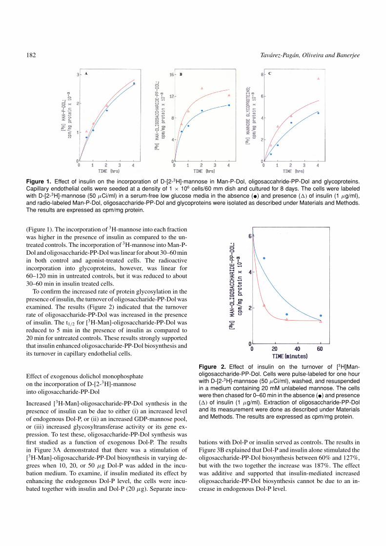
Figure 1. Effect of insulin on the incorporation of D-[2-3H]-mannose in Man-P-Dol, oligosaccahride-PP-Dol and glycoproteins.Capillary endothelial cells were seeded at a density of 1 × 10⁶ cells/60 mm dish and cultured for 8 days. The cells were labeledwith D-[2-3H]-mannose (50 µCi/ml) in a serum-free low glucose media in the absence (•) and presence (�) of insulin (1 µg/ml),and radio-labeled Man-P-Dol, oligosaccharide-PP-Dol and glycoproteins were isolated as described under Materials and Methods.The results are expressed as cpm/mg protein.
(Figure 1). The incorporation of 3H-mannose into each fractionwas higher in the presence of insulin as compared to the un-treated controls. The incorporation of 3H-mannose into Man-P-Dol and oligosaccharide-PP-Dol was linear for about 30–60 minin both control and agonist-treated cells. The radioactiveincorporation into glycoproteins, however, was linear for60–120 min in untreated controls, but it was reduced to about30–60 min in insulin treated cells.
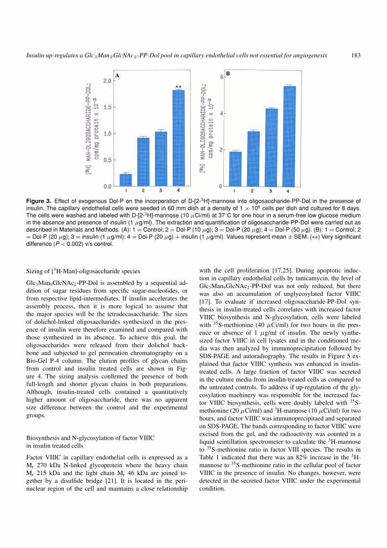
To confirm the increased rate of protein glycosylation in thepresence of insulin, the turnover of oligosaccharide-PP-Dol wasexamined. The results (Figure 2) indicated that the turnoverrate of oligosaccharide-PP-Dol was increased in the presenceof insulin. The t1/2 for [3H-Man]-oligosaccharide-PP-Dol wasreduced to 5 min in the presence of insulin as compared to20 min for untreated controls. These results strongly supportedthat insulin enhanced oligosaccharide-PP-Dol biosynthesis andits turnover in capillary endothelial cells.
Effect of exogenous dolichol monophosphateon the incorporation of D-[2-3H]-mannoseinto oligosaccharide-PP-Dol
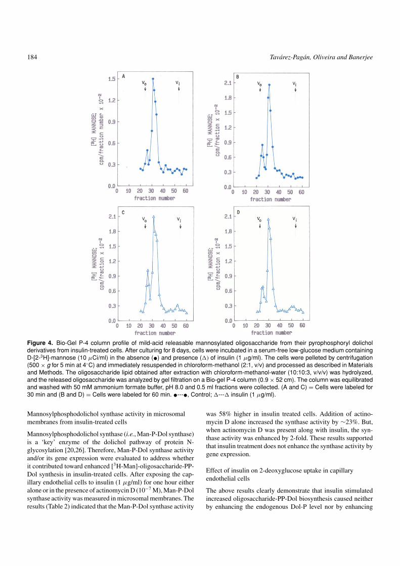
Increased [3H-Man]-oligosaccharide-PP-Dol synthesis in thepresence of insulin can be due to either (i) an increased levelof endogenous Dol-P, or (ii) an increased GDP-mannose pool,or (iii) increased glycosyltransferase activity or its gene ex-pression. To test these, oligosaccharide-PP-Dol synthesis wasfirst studied as a function of exogenous Dol-P. The resultsin Figure 3A demonstrated that there was a stimulation of[3H-Man]-oligosaccharide-PP-Dol biosynthesis in varying de-grees when 10, 20, or 50 µg Dol-P was added in the incu-bation medium. To examine, if insulin mediated its effect byenhancing the endogenous Dol-P level, the cells were incu-bated together with insulin and Dol-P (20 µg). Separate incu-
Figure 2. Effect of insulin on the turnover of [3H]Man-oligosaccharide-PP-Dol. Cells were pulse-labeled for one hourwith D-[2-3H]-mannsoe (50 µCi/ml), washed, and resuspendedin a medium containing 20 mM unlabeled mannose. The cellswere then chased for 0–60 min in the absence (•) and presence(�) of insulin (1 µg/ml). Extraction of oligosaccharide-PP-Doland its measurement were done as described under Materialsand Methods. The results are expressed as cpm/mg protein.
bations with Dol-P or insulin served as controls. The results inFigure 3B explained that Dol-P and insulin alone stimulated theoligosaccharide-PP-Dol biosynthesis between 60% and 127%,but with the two together the increase was 187%. The effectwas additive and supported that insulin-mediated increasedoligosaccharide-PP-Dol biosynthesis cannot be due to an in-crease in endogenous Dol-P level.
Insulin up-regulates a Glc3 Man9 GlcNAc2 -PP-Dol pool in capillary endothelial cells not essential for angiogenesis 183
Figure 3. Effect of exogenous Dol-P on the incorporation of D-[2-3H]-mannose into oligosaccharide-PP-Dol in the presence ofinsulin. The capillary endothelial cells were seeded in 60 mm dish at a density of 1 × 10⁶ cells per dish and cultured for 8 days.The cells were washed and labeled with D-[2-3H]-mannose (10 µCi/ml) at 37◦C for one hour in a serum-free low glucose mediumin the absence and presence of insulin (1 µg/ml). The extraction and quantification of oligosaccharide-PP-Dol were carried out asdescribed in Materials and Methods. (A): 1 = Control; 2 = Dol-P (10 µg); 3 = Dol-P (20 µg); 4 = Dol-P (50 µg). (B): 1 = Control; 2= Dol-P (20 µg); 3 = insulin (1 µg/ml); 4 = Dol-P (20 µg) + insulin (1 µg/ml). Values represent mean ± SEM. (∗∗) Very significantdifference (P < 0.002) v/s control.
Sizing of [3H-Man]-oligosaccharide species
Glc3Man9GlcNAc2-PP-Dol is assembled by a sequential ad-dition of sugar residues from specific sugar-nucleotides, orfrom respective lipid-intermediates. If insulin accelerates theassembly process, then it is more logical to assume thatthe major species will be the tetradecasaccharide. The sizesof dolichol-linked oligosaccharides synthesized in the pres-ence of insulin were therefore examined and compared withthose synthesized in its absence. To achieve this goal, theoligosaccharides were released from their dolichol back-bone and subjected to gel permeation chromatography on aBio-Gel P-4 column. The elution profiles of glycan chainsfrom control and insulin treated cells are shown in Fig-ure 4. The sizing analysis confirmed the presence of bothfull-length and shorter glycan chains in both preparations.Although, insulin-treated cells contained a quantitativelyhigher amount of oligosaccharide, there was no apparentsize difference between the control and the experimentalgroups.
Biosynthesis and N-glycosylation of factor VIIICin insulin treated cells
Factor VIIIC in capillary endothelial cells is expressed as aMr 270 kDa N-linked glycoprotein where the heavy chainMr 215 kDa and the light chain Mr 46 kDa are joined to-gether by a disulfide bridge [21]. It is located in the peri-nuclear region of the cell and maintains a close relationship
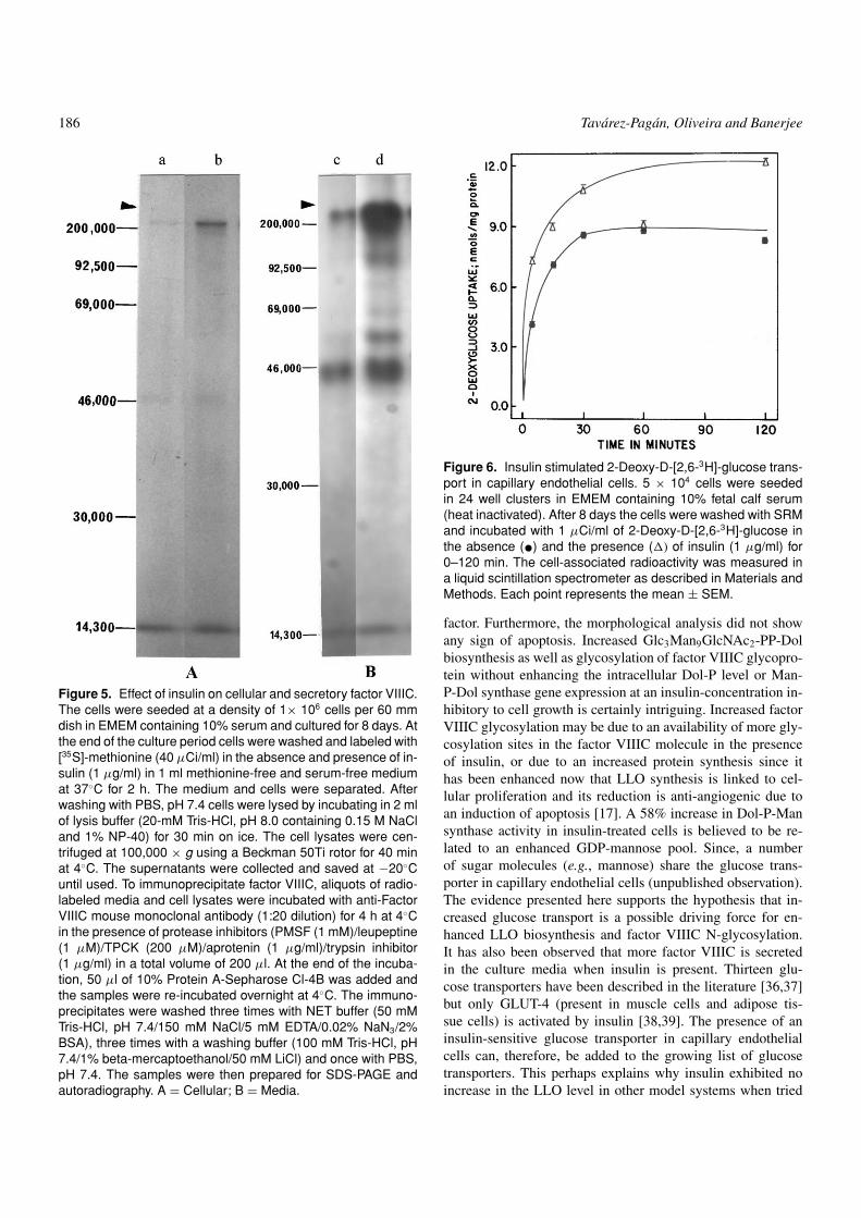
with the cell proliferation [17,25]. During apoptotic induc-tion in capillary endothelial cells by tunicamycin, the level ofGlc3Man9GlcNAc2-PP-Dol was not only reduced, but therewas also an accumulation of unglycosylated factor VIIIC[17]. To evaluate if increased oligosaccharide-PP-Dol syn-thesis in insulin-treated cells correlates with increased factorVIIIC biosynthesis and N-glycosylation, cells were labeledwith 35S-methionine (40 µCi/ml) for two hours in the pres-ence or absence of 1 µg/ml of insulin. The newly synthe-sized factor VIIIC in cell lysates and in the conditioned me-dia was then analyzed by immunoprecipitation followed bySDS-PAGE and autoradiography. The results in Figure 5 ex-plained that factor VIIIC synthesis was enhanced in insulin-treated cells. A large fraction of factor VIIIC was secretedin the culture media from insulin-treated cells as compared tothe untreated controls. To address if up-regulation of the gly-cosylation machinery was responsible for the increased fac-tor VIIIC biosynthesis, cells were doubly labeled with 35S-methionine (20 µCi/ml) and 3H-mannose (10 µCi/ml) for twohours, and factor VIIIC was immunoprecipitaed and separatedon SDS-PAGE. The bands corresponding to factor VIIIC wereexcised from the gel, and the radioactivity was counted in aliquid scintillation spectrometer to calculate the 3H-mannoseto 35S-methionine ratio in factor VIII species. The results inTable 1 indicated that there was an 82% increase in the 3H-mannose to 35S-methionine ratio in the cellular pool of factorVIIIC in the presence of insulin. No changes, however, weredetected in the secreted factor VIIIC under the experimentalcondition.
184 Tavarez-Pagan, Oliveira and Banerjee
Figure 4. Bio-Gel P-4 column profile of mild-acid releasable mannosylated oligosaccharide from their pyrophosphoryl dolicholderivatives from insulin-treated cells. After culturing for 8 days, cells were incubated in a serum-free low-glucose medium containingD-[2-3H]-mannose (10 µCi/ml) in the absence (•) and presence (�) of insulin (1 µg/ml). The cells were pelleted by centrifugation(500 × g for 5 min at 4◦C) and immediately resuspended in chloroform-methanol (2:1, v/v) and processed as described in Materialsand Methods. The oligosaccharide lipid obtained after extraction with chloroform-methanol-water (10:10:3, v/v/v) was hydrolyzed,and the released oligosaccharide was analyzed by gel filtration on a Bio-gel P-4 column (0.9 × 52 cm). The column was equilibratedand washed with 50 mM ammonium formate buffer, pH 8.0 and 0.5 ml fractions were collected. (A and C) = Cells were labeled for30 min and (B and D) = Cells were labeled for 60 min. •---•, Control; �---� insulin (1 µg/ml).
Mannosylphosphodolichol synthase activity in microsomalmembranes from insulin-treated cells
Mannosylphosphodolichol synthase (i.e., Man-P-Dol synthase)is a ‘key’ enzyme of the dolichol pathway of protein N-glycosylation [20,26]. Therefore, Man-P-Dol synthase activityand/or its gene expression were evaluated to address whetherit contributed toward enhanced [3H-Man]-oligosaccharide-PP-Dol synthesis in insulin-treated cells. After exposing the cap-illary endothelial cells to insulin (1 µg/ml) for one hour eitheralone or in the presence of actinomycin D (10−7 M), Man-P-Dolsynthase activity was measured in microsomal membranes. Theresults (Table 2) indicated that the Man-P-Dol synthase activity
was 58% higher in insulin treated cells. Addition of actino-mycin D alone increased the synthase activity by ∼23%. But,when actinomycin D was present along with insulin, the syn-thase activity was enhanced by 2-fold. These results supportedthat insulin treatment does not enhance the synthase activity bygene expression.
Effect of insulin on 2-deoxyglucose uptake in capillaryendothelial cells
The above results clearly demonstrate that insulin stimulatedincreased oligosaccharide-PP-Dol biosynthesis caused neitherby enhancing the endogenous Dol-P level nor by enhancing
Insulin up-regulates a Glc3 Man9 GlcNAc2 -PP-Dol pool in capillary endothelial cells not essential for angiogenesis 185
Table 1. Effect of insulin on the ratio of [3H]-mannose to [35S]-methionine incorporation into Factor VIIICa
Cellular Media
Total
Sample Mean ± SEM% Increaseover control Mean ± SEM
% Increaseover control Cellular + Media
Control 1.00 ± 0.14 – 1.00 ± 0.27 – –Insulin (1 µg/ml) 1.82 ± 0.15 82 1.00 ± 0.13 0 82
aCapillary endothelial cells (1 × 10⁶ cells/60 mm dish) were cultured for 8 days in a regular EMEM. At the end, the cells were washed andincubated in the presence and absence of insulin (1 µg/ml) in 1 ml of low glucose, methionine-free medium containing aprotinine (1 µg/ml)together with [35S]-methionine (20 µCi/ml; Sp. Act. 1400 Ci/mmol) and D-[2-3H]-mannose (10 µCi/ml; Sp. Act. 18.5 Ci/mmol) at 37◦C for 2 h ina CO2 incubator. After separating the culture medium, the cells were lysed in the lysis buffer as described in the Materials and Methods. FactorVIIIC was immunoprecipitated from media and cell lysates, and subjected to SDS-PAGE followed by autoradiography. The radioactivities in factorVIIIC bands were quantified by counting in a liquid scintillation spectrometer, and the ratio of [3H] to [35S] was determined. The results are mean± SEM from three experiments.
Table 2. Effect of insulin on Man-P-Dol synthase activity incapillary endothelial cellsa
Man-P-Dol synthase activityTreatment (pmol/mg protein/5 min)
Control 8.4 (7.5; 9.3)Insulin (1 µg/ml) 13.3 (18.1; 9.06)Actinomycin D (2 × 10⁻³ M) 9.7 (11.6; 7.9)Insulin (1 µg/ml) + 17.1 (12.4; 22.4)
Actinomycin D (2 × 10⁻³ M)
aMicrosomal membranes from control and insulin treated (1 µg/ml)capillary endothelial cells were incubated in 5 mM Tris-HCl, pH 7.0 con-taining 12.5 mM sucrose, 5 µm EDTA, 5 mM MnCl2, 4 mM 5’-AMPand 2.5 µM GDP-[U-14C]mannose (Sp. Act. 412 cpm/pmol) in 100 µlfor 5 min at 37◦C, and Man-P-Dol was extracted as described in Mate-rials and Methods. The results are an average from two measurements.The numbers in the parentheses are the two numbers).
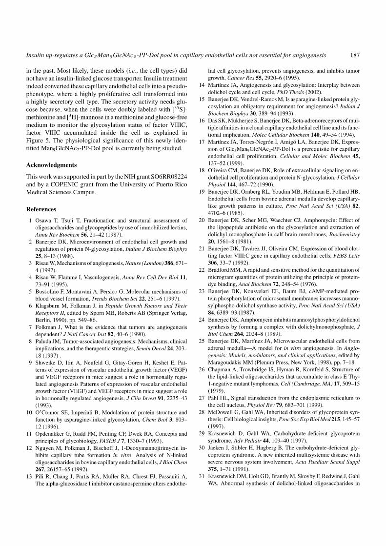
the Man-P-Dol synthase activity. Therefore, the attention wasturned toward addressing the size of the sugar nucleotide pool.Since sugar nucleotide synthesis requires the availability of in-tracellular sugar molecules, we, therefore, studied the entry of2-deoxy-D-[2,6-3H]-glucose into the cell in the presence andabsence of insulin. As shown in Figure 6, the transport of 2-deoxy-D-[2,6-3H]-glucose across the endothelial cell plasmamembrane was a time-dependent process. It also explainedthat the transport was linear approximately for 15 min andthen reached a steady state. Addition of insulin (1 µg/ml) in-creased the rate of 2-deoxyglucose transport into the capillaryendothelial cells.
Discussion
Insulin, a growth factor, has been found to down-regulate theproliferation of capillary endothelial cells [18]. To delineatethis reciprocal relationship, we have studied the modulationof Glc3Man9GlcNAc2-PP-Dol assembly in the presence of in-sulin. The results suggested that insulin signaling of capillary
endothelial cell growth inhibition was not mediated by down-regulation of LLO biosynthesis. In fact, LLO biosynthesis wasup-regulated in insulin treated capillary endothelial cells.
The roles of glycoprotein glycans span from protein struc-ture to cell function. The glycan chain assembly, therefore, mustrespond to environmental as well as humoral factors. For ex-ample, activation of β-adrenoreceptor enhanced capillary en-dothelial cell proliferation and capillary lumen formation byactivating the protein N-glycosylation pathway mediated by in-tracellular cAMP [15,16]. A recent report from our laboratoryalso supported that down-regulation of Glc3Man9GlcNAc2-PP-Dol synthesis in capillary endothelial cells by inhibitingGcNAc-1phosphate transferase with a pyrimidine nucleoside,tunicamycin, or tying up intracellular dolichylmonophosphate(Dol-P) with a lipopeptide, amphomycin, reduced angiogenesisby arresting cells in G1 phase and induced apoptosis. The cellsused a signaling mechanism of Unfolded Protein Response [27].In addition, fatal disorders of glycoprotein biosynthesis such ascongenital disorders of glycosylation (CDG) are a result of de-fects in the synthesis of N-linked glycans [28–30]. This defectmay result in the absence of entire N-linked glycan chains frommany proteins or from under-glycosylation due to synthesis ofa truncated oligosaccharide-lipd (LLO) precursor because of ashortage of Man-P-Dol [15,20,27,31–34].
The capillary endothelial cells line blood vessels. Therefore,angiogenesis is an essential physiological event during growthand development, as well as during wound healing. Failing toinduce capillary endothelial cell proliferation by insulin [18],irrespective of the presence of 210,000 high affinity receptorsper cell [35], raised a question about the modulation of thedolichol cycle of the protein N-glycosylation by insulin. Theresults presented here clearly demonstrate the presence of areciprocal relationship between the inhibition of capillary en-dothelial cell proliferation and the Glc3Man9GlcNAc2-PP-Dolbiosynthesis in cells treated with insulin. The exact molecularmechanism of the growth inhibitory action of insulin in capil-lary endothelial cells is currently not understood, but it is be-lieved that increased cell-doubling time is the main contributing
186 Tavarez-Pagan, Oliveira and Banerjee
Figure 5. Effect of insulin on cellular and secretory factor VIIIC.The cells were seeded at a density of 1× 10⁶ cells per 60 mmdish in EMEM containing 10% serum and cultured for 8 days. Atthe end of the culture period cells were washed and labeled with[35S]-methionine (40 µCi/ml) in the absence and presence of in-sulin (1 µg/ml) in 1 ml methionine-free and serum-free mediumat 37◦C for 2 h. The medium and cells were separated. Afterwashing with PBS, pH 7.4 cells were lysed by incubating in 2 mlof lysis buffer (20-mM Tris-HCl, pH 8.0 containing 0.15 M NaCland 1% NP-40) for 30 min on ice. The cell lysates were cen-trifuged at 100,000 × g using a Beckman 50Ti rotor for 40 minat 4◦C. The supernatants were collected and saved at −20◦Cuntil used. To immunoprecipitate factor VIIIC, aliquots of radio-labeled media and cell lysates were incubated with anti-FactorVIIIC mouse monoclonal antibody (1:20 dilution) for 4 h at 4◦Cin the presence of protease inhibitors (PMSF (1 mM)/leupeptine(1 µM)/TPCK (200 µM)/aprotenin (1 µg/ml)/trypsin inhibitor(1 µg/ml) in a total volume of 200 µl. At the end of the incuba-tion, 50 µl of 10% Protein A-Sepharose Cl-4B was added andthe samples were re-incubated overnight at 4◦C. The immuno-precipitates were washed three times with NET buffer (50 mMTris-HCl, pH 7.4/150 mM NaCl/5 mM EDTA/0.02% NaN3/2%BSA), three times with a washing buffer (100 mM Tris-HCl, pH7.4/1% beta-mercaptoethanol/50 mM LiCl) and once with PBS,pH 7.4. The samples were then prepared for SDS-PAGE andautoradiography. A = Cellular; B = Media.
Figure 6. Insulin stimulated 2-Deoxy-D-[2,6-3H]-glucose trans-port in capillary endothelial cells. 5 × 10⁴ cells were seededin 24 well clusters in EMEM containing 10% fetal calf serum(heat inactivated). After 8 days the cells were washed with SRMand incubated with 1 µCi/ml of 2-Deoxy-D-[2,6-3H]-glucose inthe absence (•) and the presence (�) of insulin (1 µg/ml) for0–120 min. The cell-associated radioactivity was measured ina liquid scintillation spectrometer as described in Materials andMethods. Each point represents the mean ± SEM.
factor. Furthermore, the morphological analysis did not showany sign of apoptosis. Increased Glc3Man9GlcNAc2-PP-Dolbiosynthesis as well as glycosylation of factor VIIIC glycopro-tein without enhancing the intracellular Dol-P level or Man-P-Dol synthase gene expression at an insulin-concentration in-hibitory to cell growth is certainly intriguing. Increased factorVIIIC glycosylation may be due to an availability of more gly-cosylation sites in the factor VIIIC molecule in the presenceof insulin, or due to an increased protein synthesis since ithas been enhanced now that LLO synthesis is linked to cel-lular proliferation and its reduction is anti-angiogenic due toan induction of apoptosis [17]. A 58% increase in Dol-P-Mansynthase activity in insulin-treated cells is believed to be re-lated to an enhanced GDP-mannose pool. Since, a numberof sugar molecules (e.g., mannose) share the glucose trans-porter in capillary endothelial cells (unpublished observation).The evidence presented here supports the hypothesis that in-creased glucose transport is a possible driving force for en-hanced LLO biosynthesis and factor VIIIC N-glycosylation.It has also been observed that more factor VIIIC is secretedin the culture media when insulin is present. Thirteen glu-cose transporters have been described in the literature [36,37]but only GLUT-4 (present in muscle cells and adipose tis-sue cells) is activated by insulin [38,39]. The presence of aninsulin-sensitive glucose transporter in capillary endothelialcells can, therefore, be added to the growing list of glucosetransporters. This perhaps explains why insulin exhibited noincrease in the LLO level in other model systems when tried
Insulin up-regulates a Glc3 Man9 GlcNAc2 -PP-Dol pool in capillary endothelial cells not essential for angiogenesis 187
in the past. Most likely, these models (i.e., the cell types) didnot have an insulin-linked glucose transporter. Insulin treatmentindeed converted these capillary endothelial cells into a pseudo-phenotype, where a highly proliferative cell transformed intoa highly secretory cell type. The secretory activity needs glu-cose because, when the cells were doubly labeled with [35S]-methionine and [3H]-mannose in a methionine and glucose-freemedium to monitor the glycosylation status of factor VIIIC,factor VIIIC accumulated inside the cell as explained inFigure 5. The physiological significance of this newly iden-tified Man9GlcNAc2-PP-Dol pool is currently being studied.
Acknowledgments
This work was supported in part by the NIH grant SO6RR08224and by a COPENIC grant from the University of Puerto RicoMedical Sciences Campus.
References
1 Osawa T, Tsuji T, Fractionation and structural assessment ofoligosaccharides and glycopeptides by use of immobilized lectins,Annu Rev Biochem 56, 21–42 (1987).
2 Banerjee DK, Microenvironment of endothelial cell growth andregulation of protein N-glycosylation, Indian J Biochem Biophys25, 8–13 (1988).
3 Risau W, Mechanisms of angiogenesis, Nature (London) 386, 671–4 (1997).
4 Risau W, Flamme I, Vasculogenesis, Annu Rev Cell Dev Biol 11,73–91 (1995).
5 Bussolino F, Montavani A, Persico G, Molecular mechanisms ofblood vessel formation, Trends Biochem Sci 22, 251–6 (1997).
6 Klagsburn M, Folkman J, in Peptide Growth Factors and TheirReceptors II, edited by Sporn MB, Roberts AB (Springer Verlag,Berlin, 1990), pp. 549–86.
7 Folkman J, What is the evidence that tumors are angiogenesisdependent? J Natl Cancer Inst 82, 40–6 (1990).
8 Paluda JM, Tumor-associated angiogenesis: Mechanisms, clinicalimplications, and the therapeutic strategies, Semin Oncol 24, 203–18 (1997) .
9 Shweike D, Itin A, Neufeld G, Gitay-Goren H, Keshet E, Pat-terns of expression of vascular endothelial growth factor (VEGF)and VEGF receptors in mice suggest a role in hormonally regu-lated angiogenesis Patterns of expression of vascular endothelialgrowth factor (VEGF) and VEGF receptors in mice suggest a rolein hormonally regulated angiogenesis, J Clin Invest 91, 2235–43(1993).
10 O’Connor SE, Imperiali B, Modulation of protein structure andfunction by asparagine-linked glycosylation, Chem Biol 3, 803–12 (1996).
11 Opdenakker G, Rudd PM, Penting CP, Dwek RA, Concepts andprinciples of glycobiology, FASEB J 7, 1330–7 (1993).
12 Nguyen M, Folkman J, Bischoff J, 1-Deoxymannojirimycin in-hibits capillary tube formation in vitro. Analysis of N-linkedoligosaccharides in bovine capillary endothelial cells, J Biol Chem267, 26157–65 (1992).
13 Pili R, Chang J, Partis RA, Muller RA, Chrest FJ, Passaniti A,The alpha-glucosidase I inhibitor castanospermine alters endothe-
lial cell glycosylation, prevents angiogenesis, and inhibits tumorgrowth, Cancer Res 55, 2920–6 (1995).
14 Martınez JA, Angiogenesis and glycosylation: Interplay betweendolichol cycle and cell cycle, PhD Thesis (2002).
15 Banerjee DK, Vendrel-Ramos M, Is asparagine-linked protein gly-cosylation an obligatory requirement for angiogenesis? Indian JBiochem Biophys 30, 389–94 (1993).
16 Das SK, Mukherjee S, Banerjee DK, Beta-adrenoreceptors of mul-tiple affinities in a clonal capillary endothelial cell line and its func-tional implication, Molec Cellular Biochem 140, 49–54 (1994).
17 Martınez JA, Torres-Negron I, Amigo LA, Banerjee DK, Expres-sion of Glc3Man9GlcNAc2-PP-Dol is a prerequisite for capillaryendothelial cell proliferation, Cellular and Molec Biochem 45,137–52 (1999).
18 Oliveira CM, Banerjee DK, Role of extracellular signaling on en-dothelial cell proliferation and protein N-glycosylation, J CellularPhysiol 144, 467–72 (1990).
19 Banerjee DK, Ornberg RL, Youdim MB, Heldman E, Pollard HB,Endothelial cells from bovine adrenal medulla develop capillary-like growth patterns in culture, Proc Natl Acad Sci (USA) 82,4702–6 (1985).
20 Banerjee DK, Scher MG, Waechter CJ, Amphomycin: Effect ofthe lipopeptide antibiotic on the glycosylation and extraction ofdolichyl monophosphate in calf brain membranes, Biochemistry20, 1561–8 (1981).
21 Banerjee DK, Tavarez JJ, Oliveira CM, Expression of blood clot-ting factor VIII:C gene in capillary endothelial cells, FEBS Letts306, 33–7 (1992).
22 Bradford MM, A rapid and sensitive method for the quantitation ofmicrogram quantities of protein utilizing the principle of protein-dye binding, Anal Biochem 72, 248–54 (1976).
23 Banerjee DK, Kousvelari EE, Baum BJ, cAMP-mediated pro-tein phosphorylation of microsomal membranes increases manno-sylphospho dolichol synthase activity, Proc Natl Acad Sci (USA)84, 6389–93 (1987).
24 Banerjee DK, Amphomycin inhibits mannosylphosphoryldolicholsynthesis by forming a complex with dolichylmonophosphate, JBiol Chem 264, 2024–8 (1989).
25 Banerjee DK, Martınez JA, Microvascular endothelial cells fromadrenal medulla—A model for in vitro angiogenesis. In Angio-genesis: Models, modulators, and clinical applications, edited byMaragoudakis MM (Plenum Press, New York, 1998), pp. 7–18.
26 Chapman A, Trowbridge IS, Hyman R, Kornfeld S, Structure ofthe lipid-linked oligosaccharides that accumulate in class E Thy-1-negative mutant lymphomas, Cell (Cambridge, MA) 17, 509–15(1979).
27 Pahl HL, Signal transduction from the endoplasmic reticulum tothe cell nucleus, Physiol Rev 79, 683–701 (1999).
28 McDowell G, Gahl WA, Inherited disorders of glycoprotein syn-thesis: Cell biological insights, Proc Soc Exp Biol Med 215, 145–57(1997).
29 Krasnewich D, Gahl WA, Carbohydrate-deficient glycoproteinsyndrome, Adv Pediatr 44, 109–40 (1997).
30 Jaeken J, Stibler H, Hagberg B, The carbohydrate-deficient gly-coprotein syndrome. A new inherited multisystemic disease withsevere nervous system involvement, Acta Paediatr Scand Suppl375, 1–71 (1991).
31 Krasnewich DM, Holt GD, Brantly M, Skovby F, Redwine J, GahlWA, Abnormal synthesis of dolichol-linked oligosaccharides in
188 Tavarez-Pagan, Oliveira and Banerjee
carbohydrate-deficient glycoprotein syndrome, Glycobiology 5,503–10 (1995).
32 Korner C, Lehle L, von Figura K, Abnormal synthesis of mannose1-phosphate derived carbohydrates in carbohydrate-deficient gly-coprotein syndrome type I fibroblasts with phosphomannomutasedeficiency, Glycobiology 8, 165–71 (1998).
33 Imbach T, Schenk B, Schollen E, Burda P, Stutz A, Grunewald S,Bailie NM, King MD, Jaeken J, Matthijs G, Berger EG, Abei M,Hennet T, Deficiency of dolichol-phosphate-mannose synthase-1causes congenital disorder of glycosylation type Ie, J Clin Invest105, 233–9 (2000).
34 Volker C, De Praeter CM, Hardt B, Breuer W, Kalz-Fuller B, VanCoster RN, Bause E, Processing of N-linked carbohydrate chainsin a patient with glucosidase I deficiency (CDG type Iib), Glyco-biology 12, 473–83 (2002).
35 Brush JS, Tavarez-Pagan JJ, Banerjee DK, Insulin and IGF-1 man-ifest differential effects in a clonal capillary endothelial cell line,Biochem Int 25, 537–45 (1991).
36 Doege H, Bocianski A, Scheepers A, Axer H, Eckel J, JoostH-G, Schurmann A, Characterization of human glucose transporter(GLUT) 11 (encoded by SLC2A11), a novel sugar-transport facil-itator specifically expressed in heart and skeletal muscle, BiochemJ 359, 443–9 (2001).
37 Uldry M, Ibberson M, Horisberger J-D, Chatton J-Y, Riederer BM,Thorens B, Identification of a mammalian H(+)-myo-inositol sym-porter expressed predominantly in the brain, The EMBO J 20,4467–77 (2001).
38 Sampaio de Freitas M, Garcia De Souza EP, Vargas daSilva S, da Rocha Kaezer A, da Silva Vieira R, SanchezMA, Barja-Fidalgo C, Up-regulation of phosphatidylinositol 3-kinase and glucose transporter 4 in muscle of rats subjectedto maternal undernutrition, Biochim Biophys Acta 1639, 8–16(2003).
39 Kahn HR, Pessian JE, Insulin regulation of glucose uptake: A com-plex interplay of intracellular signalling pathways, Diabetologia45, 1475–83 (2002).


![Characterization of glycosphingolipids from Schistosoma mansoni eggs carrying Fuc(��1-3)GalNAc, GalNAc(��1-4)[Fuc(��1-3)]GlcNAc- and Gal(��1-4)[Fuc(��1-3)]GlcNAc-](https://static.fdokumen.com/doc/165x107/63176e09b6c3e3926d0ddfa3/characterization-of-glycosphingolipids-from-schistosoma-mansoni-eggs-carrying-fuc1-3galnac.jpg)