Tumor Necrosis Factor-alpha and Muc2 Mucin Play Major Roles in Disease Onset and Progression in...
13
Tumor Necrosis Factor-a and Muc2 Mucin Play Major Roles in Disease Onset and Progression in Dextran Sodium Sulphate-Induced Colitis Poonam Dharmani, Pearl Leung, Kris Chadee* Department of Microbiology, Immunology and Infectious Diseases, Gastrointestinal Research Group, Health Sciences Centre, University of Calgary, Calgary, Alberta, Canada Abstract The sequential events and the inflammatory mediators that characterize disease onset and progression of ulcerative colitis (UC) are not well known. In this study, we evaluated the early pathologic events in the pathogenesis of colonic ulcers in rats treated with dextran sodium sulfate (DSS). Following a lag phase, day 5 of DSS treatment was found clinically most critical as disease activity index (DAI) exhibited an exponential rise with severe weight loss and rectal bleeding. Surprisingly, on days 1-2, colonic TNF-a expression (70-80-fold) and tissue protein (50-fold) were increased, whereas IL-1b only increased on days 7-9 (60-90-fold). Days 3-6 of DSS treatment were characterized by a prominent down regulation in the expression of regulatory cytokines (40-fold for IL-10 and TGFb) and mucin genes (15-18 fold for Muc2 and Muc3) concomitant with depletion of goblet cell and adherent mucin. Remarkably, treatment with TNF-a neutralizing antibody markedly altered DSS injury with reduced DAI, restoration of the adherent and goblet cell mucin and IL-1b and mucin gene expression. We conclude that early onset colitis is dependent on TNF-a that preceded depletion of adherent and goblet cell mucin prior to epithelial cell damage and these biomarkers can be used as therapeutic targets for UC. Citation: Dharmani P, Leung P, Chadee K (2011) Tumor Necrosis Factor-a and Muc2 Mucin Play Major Roles in Disease Onset and Progression in Dextran Sodium Sulphate-Induced Colitis. PLoS ONE 6(9): e25058. doi:10.1371/journal.pone.0025058 Editor: Irun R. Cohen, Weizmann Institute of Science, Israel Received August 18, 2011; Accepted August 26, 2011; Published September 19, 2011 Copyright: ß 2011 Dharmani et al. This is an open-access article distributed under the terms of the Creative Commons Attribution License, which permits unrestricted use, distribution, and reproduction in any medium, provided the original author and source are credited. Funding: This work was supported by a grant from the Canadian Institute for Health Research (CIHR, KC). PD is funded by a Canadian Association of Gastroenterology-AstraZeneca-CIHR Research and Fellowship Award. The funders had no role in study design, data collection and analysis, decision to publish, or preparation of the manuscript. Competing Interests: The authors have declared that no competing interests exist. * E-mail: [email protected] Introduction Inflammatory bowel disease (IBD), an umbrella term that includes Crohn’s disease and ulcerative colitis (UC), are chronic relapsing inflammatory disorders of the gut that are believed to occur in genetically predisposed individuals due to exposure of unknown environmental and microbial agents [1]. A normal healthy intestine exhibits homeostasis where the mucosal immune system escalates an immune response against pathogens but remains tolerant to antigens derived from food and commensal microbes. Loss of mucosal tolerance is due to an uncontrolled inflammatory cascade resulting from a number of mutual and probably sequential events involving both immune (gut associated lymphoid tissues, GALT and professional antigen presenting cells, APC) and non-immune cells/molecules (epithelial cells of gut and resident microflora) [1,2]. However, the precise etiology of the pathogenesis of UC is still not known. To date, studies to unravel the pathogenesis of UC have been focused on various mucosal models of inflammation that closely resembles human colitis. One of the most comprehensively illustrated models of experimental colitis is Dextran Sodium Sulphate (DSS) induced colitis which mimics the clinical and histological features of human UC as the colonic lesions exhibits high homogeneity and reproducibility [3]. Acute and chronic colitis induced by DSS has been used to study changes in metabolically labeled and tissue mucin content [4] and/or changes in epithelial permeability, MPO and pro-inflammatory cytokines [4]. However, the inflammatory mediators that play a role in disease onset and progression of colitis are poorly defined. Clinical and experimental studies using DSS models of colitis suggests that the key contributors in disease pathogenesis include: (i) an alteration in the mucosal barrier integrity and function; (ii) reallocation in the role of pathogen recognition receptors (PRRs) of APCs and, (iii) an immune response skewed towards effector cell function (Th1 and probably Th2) [2,5]. Despite these advances, it is still not clear which mediator(s) play a central role in disease onset and/or progression of colitis. As TNF-a and adherent and goblet cell mucin are two major components that are altered in UC, we reasoned that both of these components play major roles in epithelial barrier function and may be selectively altered prior to epithelial cell damage in DSS- induced colitis. In the present study, we used a protocol-treating animal for 9 consecutive days with DSS to characterize the earliest events in disease onset and progression to acute colitis. In particular, we focused on the contribution of TNF-a and colonic mucin in innate host defense prior to epithelial cell damage and investigated whether TNF-a neutralizing antibody can alter disease onset and/or progression in DSS-induced colitis in rats. Results Disease Activity Index (DAI) and Tissue damage DAI is a cumulative index of body weight loss, rectal bleeding and stool malformation and is considered as the best measure of PLoS ONE | www.plosone.org 1 September 2011 | Volume 6 | Issue 9 | e25058
Transcript of Tumor Necrosis Factor-alpha and Muc2 Mucin Play Major Roles in Disease Onset and Progression in...

Tumor Necrosis Factor-a and Muc2 Mucin Play MajorRoles in Disease Onset and Progression in DextranSodium Sulphate-Induced ColitisPoonam Dharmani, Pearl Leung, Kris Chadee*
Department of Microbiology, Immunology and Infectious Diseases, Gastrointestinal Research Group, Health Sciences Centre, University of Calgary, Calgary, Alberta,
Canada
Abstract
The sequential events and the inflammatory mediators that characterize disease onset and progression of ulcerative colitis(UC) are not well known. In this study, we evaluated the early pathologic events in the pathogenesis of colonic ulcers in ratstreated with dextran sodium sulfate (DSS). Following a lag phase, day 5 of DSS treatment was found clinically most critical asdisease activity index (DAI) exhibited an exponential rise with severe weight loss and rectal bleeding. Surprisingly, on days1-2, colonic TNF-a expression (70-80-fold) and tissue protein (50-fold) were increased, whereas IL-1b only increased on days7-9 (60-90-fold). Days 3-6 of DSS treatment were characterized by a prominent down regulation in the expression ofregulatory cytokines (40-fold for IL-10 and TGFb) and mucin genes (15-18 fold for Muc2 and Muc3) concomitant withdepletion of goblet cell and adherent mucin. Remarkably, treatment with TNF-a neutralizing antibody markedly altered DSSinjury with reduced DAI, restoration of the adherent and goblet cell mucin and IL-1b and mucin gene expression. Weconclude that early onset colitis is dependent on TNF-a that preceded depletion of adherent and goblet cell mucin prior toepithelial cell damage and these biomarkers can be used as therapeutic targets for UC.
Citation: Dharmani P, Leung P, Chadee K (2011) Tumor Necrosis Factor-a and Muc2 Mucin Play Major Roles in Disease Onset and Progression in Dextran SodiumSulphate-Induced Colitis. PLoS ONE 6(9): e25058. doi:10.1371/journal.pone.0025058
Editor: Irun R. Cohen, Weizmann Institute of Science, Israel
Received August 18, 2011; Accepted August 26, 2011; Published September 19, 2011
Copyright: � 2011 Dharmani et al. This is an open-access article distributed under the terms of the Creative Commons Attribution License, which permitsunrestricted use, distribution, and reproduction in any medium, provided the original author and source are credited.
Funding: This work was supported by a grant from the Canadian Institute for Health Research (CIHR, KC). PD is funded by a Canadian Association ofGastroenterology-AstraZeneca-CIHR Research and Fellowship Award. The funders had no role in study design, data collection and analysis, decision to publish, orpreparation of the manuscript.
Competing Interests: The authors have declared that no competing interests exist.
* E-mail: [email protected]
Introduction
Inflammatory bowel disease (IBD), an umbrella term that
includes Crohn’s disease and ulcerative colitis (UC), are chronic
relapsing inflammatory disorders of the gut that are believed to
occur in genetically predisposed individuals due to exposure of
unknown environmental and microbial agents [1]. A normal
healthy intestine exhibits homeostasis where the mucosal immune
system escalates an immune response against pathogens but
remains tolerant to antigens derived from food and commensal
microbes. Loss of mucosal tolerance is due to an uncontrolled
inflammatory cascade resulting from a number of mutual and
probably sequential events involving both immune (gut associated
lymphoid tissues, GALT and professional antigen presenting cells,
APC) and non-immune cells/molecules (epithelial cells of gut and
resident microflora) [1,2]. However, the precise etiology of the
pathogenesis of UC is still not known.
To date, studies to unravel the pathogenesis of UC have been focused
on various mucosal models of inflammation that closely resembles
human colitis. One of the most comprehensively illustrated models of
experimental colitis is Dextran Sodium Sulphate (DSS) induced colitis
which mimics the clinical and histological features of human UC as the
colonic lesions exhibits high homogeneity and reproducibility [3]. Acute
and chronic colitis induced by DSS has been used to study changes in
metabolically labeled and tissue mucin content [4] and/or changes in
epithelial permeability, MPO and pro-inflammatory cytokines [4].
However, the inflammatory mediators that play a role in disease onset
and progression of colitis are poorly defined. Clinical and experimental
studies using DSS models of colitis suggests that the key contributors in
disease pathogenesis include: (i) an alteration in the mucosal barrier
integrity and function; (ii) reallocation in the role of pathogen
recognition receptors (PRRs) of APCs and, (iii) an immune response
skewed towards effector cell function (Th1 and probably Th2) [2,5].
Despite these advances, it is still not clear which mediator(s) play a
central role in disease onset and/or progression of colitis.
As TNF-a and adherent and goblet cell mucin are two major
components that are altered in UC, we reasoned that both of these
components play major roles in epithelial barrier function and
may be selectively altered prior to epithelial cell damage in DSS-
induced colitis. In the present study, we used a protocol-treating
animal for 9 consecutive days with DSS to characterize the earliest
events in disease onset and progression to acute colitis. In
particular, we focused on the contribution of TNF-a and colonic
mucin in innate host defense prior to epithelial cell damage and
investigated whether TNF-a neutralizing antibody can alter
disease onset and/or progression in DSS-induced colitis in rats.
Results
Disease Activity Index (DAI) and Tissue damageDAI is a cumulative index of body weight loss, rectal bleeding
and stool malformation and is considered as the best measure of
PLoS ONE | www.plosone.org 1 September 2011 | Volume 6 | Issue 9 | e25058

clinical activity of colitis [6]. DAI during the first 4 days was not
associated with significant change in the body weight of control
and DSS treated animals. However, on day 5 there was an
exponential increase in DAI that continued up to day 9 (Fig. 1A).
Clinically, day 5 of DSS treatment was a critical turning point as
DAI strongly correlated with weight loss (Fig. 1B) in comparison to
control animals. An approximate reduction of 18–20% total body
weight was observed in DSS treated animals on day 9. After 9 days
of continuous 5% DSS treatment, animals suffered severe rectal
bleeding (30%) and/or deaths (10% of animals).
To determine the earliest histological alterations during DSS-
induced colitis, serial sections of the distal colon predicted to
develop ulcers were evaluated on a daily basis. On days 2–4, no
histological alterations were observed (Fig. 2B), but as early as day
5, focal erosions of the epithelium with acute inflammatory
infiltrate (Fig. 2C) including lymphocyte and polymorphonuclear
lymphocytes were seen in DSS treated animals. In particular, crypt
dysplasia (Fig. 2D) was evident during the development of colitis
from days 5–9. Notably, moderate to severe submucosal edema,
hyperemia and erosions were observed in the colon in DSS treated
animals on day 9 (Fig. 2E). Typical histological alteration in the
mucosa resembled active UC with severe mucosal and submucosal
lesions.
DSS Alters Colonic Mucin and Muc2/3 ExpressionMucin is the first line of host defense and its alteration severely
affects epithelial barrier function [7]. Defects in mucosal cell barrier
function are related to permeability to macromolecules, increased
bacterial invasion and/or translocation which primarily depends
upon depletion of the thick viscous mucin layer due to severe mucus
secretagogues activity, depletion of mucin by goblet cells and mucin
wash out due to mucosal inflammation and diarrhea [8]. As loss of
the protective mucus barrier and goblet cell mucin may be the initial
inciting event that underlies injury and inflammation in UC, we
quantified randomly the number of goblet cells in the crypts that
were filled/empty or releasing mucin in DSS-treated rats. In control
animals there was a thick adherent mucus layer on the epithelium
and well-organized long crypts with dense mucin-filled goblet cells
(Fig. 3A). Morphologically, in control animals, 84% of goblet cells
were filled with mucin and only 4% of empty goblet cells were seen.
However, in DSS-treated animals a significant temporal change in
the number and morphology of mucin secreting activity of goblet
cells were observed. On day 2 of DSS treatment, goblet cells were
filled with mucin (87%) accompanied by a thick adherent and loose
mucus exudate in the lumen in the absence of tissue injury or
abnormal cellular infiltrate (Fig. 3B and Table 1). However, on day
5, intense mucus secretagogue activity resulted in goblet cells
depleted of mucin and in other areas mucus streaming for goblet
cells with a thick none adherent mucus layer on the surface
epithelium (Fig. 3C). As shown in Table 1, there was a significant
decrease in the number of filled goblet cells (21% in comparison to
84% of control) with a corresponding rise in number of empty
goblet cells (49% in comparison to 4% of control) and goblet cells
releasing mucin (31% in comparison to 12% of control). The mucus
cap was layered on the injured surface focal lesions. A curious
finding was that goblet cells in the lower half of the crypts were
devoid of mucin (Fig. 3C). This time point of high mucin
secretagogue activity also coincided with a sharp increase in DAI
(day 5). On day 7 of DSS treatment, few goblet cells were seen at the
site of well-developed ulcers formation, the mucus cap was
completely lost and goblet cells in areas adjacent to the ulcers had
very little mucin (Fig. 3D). In particular, there was a significant
increase in the number of empty goblet cells (77% in comparison to
4% of control, Table 1). On the day 9 of DSS treatment, goblet cells
were almost absent with a paucity of PAS positive proteins in the
ulcerated site. In the adjacent areas to the ulcers, crypts were
Figure 1. Disease Activity Index (A, DAI) and change in body weight (B) during the progressive development of DSS-induced colitisin rats. Animals received 5% DSS in drinking water for 1–9 days. Note, a striking difference in DAI (A) was observed from day 5 onwards (arrow).Changes in the body weight (B) of control (asterisk) and DSS treated animals (circles). Loss in the body weight coincides well with increase in DAI. Datarepresents the means 6 SEM from 6 animals per day.doi:10.1371/journal.pone.0025058.g001
Pathogenesis of DSS-Induced Colitis
PLoS ONE | www.plosone.org 2 September 2011 | Volume 6 | Issue 9 | e25058

damaged and the few goblet cells contained insignificant amount of
mucin (Fig. 3E).
As a decrease in luminal mucin content may reflect a differential
expression of mucin genes, we determine whether the expression
of secretory (Muc2) and membrane bound (Muc3) mucin were also
altered during the onset of ulcer formation. As shown in Fig. 3F,
following a significant increase in Muc2 (,22 fold) and Muc3 (,8
fold) gene expression between days 1–2, there was a marked down
regulation of Muc2/3 from day 4 onwards (15- and 18-fold lower
respectively). No significant difference in the expression of Muc1
was observed in DSS treated and control animals.
DSS Treatment Alters the Expression Pattern of TLRsTLRs are sensors on epithelial cells/APCs that identify and
respond to microbes by eliciting effector, regulatory or cytopro-
tective responses [7]. As changes in TLR expression pattern are
critical for the induction of both mucosal effector and regulatory
cell responses, the expression of TLR2, TLR4, TLR5 and TLR9
genes involved in microbial recognition was examined. Surpris-
ingly, the expression profiles for TLR2/4 (Fig. 4A) and TLR5/9
(Fig. 4B) genes in DSS treated animals increased 60–80-fold and
10–20-fold, respectively, on days 1–3. The increase in TLR2/4
expression was evidenced through all 9 days of DSS treatment,
albeit was not as prominent as during the first three days (Fig. 4A).
TLR5/9 expression levels returned to normal on day 5 and
remained low up to day 9 (Fig. 4B). DSS was a potent
inflammatory insult for TLR expression in the onset of disease.
DSS Stimulates the Production of Pro-inflammatory andRegulatory Cytokines
A break in tolerance to enteric bacteria and an aberrant response
to normal luminal flora leading to an immunological imbalance is
Figure 2. Histopathological characterization of DSS-induced colitis. A: Normal control rat colon on day 9 showing well organized crypts andlamina propria and submucosal structures. B: DSS treatment on day 2 showing intact mucosal and sub-mucosal structures. C: DSS treatment on day 5showing focal erosions of the epithelium with an acute inflammatory infiltrate. D: DSS treatment on day 7 showing crypt dysplasia and edema of thesubmucosa. E: DSS treatment on day 9 showing complete denuding of the surface epithelium, dense cellular inflammatory infiltrate in the laminapropria and loss of crypt structures (Scale bar represents 25 mm; all sections were stained with H&E).doi:10.1371/journal.pone.0025058.g002
Pathogenesis of DSS-Induced Colitis
PLoS ONE | www.plosone.org 3 September 2011 | Volume 6 | Issue 9 | e25058

the hallmark of UC pathogenesis. This imbalance represents both
effector and regulatory mucosal immune responses. We therefore
next considered the impact of DSS treatment on production of the
important pro-inflammatory (TNF-a and IL-1b, Fig. 5A) and
regulatory cytokines (IL-10 and TGFb, Fig. 5B). Pro-inflammatory
cytokine expression exhibited a bimodal expression profile (Fig. 5A),
which was initially led by a significant increase in TNF-a (,70–80-
fold increase on days 1–2), while acute disease was dominated by
significant high expression of IL-1b (,60–90-fold increase on days
6–9). Even though TNF-a expression decreased to 30-fold on day 3,
its expression remained significantly high up to day 9 (Fig. 5A).
Predictable, colonic tissues also showed high levels of TNF-a protein
(Fig. 5C) and mirrored the TNF-a gene expression profile.
Interestingly, the regulatory cytokines IL-10 and TGFb were
significant up regulated during the onset of disease (days 2–4) but at
day 5, there was a sharp decline in the expression of both regulatory
cytokines that stayed low up to day 9 (Fig. 5B). Early onset and acute
colitis was dominated by a marked up-regulation of TNF-a,
whereas, IL-1b was prominent in acute disease.
DSS Treatment Affected MPO Activity and ChemokineExpression
Colonic myeloperoxidase (MPO) activity is an indicator of
neutrophil infiltration and inflammation. DSS treated animals
showed a significant rise in MPO activity from day 6 onwards that
peaked on day 8–9 (Fig. 6A). This rise in MPO activity during later
stages of DSS-induced colitis were further corroborated by the
alteration in the gene expression of CINC-1, an analogue of human
IL-8 and a rat chemokine that has potent chemo attractant effects
on neutrophils [9]. Gene expression analysis of CINC-1 depicted a
baseline profile during the onset of disease (days 1–5) but between
days 6–9 of acute phase of the disease, a 4–5-fold increase in
expression of CINC-1 was observed (Fig. 6B). As expected, MPO
activity and CINC-1 expression were highly correlated with DAI.
The Effect of TNF-a Neutralizing Antibody on DiseaseOnset and Progression
As TNF-a mRNA expression and colonic tissue protein were
markedly up regulated in disease onset (Days 2–3, Fig. 5)
associated with alterations in Muc2 expression (Fig. 3F) which
preceded goblet and luminal mucin alterations, we determined if
TNF-a neutralizing antibody could alter the course of the disease.
As shown in Fig. 7A, TNF-a neutralizing antibody significant
decreased DAI on day 5 and 9 as compared to untreated controls.
Notably, the TNF-a neutralizing antibody treated group had
significantly lower DAI (Fig. 7A) and higher body weight (Fig. 7B)
at the critical day 5 time point associated with the exponential rise
in the DAI observed in the DSS treated group. As expected, there
was a significant reduction in the levels of TNF-a protein in
colonic tissues on days 2, 5 and 9 in the TNF-a neutralizing
antibody treated groups as compared to DSS untreated treated
animals (Fig. 7C). Consistent with the protein level, TNF-aneutralizing antibody treatment significantly inhibited the up
regulation of TNF-a mRNA expression seen in the DSS treated
rats (Fig. 7D). The most prominent effect of TNF-a neutralizing
antibody treatment was observed on the expression of IL-1b gene
expression. IL-1b gene expression was 32-fold higher on day 9 in
the TNF-a neutralizing antibody treated group compared to
controls (Fig. 7E). In comparison, DSS-treated rats showed 85-fold
increase in IL-1b gene expression on day 9 of DSS treatment as
compared to controls (Fig. 7E). These results suggest a TNF-adependent cytokine network in the pathogenesis of DSS-induced
colitis.
In addition to the effect on pro-inflammatory cytokine
production, TNF-a neutralizing antibody also showed significant
cessation in the alteration of mucin expression and mucus
production triggered by DSS-treatment. Muc2 gene expression
was only 12-fold higher on day 2 and 6-fold lower on day 9 in the
TNF-a neutralizing antibody treated group as compared to
controls (Fig. 7F). In comparison, DSS-treated rats showed a 22-
fold increase in the Muc2 gene expression on day 2 and a 10-fold
decrease on day 9 when compared to controls. H&E and PAS
staining showed less focal erosions and inflammatory cellular
infiltrate (Fig. 8 and Fig. 9), more adherent mucus in the lumen
and organized long crypts containing mucin-filled goblet cells on
day 2 and 5 in the TNF-a neutralizing antibody treated group.
Even on day 9, mucins filled goblet cells were seen in the colonic
tissue of the TNF-a neutralizing antibody treated group whereas
untreated DSS treated rats showed mucin-devoid goblet cells from
day 5 onwards (Fig. 9F). In particular, on day 5 there were more
filled goblet cells (60% in comparison to 37% of DSS treated
group) and less numbers of empty goblet cells (29% in comparison
to 40% of DSS treated group) in the TNF-a neutralizing antibody
treated group compared to the DSS only treated group (Table 2).
Even on day 9, there were goblet cells in ulcerated area, whereas
in the DSS only treated group we were no goblet cells in the
ulcerated areas. The ulcerated areas in the TNF-a neutralizing
antibody treated group on day 9 seem restricted to the surface
epithelium (Fig. 9F) with well-organized crypts with mucin filled
goblet cells. These data suggest that neutralizing TNF-a markedly
affected mucin release, mucus depletion and crypt inflammation to
restrict the mucosal damaging effects of DSS.
Discussion
This is the first comprehensive study to quantify the salient
features of early onset and acute progressive events in the
pathogenesis of DSS induced colitis. Onset of disease (days 1–5)
was characterized by elevated levels of TNF-a mRNA expression,
protein production, depletion of luminal adherent mucin and
goblet cell mucin stores prior to the appearance of focal erosions
on mucosal epithelial cells. These early events resulted in a
progressive increase in DAI (from day 5 onwards) and weight loss
Table 1. Morphology of goblet cells from rat tissue treatedwith DSS for different time point.
Percentage of total goblet cells
DSS Treatment Filled Releasing Mucus Empty
Control 84.061.3 12.060.4 4.060.2
Day 2 86.560.5 10.460.4 3.160.5
Day 5 20.960.6*** 30.560.5* 48.660.9**
Day 7 17.160.64*** 5.760.4 77.161.4***
Day 9 ND ND ND
Data are presented as % of goblet cells 6 SEM. *P,0.05, **P,0.01,***P,0.0001 compared to control group. Goblet cell morphology wasquantified from randomly selected crypts of PAS stained sections. A minimumnumber of 100 (100–110) goblet cells were counted under 40 X magnificationfrom each section. Goblet cell morphology was designated as adapted aspreviously described [28]. Filled goblet cells: goblet cells with intact mucusgranules; Releasing mucus: cells releasing mucus with PAS-stained mucusemerging as a thick stream; Empty goblet cells with PAS stained mucus absentfrom cells exhibiting a deep concave cavitation of the apical surface. ND: Notdetermined as the epithelial layer and crypts on day 9 DSS treatment wasdestroyed or too damaged in the ulcerated site.doi:10.1371/journal.pone.0025058.t001
Pathogenesis of DSS-Induced Colitis
PLoS ONE | www.plosone.org 4 September 2011 | Volume 6 | Issue 9 | e25058

associated with rectal bleeding and organized ulcers in the distal
colon. Treatment with TNF-a neutralizing antibody significantly
decreased DAI, delayed the acute phase of colitis and effectively
curtailed alterations in the expression and production of TNF-aand mucins. These results suggest that both increased TNF-a and
mucin depletion was a prerequisite for the development of focal
erosions. The acute phase of disease was dominated by loss of
luminal and cellular mucin stores, down regulation of regulatory
cytokines, elevated levels of MPO, CINC-1 and IL-1b expression.
The most dominant biomarker in the onset of disease was the
pro-inflammatory cytokine-TNF-a, which was increased 70–80-
fold suggesting that it played a major role in innate host defense.
TNF-a is a 17-kda pro-inflammatory cytokine produced by
monocytes, macrophages, and T cells. Our data suggests that
TNF-a target epithelial cells (and perhaps lymphocytes) during the
initial phase of colitis to trigger a cytokine network as well as to
enhance mucin production. Treatment with TNF-a neutralizing
antibody significantly reduced both DAI and IL-1b and Muc2
gene expression induced by DSS treatment. Our findings are in
contrast with a report that shows an exacerbated DSS-induced
colitis in TNF-a knockout mice [10]. We speculate that this might
be due to the partial blockage of TNF-a achieved with TNF-a
neutralizing antibody in our study and that complete knocking out
of the TNF-a gene may have triggered other pro-inflammatory
responses [10]. Another critical characteristic of this phase is high
mucin content in goblet cells and a significant up regulation Muc2
and Muc3 gene expression. This is contradictory to the view that
impairment of mucosal barriers via depletion of mucin layer and/
or downregulation of mucin producing genes may be an early
event of pathogenesis. The up regulation of mucin genes could also
be an early event of inflammation triggered by pro-inflammatory
cytokines including TNF-a [11]. Perhaps mucin production is a
component of the inflammatory responses of epithelial tissue
[12,13]. Several studies have shown that mucin secretion is
increased upon IL-1b, IL-4, IL-13 or TNF-a stimulation [14–16].
Other noteworthy changes during the first 1-3 days are an
extensive up regulation of TLR2 and 4 and controlled up
regulation of TLR5 and TLR9. In vivo and in vitro studies have
shown an exaggerated TLR expression (especially TLR4) that
leads to an uncontrolled immune response (Th1 or Th17
mediated) against resident microflora [17]. It could be noted that
no significant change in DAI or histological damage of colonic
mucosa was seen in the early phase of colitis suggesting that the
clinical sign of colitis are not evident unless there is complete
Figure 3. The effect of DSS treatment on adherent and goblet cell mucin content and mucin gene expression. Colonic tissues sectionswere stained with Periodic acid Schiff reagent to visualize adherent and goblet cell mucin content. A: Control rat colon on day 0 showing goblet cellswith high mucin content from the base to the tip of the crypts (magenta color). B: DSS treatment on day 2 showing mucin filled goblet cells as well aslarge amount of secreted adherent mucus (single head arrow) in the lumen. C: DSS treatment on day 5 showing disrupted elongated basal cryptswith little mucin. Goblet cells at the tips of the crypts show intense mucus secretagogue activity with loose disorganized luminal mucus (double headarrow). D: DSS treatment on day 7 demonstrating loss of goblet cells at the site of ulcer formation (single head arrow), dense cellular infiltrate and lossof the adherent mucus barrier. E: DSS treated rat on day 9 showing ruptured mucosa (single head arrow) with no evident of goblet cells at the site ofdamage or in the adjacent areas. Scale bar represents 25 mm. F: The effect of DSS treatment on Muc2 and Muc3 gene expression. The relative geneexpression levels were determined by real time PCR for using mRNA extracted from control and DSS treated rats during the 9 consecutive days of DSStreatment. Expression levels were normalized using GAPDH as housekeeping gene and the mRNA levels plotted as fold change over control. Datashown are the means 6 SEM of 6 animals/day. *P,0.05 and **P,0.001 compared to control colon.doi:10.1371/journal.pone.0025058.g003
Pathogenesis of DSS-Induced Colitis
PLoS ONE | www.plosone.org 5 September 2011 | Volume 6 | Issue 9 | e25058

destruction of mucosal homeostasis. Importantly, this phase with
no significant DAI was prolonged in rats treated with TNF-aneutralizing antibody. The pro-inflammatory cytokine TNF-a was
the most prominent early biomarker of DSS-induced colitis based
on its extensive up regulation during the onset of colitis induction
and the fact that treatment of TNF-a neutralizing antibody
significantly reduced DSS induced DAI.
Our data suggests that days 4–6 are the most crucial in the
induction of DSS induced colitis. Unlike a universal up-regulation
of different genes seen in the onset of disease, days 4–6 showed a
differential expression profile of various genes suggesting an
alteration in factors responsible for mucosal homeostasis. For
example, there was exponential rise in DAI (mainly due to
extensive drop in the body weight) and the presence of focal lesions
from day 5 onwards in DSS animals. Curiously, this phase was
dominated by the down regulation of regulatory and cytoprotec-
tive factors. In particular, the regulatory cytokines IL-10 and
TGFb were significant down regulated in DSS treatment (40-fold
less than controls). Down regulation of regulatory T cell activation
including Treg and Th3 cells (secreting TGFb and IL-10) is a
major predisposing factor in the pathogenesis of IBD [18,19].
Decrease in TGFb leads to diminished regulation of Th1, Th2 and
Th17 effector T cell activation and also more epithelial cell
apoptosis, while lowered IL-10 expression leads to more aggressive
macrophage activity. An initial upregulation of mucin genes was
replaced by sudden down regulation of both Muc2 and Muc3
genes supporting the fact impairment of the intestinal mucosal
barrier may lead to high mucosal permeability and diminished
epithelial protection [20] that probably leads to later immune
assault. A number of reports have documented that inflamed and
non-inflamed intestinal tissues in UC and CD have impaired and
permeable mucosal barriers [20,21]. Another critical aspect is the
change in the expression profile of various TLR genes. TLR2 and
4 continued to be up regulated though not as extensive as in the
first phase, but TLR5 and 9 showed 5–15 fold down regulation
from day 5 onward. The results points toward the putative
tolerogenic role for the two PRRs. It has been reported earlier that
low expression of TLR5 is seen in both forms of IBD [22]. The
pro-inflammatory cytokines continued to be up-regulated but in
more controlled fashion than the initial phase. Significant delay in
reaching the acute phase, relatively intact mucosal architecture,
significant mucin content in the goblet cells and limited down
regulation of Muc2 gene in TNF-a neutralizing antibody treated
rats on day 5 and 9 of DSS treatment further suggests that
depletion of mucosal barrier is a key event during transition of
initial to acute phase. Together these results reinforces that day 5
of DSS treatment is a critical time point exhibiting a sharp rise in
DAI complemented by extensive change of trend for the
Figure 4. The effect of DSS treatment on the TLR gene expression. Relative gene expression of (A) TLR2 (asterisks) and TLR4 (circles) and (B)TLR5 (asterisks) and TLR9 (circles) genes in DSS treated rats over controls during 9 days of DSS treatment. The expression levels of TLR genes weredetermined by real time PCR and normalized using GAPDH as housekeeping gene. The mRNA levels are plotted as fold change over control. Datashown are the means 6 SEM of 6 animals/day. *P,0.05 and **P,0.001 compared to control colon.doi:10.1371/journal.pone.0025058.g004
Pathogenesis of DSS-Induced Colitis
PLoS ONE | www.plosone.org 6 September 2011 | Volume 6 | Issue 9 | e25058

expression/production of TLR, cytokine and mucin genes. DAI,
mucosal depletion and regulatory cytokines, by virtue of their
prominent down-regulation appears to be the most prominent and
indicative biomarker at this stage in the pathogenesis of DSS-
induced colitis.
Acute inflammation on days 7–9 was dominated by an extensive
increase in the expression of pro-inflammatory cytokines with IL-
1b taking up the center stage (an increase of 80-fold higher
expression than controls on day 9) and TNF-a showing second
most prominent change in expression (50-fold higher expression
than controls on day 9). The results are suggestive of the
predominating role of T cells in the later stages of colitis. Genes
encoding mucin, IL-10, TGFb, TLR5 and TLR9 continued to be
down regulated during this phase. Colitis was well documented at
this stage with the highest DAI score on day 9 and histological
analysis showed crypt damage, dysplasia, inflammatory infiltrates
Figure 5. The effect of DSS treatment on pro-inflammatory and regulatory cytokine gene expression. The relative gene expressionlevels were determined by real time PCR for (A) TNF-a (circle) and IL-1b (asterisks) and (B) TGFb (circle) and IL-10 (asterisks) genes using mRNAextracted from control and DSS treated rats during 9 days of DSS treatment. Expression levels of all genes were normalized using GAPDH ashousekeeping gene. The mRNA levels are plotted as fold change over control. Data shown are the means 6 SEM of 6 animals/day. *P,0.05 and**P,0.001 compared to control colon. C: The effect of DSS treatment on the TNF-a protein secretion as measured by ELISA. TNF-a protein in DSStreated rats (circle) and controls (asterisks) are plotted as pg/mg of tissue. Data shown are the means 6 SEM of 6 animals/day. *P,0.05 and**P,0.001 compared to control colon.doi:10.1371/journal.pone.0025058.g005
Pathogenesis of DSS-Induced Colitis
PLoS ONE | www.plosone.org 7 September 2011 | Volume 6 | Issue 9 | e25058

and ulcerations in the mucosa of the DSS treated group. In UC,
leucocytes numbers are increased associated with high migration
from the vasculature into the intestinal mucosa mediated by
several chemokines including IL-6, RANTES, MCP1 and MCP2
mediated by various adhesion molecules [23,24]. This is followed
by high release of tissue damaging deleterious metabolites and
mediators including nitric oxide, free oxygen radicals, PGs,
leukotrienes, histamine, proteases and MMPs by macrophages
and other immune cells [25,26], which cause extensive mucosal
damage analogous to what is observed in DSS colitis.
In conclusion, this study demonstrates for the first time that
mucosal TNF-a and alteration of the adherent mucus barrier are
predisposing factors for early onset epithelial cells damage in DSS
colitis. In contrast, high-sustained levels of TNF-a and depletion of
adherent and goblet cell mucin are necessary for maintenance of
acute colitis. Treatment with TNF-a neutralizing antibody
significant altered the onset and severity of disease and prevented
the loss of the adherent mucus layer and goblet cell mucin.
Materials and Methods
AnimalsSix-week-old male Sprague–Dawley rats weighing between 250
and 300 g (Charles River, St. Constant, Quebec) were housed in
cages 2 per group at a constant room temperature, with 12-h light
and dark cycles, and fed standard rodent chow and water ad
libitum. Following a 7-day acclimation period, rats were random-
ized into experimental and control groups for induction of colitis.
The Animal Experiment Ethics Committee of the University of
Calgary, Canada approved this study (ID MO8123).
Experimental Design and Induction of ColitisTo study the earliest events in disease onset and progression of
DSS induced colitis rats were divided into two groups, controls
which had free access to water and the DSS colitis group which had
free access to a water containing 5% DSS (wt/vol; molecular weight
50 KDa; Fisher Biotech, Canada) for 9 consecutive days. Animals
were sacrificed on all consecutive days of DSS treatment from day 0
to day 9. A total of 18 animals were utilized for each time point
(N = 6 for each trial/day). To study the effect of anti-TNF-aneutralizing antibody on disease onset and progression of colitis a
third group of rats were treated with neutralizing TNF-a antibody
in addition to the above-mentioned control and DSS colitis group.
The TNF-a antibody treated group were administered the antibody
at a dose of 100 mg/animal/day (for rationale, see the section on
TNF-a neutralizing antibody treatment) and had free access to
water containing 5% DSS for 9 consecutive days. A total of 18
animals were utilized for each time point (N = 6 for each trial/day).
On the day of sacrifice, animals were given sodium pentobarbital
anesthesia (35 mg/kg body weight). Blood specimens were collected
Figure 6. The effect of DSS treatment on the expression and activity of pro-inflammatory mediators. A: Myeloperoxidase activity (MPO)was measured in the colonic mucosa of rats administered 5% DSS in drinking water for 1–9 days. MPO activity is expressed as unit per mg tissue andall values are the means 6 SEM of 6 animals/day. *P,0.05 compared with normal control groups. B: The effect of DSS treatment on CINC expressions.The relative gene expression levels were determined by real time PCR for CINC gene using mRNA extracted from control and DSS treated rats during9 days of DSS treatment. Expression levels of all genes were normalized using GAPDH as housekeeping gene. The mRNA levels are plotted as foldchange over control. Data shown are the means 6 SEM of 6-animals/day. *P,0.05 and **P,0.001 compared to control colon.doi:10.1371/journal.pone.0025058.g006
Pathogenesis of DSS-Induced Colitis
PLoS ONE | www.plosone.org 8 September 2011 | Volume 6 | Issue 9 | e25058
by cardiac puncture for flow cytometric enumeration of circulating
leukocyte and T cell subset counts. Colons were immediately
excised, rinsed with ice-cold phosphate-buffered saline, and placed
on ice and four cross sections of the each proximal, middle and distal
colon (50–100 mg) were collected. Three cross sections were snap-
frozen in liquid nitrogen and stored at 270uC for RNA isolation,
protein preparation and analysis of myeloperoxidase activity. The
fourth cross section was immediately fixed in 10% neutral buffered
formalin for histological analysis.
Disease Activity Index and Pathological Evaluation ofColitis
Disease Activity Index (DAI) was quantified using the parameters
of animal weight loss, stool consistency, and gross blood in the feces,
which were recorded daily for each animal. These parameters were
each assigned a score, which was utilized to calculate an average
daily (DAI) for each animal, as previously described [27].
Macroscopic and Histological ExaminationThe proximal, middle and distal colon were examined
macroscopically and reported as showing: no mucosal lesions,
hyperemia, edema or small area of erosion/ulceration and
extensive, marked erosion/ulceration. Assessment of body weight
and evaluation of stool consistency (diarrhea) and rectal bleeding
were performed on a daily basis. Body weight was assessed at
baseline and every day for the duration of the experiment in the
control and DSS-treated groups. Weight change was calculated as
percentage change in weight compared with baseline. Animals
were monitored for rectal bleeding, diarrhea and general signs of
morbidity. In three separate preliminary experiments, we
consistently observed that most ulcers developed in the distal
colon 1–2 cm from the anus. We therefore designated this area for
tissues examination for all subsequent experiments listed below.
Tissue sections from the distal colon were fixed in 10% buffered
formalin, embedded in paraffin and 6 mm sections were stained
with hematoxylin and eosin (H&E) and Periodic acid-Schiff (PAS).
Microscopically, H&E tissues were reported as showing: a normal
appearance, mild infiltrates of small round cells and polymorpho-
nuclear leukocytes into the lamina propria mucosa, with either no
or only shallow erosion, or deep erosion, ulceration and marked
infiltration with small round cells and polymorphonuclear
leukocytes, often including crypt abscess formation. PAS was used
to visualize pre-formed mucin in goblet cells and mucin that were
Figure 7. The effect of TNF-a neutralizing antibody treatment on disease onset and progression in DSS induced colitis. A:Comparison of disease activity index (DAI) on day 2, 5 and 9 of DSS treatment in animals that received 5% DSS in drinking water alone (black circles)or with TNF-a neutralizing antibody (grey diamonds). B: Body weight change plotted for control (asterisks), DSS only (black circles) or DSS + TNF-aneutralizing antibody treatment (grey diamonds). Data represents the means 6 SEM from 6 animals per day. C: TNF-a protein secretion measured byELISA on days 2, 5 and 9 of DSS treatment in animals that received 5% DSS in drinking water alone (black circles) or with TNF-a neutralizing antibody(grey diamonds). Data shown are the means 6 SEM of 6-animals/day. *P,0.05 and **P,0.001 compared to control colon. D–F: The relative geneexpression levels determined by real time PCR for TNF-a (D), IL-1b (E) and Muc2 (F) genes on day 2, 5 and 9 of DSS treatment in animals that received5% DSS in drinking water alone (black circles) or with TNF-a neutralizing antibody (grey diamonds). The expression levels of all genes werenormalized using GAPDH as housekeeping gene and mRNA levels are as fold change over control. Data shown are the means 6 SEM of 6-animals/day. *P,0.05 and **P,0.001 compared to control colon.doi:10.1371/journal.pone.0025058.g007
Pathogenesis of DSS-Induced Colitis
PLoS ONE | www.plosone.org 9 September 2011 | Volume 6 | Issue 9 | e25058

secreted and/or in the mucus gel in the lumen. Mucins in goblet
cells were quantified as previously described [28]. Filled goblet
cells: goblet cells with intact mucus granules; Releasing mucus:
cells releasing mucus with PAS-stained mucus emerging as a thick
stream; Empty goblet cells with PAS stained mucus absent from
cells exhibiting a deep concave cavitation of the apical surface.
Figure 8. The effect of TNF-a neutralizing antibody on the development of colonic lesions in DSS treated rats. A: DSS treatment aloneon day 2 showing intact mucosal (arrow) and sub-mucosal structures. B: TNF-a neutralizing antibody plus DSS treatment on day 2 showing intactcrypts and mucosal (arrow) and submucosal structure. C: DSS treatment alone on day 5 showing focal erosions of the epithelium (arrow) with acuteinflammatory infiltrate. D: The effect of TNF-a neutralizing antibody plus DSS treatment on day 5 showing well developed crypts with no abnormalcellular infiltrate with intact mucosa (arrow). E: DSS treatment alone on day 9 showing extensive mucosal damage and deep ulceration (arrow) withlarge numbers of inflammatory cellular infiltrates. F: The effect of TNF-a neutralizing antibody plus DSS treatment on day 9 showing focal erosions(arrow) with less damaged and cellular infiltrate in the mucosal architecture adjacent to the lesion. (All sections were stained with H&E; scale barrepresents 25 mm).doi:10.1371/journal.pone.0025058.g008
Pathogenesis of DSS-Induced Colitis
PLoS ONE | www.plosone.org 10 September 2011 | Volume 6 | Issue 9 | e25058
MPO Activity in Colonic TissuesFor measurement of MPO activity tissues were weighed and
homogenized in 10 volume of 50 mM phosphate buffer (pH 6.0)
at 4uC, and centrifuged at 30,0006 g for 30 min at 4uC. The
pellet was extracted with 0.5% hexadecyltrimethylammonium
bromide (HTAB; Sigma Chemical Co.) in 50 mM phosphate
buffer (pH 6.0) at 25uC(23). Samples were sonicated for 10–
15 sec and then centrifuged at 30,0006 g for 30 min. Superna-
tants were reacted with o-dianisidine dihydrochloride (Sigma
Chemical Co.) containing 1 mL/mL of 3% H2O2, and MPO
activity was assayed in a 96-well microtiter plate by mixing 20 ml
of the supernatant. The change in absorbance was measured
spectrophotometrically. Bradford assay was used to measure
protein content in the supernatant and results are expressed as
MPO activity (mU/mg).
Quantitative Real-Time PCR AnalysisQuantitative real time PCR analysis was performed to assess
changes in the expression of genes encoding for the major
secretory and membrane bound mucins (Muc2, Muc1 and Muc3),
Toll-like receptors (TLR2, TLR4, TLR5 and TLR9), Cytokine-
induced neutrophils chemoattractant (CINC) and various pro-
inflammatory (TNF-a and IL-1b) and regulatory (IL-10 and
TGFb) cytokines. Total RNA was extracted with TriZol reagent
(Invitrogen). The yield and purity of the RNA was determined by
spectroscopic analysis. 2 mg of RNA was reverse transcribed using
M-MLV reverse transcriptase (Invitrogen) as per manufacturer’s
instructions. One microlitre of cDNA was used for real-time PCR
(Corbett Research). Real-time primers used with the specific
annealing temperatures are shown in Table 3. Amplifications were
carried out with Qiagen’s Quantitect SYBR Green PCR kit by
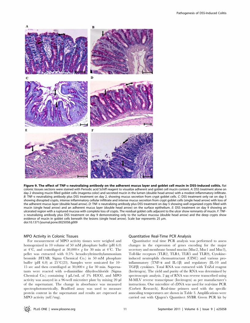
Figure 9. The effect of TNF-a neutralizing antibody on the adherent mucus layer and goblet cell mucin in DSS-induced colitis. Ratcolonic tissues sections were stained with Periodic acid Schiff reagent to visualize adherent and goblet cell mucin content. A: DSS treatment alone onday 2 showing mucin filled goblet cells (magenta color) and secreted mucin in the lumen (double head arrow) with a modest inflammatory infiltrate.B: TNF-a neutralizing antibody plus DSS treatment on day 2, showing mucus secretion from crypt goblet cells. C: DSS treatment only rat on day 5showing disrupted crypts, intense inflammatory cellular infiltrate and intense mucus secretion from crypt goblet cells (single head arrow) with loss ofthe adherent mucus layer (double head arrow). D: TNF-a neutralizing antibody plus DSS treatment on day 5 showing well organized crypts filled withmucin (single head arrow) and an adherent mucus layer (double head arrow) on the surface epithelium. E: DSS treatment on day 9 showing anulcerated region with a ruptured mucosa with complete loss of crypts. The residual goblet cells adjacent to the ulcer show remnants of mucin. F: TNF-a neutralizing antibody plus DSS treatment on day 9 demonstrating only to the surface mucosa (double head arrow) and the deep crypts showevidence of mucin in goblet cells beneath the lesions (single head arrow). Scale bar represents 25 mm.doi:10.1371/journal.pone.0025058.g009
Pathogenesis of DSS-Induced Colitis
PLoS ONE | www.plosone.org 11 September 2011 | Volume 6 | Issue 9 | e25058

using the following cycling conditions: 94uC hold for 15 min,
followed by 40 cycles of denaturation at 94uC for 20 s, annealing
at different temperatures for 30 s and extension at 72uC for 30 s
following the manufacturer’s instructions. Specificity of amplifica-
tion was checked by melt curve analysis. mRNA expression for the
different genes was normalized against GAPDH and fold change
over control was determined according to the ddCt method [29].
Protein Level of TNF-a in Colonic TissuesTNF-a in homogenized intestinal tissues was measured using an
ELISA kit according to the manufacturer’s protocol (rat TNF-aELISA Kit, Abcam, Canada). Colonic tissues (20 mg) were
homogenized in cold phosphate buffer saline (PBS) using a
Polytron homogenizer and centrifuged at 20,000 g for 20 min to
4uC to obtain the supernatant. Total protein concentration of the
tissue supernatant was measured using a BCA kit. TNF-a is
expressed as pg/mg of tissue.
Assessment of Administration of Anti TNF-a NeutralizingAntibody on the Onset and Progression of DSS-InducedColitis
In preliminary experiments, three doses of anti TNF-a antibody
(50, 100 and 200 mg/animal/day; ip) were administered to
animals for 9 consecutive days with free access to water containing
5% DSS. A dose of 100 mg/animal/day was found most optimal
in significantly reducing DAI (data not shown) and was used for all
the further experiments. DAI was quantified and histological
examination was performed for colitis as described above. A
quantitative real time PCR analysis was performed using RNA
isolated form colonic tissue to assess the changes in the gene
expression of TNF-a in addition to other genes (Muc2, TLR4,
TLR5, IL-1b, IL-10 and TGFb) that have shown alteration on
DSS treatment in the first set of experiments (see results). TNF-aprotein in homogenized intestinal tissues was also measured using
ELISA as described above.
Statistical AnalysisResults are expressed as means 6 SD. Significance differences
between control and the other strains were determined using
Kruskal-Wallis test with Dunns post-test to compare specific
groups. The choice of a non-parametric test (Kruskal-Wallis test)
instead of a parametric test (Analysis of Variance, ANOVA) was
based on the fact that at least one of the groups in all but two of the
comparisons was non-Gaussian. To maintain consistency, Krus-
kal-Wallis test was used for all comparisons. All statistical analyses
were performed using Graph Pad Instat software. P values .0.05
were considered significant.
Author Contributions
Conceived and designed the experiments: KC PD. Performed the
experiments: PD PL. Analyzed the data: PD PL. Contributed reagents/
materials/analysis tools: PD KC PL. Wrote the paper: PD KC.
References
1. Dharmani P, Chadee K (2008) Biologic therapies against inflammatory bowel
disease: a dysregulated immune system and the cross talk with gastrointestinal
mucosa hold the key. Curr Mol Pharmacol 1: 195–212.
2. Hendrickson BA, Gokhale R, Cho JH (2002) Clinical aspects and pathophys-
iology of inflammatory bowel disease. Clin Microbiol Rev 15: 79–94.
3. Yan Y, Kolachala V, Dalmasso G, Nguyen H, Laroui H, et al. (2009) Temporal
and spatial analysis of clinical and molecular parameters in dextran sodium
sulfate induced colitis. PLoS One 4: e6073.
4. Renes IB, Boshuizen JA, Van Nispen DJ, Bulsing NP, Buller HA, et al. (2002)
Alterations in Muc2 biosynthesis and secretion during dextran sulfate sodium-
Table 3. Rat primers sequences and their annealingtemperature.
Gene Primer SequenceAnnealingtemperature
Muc1: Forward GAGTGAATATCCTACCTACCAC 58uC
Reverse TTCACCAGGCTAACGTGGTGAC
Muc2: Forward GCCAGATCCCGAAACCA 55uC
Reverse TATAGGAGTCTCGGCAGTCA
Muc3: Forward AACTTCCAGCCCTCCCTAAG 50uC
Reverse GCTTCCAGCATCGTCTCTCT
TLR2: Forward GAGTCTGCTGTGCCCTTCTC 50uC
Reverse CATGAGGTTCTCCACCCAAT
TLR4: Forward GTTGGATGGAAAAGCCTTGA 50uC
Reverse CCTGTGAGGTCGTTGAGGTT
TLR5: Forward GAAGGCTGTGAATCTCGTTGG 50uC
Reverse CTGCCCAACCTCAGGATCTTA
TLR9: Forward CCTGGCACACAATGACATTCA 50uC
Reverse TAAGGTCCTCCTCGTCCCA
IL-1b: Forward CACCTCTCAAGCAGAGCACAG 59uC
Reverse GGGTTCCATGGTGAAGTCAAC
TNFa: Forward AAATGGGCTCCCTCTCATCAGTT 59uC
Reverse TCTGCTTGGTGGTTTGCTACGAC
IL-10 Forward GGCTCAGCACTGCTATGTTGCC 65uC
doi:10.1371/journal.pone.0025058.t003
Table 2. Goblet cell morphology in DSS + TNF-a neutralizingantibody treated group in comparison to only DSS treatment.
Groups Percentage of total goblet cells
FilledReleasingMucus Empty
Day 2 DSS only 77.463.6 16.561.2 6.160.6
DSS + anti-TNF-a 67.961.2 2260.7 10.160.5
Day 5 DSS only 37.160.9 22.660.5 40.260.7
DSS + anti-TNF-a 59.661.3* 22.860.7 29.461.6*
Day 9 DSS only ND ND ND
DSS + anti-TNF-a 19.660.4 21.660.3 58.861.0
Data are presented as % of goblet cells 6 SEM. *P,0.05 when compared to DSStreated group of the respective day. Goblet cell morphology was quantifiedfrom randomly selected crypts of PAS stained sections. A minimum number of100 (100–110) goblet cells were counted under 40 X magnification from eachsection. Only in the DSS + anti-TNFa treated group we were able to count 51goblet cells. Goblet cell morphology was designated as previously described[28]. Filled goblet cells: goblet cells with intact mucus granules; Releasingmucus: cells releasing mucus with PAS-stained mucus emerging as a thickstream; Empty goblet cells with PAS stained mucus absent from cells exhibitinga deep concave cavitation of the apical surface. ND: Not determined as theepithelial layer and crypts on day 9 of DSS treatment was destroyed or toodamaged in the ulcerated site.doi:10.1371/journal.pone.0025058.t002
Pathogenesis of DSS-Induced Colitis
PLoS ONE | www.plosone.org 12 September 2011 | Volume 6 | Issue 9 | e25058

induced colitis. Am J Physiol Gastrointest Liver Physiol 282: G382–
389.
5. Baumgart DC, Sandborn WJ (2007) Inflammatory bowel disease: clinical aspects
and established and evolving therapies. Lancet 369: 1641–1657.
6. Dieleman LA, Pena AS, Meuwissen SG, van Rees EP (1997) Role of animal
models for the pathogenesis and treatment of inflammatory bowel disease.
Scand J Gastroenterol Suppl 223: 99–104.
7. Moncada DM, Kammanadiminti SJ, Chadee K (2003) Mucin and Toll-like
receptors in host defense against intestinal parasites. Trends Parasitol 19:
305–311.
8. Clayburgh DR, Shen L, Turner JR (2004) A porous defense: the leaky epithelial
barrier in intestinal disease. Lab Invest 84: 282–291.
9. Toshina K, Hirata I, Maemura K, Sasaki S, Murano M, et al. (2000) Enprostil, a
prostaglandin-E(2) analogue, inhibits interleukin-8 production of human colonic
epithelial cell lines. Scand J Immunol 52: 570–575.
10. Xu Y, Hunt NH, Bao S (2007) The correlation between pro-inflammatory
cytokines, MAdCAM-1 and cellular infiltration in the inflamed colon from TNF-
alpha gene knockout mice. Immunol Cell Biol 85: 633–639.
11. Dharmani P, Srivastava V, Kissoon-Singh V, Chadee K (2009) Role of intestinal
mucins in innate host defense mechanisms against pathogens. J Innate Immun 1:
123–135.
12. Andrianifahanana M, Moniaux N, Batra SK (2006) Regulation of mucin
expression: mechanistic aspects and implications for cancer and inflammatory
diseases. Biochim Biophys Acta 1765: 189–222.
13. Shekels LL, Anway RE, Lin J, Kennedy MW, Garside P, et al. (2001)
Coordinated Muc2 and Muc3 mucin gene expression in Trichinella spiralis
infection in wild-type and cytokine-deficient mice. Dig Dis Sci 46: 1757–1764.
14. Enss ML, Cornberg M, Wagner S, Gebert A, Henrichs M, et al. (2000) Pro-
inflammatory cytokines trigger MUC gene expression and mucin release in the
intestinal cancer cell line LS180. Inflamm Res 49: 162–169.
15. Iwashita J, Sato Y, Sugaya H, Takahashi N, Sasaki H, et al. (2003) mRNA of
MUC2 is stimulated by IL-4, IL-13 or TNF-alpha through a mitogen-activated
protein kinase pathway in human colon cancer cells. Immunol Cell Biol 81:
275–282.
16. Kim YD, Jeon JY, Woo HJ, Lee JC, Chung JH, et al. (2002) Interleukin-1beta
induces MUC2 gene expression and mucin secretion via activation of PKC-
MEK/ERK, and PI3K in human airway epithelial cells. J Korean Med Sci 17:
765–771.
17. Franchimont D, Vermeire S, El Housni H, Pierik M, Van Steen K, et al. (2004)
Deficient host-bacteria interactions in inflammatory bowel disease? The toll-likereceptor (TLR)-4 Asp299gly polymorphism is associated with Crohn’s disease
and ulcerative colitis. Gut 53: 987–992.
18. Hahm KB, Im YH, Parks TW, Park SH, Markowitz S, et al. (2001) Loss oftransforming growth factor beta signalling in the intestine contributes to tissue
injury in inflammatory bowel disease. Gut 49: 190–198.19. Kuhn R, Lohler J, Rennick D, Rajewsky K, Muller W (1993) Interleukin-10-
deficient mice develop chronic enterocolitis. Cell 75: 263–274.
20. Soderholm JD, Olaison G, Peterson KH, Franzen LE, Lindmark T, et al. (2002)Augmented increase in tight junction permeability by luminal stimuli in the non-
inflamed ileum of Crohn’s disease. Gut 50: 307–313.21. Sun Y, Fihn BM, Sjovall H, Jodal M (2004) Enteric neurones modulate the
colonic permeability response to luminal bile acids in rat colon in vivo. Gut 53:362–367.
22. Cario E, Podolsky DK (2000) Differential alteration in intestinal epithelial cell
expression of toll-like receptor 3 (TLR3) and TLR4 in inflammatory boweldisease. Infect Immun 68: 7010–7017.
23. Charo IF, Ransohoff RM (2006) The many roles of chemokines and chemokinereceptors in inflammation. N Engl J Med 354: 610–621.
24. Goebel S, Huang M, Davis WC, Jennings M, Siahaan TJ, et al. (2006) VEGF-A
stimulation of leukocyte adhesion to colonic microvascular endothelium:implications for inflammatory bowel disease. Am J Physiol Gastrointest Liver
Physiol 290: G648–654.25. Keshavarzian A, Choudhary S, Holmes EW, Yong S, Banan A, et al. (2001)
Preventing gut leakiness by oats supplementation ameliorates alcohol-inducedliver damage in rats. J Pharmacol Exp Ther 299: 442–448.
26. Leeb SN, Vogl D, Gunckel M, Kiessling S, Falk W, et al. (2003) Reduced
migration of fibroblasts in inflammatory bowel disease: role of inflammatorymediators and focal adhesion kinase. Gastroenterology 125: 1341–1354.
27. Hogan SP, Seidu L, Blanchard C, Groschwitz K, Mishra A, et al. (2006)Resistin-like molecule beta regulates innate colonic function: barrier integrity
and inflammation susceptibility. J Allergy Clin Immunol 118: 257–268.
28. Chadee K, Keller K, Forstner J, Innes DJ, Ravdin JI (1991) Mucin andnonmucin secretagogue activity of Entamoeba histolytica and cholera toxin in
rat colon. Gastroenterology 100: 986–997.29. Livak KJ, Schmittgen TD (2001) Analysis of relative gene expression data using
real-time quantitative PCR and the 2(-Delta Delta C(T)) Method. Methods 25:402–408.
Pathogenesis of DSS-Induced Colitis
PLoS ONE | www.plosone.org 13 September 2011 | Volume 6 | Issue 9 | e25058