Clinical Management of Foreign Bodies of the Esophagus as ...
78
CLINICAL MANAGEMENT OF FOREIGN BODIES OF THE ESOPHAGUS AS SEEN AT THE KENYATTA NATIONAL HOSPITAL. PRINCIPAL RESEARCHER: DR.OUYAH WILSON LIBUTSI REG.NO.H58/63689/2010 M.MED OTORHINOLARYNGOLOGY-HEAD AND NECK SURGERY RESIDENT. UNIVERSITY OF NAIROBI SUPERVISORS: PROF ISAAC MUTHURE MACHARIA, MBChB,MMed (ENT), FCS (ECSA) Professor and Consultant Otorhinolaryngologist-Head and Neck Surgeon, Department of Surgery, University of Nairobi. DR.PETER MASINDE MBChB, MMed (ENT) Head of ENT Department Kenyatta National Hospital, Consultant Otorhinolaryngologist-Head & Neck Surgeon, Kenyatta National Hospital. A proposal for dissertation as partial fulfillment of requirements of the University of Nairobi, for the Award of the Degree of Masters in Medicine in Otorhinolaryngology, Head and Neck Surgery. 2017
-
Upload
khangminh22 -
Category
Documents
-
view
0 -
download
0
Transcript of Clinical Management of Foreign Bodies of the Esophagus as ...
CLINICAL MANAGEMENT OF FOREIGN BODIES OF THE ESOPHAGUS AS SEEN
AT THE KENYATTA NATIONAL HOSPITAL.
PRINCIPAL RESEARCHER:
DR.OUYAH WILSON LIBUTSI
REG.NO.H58/63689/2010
M.MED OTORHINOLARYNGOLOGY-HEAD AND NECK SURGERY RESIDENT.
UNIVERSITY OF NAIROBI
SUPERVISORS:
PROF ISAAC MUTHURE MACHARIA, MBChB,MMed (ENT), FCS (ECSA)
Professor and Consultant Otorhinolaryngologist-Head and Neck Surgeon,
Department of Surgery,
University of Nairobi.
DR.PETER MASINDE MBChB, MMed (ENT)
Head of ENT Department Kenyatta National Hospital,
Consultant Otorhinolaryngologist-Head & Neck Surgeon,
Kenyatta National Hospital.
A proposal for dissertation as partial fulfillment of requirements of the University of Nairobi, for
the Award of the Degree of Masters in Medicine in Otorhinolaryngology, Head and Neck Surgery.
2017
ii
DECLARATION
I hereby certify that this proposal is my original work and has not been submitted for any degree
at any other institution.
Dr. Ouyah Wilson Libutsi
Signed……………………………………………………………………………………………
Date……………………………………………………………………………………………...
iii
DEDICATION
This dissertation is dedicated to my parents Mr and Mrs J. Ouyah, my siblings Dorothy, Frankline,
Caroline and Vincent for their support. Many thanks to my wife Miriam Libutsi and my children
Victor, Linda, Andrew and William for their encouragement and support throughout the study.

iv
ACKNOWLEDGEMENT
I would like to express my deepest gratitude to my supervisors, Prof. I. M Macharia and
Dr.Masinde P, for their support, patience and expert guidance throughout this study. Am also
grateful to Mr. Ayieko P, for his help in statistical analysis of the results from the study.
v
APPROVAL
The proposal has been submitted in partial fulfillment of the degree of master of Medicine in
Otorhinolaryngology, Head and Neck Surgery with our approval as supervisors.
1. PROF. ISAAC MUTHURE MACHARIA
Signed…………………………………………………………………………………………
Date……………………………………………………………………………………………
2. DR.PETER MASINDE
Signed…………………………………………………………………………………………
Date……………………………………………………………………………………………

vi
TABLE OF CONTENTS
DECLARATION........................................................................................................................... ii
DEDICATION................................................................................................................................v
ACKNOWLEGEMENTS ........................................................................................................... iv
APPROVAL ...................................................................................................................................v
TABLE OF CONTENTS ............................................................................................................ vi
FIGURES AND TABLES ........................................................................................................ viii
ABBREVIATIONS ...................................................................................................................... ix
ABSTRACT ....................................................................................................................................x
1.0 BACKGROUND ................................................................................................................. 1
1.1 Introduction .......................................................................................................................... 1
1.2 Anatomy And Physiology .................................................................................................... 2
1.3 Clinical Manifestations ......................................................................................................... 3
1.4 Management ......................................................................................................................... 4
1.5 Techniques For Esophageal Foreign Body Removal ........................................................... 5
1.6 Approaches For Specific Types Of Foreign Bodies ............................................................. 6
1.7 Complications Of Esophageal Foreign Bodies .................................................................... 8
2.0 LITERATURE REVIEW .................................................................................................. 9
2.1 Study Justification .............................................................................................................. 13
2.2 Research Question .............................................................................................................. 13
2.3 Objectives ........................................................................................................................... 13
2.3.1 Broad objective .......................................................................................................... 13
2.3.2 Specific objectives .................................................................................................... 13
3.0 METHODOLOGY ........................................................................................................... 14
3.1 Study designs ...................................................................................................................... 14
3.2 Study site ............................................................................................................................. 14
3.3 Study population ................................................................................................................. 15
vii
3.4 Inclusion criteria ................................................................................................................. 15
3.5 Exclusion criteria ................................................................................................................ 15
3.6 Sample size ......................................................................................................................... 15
3.7 Sampling procedure ............................................................................................................ 16
3.8 Data Collection ................................................................................................................... 16
3.9 Standard Operating Procedure ............................................................................................ 17
3.10 Quality control .................................................................................................................... 18
3.11 Data management and analysis ........................................................................................... 18
3.12 Ethical consideration .......................................................................................................... 19
4.0 RESULTS ......................................................................................................................... 20
Discussion .......................................................................................................................... 34
Conclusion .......................................................................................................................... 37
Recommendation ................................................................................................................ 38
REFERENCES ............................................................................................................................ 39
APPENDICES ............................................................................................................................. 43
Appendix 1: General Patient Information .................................................................................... 43
Appendix 2: Consent Form ........................................................................................................... 46
Appendix 3: Assent Information Document ................................................................................. 49
Appendix 4: Assent Form ............................................................................................................. 52
Appendix 5: Kiambatisho ............................................................................................................ 54
Appendix 6: Questionnaire .......................................................................................................... 63
Appendix 7: Research Approval ...................................................................................................67
viii
LIST OF FIGURES AND TABLES
FIGURES
Figure 1: Anatomy of the esophagus ...............................................................................................3
Figure 2: Age distribution of patients presenting with FBs of esophagus in KNH .......................20
Figure 3: Sex distribution of patients with foreign bodies of the esophagus .................................21
Figure 4: Referral and non-referral cases with foreign bodies of the esophagus ...........................21
Figure 5: Proportion of foreign bodies of the esophagus seen at KNH... ......................................23
Figure 6: Duration from ingestion of FB to presentation at KNH ………….. ..............................25
Figure 7: Methods used in removal of the FB... ............................................................................29
TABLES
Table 1: Referrals from other medical facilities ............................................................................22
Table 2: Relationship between patients’ age, sex and referral to KNH with the type of FB .........24
Table 3: Relationship between patients’ residence and the duration before presentation to KNH
........................................................................................................................................................25
Table 4: Clinical presentation of patients with FB of the esophagus ...........................................26
Table 5: Association between clinical presentation and type of foreign body ..............................27
Table 6: Examination findings .......................................................................................................28
Table 7: Relationship between examination findings and type of FB ...........................................28
Table 8: Relationship between methods used to remove the esophageal FB and injuries seen in
the esophageal ................................................................................................................................31
Table 9: Association between esophageal injury and type of foreign body .................................32
Table 10: Types of FB and postoperative complications ……......................................................33
ix
ABBREVIATIONS
A&E Accident and Emergency department
ANOVA Analysis Of Variance
CT Scan Computer tomography scan
EFB Esophageal Foreign Body
ENT Clinic Ear,Nose and Throat Clinic
FB Foreign body
FBI Foreign Body Impaction
GA General Anesthesia
GIT Gastrointestinal Tract
KNH Kenyatta National Hospital
MRI Magnetic Resonance Imaging
PACU Post Anesthesia Care Unit
SD Standard Deviation
SPSS Stastical Package for the Social Sciences
TEF Tracheoesophageal Fistula
U/S Scan Ultrasound Scan

x
ABSTRACT
Background:
Foreign bodies of the esophagus are common at the Kenyatta National Hospital with an increasing
number of lithium disc batteries being seen. A local study showed two peak ages of patients with
esophageal FBs, those below 6 years (59.3%) and adults at 23.7%.
In the United States, there are over 100 000 cases of esophageal FBs per year. In 80–90% of these
cases, the FB passes spontaneously through the GIT while the remainders lodge in the esophagus
and need to be removed.
Study objective: To determine the clinical presentation and management of FBs of the esophagus
at the KNH.
Study design & setting: Hospital-based descriptive cross-sectional study, the setting being at the
KNH A&E and ENT departments
Methodology:A total of 100 cases were recruited and a detailed history of the type of FB,
presenting signs and symptoms, modes of management and complications were recorded.
Statistical Analysis:
Data analysis was conducted using SPSS (version 20). The bivariate analysis was based on
calculation of Pearson’s chi square test and statistical significance was based on a p value cut off
of 0.05.
xi
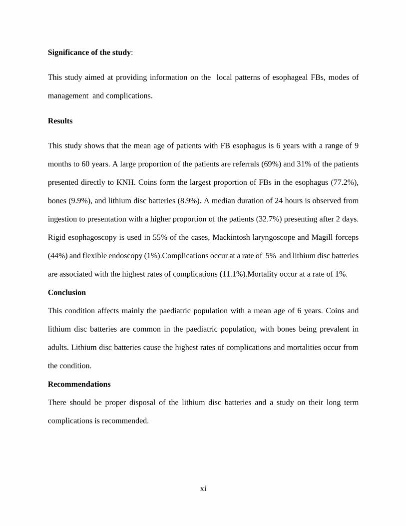
Significance of the study:
This study aimed at providing information on the local patterns of esophageal FBs, modes of
management and complications.
Results
This study shows that the mean age of patients with FB esophagus is 6 years with a range of 9
months to 60 years. A large proportion of the patients are referrals (69%) and 31% of the patients
presented directly to KNH. Coins form the largest proportion of FBs in the esophagus (77.2%),
bones (9.9%), and lithium disc batteries (8.9%). A median duration of 24 hours is observed from
ingestion to presentation with a higher proportion of the patients (32.7%) presenting after 2 days.
Rigid esophagoscopy is used in 55% of the cases, Mackintosh laryngoscope and Magill forceps
(44%) and flexible endoscopy (1%).Complications occur at a rate of 5% and lithium disc batteries
are associated with the highest rates of complications (11.1%).Mortality occur at a rate of 1%.
Conclusion
This condition affects mainly the paediatric population with a mean age of 6 years. Coins and
lithium disc batteries are common in the paediatric population, with bones being prevalent in
adults. Lithium disc batteries cause the highest rates of complications and mortalities occur from
the condition.
Recommendations
There should be proper disposal of the lithium disc batteries and a study on their long term
complications is recommended.
1
1.0 BACKGROUND
1.1 INTRODUCTION
Globally FBs in the esophagus are a common occurrence affecting both children and adults. In the
United States, more than 100,000 cases of foreign bodies in the esophagus are managed annually
and 80 percent occur in children2.Most of the foreign bodies that reach the gastrointestinal tract
pass spontaneously and only 10 to 20 percent will require endoscopic removal, and less than 1
percent require surgical intervention2. Although mortality from foreign body ingestion is
extremely low, deaths have been reported4. Morbidity from foreign body ingestion is related to the
various complications including weight loss or recurrent aspiration pneumonia ,mucosal damage leading
to strictures, or erosion of the esophageal wall creating a fistula with the trachea or other nearby structures.
Sharp objects may perforate the esophagus, and erosion into the aorta also has been reported, which can
lead to mortalities9
Locally and in the U.S, coins are the most common foreign bodies ingested by children. Other
objects, including toy parts, magnets, batteries, safety pins, screws, marbles, bones and food
boluses have been reported5
2
1.2 ANATOMY AND PHYSIOLOGY
The esophagus is a 25-cm long muscular tube that connects the pharynx to the stomach. The length
of the esophagus at birth varies between 8 and 10 cm. At 5 years it measures 12cm and at 15 years
it measures about 19 cm. It extends from the lower border of the cricoid cartilage (at the level of
the sixth cervical vertebra) to the cardiac orifice of the stomach at the side of the body of the 11th
thoracic vertebra. The upper limit in the newborn infant is found at the level of the fourth or fifth
cervical vertebra, and the lower limit is at the level of the ninth thoracic vertebra.
The esophagus has been subdivided into 3 portions. These are the cervical portion, extending from
the cricopharyngeus to the suprasternal notch, the thoracic portion extending from the suprasternal
notch to the diaphragm and the abdominal portion extending from the diaphragm to the cardiac
portion of the stomach. It’s located in the posterior mediastinum and related to organs in this
region.On the right side the esophagus is covered by the mediastinal part of the parietal pleura.
Sharp objects may perforate this layer leading to mediastinitis.On the left side of the esophagus is
the thoracic aorta. Erosion into the aorta caused by corrosive foreign bodies has been reported,
causing life-threatening gastrointestinal bleeding9
Anterior to the esophagus, is the trachea and below the level of the tracheal bifurcation, are the
right pulmonary artery and the left main bronchus. Longstanding foreign bodies may create a
fistula with the trachea. The esophagus passes immediately posteriorly to the left atrium, separated
from it only by pericardium. Other structures posterior to the esophagus include the thoracic duct,
portions of the hemiazygos veins, the right posterior intercostal vessels, and, near the diaphragm,
the thoracic aorta.

3
Courtesy of www.netterimages.com
Figure 1: Anatomy of the esophagus
The esophagus has 3 points of physiologic constrictions which are produced by the upper
esophageal sphincter (cricopharyngeus muscle), the aortic arch and the left bronchus, and the lower
esophageal sphincter. These can be seen in the diagram above. Esophageal foreign bodies tend to
lodge in these areas of constrictions.
1.3 CLINICAL MANIFESTATIONS
Usually young children with esophageal foreign bodies are brought to medical attention by their
parents because the ingestion was witnessed or reported to them7. Older children and adults may
localize the sensation of something stuck to the neck or lower chest, suggesting irritation in the
4
upper or lower esophagus, respectively. Patients of any age may present with refusal of feeds or
dysphagia, drooling, or respiratory symptoms including wheezing, stridor, or choking.
1.4 MANAGEMENT
A thorough history and physical examination are important in diagnosing an esophageal foreign
body and to the prevention of complications10.The physical examination of the neck may reveal
swelling, or crepitus, suggesting esophageal perforation has occurred. The chest examination may
reveal inspiratory stridor or expiratory wheezing, suggesting a lodged esophageal foreign body
with tracheal compression.
Imaging is important for all patients with suspected foreign body ingestion. The initial diagnostic
test should be biplane radiographs (anteroposterior and lateral) of the neck, chest, and abdomen8.
Flat objects (e.g., coins or disk batteries) usually orient in the coronal plane and appear as a circular
object on an antero-posterior projection whereas objects lodged in the trachea tend to orient in the
sagittal plane and are best seen in lateral projection. Plastic or wood, some thin metal objects, and
many types of bones are not readily seen on plain films11.
If the patient is symptomatic, or if the suspected foreign body has any dangerous characteristics
(large >2 cm in width or long >5 cm in length, or sharp), or if the type of foreign body is not
definitively known by the caretakers, then CT Scans with 3-dimensional reconstruction is
done12.MRI scans can be used for evaluation of radiolucent foreign bodies, but is contraindicated
if any metallic foreign body is present.
Ultrasonography has been used to identify the location and nature of foreign bodies in the
esophagus or stomach if appropriate expertise is available13.Contrast studies e.g. barium contrast
are avoided as they may obscure visualization of the FB on subsequent endoscopy. The contrast
5
may be aspirated if the esophagus is obstructed. Hence, endoscopy may be preferred over contrast
even if radiographs are negative13.
Urgent intervention is indicated if warning signs are present. These include when the foreign body
is sharp, long (>5 cm), and is in the esophagus or stomach, or when a disk battery is in the
esophagus,or when the patient shows signs of airway compromise and when there is evidence of
near-complete esophageal obstruction seen when patient cannot swallow secretions. Objects
lodged for more than 24 hours or for an unknown duration should be removed promptly14. After
this period, complications such as transmural erosion, perforation, and fistulae are more likely to
occur. Complications are more likely if the foreign body is a sharp or pointed object, disk battery,
non-radio-opaque, or located below the upper third of the esophagus.
1.5 TECHNIQUES FOR ESOPHAGEAL FOREIGN BODY REMOVAL
Various methods are used to remove esophageal foreign bodies.One of the commonest method
used in the West is flexible endoscopy. In this technique the foreign body can be visualized directly
and manipulated, and the surrounding gastrointestinal tract can be examined for potential
complications15. This procedure is performed under conscious sedation or general anesthesia. It is
helpful to practice grasping a duplicate of the foreign body using the retrieval tools before
beginning the procedure.
Rigid endoscopy is another technique which utilizes a non-flexible channeled device that is
introduced into the esophagus under general anesthesia. It is most useful for impacted sharp objects
that are located in the proximal esophagus, at the level of the hypopharynx and cricopharyngeus
muscle.
6
A mackintosh laryngoscope and Magill forceps can be used to extract foreign bodies impacted in
the oropharynx or upper esophagus. An endotracheal tube is placed to protect the airway, and a
mackintosh laryngoscope is used to gently open the esophagus and visualize the foreign body
which is grasped and removed using a Magill forceps.
Bougienage (passage of a dilator) has been used to push objects into the stomach and it does not
permit visualization of the esophagus and does not retrieve the foreign body. Therefore, it is most
appropriate for foreign bodies that are very likely to pass beyond the stomach without
complications, and in situations where there is a low risk of esophageal injury18.
A Foley catheter has also been used. A deflated Foley catheter is passed beyond the foreign body.
The balloon is then inflated using a radio-opaque contrast dye, and the catheter is slowly drawn
back under fluoroscopic guidance, to remove the foreign body through the mouth. The technique
can be successful with proximal esophageal foreign bodies but may cause aspiration of the foreign
body if it is inadvertently dragged into the trachea14.
1.6 APPROACHES FOR SPECIFIC TYPES OF FOREIGN BODIES
Globally coins are the most common foreign body ingested by children19.If a coin is visualized in
the esophagus and the patient is asymptomatic, he can be observed for up to 24 hours after
ingestion of the coin. In such patients, 20 to 30 percent of coins will pass into the stomach
spontaneously during the observation period. Spontaneous passage is more common in older
children and when coins are located in the distal third of the esophagus. The esophageal coin
should be removed promptly if the patient is symptomatic or if the time of ingestion is not known.
If the child is asymptomatic and the coin does not pass spontaneously by 24 hours after ingestion,
it should be removed.
7
Lithium disc batteries are associated with significant morbidity, when lodged in the esophagus
and thus are a medical emergency. In addition to direct pressure necrosis, contact of the flat
esophageal wall with both poles of the battery conducts electricity, resulting in liquefaction
necrosis and perforation of the esophagus. Retained batteries also can cause problems through
leakage of caustic material e.g. heavy metal like mercury, silver, lithium, and a strong hydroxide
of sodium or potassium20.
Sharp-pointed objects e.g. straight pins, needles, and fish bones lodged in the esophagus represent
a medical emergency because of a high risk of perforation and should be removed immediately.
When lodged in the hypopharynx, they can cause a retropharyngeal abscess8.The risk of mucosal
injury during retrieval of a sharp object can be minimized by orienting the object with the sharp-
end trailing during extraction and using a protector hood on the end of the endoscope.
Esophageal food impaction commonly impacted meat is the most common esophageal foreign
body in adults and relatively rare in children. It presents as dysphagia beginning acutely while
eating.Patients who are unable to swallow oral secretions require immediate attention and removal
of the impaction. If the patient is comfortable and able to handle oral secretions, endoscopic
intervention can be delayed, as many food impactions will pass spontaneously. However,
intervention should not be delayed beyond 24 hours. The food bolus can be removed en bloc or in
a piecemeal fashion. Once reduced in size, the bolus may be gently pushed into the stomach using
the tip of the endoscope22.
8
1.7 COMPLICATIONS OF ESOPHAGEAL FOREIGN BODIES
Longstanding esophageal foreign bodies may cause weight loss or recurrent aspiration pneumonia,
due to decreased caloric intake and poor handling of oral secretions, respectively. They also can
damage the mucosa and lead to strictures, or erosion of the esophageal wall, creating a fistula with
the trachea or other nearby structures. Sharp objects may perforate the esophagus, resulting in neck
swelling, crepitus, mediastinitis or pneumomediastinum. Erosion into the aorta also has been
reported, causing life-threatening gastrointestinal bleeding9
9
2.0 LITERATURE REVIEW
Foreign bodies in the esophagus are a common condition locally and globally and studies have
been done to ascertain the incidence, modes of management and complications arising from the
condition.
The types of FBs found in the esophagus are quite varied and this was shown by Arana A,et al8
who conducted a retrospective and found FBs to be the following in decreasing order of frequency,
coins, toy parts, jewels, batteries, sharp materials such as needles and pins, fish and chicken bones,
and "large" amounts of food. He found that 9% of these FBs were removed with a Magill forceps,
20% were removed with a magnet probe and endoscopic removal was performed in 25% of the
cases. Locally a retrospective study by Oduor P showed that majority of the FBs seen, were
metallic objects including coins which made up 67% of the cases, followed by meat and bones
which made 28.3%, vegetable material constituted 4.1% and plastics made up 1.6%.1
The location of FBs in the esophagus, is determined by various factors including the natural
esophageal constrictions, the size and shape of the FB. Athanassiadi K, et al5 in a retrospective
study showed that 57% of the FBs were located in the cervical esophagus, 26% were in the
thoracic esophagus and 17% were at the cardioesophageal junction.The location, type of FB, and
availability of expertise and equipment determines the mode of management. In a randomized
prospective study by Waltzman ML,et al23,a comparison was made between immediate endoscopic
removal of esophageal FBs and observing the patients for a period of 24hrs then followed by
removal of the FB when necessary. The results showed that 23% of the patients
taken for immediate endoscopic removal had spontaneous passage of the FB as compared to 30%
in the observation group. Similar findings were made by Sharieff GQ, et al24in a retrospective
study in which healthy patients with acute (less than 24 h) coin ingestions, were observed at home
10
with next-day follow-up. He also found that patients with acute esophageal coin ingestions may
experience spontaneous coin passage.
Another comparison of the modes of management was done by Gmeiner D,et al16 where use of a
flexible endoscope was compared to the use of a rigid endoscope. The study showed the success
rate for foreign body removal was at 93.4% using the flexible endoscope and 95.2% using the rigid
endoscope. These results are similar to those shown by Katsinelos P. et al30 who did a study on
endoscopic management of foreign body and food bolus impaction in the upper gastrointestinal
tract. He found that the overall success rate for endoscopic management was 98.6% and that
surgical removal of a foreign body was required in only 1.4% of the cases.
Other modes of management of FBs in the esophagus have been studied and Little DC,et al25
analyzed 468 cases in a retrospective study which were managed using balloon extraction with
fluoroscopy, and 80% of the objects were successfully and 8% were advanced into the
stomach.Similarly,Janik JE,et al17conducted a retrospective review of 36 children who had upper
esophageal coins extracted using a Magill forceps and found that all coins were removed without
complication which was observed in 33 cases on the first attempt and 3cases on the second attempt.
Though the sample size was small the study showed that the use of a Magill forceps minimizes
instrumentation of the esophagus and is highly successful at removing coins lodged at or
immediately below the level of the cricopharyngeus muscle.
Complications caused by FBs in the esophagus are a major cause of morbidity in the affected
patients.In a prospective study,Peters NJ,et al28 analyzed 7 cases of esophageal perforations due to
foreign body ingestion and found the sites affected to be the cervical and thoracic esophagus.
Two of these patients presented with subcutaneous emphysema and one patient had trachea-
11
esophageal fistula (TEF) after disc battery ingestion. Similarly, Ngan JH,et al29conducted a
prospective study on injuries arising from fish bone ingestion. They found that 1% of the cases
had mucosal tears from triangular bones lodged in the hypopharynx and also found that prediction
of the presence of fish bones by symptoms and radiograph was poor. The study showed that
location of symptoms was useful in guiding the endoscopist to the site of lodgment and concluded
that rigid laryngoesophagoscopy was the appropriate means of removing triangular bones lodged
in the hypopharynx.
In addition, Shivakumar AM, et al26 analyzed a total number of 104 cases and found that coins
were the most frequent offending agents in children making 87.5% of the cases and
retropharyngeal abscess as a complication was seen in 1.92% which was associated with ingestion
of sharp FBs. Denney W, et al27 did a 10-year retrospective analysis of foreign body and caustic
ingestions showing that mucosal ulceration, seen in 30% of the cases, was related to a complaint
of substernal pain and was related to duration of impaction and the unexpected finding of FB
during chest radiograph. The study also found that esophageal FBs unexpectedly found on chest
radiograph or known to be present greater than 72 hours were more likely to have esophageal
ulceration.
Lithium disc batteries in the esophagus are an important cause of complications and Kimball SJ,et
al20 conducted a retrospective review of esophageal disc battery ingestions over a 10 year period.
He analyzed 10 pediatric patients who had ingested disc battery which were lodged in the
esophagus and found that 3 patients had minimal esophageal damage and 7 sustained severe
esophageal damage which involved the muscularis layer. One patient in the latter group had an
extensive injury that extended into the trachea resulting in a tracheoesophageal fistula and though

12
the sample size was small they concluded due to rapid and severe injury that occur following disc
battery ingestion emergency endoscopic removal is necessary.
13
2.1 STUDY JUSTIFICATION
Foreign body impaction in the esophagus is common in the ENT department at KNH with an
average of 25 cases seen and admitted on a monthly basis with a large proportion of these being
referrals from other hospitals. In the recent years, new foreign bodies like lithium disc batteries
have emerged. These are associated with certain complications including tracheosophageal
fistulae, mediastinitis and esophageal strictures, which cause considerable morbidity to the
affected patients. There was no current data available locally on these types of FBs.
This study aimed at providing insight into the types of FBs in the esophagus seen in KNH and the
demographics of the patients involved. This information can be utilized in educating the public on
the most appropriate preventive measures. In addition, the type of interventions can be adjusted
according to the types of foreign bodies to minimize complications.
2.2 RESEARCH QUESTION
What are the types of foreign bodies and how are patients presenting with foreign body esophagus
managed at The Kenyatta National Hospital?
2.3 OBJECTIVES
2.3.1 Broad objective
To determine the types, clinical presentation, and management of foreign bodies in the esophagus
at the KNH
2.3.2 Specific objectives
1. To determine the demographic pattern including region of origin of pediatric and adult patients
presenting with foreign bodies in the esophagus at the KNH A&E.
14
2. To determine the types of foreign bodies in the esophagus of pediatric and adult patients as seen
at the KNH.
3. To determine the clinical presentation of the cases with esophageal FB in pediatric and adult
patients as seen at the KNH.
4. To determine modes of management and outcomes of esophageal FB in patients during
admission and inpatient stay at the KNH.
5. To determine complications caused by the FBs and the predisposing risk factors in patients
admitted at the KNH.
3.0 METHODOLOGY
3.1 Study designs
This was a hospital based descriptive cross-sectional study, as all patients diagnosed or suspected
to suffer from FB impaction in the esophagus and fulfill the inclusion criteria were recruited at
the A&E and followed up from admission to the day of discharge. Any form of subsequent
management provided to the patient while admitted at the KNH and complications arising from
the FB were captured.
3.2 Study site
This study was undertaken at the KNH A&E department, ENT clinic, ENT wards and operating
theatres. Patients were initially assessed at A&E and ENT clinic and radiological tests were
requested for at this point and the patients were admitted for specific management. Patients were
initially admitted to the ENT ward after being diagnosed with FB in the esophagus from where
they were taken to theatre for removal of the FB under general anesthesia. Patients were then
15
returned to the ward postoperatively for monitoring and observation for any complication and were
retained there until they fully recovered after which they were discharged home.
3.3 Study population
All patients both children and adults presenting with foreign bodies in the esophagus presenting at
A&E and ENT department in KNH during the period of the study were recruited into the study.
3.4 Inclusion criteria
All patients both children and adults presenting with a history of suspected foreign body ingestion
presenting at A&E and ENT department in KNH who or their parents /guardians consented or
assented, were included in the study.
3.5 Exclusion criteria
Patients who declined to give consent for the study or whose parent/guardian declined to consent
or assent to their participation in the study.
3.6 Sample size
Cochran’s formula31 for calculating the sample was used.
𝑛 = 𝑁𝑍2𝑃(1 − 𝑃)
𝑑2(𝑁 − 1) + 𝑍2𝑃(1 − 𝑃)
N = population of patients attending KNH with FB in the esophagus is estimated at 25 to 30 per
month. Based on this assumption and the projected duration of data collection for the current
project a population size of 135 FB patients was used in this calculation.

16
P = Prevalence of FB in the esophagus was estimated at 50% to allow for the most conservative
estimate of sample size
1-P = 1 minus the prevalence various patterns of FB in the esophagus
Z = Z statistic representing 95% level of confidence (1.96)
d = desired level of precision set to 5%
𝑛 = 135 × 1.9620.5(1 − 0.5)
0.052(135 − 1) + 1.962 × 0.5(1 − 0.5)
n = 100
3.7 Sampling procedure
Patients, both children and adults who presented to A&E and ENT departments at the KNH with
a history of suspected foreign body in the esophagus or those observed to have swallowed a FB
were informed about the study by the principal investigator. Parents or guardians of the affected
children were informed about the study. Once the patient and the parent/guardian were agreeable
to the study they were requested to consent or give assent to the study.
The study employed consecutive sampling involving recruiting each eligible patient until the
desired sample size was achieved.
3.8 Data Collection
The patients and parents/guardians who consented to the study underwent a physical examination
by the principal investigator and the findings were recorded in a preformed structured
questionnaire. This was done in three stages:

17
Stage 1, Preoperative stage: the patient or the parent/guardian was taken through the consent
explanation elaborating details of the study. If satisfied by the explanation and are agreeable, the
patient or guardian signed the consent and the patient was recruited to participate in the study
Demographic data of the patient were taken and details about the type of FB taken and time of
ingestion was recorded. The period from ingestion of the FB to presentation to A&E or ENT clinic
at the KNH was recorded. The signs and symptoms which occurred after ingestion of the FB were
recorded. Mode of investigation for the FB was recorded. Details of any initial management like
forced feeding and subsequent referral was recorded.
Patients who were not taken to theatre and passed the FB spontaneously were captured at this
stage.
Stage 2, Preoperative stage: an operative record questionnaire was provided for the surgeon to
indicate the type of FB removed and the method used for removal intraoperatively. Details of the
level at which the FB was found, any injuries to the esophagus observed intraoperatively and if a
nasogastric tube (NGT) was placed were recorded in the questionnaire.
Stage 3, Postoperative stage: the number of days the patient was admitted in the ENT ward was
recorded i.e. from the date of FB removal to the discharge date. Any complications arising from
the FB in the esophagus was also captured in this stage.
3.9 Standard Operating Procedure
The Standard Operating Procedure for removal of esophageal FBs at KNH was observed for all
the patients undergoing removal of the FB under general anesthesia(GA).All the patients were
starved prior to the procedure and GA was given following the laid down protocol in KNH.
18
Reversal from the GA followed the protocol and all patients were observed in Post anesthesia Care
Unit(PACU) before being tranferred to the ENT ward.
3.10 Quality control
Patient selection, history taking, examination and assessment of the imaging modalities were done
by the principal researcher to prevent observer bias. Data collection was conducted using
procedures outlined in a standard operating procedure (SOP). The SOP contained details on codes
to be used during data collection and standardization of medical and surgical terms that are
commonly used in the study setting
The questionnaires were pretested before use and appropriate adjustments implemented. At the
end of each interview or data abstraction the investigator inspected all the fields in the
questionnaire to ensure data completeness and minimize missing data. A specific code (999) was
used to identify truly missing information. A database for data was designed during the pilot phase
and tested to ensure filters for minimizing data entry errors work as desired.
3.11 Data management and analysis
Data was collected using paper questionnaires and entered into Microsoft Excel worksheets before
being transferred to SPSS (version 20) for analysis. Data cleaning was conducted in SPSS using
functions for univariable analysis to produce frequency tables and summaries. The main outcome
in the analysis was the percentage of patients with the leading clinical presentations of FB and the
frequencies (percentages) reported for each of the main modalities of FB management. The
demographics of patients presenting with foreign bodies were determined by calculating
descriptive statistics including mean (SD) for continuous variables e.g. age and counts and
percentages for categorical characteristics e.g. sex. Methods for univariate analysis of categorical
19
factors were used to calculate frequency (counts) and relative frequency (percentage) for
objectives related to: types of FB in the esophagus seen at KNH, clinical presentation of cases,
early indicators of complications and modes of management. The next stage of analysis used
statistical inference to conduct bivariate analysis of association between (i) specific FBs and
patient demographics, (ii) regional variation in types of esophageal FB, and (iii) both clinician and
patient predisposing risk factors for FB complications. The bivariate analysis was based on cross
tabulations and calculation of Pearson’s chi square test for independence. The level of statistical
significance was based on a p value cut off of 0.05.
Results are presented using frequencies and frequency distributions, cross-tabulations, pie charts
and graphs.
3.12 Ethical consideration
The study was carried out only after approval by the KNH/UON ethics and research committee.
The ethical committee approval number is P712/10/2016.Those recruited in the study were
required to give informed consent or assent. The study was carried out after permission from the
KNH administration. Confidentiality was maintained at all times and the results will be published
in medical journals and presented in medical conferences. They may also be published in print or
electronic media where applicable.There is no monetary gain by the researcher. The patient or
parent/guardian reserved the right to withdraw from the research without victimization and the
participant (patient) incurred no extra financial costs and the investigator has no conflict of interest.
20
4.0 RESULTS
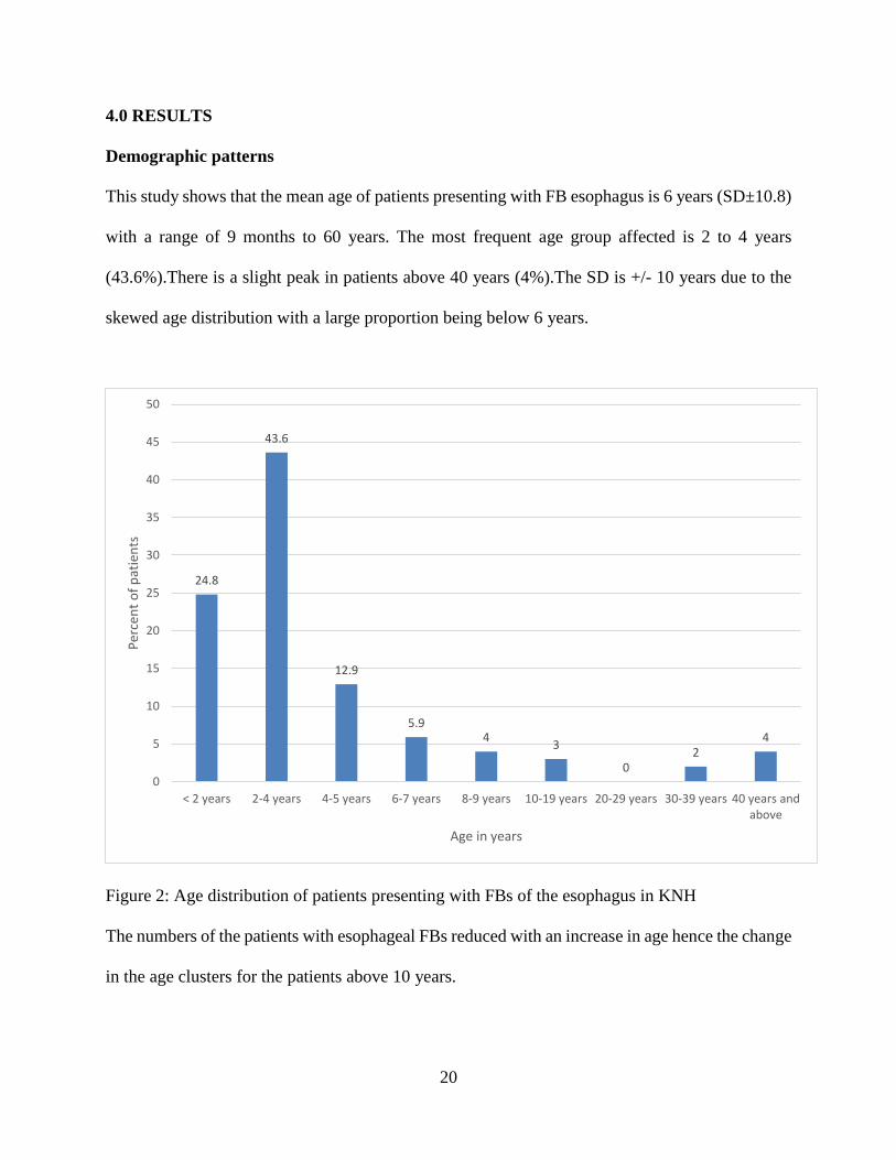
Demographic patterns
This study shows that the mean age of patients presenting with FB esophagus is 6 years (SD±10.8)
with a range of 9 months to 60 years. The most frequent age group affected is 2 to 4 years
(43.6%).There is a slight peak in patients above 40 years (4%).The SD is +/- 10 years due to the
skewed age distribution with a large proportion being below 6 years.
Figure 2: Age distribution of patients presenting with FBs of the esophagus in KNH
The numbers of the patients with esophageal FBs reduced with an increase in age hence the change
in the age clusters for the patients above 10 years.
24.8
43.6
12.9
5.94
3
02
4
0
5
10
15
20
25
30
35
40
45
50
< 2 years 2-4 years 4-5 years 6-7 years 8-9 years 10-19 years 20-29 years 30-39 years 40 years andabove
Per
cen
t o
f p
atie
nts
Age in years
21
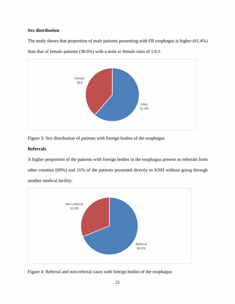
Sex distribution
The study shows that proportion of male patients presenting with FB esophagus is higher (61.4%)
than that of female patients (38.6%) with a male to female ratio of 1.6:1.
Figure 3: Sex distribution of patients with foreign bodies of the esophagus
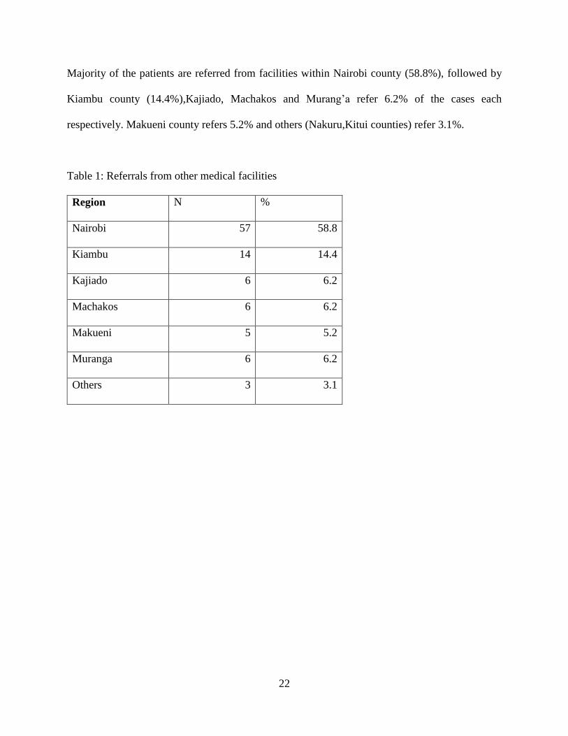
Referrals
A higher proportion of the patients with foreign bodies in the esophagus present as referrals from
other counties (69%) and 31% of the patients presented directly to KNH without going through
another medical facility.
Figure 4: Referral and non-referral cases with foreign bodies of the esophagus
Male61.4%
Female38.6
Referral69.0%
Non-referral31.0%
22
Majority of the patients are referred from facilities within Nairobi county (58.8%), followed by
Kiambu county (14.4%),Kajiado, Machakos and Murang’a refer 6.2% of the cases each
respectively. Makueni county refers 5.2% and others (Nakuru,Kitui counties) refer 3.1%.
Table 1: Referrals from other medical facilities
Region N %
Nairobi 57 58.8
Kiambu 14 14.4
Kajiado 6 6.2
Machakos 6 6.2
Makueni 5 5.2
Muranga 6 6.2
Others 3 3.1
23
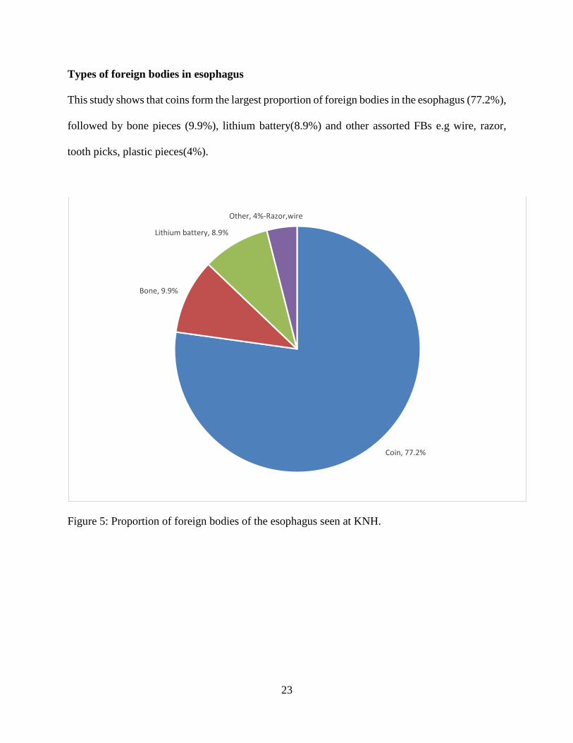
Types of foreign bodies in esophagus
This study shows that coins form the largest proportion of foreign bodies in the esophagus (77.2%),
followed by bone pieces (9.9%), lithium battery(8.9%) and other assorted FBs e.g wire, razor,
tooth picks, plastic pieces(4%).
Figure 5: Proportion of foreign bodies of the esophagus seen at KNH.
Coin, 77.2%
Bone, 9.9%
Lithium battery, 8.9%
Other, 4%-Razor,wire
24
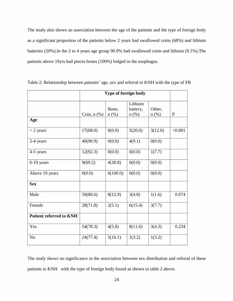
The study also shows an association between the age of the patients and the type of foreign body
as a significant proportion of the patients below 2 years had swallowed coins (68%) and lithium
batteries (20%).In the 2 to 4 years age group 90.9% had swallowed coins and lithium (9.1%).The
patients above 19yrs had pieces bones (100%) lodged in the esophagus.
Table 2: Relationship between patients’ age, sex and referral to KNH with the type of FB
Type of foreign body
Coin, n (%)
Bone,
n (%)
Lithium
battery,
n (%)
Other,
n (%) P
Age
< 2 years 17(68.0) 0(0.0) 5(20.0) 3(12.0) <0.001
2-4 years 40(90.9) 0(0.0) 4(9.1) 0(0.0)
4-5 years 12(92.3) 0(0.0) 0(0.0) 1(7.7)
6-19 years 9(69.2) 4(30.8) 0(0.0) 0(0.0)
Above 19 years 0(0.0) 6(100.0) 0(0.0) 0(0.0)
Sex
Male 50(80.6) 8(12.9) 3(4.8) 1(1.6) 0.074
Female 28(71.8) 2(5.1) 6(15.4) 3(7.7)
Patient referred to KNH
Yes 54(78.3) 4(5.8) 8(11.6) 3(4.3) 0.234
No 24(77.4) 5(16.1) 1(3.2) 1(3.2)
The study shows no significance in the association between sex distribution and referral of these
patients to KNH with the type of foreign body found as shown in table 2 above.
25
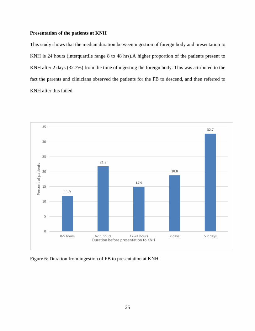
Presentation of the patients at KNH
This study shows that the median duration between ingestion of foreign body and presentation to
KNH is 24 hours (interquartile range 8 to 48 hrs).A higher proportion of the patients present to
KNH after 2 days (32.7%) from the time of ingesting the foreign body. This was attributed to the
fact the parents and clinicians observed the patients for the FB to descend, and then referred to
KNH after this failed.
Figure 6: Duration from ingestion of FB to presentation at KNH
11.9
21.8
14.9
18.8
32.7
0
5
10
15
20
25
30
35
0-5 hours 6-11 hours 12-24 hours 2 days > 2 days
Per
cen
t o
f p
atie
nts
Duration before presentation to KNH
26
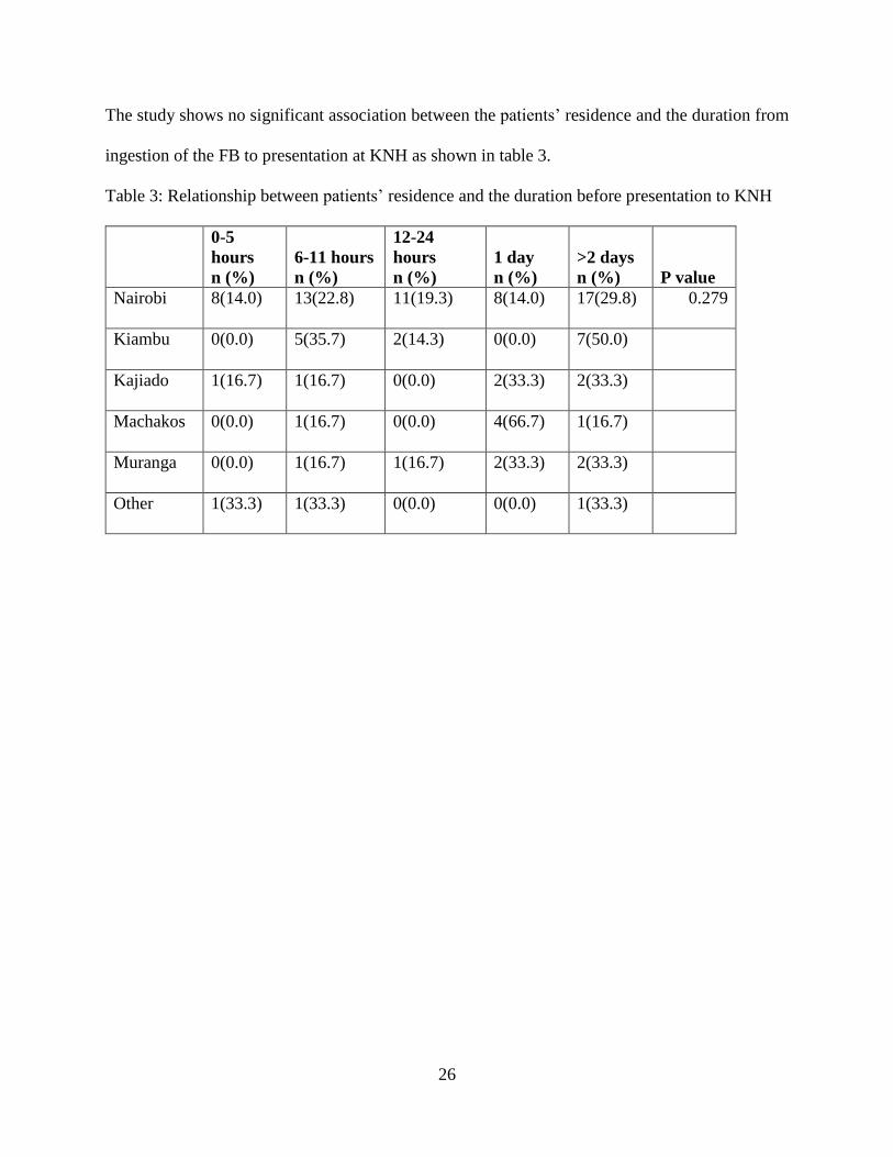
The study shows no significant association between the patients’ residence and the duration from
ingestion of the FB to presentation at KNH as shown in table 3.
Table 3: Relationship between patients’ residence and the duration before presentation to KNH
0-5
hours
n (%)
6-11 hours
n (%)
12-24
hours
n (%)
1 day
n (%)
>2 days
n (%) P value
Nairobi 8(14.0) 13(22.8) 11(19.3) 8(14.0) 17(29.8) 0.279
Kiambu 0(0.0) 5(35.7) 2(14.3) 0(0.0) 7(50.0)
Kajiado 1(16.7) 1(16.7) 0(0.0) 2(33.3) 2(33.3)
Machakos 0(0.0) 1(16.7) 0(0.0) 4(66.7) 1(16.7)
Muranga 0(0.0) 1(16.7) 1(16.7) 2(33.3) 2(33.3)
Other 1(33.3) 1(33.3) 0(0.0) 0(0.0) 1(33.3)
27
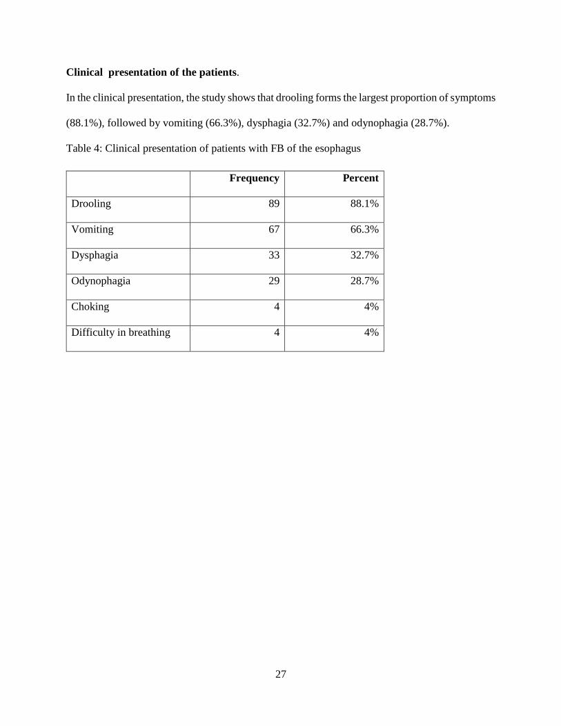
Clinical presentation of the patients.
In the clinical presentation, the study shows that drooling forms the largest proportion of symptoms
(88.1%), followed by vomiting (66.3%), dysphagia (32.7%) and odynophagia (28.7%).
Table 4: Clinical presentation of patients with FB of the esophagus
Frequency Percent
Drooling 89 88.1%
Vomiting 67 66.3%
Dysphagia 33 32.7%
Odynophagia 29 28.7%
Choking 4 4%
Difficulty in breathing 4 4%
28
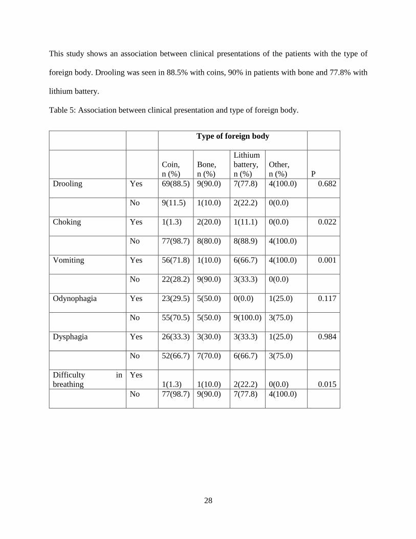
This study shows an association between clinical presentations of the patients with the type of
foreign body. Drooling was seen in 88.5% with coins, 90% in patients with bone and 77.8% with
lithium battery.
Table 5: Association between clinical presentation and type of foreign body.
Type of foreign body
Coin,
n (%)
Bone,
n (%)
Lithium
battery,
n (%)
Other,
n (%) P
Drooling Yes 69(88.5) 9(90.0) 7(77.8) 4(100.0) 0.682
No 9(11.5) 1(10.0) 2(22.2) 0(0.0)
Choking Yes 1(1.3) 2(20.0) 1(11.1) 0(0.0) 0.022
No 77(98.7) 8(80.0) 8(88.9) 4(100.0)
Vomiting Yes 56(71.8) 1(10.0) 6(66.7) 4(100.0) 0.001
No 22(28.2) 9(90.0) 3(33.3) 0(0.0)
Odynophagia Yes 23(29.5) 5(50.0) 0(0.0) 1(25.0) 0.117
No 55(70.5) 5(50.0) 9(100.0) 3(75.0)
Dysphagia Yes 26(33.3) 3(30.0) 3(33.3) 1(25.0) 0.984
No 52(66.7) 7(70.0) 6(66.7) 3(75.0)
Difficulty in
breathing
Yes
1(1.3) 1(10.0) 2(22.2) 0(0.0) 0.015
No 77(98.7) 9(90.0) 7(77.8) 4(100.0)
29
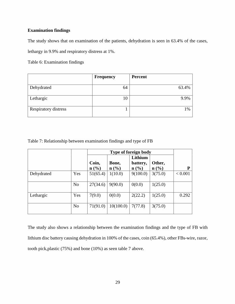
Examination findings
The study shows that on examination of the patients, dehydration is seen in 63.4% of the cases,
lethargy in 9.9% and respiratory distress at 1%.
Table 6: Examination findings
Frequency Percent
Dehydrated 64 63.4%
Lethargic 10 9.9%
Respiratory distress 1 1%
Table 7: Relationship between examination findings and type of FB
Type of foreign body
P
Coin,
n (%)
Bone,
n (%)
Lithium
battery,
n (%)
Other,
n (%)
Dehydrated Yes 51(65.4) 1(10.0) 9(100.0) 3(75.0) < 0.001
No 27(34.6) 9(90.0) 0(0.0) 1(25.0)
Lethargic Yes 7(9.0) 0(0.0) 2(22.2) 1(25.0) 0.292
No 71(91.0) 10(100.0) 7(77.8) 3(75.0)
The study also shows a relationship between the examination findings and the type of FB with
lithium disc battery causing dehydration in 100% of the cases, coin (65.4%), other FBs-wire, razor,
tooth pick,plastic (75%) and bone (10%) as seen table 7 above.
30
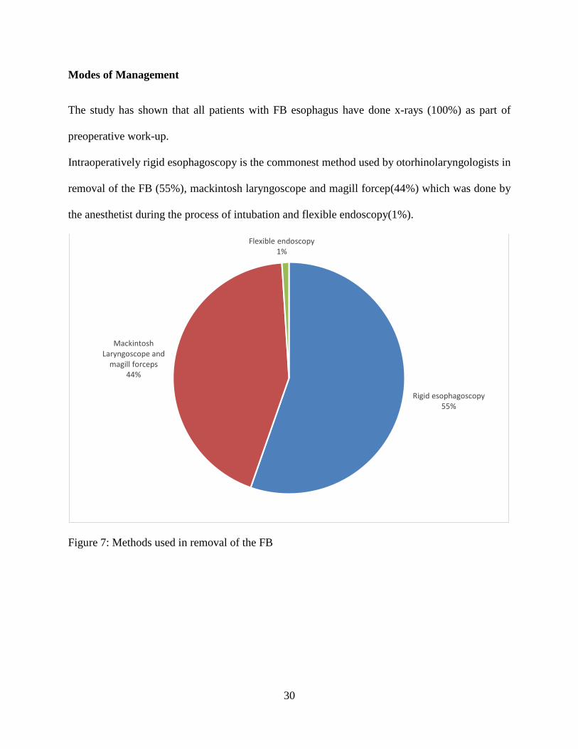
Modes of Management
The study has shown that all patients with FB esophagus have done x-rays (100%) as part of
preoperative work-up.
Intraoperatively rigid esophagoscopy is the commonest method used by otorhinolaryngologists in
removal of the FB (55%), mackintosh laryngoscope and magill forcep(44%) which was done by
the anesthetist during the process of intubation and flexible endoscopy(1%).
Figure 7: Methods used in removal of the FB
Rigid esophagoscopy55%
Mackintosh Laryngoscope and
magill forceps44%
Flexible endoscopy1%
31
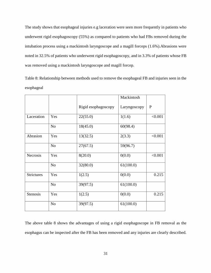
The study shows that esophageal injuries e.g laceration were seen more frequently in patients who
underwent rigid esophagoscopy (55%) as compared to patients who had FBs removed during the
intubation process using a mackintosh laryngoscope and a magill forceps (1.6%).Abrasions were
noted in 32.5% of patients who underwent rigid esophagoscopy, and in 3.3% of patients whose FB
was removed using a mackintosh laryngoscope and magill forcep.
Table 8: Relationship between methods used to remove the esophageal FB and injuries seen in the
esophageal
Rigid esophagoscopy
Mackintosh
Laryngoscopy P
Laceration Yes 22(55.0) 1(1.6) <0.001
No 18(45.0) 60(98.4)
Abrasion Yes 13(32.5) 2(3.3) <0.001
No 27(67.5) 59(96.7)
Necrosis Yes 8(20.0) 0(0.0) <0.001
No 32(80.0) 61(100.0)
Strictures Yes 1(2.5) 0(0.0) 0.215
No 39(97.5) 61(100.0)
Stenosis Yes 1(2.5) 0(0.0) 0.215
No 39(97.5) 61(100.0)
The above table 8 shows the advantages of using a rigid esophagoscope in FB removal as the
esophagus can be inspected after the FB has been removed and any injuries are clearly described.
32
Hence lacerations, abrasions, necrosis or strictures can be noted as compared to the use of
Mackintosh laryngoscope and magill forceps where the injuries may not be seen.
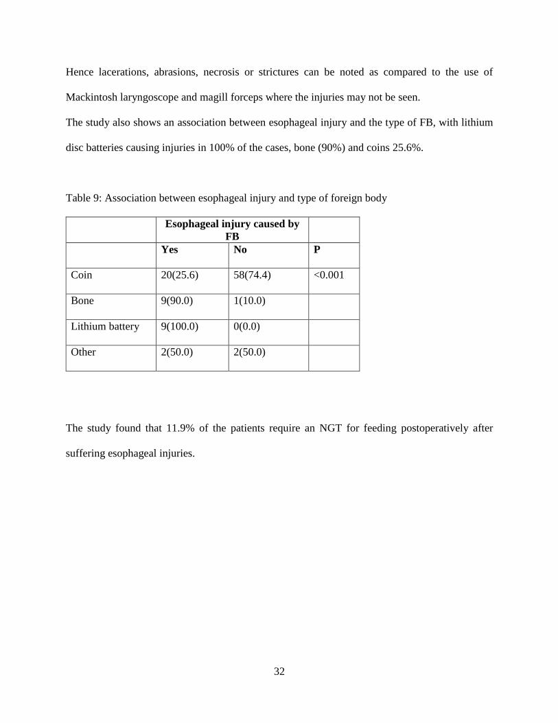
The study also shows an association between esophageal injury and the type of FB, with lithium
disc batteries causing injuries in 100% of the cases, bone (90%) and coins 25.6%.
Table 9: Association between esophageal injury and type of foreign body
Esophageal injury caused by
FB
Yes No P
Coin 20(25.6) 58(74.4) <0.001
Bone 9(90.0) 1(10.0)
Lithium battery 9(100.0) 0(0.0)
Other 2(50.0) 2(50.0)
The study found that 11.9% of the patients require an NGT for feeding postoperatively after
suffering esophageal injuries.
33
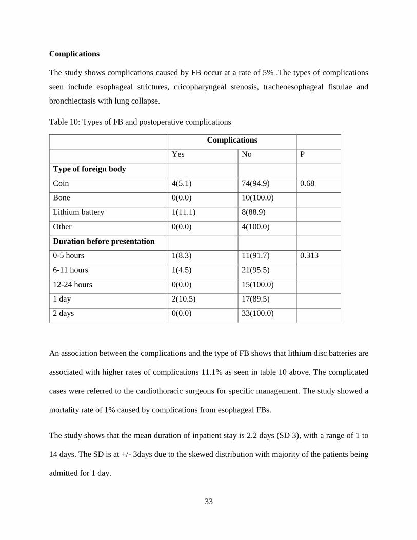
Complications
The study shows complications caused by FB occur at a rate of 5% .The types of complications
seen include esophageal strictures, cricopharyngeal stenosis, tracheoesophageal fistulae and
bronchiectasis with lung collapse.
Table 10: Types of FB and postoperative complications
Complications
Yes No P
Type of foreign body
Coin 4(5.1) 74(94.9) 0.68
Bone 0(0.0) 10(100.0)
Lithium battery 1(11.1) 8(88.9)
Other 0(0.0) 4(100.0)
Duration before presentation
0-5 hours 1(8.3) 11(91.7) 0.313
6-11 hours 1(4.5) 21(95.5)
12-24 hours 0(0.0) 15(100.0)
1 day 2(10.5) 17(89.5)
2 days 0(0.0) 33(100.0)
An association between the complications and the type of FB shows that lithium disc batteries are
associated with higher rates of complications 11.1% as seen in table 10 above. The complicated
cases were referred to the cardiothoracic surgeons for specific management. The study showed a
mortality rate of 1% caused by complications from esophageal FBs.
The study shows that the mean duration of inpatient stay is 2.2 days (SD 3), with a range of 1 to
14 days. The SD is at +/- 3days due to the skewed distribution with majority of the patients being
admitted for 1 day.
34
DISCUSSION
Foreign bodies of the esophagus are common at the Kenyatta National Hospital affecting both
children and adults and this study has shown the current trend and burden of this condition. In this
study a total of 100 patients with FBs of the esophagus were recruited. The mean age of the patients
is found to be 6 years with a range of 9 months to 60 years.
The most frequent age group affected is 2 to 4 years (43.6%) while the adults comprise 6% of the
cases with a peak in patients above 40 years. These results compare to a local study by P.Oduor1
for the period between 1981 and 1986, which showed two peak ages for FBs in the esophagus of
patients below 6 years comprising 59.3% of the patients and adults comprising 23.7% of the cases,
a much higher percentage than what this study has shown. This study has shown that the proportion
of male patients was higher (61.4%) than that of female patients (38.6%) with a male to female
ratio of 1.6:1.
This study shows that a higher proportion of the patients with foreign bodies in the esophagus
presented as referrals from counties neighbouring Nairobi county (69%) and 31% of the patients
presented directly to KNH without going through another medical facility. Majority of the patients
(58.8%) were referred from facilities within Nairobi county, followed by Kiambu county
(14.4%),Kajiado, Machakos and Murang’a refer 6.2% of the cases each. Makueni refers 5.2% and
other counties (Nakuru, Nyeri) refer 1% of the cases.
This study shows that coins form the largest proportion of foreign bodies in the esophagus
(77.2%),followed by bone pieces(9.9%),lithium disc battery(8.9%) and other assorted FBs e.g
wire, razor, tooth picks, plastics(4%).This compares with a study done by Arana A,et al8 who
found FBs to be the following in decreasing order of frequency, coins, toy parts, jewels, batteries,
35
sharp materials such as needles and pins, fish and chicken bones, and "large" amounts of food.
Locally, Oduor P showed that majority of the FBs seen, were metallic objects including coins
which made up 67% of the cases, followed by meat and bones which made 28.3%, vegetable
material constituted 4.1% and plastics made up 1.6%.1
This study also shows an association between age of the patients and the type of foreign body with
a significant proportion of the patients below 2 years shown to have swallowed coins(68%) and
lithium batteries(20%).In the 2 to 4 years age group, 90.9% had swallowed coins and
lithium(9.1%).Patients above 19yrs had bones(100%).
The study also analyzed the duration from ingestion to presentation at KNH and shows a median
duration of 24 hours (interquartile range 8 to 48 hrs).It shows that a higher proportion of the
patients present to KNH after 2 days (32.7%) from the time of ingesting the foreign body. This is
attributed to the fact the parents and clinicians observe the patients at home or in the hospital,
waiting for the FB to descend, and were referred to KNH when this failed. Sharieff GQ,et al24
studied healthy patients with acute (less than 24 h) coin ingestions, they were observed at home
with next-day follow-up and he found that patients with acute esophageal coin ingestions may
experience spontaneous coin passage.
In the clinical presentation of these cases, this study shows that drooling forms the largest
proportion of the symptoms (88.1%), followed by vomiting (66.3%), dysphagia (32.7%) and
odynophagia (28.7%).An association is made between clinical presentations of the patients with
the type of foreign body and drooling is seen in 88.5% of cases with coins, 90% in patients with
bone and 77.8% with lithium battery. On general examination of the patients dehydration is seen
in 63.4% of the cases, lethargy in 9.9% and respiratory distress was seen in 1% of the cases. This
study shows an association between the clinical signs and the type of FB, with lithium disc battery
36
causing dehydration in 100% of the cases, coin (65.4%), other FBs-wire, razor, tooth pick, plastics
(75%) and bone (10%).
On management, this study shows that all the patients have x-ray (100%) done as part of
preoperative work-up. Intraoperatively, this study shows that rigid esophagoscopy is used in 55%
of the cases, mackintosh laryngoscope and magill forcep(44%) which was used during the process
of intubation by the anesthetist and flexible endoscopy (1%).The modes of management were all
successful in removing the FB, similar to results shown by Gmeiner D,et al16 who compared use
of a flexible endoscope and a rigid endoscope. He showed the success rate for foreign body
removal was at 93.4% using the flexible endoscope and 95.2% using the rigid endoscope. Our
results are also comparable to those that Janik JE,et al17got in a retrospective review of 36 children
who had upper esophageal coins extracted using a Magill forceps. All coins were removed without
any complication.
This study also shows that injuries to the esophagus are seen in 55% of patients who undergo rigid
esophagoscopy as compared to laryngoscopy (1.6%).This was a setback in the study as a
reexamination of the esophagus was not done in all cases after the FB was removed using a
mackintosh and magill forcep. The study also shows an association between esophageal injury and
the type of FB with lithium disc battery causing injuries in 100% of the cases, bone (90%) and
coins 25.6%.It also shows that postoperatively 11.9% of the patients require an NGT for feeding
due to the severity of the esophageal injuries caused by the FB.
37
The study shows that the mean duration of inpatient stay is 2.2 days (SD+/- 3), with a range of 1
to 14 days. The SD is at +/- 3days due to the skewed distribution with majority of the patients
being admitted for 1 day. Another setback of the study was that the patients were not followed up
after discharge to monitor their progress and any complications which may arise thereafter.
The study shows that complications caused by FB are at 5%.These were esophageal strictures,
cricopharyngeal stenosis, tracheoesophageal fistulae and bronchiectasis with lung collapse. An
association between the complications and the type of FB is made and lithium disc battery is
shown to be associated with the highest rates of complications 11.1%.Kimball SJ,et al20also
concluded that lithium battery are a major cause of complications in the esophagus. These
complicated cases were referred to the cardiothoracic department who managed them until they
recovered.
The study showed a mortality rate of 1% caused by complications from esophageal FBs.
CONCLUSION
Foreign bodies of the esophagus are a common condition affecting mainly the paediatric
population with a mean age of 6 years and the age group between 2-4 years being the most affected.
Most of these cases are referred from primary health care facilities to KNH. Coins and lithium
batteries are common in the paediatric population with pieces of bones being prevalent in the adult
population. Esophageal injuries are best assessed using a rigid esophagoscope and Lithium disc
batteries cause the highest rates of complications which can eventually lead to mortalities.
38
RECOMMENDATIONS
In line with the findings of this study, lithium disc batteries should be disposed off safely away
from children and public education on the effect of swallowing the batteries should be done to
reduce the incidence of these cases. This can be done through posters, radio programs,
newspapers, MCH clinics and in schools.
A study should be done on the long term effects of the lithium disc batteries on the patients who
did not develop immediate complications. Long-term follow-up of these patients is recommended.
39
REFERENCES
1. Peter Oduor, Foreign bodies in tracheo-bronchial tree and the esophagus at Kenyatta National
Hospital. A retrospective study from January, 1st 1982 to December 1986. MMED
dissertation, University of Nairobi
2. Kramer RE, Lerner DG, Lin T, et al. Management of ingested foreign bodies in children: a
clinical report of the NASPGHAN Endoscopy Committee. J PediatrGastroenterolNutr 2015;
60:562.
3. Wyllie R. Foreign bodies in the gastrointestinal tract. CurrOpinPediatr 2006; 18:563.
4. Yardeni D, Yardeni H, Coran AG, et al. Severe esophageal damage due to button battery
ingestion: can it be prevented? PediatrSurgInt 2004; 20:496.
5. Athanassiadi K, Gerazounis M, Metaxas E, et al. Management of esophageal foreign bodies:
a retrospective review of 400 cases. Eur J CardiothoracSurg 2002; 21:653.
6. Reilly S, Carr L. Foreign body ingestion in children with severe developmental disabilities: a
case study. Dysphagia 2001; 16:68.
7. Louie JP, Alpern ER, Windreich RM. Witnessed and unwitnessed esophageal foreign bodies
in children. Pediatr Emerg Care 2005; 21:582.
8. Arana A, Hauser B,Hachimi-Idrissi S, et al. Management of ingested foreign bodies in
childhood and review of the literature. Eur J Pediatr 2001; 160:468.
9. Yamada T, Sato H, Seki M, et al. Successful salvage of aortoesophageal fistula caused by a
fish bone. Ann ThoracSurg 1996; 61:1843.
40
10. Tokar B, Cevik AA, Ilhan H. Ingested gastrointestinal foreign bodies: predisposing factors
for complications in children having surgical or endoscopic removal. PediatrSurgInt 2007;
23:135.
11. Eisen GM, Baron TH, Dominitz JA, et al. Guideline for the management of ingested foreign
bodies. GastrointestEndosc 2002; 55:802.
12. Kazam JK, Coll D, Maltz C. Computed tomography scan for the diagnosis of esophageal
foreign body. Am J Emerg Med 2005; 23:897.
13. Salmon M, Doniger SJ. Ingested foreign bodies: a case series demonstrating a novel
application of point-of-care ultrasonography in children. PediatrEmerg Care 2013; 29:870.
14. Uyemura MC. Foreign body ingestion in children. AmFam Physician 2005; 72:287.
15. Soprano JV, Fleisher GR, Mandl KD. The spontaneous passage of esophageal coins in
children. Arch PediatrAdolesc Med 1999; 153:1073.
16. Gmeiner D, von Rahden BH, Meco C, et al. Flexible versus rigid endoscopy for treatment of
foreign body impaction in the esophagus. SurgEndosc 2007; 21:2026.
17. Janik JE, Janik JS. Magill forceps extraction of upper esophageal coins. J PediatrSurg 2003;
38:227.
18. Arms JL, Mackenberg-Mohn MD, Bowen MV, et al. Safety and efficacy of a protocol using
bougienage or endoscopy for the management of coins acutely lodged in the esophagus: a
large case series. Ann Emerg Med 2008; 51:367.
19. Waltzman ML. Management of esophageal coins. CurrOpinPediatr 2006; 18:571.
41
20. Kimball SJ, Park AH, Rollins MD 2nd, et al. A review of esophageal disc battery ingestions
and a protocol for management. Arch Otolaryngol Head Neck Surg 2010; 136:866.
21. Lao J, Bostwick HE, Berezin S, et al. Esophageal food impaction in children. PediatrEmerg
Care 2003; 19:402.
22. Hurtado CW, Furuta GT, Kramer RE. Etiology of esophageal food impactions in children. J
PediatrGastroenterolNutr 2011; 52:43.
23. Waltzman ML, Baskin M, WypijD, etal.A randomized clinical trial of the management of
esophageal coins in children.Pediatrics. 2005;116(3):614
24. Sharieff GQ, Brousseau TJ, Bradshaw JA, et al,Acute esophageal coin ingestions: is
immediate removal necessary?PediatrRadiol. 2003;33(12):859
25. Little DC, Shah SR, St Peter SD, et al.Esophageal foreign bodies in the pediatric population:
our first 500 cases.JPediatr Surg. 2006;41(5):914.
26. Shivakumar AM, Naik AS, Prashanth KB, etal.Foreign body in upper digestive tract.Indian J
Pediatr. 2004;71(8):689.
27. Denney W, Ahmad N, Dillard B, etal.Children will eat the strangest things: a 10-year
retrospective analysis of foreign body and caustic ingestions from a single academic
center.Pediatr Emerg Care. 2012 Aug;28(8):731-4.
28. Peters NJ, Mahajan JK, Bawa M, et al.Esophageal perforations due to foreign body impaction
in children.JPediatr Surg. 2015;50(8):1260.
29. Ngan JH, Fok PJ, Lai EC, etal.A prospective study on fish bone ingestion. Experience of 358
patients.Ann Surg. 1990;211(4):459.

42
30. Katsinelos P, Kountouras J, Paroutoglou G, et al. Endoscopic techniques and management of
foreign body ingestion and food bolus impaction in the upper gastrointestinal tract: a
retrospective analysis of 139 cases. JClinGastroenterol. 2006;40(9):784.
31. Daniel WW, 1999. Biostatistics: A foundation for analysis in the health sciences. 7th Edition.
New York. John Wiley & Sons.
43
APPENDICES
APPENDIX 1: GENERAL PATIENT INFORMATION
1. Introduction
I am a senior house officer in ENT-Head & Neck Surgery department. I am requesting for your
consent to participate in a study on the clinical management of foreign bodies of the esophagus as
seen in Kenyatta National Hospital.
2. How you will participate
a) I will ask you questions regarding the foreign body swallowed and any symptoms which
occurred soon after swallowing it.
b) I will carry out a complete Ear, Nose, Throat, Head and Neck examination.
c) I will record the imaging modality that was done.
d) I will record the method used to remove the foreign body any injuries caused by the foreign
body and the post-operative management if any.
e) I will record the number of days you will spend in hospital after the FB has been removed.
f) There will be no monetary benefits for participating in the study and it will be purely on
voluntary basis.
g) You will incur no extra financial costs and confidentiality will be maintained at all times.
h) You will reserve the right to withdraw from the study at any time without any penalty.
i) You will be informed about investigations and importance of the results.
44
3. How will participation affect you?
The study does not affect you negatively in any way because:
a) All the information you give will be confidential.
b) The conclusions drawn from the study shall be useful to improve the management of foreign
bodies in the esophagus.
4. What do we do with the information we get?
The information we get will help us understand the pattern of foreign bodies in the esophagus and
in the management of these cases.
We may publish our findings in scientific journals or present them in scientific meetings.
5.Are you satisfied with the information given?
If you are satisfied with our explanation and you are willing to participate in the study, then please
sign the consent form below.
If you have any questions or need further clarification about the study, kindly contact the
following:
Principal investigator:
Dr.Ouyah W.Libutsi,
Department of Surgery, College of Health Sciences,
University of Nairobi,P.O. Box 2134-00100,Nairobi.
Phone number: 0723541968, Email:[email protected]
45
2. Supervisors:
Prof.Isaac Muthure Macharia,
Professor and Consultant ENT-Head and Neck Surgeon,
Department of Surgery,
University of Nairobi.
Dr.Peter Masinde
Head of ENT Department and Consultant ENT-Head and Neck Surgeon,
Kenyatta National Hospital.
The Chairman KNH-UON Ethics and Research Committee,
Kenyatta National Hospital,Nairobi
46
47
APPENDIX 2: CONSENT FORM
I……………………………………………………..of ……………………………………..…do
hereby give consent to be included in this study on the patterns and management of foreign bodies
in the esophagus as seen in Kenyatta National Hospital.
The nature of the study has been explained to me by Dr. …………………………………………
Date……………………………………………………..Signed…………………………………
I Dr. …………………………………………………confirm that I have explained to the patient
the nature of the study.
Date……………………………………………………..Signed…………………………………
For any further clarifications, contact any of the following:
1.Principal investigator:
Dr.Ouyah W.Libutsi, Resident in ENT-Head & Neck Surgery, Department of surgery,
University of Nairobi, P.O Box 2134-00100 Nairobi.
Phone number: 0723541968
Email:[email protected]
2.Supervisors:
Prof. Isaac Muthure Macharia,
Professor and Consultant ENT-Head and Neck Surgeon,
Department of Surgery, University of Nairobi.

48
Dr.Peter Masinde
Head of ENT Department and Consultant ENT-Head and Neck Surgeon,
Kenyatta National Hospital.
The Chairman KNH-UON Ethics and Research Committee,
Kenyatta National Hospital,Nairobi
Tel.2726300 Ext.44355
49
APPENDIX 3: ASSENT INFORMATION DOCUMENT
1. Introduction
I am a senior house officer in ENT-Head & Neck Surgery department. I am requesting for your
assent for the patient under your care to participate in a study on the clinical management of foreign
bodies of the esophagus as seen in Kenyatta National Hospital.
2. How you will participate
j) I will ask you questions regarding the foreign body swallowed and any symptoms which
occurred soon after swallowing it.
k) I will carry out a complete Ear, Nose, Throat, Head and Neck examination on the patient.
l) I will record the imaging modality that was done.
m) I will record the method used to remove the foreign body any injuries caused by the foreign
body and the post-operative management if any.
n) I will record the number of days your patient will spend in hospital after the FB has been
removed.
o) There will be no monetary benefits for participating in the study and it will be purely on
voluntary basis.
p) You will incur no extra financial costs and confidentiality will be maintained at all times.
q) You will reserve the right to withdraw from the study at any time without any penalty.
r) You will be informed about investigations and importance of the results.
50
3. How will participation affect you?
The study does not affect the patient under your care negatively in any way because:
c) All the information you give will be confidential.
d) The conclusions drawn from the study shall be useful to improve the management of foreign
bodies in the esophagus.
4. What do we do with the information we get?
The information we get will help us understand the pattern of foreign bodies in the esophagus any
complications and the management of these cases.
We may publish our findings in scientific journals or present them in scientific meetings.
5.Are you satisfied with the information given?
If you are satisfied with our explanation and you are willing to participate in the study, then please
sign the assent form below.
If you have any questions or need further clarification about the study, kindly contact the
following:
Principal investigator:
Dr.Ouyah W.Libutsi,
Department of Surgery, College of Health Sciences,
University of Nairobi, P.O. Box 2134-00100, Nairobi.
Phone number: 0723541968, Email:[email protected]

51
2. Supervisors:
Prof. Isaac Muthure Macharia,
Professor and Consultant ENT-Head and Neck Surgeon,
Department of Surgery,
University of Nairobi.
Dr.Peter Masinde
Head of ENT Department and Consultant ENT-Head and Neck Surgeon,
Kenyatta National Hospital.
The Chairman KNH-UON Ethics and Research Committee,
Kenyatta National Hospital,Nairobi
52
APPENDIX 4: ASSENT FORM
I................................................................parent/guardian to..........................................................
do hereby give assent for the patient to be included in this study on the patterns and management
of foreign bodies in the esophagus as seen in Kenyatta National Hospital.
The nature of the study has been explained to me by
Dr.…………………………………………………………………………………………………
Date……………………………………………………..Signed…………………………………
I Dr.…………………………………………………confirm that I have explained to the
parent/guardian the nature of the study.
Date……………………………………………………..Signed…………………………………
For any further clarifications, contact any of the following:
1.Principal investigator:
Dr.Ouyah W.Libutsi, Resident in ENT-Head & Neck Surgery, Department of surgery,
University of Nairobi,
P.O Box 2134-00100 Nairobi.
Phone number:0723541968
Email:[email protected]
53
2.Supervisors:
Prof. Isaac Muthure Macharia,
Professor and Consultant ENT-Head and Neck Surgeon,
Department of Surgery, University of Nairobi.
Dr.Peter Masinde
Head of ENT Department and Consultant ENT-Head and Neck Surgeon,
Kenyatta National Hospital.
The Chairman KNH-UON Ethics and Research Committee,
Kenyatta National Hospital,Nairobi
Tel.2726300 Ext.44355
54
APPENDIX 5: KIAMBATISHO
KIAMBATISHO 1:
MAELEZO KUHUSUIDHINI YA MGONJWA
1. Kitangulizi
Mimi ni daktari ninaye endelea na masomo ya juu kwa kitengo cha upasuaji wa
masikio,mapua,koo na shingo katika Chuo Kikuu cha Nairobi.Ningependa kuomba idhini yako ya
kushiriki katika utafiti wenye lengo la kujua mwenendo wa vitu visivyo faa kuwa kwenye koromeo
na vile ya kuviondoa kwenye koromeo katika hosipitali kuu ya Kenyatta.
2.Jinsi ya kushiriki
a) Nitakuuliza maswali kuhusu kitu ulichomeza na mambo yaliyofanyika baadaye kama
kutapika,kushindwa kumeza au kupumua, ama uchungu.
b) Nitafanya uchunguzi wa kikamilifu wa masikio,pua na koo.
c) Nitaandika njia iliyotumika kuondoa kitu ulichomeza kutoka kwenye koromeo,majeraha
yaliyotokana na kitu ulichomeza na njia iliyotumika kukutibu baada ya kuondoa kitu
ulichomeza.
d) Utafiti huu utafanywa kwa hiari ya mgonjwa na hakutakuwepo na faida ya fedha au fidia
kwa kushiriki.
e) Hakutakua na malipo yoyote ya ziada au gharama utakayo hitajika kulipa na siri za mgonjwa
zitaendelezwa wakati wowote.
f) Unahaki ya kujiondoa kutoka kwa utafiti huu wakati wowote bila adhabu yoyote.
g) Utapewa taarifa au habari kuhusu uchunguzi utakaofanywa na umuhimu wa matokeo.
55
3. Jinsi gani kushiriki kwako kunaweza kuleta madhara
Utafiti hautakudhuru kwa njia yoyote kwa vile:
a) Taarifa yote kuhusu mgonjwa itakuwa ni ya siri
b) Utambulisho hautatangazwa.
c) Baada ya kuhitimisha utafiti huu maarifa yatakayopatikana yatakuwa yamanufaa na yanaweza
kutumika kusaidia kuboresha matibabu ya hali hii.
4.Je,kuna hatari ya kushiriki au kutoshiriki?
a) Hakuna hatari yoyote itakayo jiri kwa kushiriki au kutoshiriki.
b) Kujiondoa wakati wowote au kupinga sehemu ya utafiti hakutaathiri matibabu au ubora wa
huduma ya afya utakayopokea.
5.Je,tutafanyia nini matokeo ya utafiti huu?
Habari itakayotokana na utafiti huu pengine haitakifaidi binafsi lakini itatupa maarifa ambayo
yataboresha matibabu ya vitu vilivy katalia kwenye koromeo.
Kuna uwezekano wa kuchapishwa kwa matokeo ya utafiti huu katika majarida ya kisayansi au
kuwekwa katika mikutano ya kisayansi.
6. Je,umeridhika?
Ukiridhika na maelezo yangu na uko tayari kushiriki,tafadhali weka sahihi yako kwenye fomu ya
idhini.
56
KIAMBATISHO 2:Fomu ya Kukubali kwa mgonjwa
Mimi………………………………………………………………….kutoka……………………
………………………..ninakubali kushirikishwa katika utafiti huu wenye lengo la kujua
mienendo na matibabu vitu visivyo faa kuwa kwenye koromeo katika hosipitali kuu ya Kenyatta.
Nimeelezewa na daktari…………………………………………………………………………...
Tarehe…………………………………………………………..Sahihi……………………………
Mimi daktari……………………………………nahakikisha ya kwamba nimeelezea mgonjwa juu
ya utafiti huu
Tarehe…………………………………………………………..Sahihi……………………………
Mawasiliano:
Mtafiti mkuu
Daktari Ouyah Wilson Libutsi, mwanafunzi wa upasuaji wa masikio,mapua na koo,
Chuo kikuu cha Nairobi,anwani 2134-00100,Nairobi
Simu 0723541968
Barua pepe:[email protected]
Wasimamizi
Profesa Isaac Muthure Macharia
Idara ya upasuaji,kitengo cha upasuaji waMasikio,Mapuana Koo
Chuo kikuu cha Nairobi,anwani 2134-00100,Nairobi,
Daktari Peter Masinde,
Mkuu wa kitengo cha upasuaji wa Masikio,mapua na koo,Hosipitali kuu ya Kenyatta
57
Mwenyekiti
KNH/UON Ethical and Research Committee
Hosipitali kuu ya Kenyatta,
Simu 2726300-9 Ext.44355
58
KIAMBATISHO3 : MAELEZO KUHUSU IDHINI YA MZAZI WA MGONJWA
1. Kitangulizi
Mimi ni daktari ninaye endelea na masomo ya juu kwa kitengo cha upasuaji wa
masikio,mapua,koo na shingo katika Chuo Kikuu cha Nairobi.Ningependa kuomba idhini yako ya
kushirikisha mtoto wako katika utafiti wenye lengo la kujua mwenendo wa vitu visivyo faa kuwa
kwenye koromeo na vile ya kuviondoa kwenye koromeo katika hosipitali kuu ya Kenyatta.
2. Jinsiya kushiriki
a) Nitakuuliza maswali kuhusu kitu mtoto alichomeza na mambo yaliyofanyika baadaye kama
kutapika, kushindwa kumeza au kupumua ,ama uchungu.
b) Nitafanya uchunguzi wa kikamilifu wa masikio,pua na koo ya mtoto.
c) Nitaandika njia iliyotumika kuondoa kitu mgonjwa alichomeza kutoka kwenye koromeo,
majeraha yaliyotokana na kitu alichomeza na njia iliyotumika kumtibu baadaya kuondoa kitu
alichomeza.
d) Utafiti huu utafanywa kwa hiari ya mzazi wa mgonjwa na hakutakuwepo na faida ya fedha
au fidia kwa kushiriki.
e) Hakutakua na malipo yoyote ya ziada au gharama utakayo hitajika kulipa na siri za mgonjwa
zitaendelezwa wakati wowote.
f) Unahaki ya kujiondoa kutoka kwa utafiti huu wakati wowote bila adhabu yoyote.
g) Utapewa taarifa au habari kuhusu uchunguzi utakaofanywa na umuhimu wa matokeo.
59
60
3. Jinsi gani kushiriki kwa mtoto wako kunaweza kuleta madhara
Utafiti hautamdhuru mgonjwa kwa njia yoyote kwa vile:
a. Taarifa yote kuhusu mgonjwa itakuwa ni ya siri
b. Utambulisho hautatangazwa.
c. Baada ya kuhitimisha utafiti huu maarifa yatakayopatikana yatakuwa ya manufaa na yanaweza
kutumika kusaidia kuboresha matibabu ya hali hii.
4.Je, kuna hatari ya kushiriki au kutoshiriki?
a. Hakuna hatari yoyote itakayo jiri kwa mgonjwa kwa kushiriki au kutoshiriki.
b. Kujiondoa wakati wowote au kupinga sehemu ya utafiti hakutaathiri matibabu au ubora wa
huduma ya afya utakayopokea.
5.Je,tutafanyia nini matokeo ya utafiti huu?
Habari itakayotokana na utafiti huu pengine haitakufaidi binafsi lakini itatupa maarifa ambayo
yataboresha matibabu ya vitu vilivyo katalia kwenye koromeo na namna ya kuvitibu.
Kuna uwezekano wa kuchapishwa kwa matokeo ya utafiti huu katika majarida ya kisayansi au
kuwekwa katika mikutano ya kisayansi.
6.Je, umeridhika?
Ukiridhika na maelezo yangu na uko tayari kwa mtoto wako kushiriki,tafadhali weka sahihi yako
kwenye fomu ya idhini.Kama uko na maswali yoyote tafadhali wasiliana nasi kupitia njia
zifwatazo:
Mtafiti mkuu
Daktari Ouyah Wilson Libutsi, mwanafunzi wa upasuaji wa masikio, mapua na koo,
Chuo kikuu cha Nairobi, anwani 2134-00100, Nairobi
Simu 0723541968
Barua pepe:[email protected]
Wasimamizi
Profesa Isaac Muthure Macharia
Idara ya upasuaji,kitengo cha upasuaji wa Masikio, Mapua na Koo
Chuo kikuu cha Nairobi, anwani 2134-00100, Nairobi,
61
KIAMBATISHO 4:Fomu ya Kukubali kwa mzazi wa mgonjwa
Mimi……………………………………mzazi wa………………………………ninakubali mtoto
wangu kushirikishwa katika utafiti huu wenye lengo la kujua mienendo na matibabu ya vitu
visivyofaa kuwa kwenye koromeo katika hosipitali kuu ya Kenyatta.
Nimeelezewa na daktari…………………………………………………………………………...
Tarehe…………………………………………………………..Sahihi……………………………
Mimi daktari……………………………………nahakikisha ya kwamba nimeelezea mzazi wa
mgonjwa kuhusu utafiti huu
Tarehe…………………………………………………………..Sahihi……………………………
Mawasiliano:
Mtafiti mkuu
Daktari Ouyah Wilson Libutsi, mwanafunzi wa upasuaji wa masikio,mapua na koo,
Chuo kikuu cha Nairobi,anwani 2134-00100,Nairobi
Simu 0723541968
Barua pepe:[email protected]
Wasimamizi
Profesa Isaac Muthure Macharia
Idara ya upasuaji,kitengo cha upasuaji waMasikio,Mapua na Koo
Chuo kikuu cha Nairobi, anwani 2134-00100,Nairobi,
Daktari Peter Masinde,
Mkuu wa kitengo cha upasuaji wa Masikio,mapua na koo
Hosipitali kuu ya Kenyatta
62
Mwenyekiti
KNH/UON Ethical and Research Committee
Hosipitali kuu ya Kenyatta,
Simu 2726300-9 Ext.44355
63
APPENDIX 6: QUESTIONNAIRE
Study number……………………………………………………….Date………………………
Section A:
Patient Biodata
I.P No……………….......Age………………………………..Gender………………………….
Section B:
History
1. What foreign body did you or the patient swallow…………………………………………....?
2. What is the period between swallowing the foreign body and presentation to A& E department
in KNH …………………………………………………………………………..?
3. Did you seek medical assistance elsewhere prior to presenting at the A&E in KNH?
a)YES b)NO
If YES,where………………………………………………………………………………….?
Was there any form of management given before being referred.....………………………….?
4. Did you or the patient have any of the following symptoms soon after swallowing the foreign
body?
a) Drooling
b) Choking
64
c) Vomiting
d) Odynophagia
e) Dysphagia
f) Difficulties in breathing
Others………………………………………………………………………………………..
Management:
5. What are the findings on examination of the patient?
a) Dehydrated
b) Lethargic
c) Respiratory distress
d) Emaciated
Other findings……………………………………….
6. What the vital signs on examination of the patient?
a)Temperature………….. b)Pulse rate………………….. c) Respiratory rate……………
7. Has the patient undergone any radiological investigations?
a) X-ray
65
b) CT Scan
c) MRI
d) U/S Scan
Operative Record
8. What method was used to remove the foreign body intraoperatively?
a) Rigid Esophagoscopy
b) Direct hypopharyngoscopy
c) Laryngoscopy and Macgill forceps
d) Flexible endoscopy
e) Foleys catheter
9. How far is the Foreign Body located from the frontal incisors in centimeters?....................cm
10. What type of foreign body was found during esophagoscopy………………………………...?
11. Are there any injuries to the esophagus caused by the foreign body?
a) YES b)NO
If YES, what injuries were observed?
a) Lacerations
b) Abrasion
c) Perforations
d) Necrosis
e) Strictures
f) Adhesions
g) Stenosis
Others…………………………………………………………………………..
12. Does the patient require NGT postoperatively?
a)YES b)NO
Postoperative management:
13. Did the patient suffer any postoperative complication?
a)YES b)NO
If YES,please specify ………………………………………………………………………
14. How many days was the patient admitted for in the ward postoperatively?............................

66
APPENDIX 7: RESEARCH APPROVAL

67