Clinical Management of Foreign Bodies of the Esophagus as ...
Upload
khangminh22Category
view
5download
0
Morphology and histologyof the esophagus and the stomach
Ph.D Dr. David LendvaiAnatomy, Histology and Embryology Institute
2019.Painting: Szinyei Merse Pál, Majális (1873.)

ESOPHAGUS

Esophagus
cervical partthoracic partabdominal part
C6 - Th11 left side
„upper esophagus sphincter”(m. cricopharyngeus)
„lower esophagus sphincter” (cardia)
narrowings:• cricoid cartilage• aortis arch• left principal bronchus• diaphragm, esophageal hiatus
narrowings from the incisors:15 cm23 cm24 cm40 cm

Esophagus
• anterior to prevertabral (alar) fascia & buccopharyngeal fascia
• behind trachea
• right to aortic arch
• medial to right lung
• behind left atrium
• anterior and right to descendingaorta
• diaphragm, esophageal hiatus
• recurrent laryngeus n.vagus n. (left ant., right post.)

Esophagus, cervical spaces
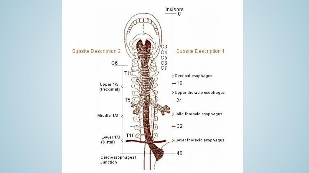
inferior thyroidea a.
esophageal r. – thoracica aorta
esophageal r. – left inf. phrenic a.
esophageal r. – left gastric a.
Esophagus blood supply


http://img.tfd.com/mk/S/X2604-S-18.png
Sengstaken-Blakemore tube
Esophagus – veinsEsophageal varix

Esophagus innervation
vagus n.sympathetic trunc
plexus esophagei

Lymphatic drainage of esophagus
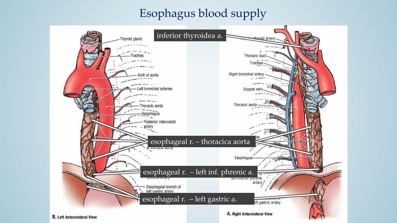
http://www.summitgastro.com/Media/Default/endoscopic-procedures/egd.png
https://en.wikipedia.org/wiki/Gastroesophageal_reflux_disease#/media/File:GERD.pnghttps://commons.wikimedia.org/wiki/File%3ABarretts_esophagus.jpg
GastroscopyGastro-esophageal reflux

Throughout the remainder of the digestive system, the histological composition of the alimentary canal canbe described by the following blue-print:

Histology of esophagus


Esophagus histology

ESOPHAGUSHISTOLOGY
Tunica mucosa, epithelium: stratified non-keratinizedsquamous epithelium (1)
Longitudinal folds by mucosa and submucosa
Lamina propria mucosae (2) cardiaglands in the distal part of esophagus.
Lamina muscularis mucosae (3) longitudinal smooth muscle
Tunica submucosa (4): mixed ormucous small salivary glands
Tunica muscularis (5): innercircular, outer longitudinal. Upperpart: striated muscle, middle: mixed, lower part: smooth muscle
Tunica adventitia (abdominal part: tunica serosa)
1
2
5
4
3
www.daganatok.hu
www.szimpatika.hu
www.chewitwell.hu
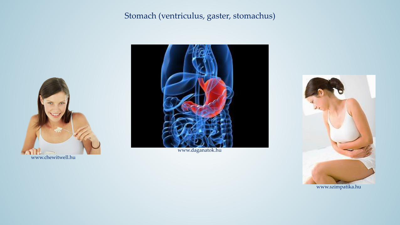
Stomach (ventriculus, gaster, stomachus)

Parts
Cardiac notch of stomach(incisure, Angle of His)
Angular incisure
Angulus gastricus
Surfaces (walls):
paries anterior
paries posterior
Margins:
curvatura minor (minor curvature)
curvatura major (major curvature)
Spalteholz
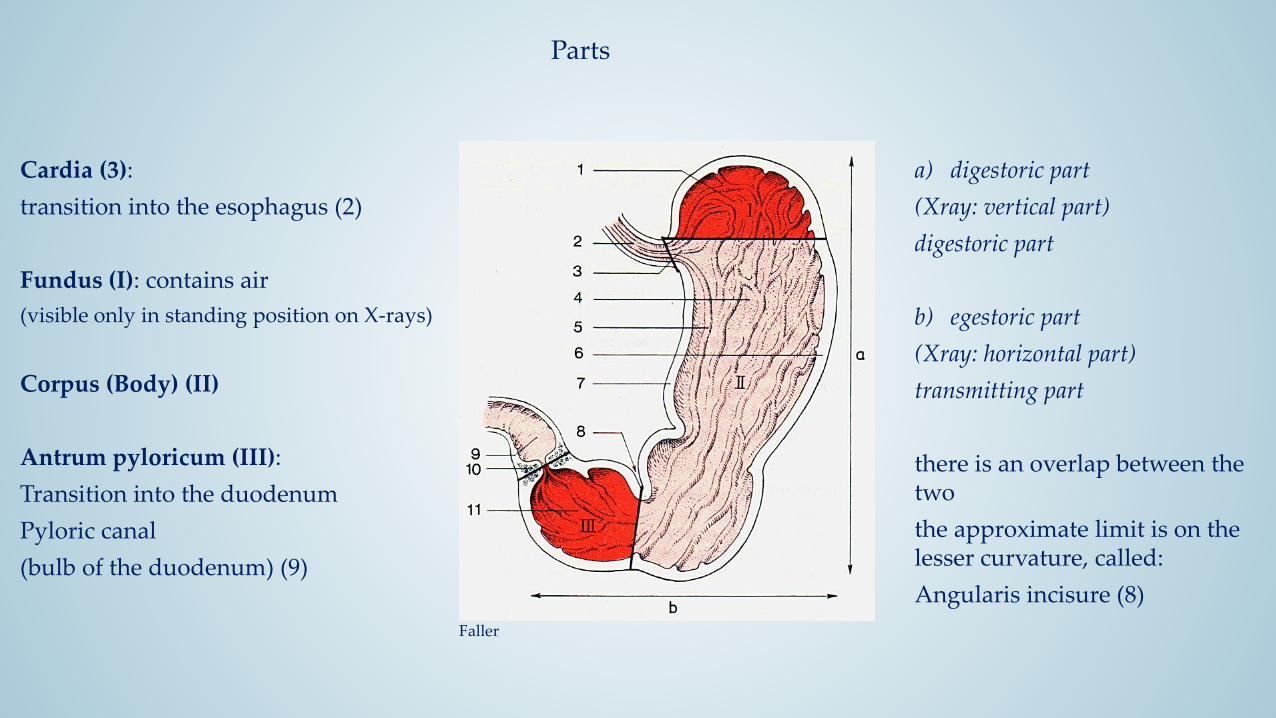
Parts
Cardia (3):
transition into the esophagus (2)
Fundus (I): contains air
(visible only in standing position on X-rays)
Corpus (Body) (II)
Antrum pyloricum (III):
Transition into the duodenum
Pyloric canal
(bulb of the duodenum) (9)
Faller
a) digestoric part
(Xray: vertical part)
digestoric part
b) egestoric part
(Xray: horizontal part)
transmitting part
there is an overlap between the two
the approximate limit is on the lesser curvature, called:
Angularis incisure (8)

Cardiac notch of stomach or the angle of His
Szél
Szél
www.scielo.isciii.es

Stomach - musculatureSobotta
Faller
Typically to hollow organs into muscular tunica organised
histology: from smooth muscle – autonomous innervation
3 layers:
(a) Longitudinal stratum – outer, longitudinal
Strongest at the lesser curvature, missing at the cardiac notch (3)
(b) Circular stratum – middle , circular continuous
, closed layer
Strengthening: sphincter pyloric muscle
(c) Oblique fibres – innermost „layer”
Typically from the the cardiac notch (His) to the greatercurvature

Stomach – inner surface
At the cardia
Sharp esophageal-stomach
transition
(endoscopy called „Z-line”)
Epithelium exchange zone! (tumors )
By the lesser curvaturelongitudinal corrugations
(longitudinal folds)
To help the fast transition of liquids into the duodenum
Between folds:
areae gastricae
Sobotta
www.cures4heartburn.com
www.kolumbus.fi
www.kolumbus.fi

Stomach – blood supply Faller
Spalteholz
The stomach develops from the foregut
blood supply: from the abdominal arteries of the foregut, called celiac trunk
(or Haller's tripod)
Anastomosing branches from left and right along the lesser and greater curvature, or to the fundus; direct or indirect supply from the celiac trunk
(12) left gastric a.: directly from the celiac trunk, „left up”
(7) rigth gastric a.: from the proper hepatic a.
(11) Right gastroepiploic a. (seu gastroomental) from the gastroduodenalis a.
(16) Left gastroepiploica a. (seu gastroomental) from the splenic a.
(14) Short gastric aa. from the splenic a., to the fundus
12 and 7 along the lesser curvature, 11 and16 along the grater curvature forms an arched anastomosis
A rich blood supply for reservein shape and size changes of the stomach, or isolated surgery

Stomach – venous drainageFaller
As from all the unpaired abdominal organs, the blood flows into the
liver via the portal vein
Fundus veins are also called coronal vv.
www.aafp.org
Szél
Porto-caval anastomosis between the portal v. and the superior v. cava is the left gastric v., by the abdominal oesophageal and the azygos/hemiazygos vv.: stagnancy of the blood in these submucosal veins, can cause dilations (varicosis). It can lead to dangerous bleedings (rupture)!!!

Stomach – lymphatic drainageLittmann
Faller
Lymphatic drainage sectors, characteric lymphatic flow:
(6) nodi lymph. gastrici sinistri (two-way lymphatic flow!!!):
chisterna chyli or left supraclavium (Virchow-lymph node)
(5) nodi lymph. gastrici dextri (it can flow in the opposite:
gastric cancer -> liver metastasis)
(7) nodi lymph. gastroepiploici sinistri
(8) nodi lymph. gastroepiploici dextri
(3) nodi lymp. pylorici
Other lymphatic drainage:
nodi lymph. pancreaticolienales
nodi lymph. lienales
nodi lymph. hepatici
nodi lymph. coeliaci
DOI: 10.1056/NEJMicm1204740

Stomach - innervationAutonomic innervation
SY: celiac ggl.
Decrease the acid secretion and the
peristaltik motility
PSY: vagus n. dexter et sinister
(Abdo part of X. cranial nerve)
Increase the acid secretion and the
peristaltik motility
The most important coordinator of the gastric movements (motility) but myenteric plexus of Auerbach is the autonomic rhythm-generator
Pernkopf
Szél
Earlier ulcers were cured by different vagotomy
The final positions of the two vagus nn., can be explained by the development of the stomach gyomor (rotation to the right)Faller

stomach – shape, position
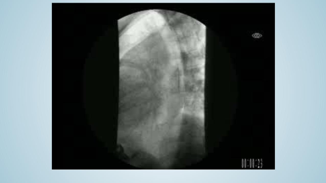
Radiological changes of the stomach
(In a standing position, there is air in the fundus)
Szél
Faller
lying
standing
• The situation of the fundus is
Relatively fixed below the left
dome of the diaphragm
• The cardia is fixed at level
Th11/12 or Th10/11 on the left
side of the vertebral column
Depending on: load, position, muscle tone, temper, ageing
• The situation of the pylorus is: lying position at L1, standing position at L2
on the right side of the vertebral column (transpyloric plain)

Stomach - topography
left hypochondrium
epigastrium
Loaded condition or in ptosis:
In the umbilical region
Faller

Stomach - topography
Partially covered, limited physical examination:
a) hepatic surface (totally covered by the liver)
b) diaphragmatic surface (Traube-space)
c) free surface or triangle of Labbe (here can be touched vie the abdominal wall
Borders: left lobe of the liver, greater curvature, left arch of the rib cage
Pernkopf
Faller
Downwards facing the transversal colon, to the left
spleen and diaphragm
Faller
Traube-space: physical examination of the thorax
Diagnostic importance percussion of pleural effusion
(percussion sound dims in the presence of liquid)

Stomach - topography
Pernkopf
Szentágothai - Réthelyi
Behind the stomach pancreas can be found, separated by the
peritoneum (abdominal surgery!)The space between the stomach
and the pancreas: bursa omentalis
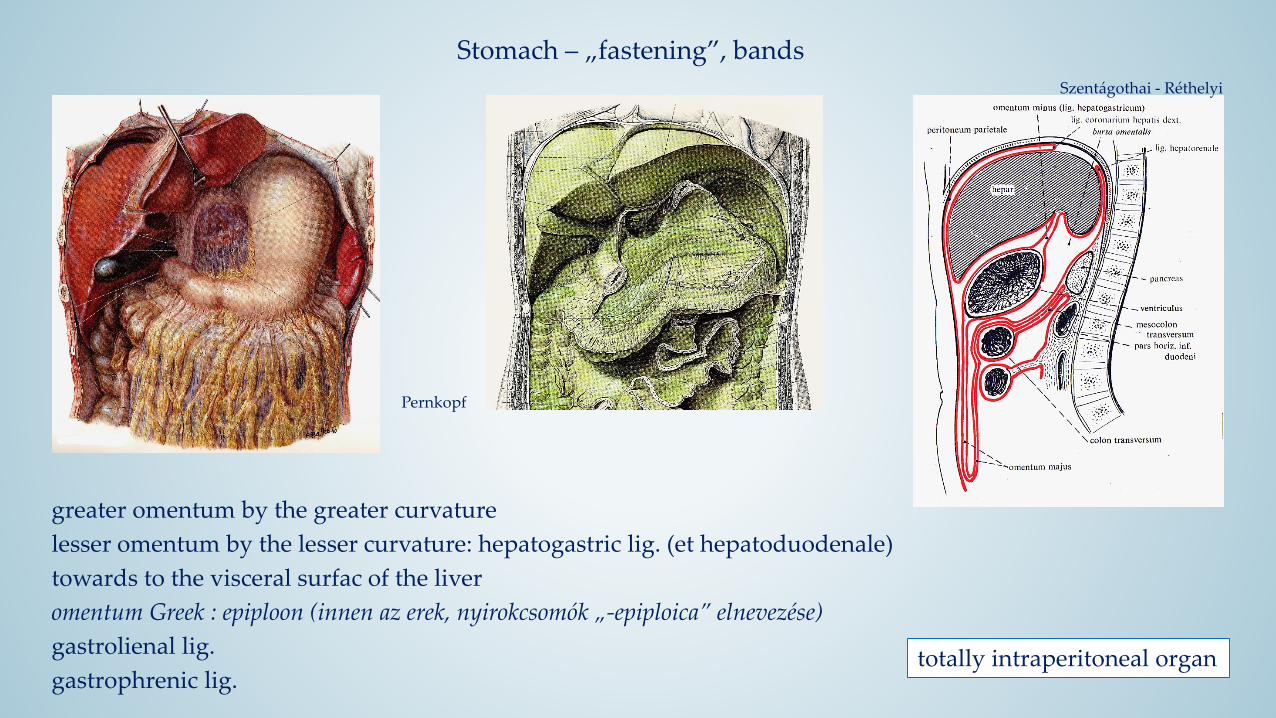
Stomach – „fastening”, bands
Szentágothai - Réthelyi
Pernkopf
greater omentum by the greater curvature
lesser omentum by the lesser curvature: hepatogastric lig. (et hepatoduodenale)
towards to the visceral surfac of the liver
omentum Greek : epiploon (innen az erek, nyirokcsomók „-epiploica” elnevezése)
gastrolienal lig.
gastrophrenic lig.totally intraperitoneal organ

Histology of the stomach
• typically to hollow organs: stratified wall
• certain areas of the stomach are different, especially the mucosa,
mainly in the shape of the glands and the cell components
Faller
Sobotta
Later will be important in oncological
Grading and staging

Histology of the cardia www.cures4heartburn.com
Sharp epithelial changing, the SNCE continues :
• simple columnar epithelium (endoscopic: Z-line)
• Typical locations of early staged tumours (metaplasy),
Barret (app. 2 cm above)
• Mixed tubular glands embedded in the lamina, mostly mucous: cardia glands
• branched, tubular glands, short and wide lumen
• wide foveola
• dense, protective mucus
• Some cells are producing gastrin

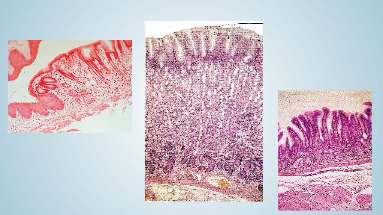
Fundus, corpus histology
Wide lamina propria of the mucosa
Elongated tubular, sometimes branched fundus glands.
Characteristic cells of the fundus glands:
1. Undifferentiated cells:
• Gastric pits
• Upper columnar epithelium replacement
• Mucus producement
2. Mucous cells:
• Mostly nin the neck and isthmus
• secretory granuls apically
• Liquid , mucus producement
3. Parietal cells:
• As they „cover” the gland
• Extremely apical surface increase
• HCl, KCl, intrinsic factor
4. Chief cells
• zymogenic cells
• pepszinogen, lipase
• At the basal part of the gland
5. enteroendocrine cells
gastrine, somatostatine,
glukagon, serotonin, histamin
Röhlich
Faller

fundus, corpus histology
fundus, PAS – Kongo - Hematoxilin
• PAS – undifferentiated and mucous cell
• Kongo(vörös) – parietal cells
• Hematoxilin – chief cells
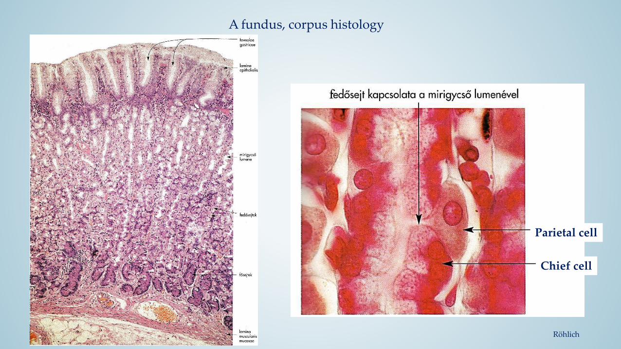
A fundus, corpus histology
Röhlich
Parietal cell
Chief cell
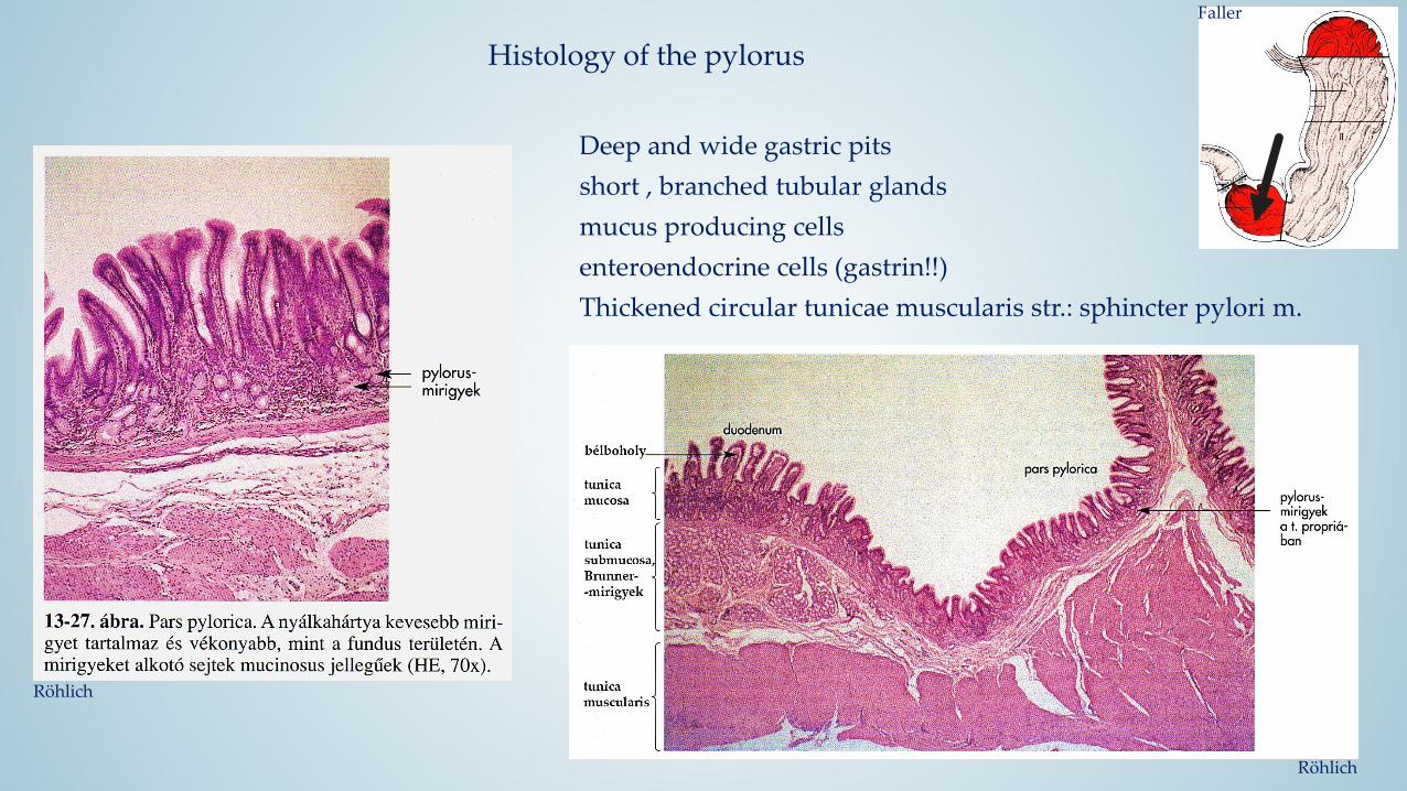
Histology of the pylorus
Faller
Röhlich
Deep and wide gastric pits
short , branched tubular glands
mucus producing cells
enteroendocrine cells (gastrin!!)
Thickened circular tunicae muscularis str.: sphincter pylori m.
Röhlich

Littmann
Copyright © 2022 FDOKUMEN