Global transcriptional response to carbonic anhydrase IX deficiency in the mouse stomach
14
Kallio et al. BMC Genomics 2010, 11:397 http://www.biomedcentral.com/1471-2164/11/397 Open Access RESEARCH ARTICLE © 2010 Kallio et al; licensee BioMed Central Ltd. This is an Open Access article distributed under the terms of the Creative Commons Attribution License (http://creativecommons.org/licenses/by/2.0), which permits unrestricted use, distribution, and reproduction in any medium, provided the original work is properly cited. Research article Global transcriptional response to carbonic anhydrase IX deficiency in the mouse stomach Heini Kallio* 1,2 , Mika Hilvo 3 , Alejandra Rodriguez 1,2 , Eeva-Helena Lappalainen 1,2 , Anna-Maria Lappalainen 1,2 and Seppo Parkkila 1,2 Abstract Background: Carbonic anhydrases (CAs) are a family of enzymes that regulate pH homeostasis in various tissues. CA IX is an exceptional member of this family because in addition to the basic CA function, it has been implicated in several other physiological and pathological processes. Functions suggested for CA IX include roles in cell adhesion and malignant cell invasion. In addition, CA IX likely regulates cell proliferation and differentiation, which was demonstrated in Car9 -/- mice. These mice had gastric pit cell hyperplasia and depletion of chief cells; however, the specific molecular mechanisms behind the observed phenotypes remain unknown. Therefore, we wanted to study the effect of CA IX deficiency on whole-genome gene expression in gastric mucosa. This was done using Illumina Sentrix ® Mouse-6 Expression BeadChip arrays. The expression of several genes with notable fold change values was confirmed by QRT- PCR. Results: CA IX deficiency caused the induction of 86 genes and repression of 46 genes in the gastric mucosa. There was 92.9% concordance between the results obtained by microarray analysis and QRT-PCR. The differentially expressed genes included those involved in developmental processes and cell differentiation. In addition, CA IX deficiency altered the expression of genes responsible for immune responses and downregulated the expression of several digestive enzymes. Conclusions: Microarray analysis identified several potential genes whose altered expression could explain the disturbed cell lineage phenotype in the Car9 -/- gastric mucosa. The results also indicated a novel role for CA IX in the regulation of immunologic processes and digestion. These findings reinforce the concept that the main role of CA IX is not the regulation of pH in the stomach mucosa. Instead, it is needed for proper function of several physiological processes. Background The carbonic anhydrases (CAs) are a family of zinc-con- taining metalloenzymes that catalyze the reversible hydration of carbon dioxide in the reaction: CO 2 + H 2 O H + + HCO 3 - . They participate in several physiological processes, such as acid-base balance, CO 2 and HCO 3 - transport, respiration, bone resorption, ureagenesis, glu- coneogenesis, lipogenesis, production of body fluids, and fertilization [1,2]. The CA family consists of 13 active isozymes in mammals, 12 of which are expressed and function in humans [3]. The CA isozymes have diverse tissue expression patterns, characteristic subcellular localizations, as well as unique kinetic and inhibitory properties. CA IX is a dimeric protein associated with the cell membrane [4,5]. In its mature form, CA IX is composed of an N-terminal proteoglycan (PG) domain, a CA cata- lytic domain, a transmembrane region, and a short intra- cytoplasmic tail at the C-terminus [6]. CA IX is the only member of the CA family containing a PG domain in addition to the CA domain. Consequently, CA IX has been suggested to participate in cell adhesion processes. In fact, using MDCK (Madin-Darby canine kidney) epi- thelial cells, it was shown that CA IX reduces E-cadherin- mediated cell-cell adhesion by interacting with β-catenin [7]. * Correspondence: [email protected] 1 Institute of Medical Technology and School of Medicine, University of T ampere, Biokatu 6, FI-33520 Tampere, Finland Full list of author information is available at the end of the article
Transcript of Global transcriptional response to carbonic anhydrase IX deficiency in the mouse stomach

Kallio et al. BMC Genomics 2010, 11:397http://www.biomedcentral.com/1471-2164/11/397
Open AccessR E S E A R C H A R T I C L E
Research articleGlobal transcriptional response to carbonic anhydrase IX deficiency in the mouse stomachHeini Kallio*1,2, Mika Hilvo3, Alejandra Rodriguez1,2, Eeva-Helena Lappalainen1,2, Anna-Maria Lappalainen1,2 and Seppo Parkkila1,2
AbstractBackground: Carbonic anhydrases (CAs) are a family of enzymes that regulate pH homeostasis in various tissues. CA IX is an exceptional member of this family because in addition to the basic CA function, it has been implicated in several other physiological and pathological processes. Functions suggested for CA IX include roles in cell adhesion and malignant cell invasion. In addition, CA IX likely regulates cell proliferation and differentiation, which was demonstrated in Car9-/- mice. These mice had gastric pit cell hyperplasia and depletion of chief cells; however, the specific molecular mechanisms behind the observed phenotypes remain unknown. Therefore, we wanted to study the effect of CA IX deficiency on whole-genome gene expression in gastric mucosa. This was done using Illumina Sentrix®Mouse-6 Expression BeadChip arrays. The expression of several genes with notable fold change values was confirmed by QRT-PCR.
Results: CA IX deficiency caused the induction of 86 genes and repression of 46 genes in the gastric mucosa. There was 92.9% concordance between the results obtained by microarray analysis and QRT-PCR. The differentially expressed genes included those involved in developmental processes and cell differentiation. In addition, CA IX deficiency altered the expression of genes responsible for immune responses and downregulated the expression of several digestive enzymes.
Conclusions: Microarray analysis identified several potential genes whose altered expression could explain the disturbed cell lineage phenotype in the Car9-/- gastric mucosa. The results also indicated a novel role for CA IX in the regulation of immunologic processes and digestion. These findings reinforce the concept that the main role of CA IX is not the regulation of pH in the stomach mucosa. Instead, it is needed for proper function of several physiological processes.
BackgroundThe carbonic anhydrases (CAs) are a family of zinc-con-taining metalloenzymes that catalyze the reversiblehydration of carbon dioxide in the reaction: CO2 + H2O �H+ + HCO3
-. They participate in several physiologicalprocesses, such as acid-base balance, CO2 and HCO3
-
transport, respiration, bone resorption, ureagenesis, glu-coneogenesis, lipogenesis, production of body fluids, andfertilization [1,2]. The CA family consists of 13 activeisozymes in mammals, 12 of which are expressed andfunction in humans [3]. The CA isozymes have diverse
tissue expression patterns, characteristic subcellularlocalizations, as well as unique kinetic and inhibitoryproperties.
CA IX is a dimeric protein associated with the cellmembrane [4,5]. In its mature form, CA IX is composedof an N-terminal proteoglycan (PG) domain, a CA cata-lytic domain, a transmembrane region, and a short intra-cytoplasmic tail at the C-terminus [6]. CA IX is the onlymember of the CA family containing a PG domain inaddition to the CA domain. Consequently, CA IX hasbeen suggested to participate in cell adhesion processes.In fact, using MDCK (Madin-Darby canine kidney) epi-thelial cells, it was shown that CA IX reduces E-cadherin-mediated cell-cell adhesion by interacting with β-catenin[7].
* Correspondence: [email protected] Institute of Medical Technology and School of Medicine, University of Tampere, Biokatu 6, FI-33520 Tampere, FinlandFull list of author information is available at the end of the article
© 2010 Kallio et al; licensee BioMed Central Ltd. This is an Open Access article distributed under the terms of the Creative CommonsAttribution License (http://creativecommons.org/licenses/by/2.0), which permits unrestricted use, distribution, and reproduction inany medium, provided the original work is properly cited.

Kallio et al. BMC Genomics 2010, 11:397http://www.biomedcentral.com/1471-2164/11/397
Page 2 of 14
CA IX is expressed in only few normal tissues with theexpression being strongest in human, rat, and mouse gas-tric mucosa, where it is present from the gastric pits tothe deep gastric glands [8,9]. CA IX is confined to thebasolateral surface of epithelial cells and is produced byall major cell types of the gastric epithelium [8]. CA IX isan exceptional member of the CA family because it isexpressed in several cancers that arise from CA IX nega-tive tissues including renal, lung, cervical, ovarian, esoph-ageal, and breast carcinomas [6]. However, gastric cancerand premalignant lesions have shown decreased expres-sion of CA IX [10]. In tumor tissues, CA IX is linked withthe hypoxic phenotype mediated by the hypoxia-induc-ible transcription factor 1 (HIF-1), which binds to thehypoxia responsive element, HRE, of the CA9 promoter[11]. In hypoxic conditions, cancer cells are dependentmainly on anaerobic metabolism in their energy produc-tion. This anaerobic tumor metabolism generatesexcesses of acidic products, such as lactic acid and H+
that have to be extruded from the cell interior. It has beenshown that CA IX can contribute to the acidification ofthe hypoxic extracellular milieu, thus helping tumor cellsto neutralize the intracellular pH [12]. Accordingly, CAIX overexpression often indicates poor prognosis andresistance to classical radio- and chemotherapies [13].However, CA IX is not only confined to the hypoxicregions of the tumors, indicating that there might besome other pathways that regulate its expression. In fact,the expression of CA IX can be induced under normoxicconditions by high cell density, and this regulation ismediated by phosphatidylinositol 3-kinase (PI3K) signal-ing [14]. Moreover, the mitogen-activated protein kinase(MAPK) pathway is involved in the regulation of CA IXexpression via control of the CA9 promoter by both HIF-1-dependent and -independent signals [15]. This pathwayis also interrelated with the PI3K pathway and the SP1(specificity protein 1) transcription factor.
The generation of Car9-/- mice has revealed that inaddition to pH regulation and cell adhesion, CA IX pos-sesses other functional roles [16]. These Car9 knockoutmice showed no abnormalities in growth, reproductivepotential, and life span. However, CA IX deficiencyresulted in hyperplasia of the gastric mucosa. The hyper-plastic mucosa contained an increased number and pro-portion of the mucus-producing pit cells whereas thenumber and proportion of chief cells was reduced. Thetotal number of parietal cells remained unchanged, andaccordingly, CA IX-deficient mice had normal gastric pH,acid secretion, and serum gastrin levels. Thus, these find-ings demonstrate that CA IX contributes to the control ofdifferentiation and proliferation of epithelial cell lineagesin the gastric mucosa. A previous study examinedwhether the effects of CA IX deficiency could be modi-fied by a high-salt diet, a known co-factor of carcinogene-
sis [17]. These results showed that the high-salt dietslightly increased the glandular atrophy in the bodymucosa in Car9-/- C57BL/6 mice, whereas this effect wasnot observed in BALB/c mice. However, no metaplasticor dysplastic abnormalities were seen in the gastrointesti-nal epithelium of CA IX-deficient C57BL/6 mice. Thus,these observations suggest that CA IX deficiency alonemay not be a significant carcinogenic factor but could ini-tiate a carcinogenic process by affecting cell proliferationand/or differentiation in the gastric mucosa.
This study was designed to better understand thehyperplastic phenotype of the stomach mucosa of Car9-/-
mice because the molecular mechanisms behind it arecurrently unknown. In addition, we wanted to more spe-cifically elucidate the functional roles of CA IX, as there isa growing body of evidence showing that they extend farbeyond pH regulation. For this purpose, a genome-wideexpression analysis was performed. Microarray data anal-ysis revealed several genes whose expression was changeddue to Car9 gene disruption in the gastric mucosa.
ResultsTranscriptional response to Car9 deficiency in the stomach wallStomach RNA from 6 Car9-/- mice and 6 wild-type micewas analyzed by microarray. The analysis revealed 86upregulated genes and 46 downregulated genes, using afold change cut-off of ± 1.4 for up- and downregulatedexpression, respectively (See additional file 1: List ofgenes differentially expressed in the stomach of Car9-/-
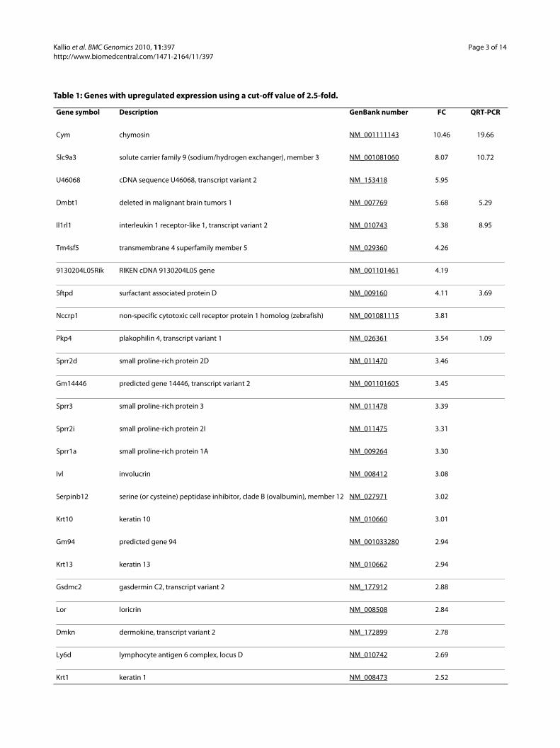
mice). This cut-off value has been proposed as an ade-quate level above which there is a high correlationbetween microarray and QRT-PCR data, regardless ofother factors such as spot intensity and cycle threshold[18]. The fold changes ranged from 10.46 to -12.14.Herein, a list of genes using a cut-off value of ± 2.5-fold isshown (Tables 1 and 2). When using these criteria, all thegenes with significantly (p < 0.05) altered expression aredisplayed, that is, 14 genes with induced expression and21 genes with repressed expression. The list of all the reg-ulated genes was functionally annotated (see additionalfile 2: Functional annotation of genes regulated in thestomach of Car9 knockout mice), showing enrichment ofhydrolase activity, developmental processes, cell differen-tiation, proteolysis, peptidase activity, structural mole-cule activity, and immune system process, among others.The functional annotation categories and gene numbersin each category are shown in Table 3.
Confirmation of microarray results by QRT-PCRChanges in the expression levels of selected genes wereconfirmed, and microarray results were validated byQRT-PCR using the same RNA samples as those used forthe microarray. Fourteen genes with notable fold change
Kallio et al. BMC Genomics 2010, 11:397http://www.biomedcentral.com/1471-2164/11/397
Page 3 of 14
Table 1: Genes with upregulated expression using a cut-off value of 2.5-fold.
Gene symbol Description GenBank number FC QRT-PCR
Cym chymosin NM_001111143 10.46 19.66
Slc9a3 solute carrier family 9 (sodium/hydrogen exchanger), member 3 NM_001081060 8.07 10.72
U46068 cDNA sequence U46068, transcript variant 2 NM_153418 5.95
Dmbt1 deleted in malignant brain tumors 1 NM_007769 5.68 5.29
Il1rl1 interleukin 1 receptor-like 1, transcript variant 2 NM_010743 5.38 8.95
Tm4sf5 transmembrane 4 superfamily member 5 NM_029360 4.26
9130204L05Rik RIKEN cDNA 9130204L05 gene NM_001101461 4.19
Sftpd surfactant associated protein D NM_009160 4.11 3.69
Nccrp1 non-specific cytotoxic cell receptor protein 1 homolog (zebrafish) NM_001081115 3.81
Pkp4 plakophilin 4, transcript variant 1 NM_026361 3.54 1.09
Sprr2d small proline-rich protein 2D NM_011470 3.46
Gm14446 predicted gene 14446, transcript variant 2 NM_001101605 3.45
Sprr3 small proline-rich protein 3 NM_011478 3.39
Sprr2i small proline-rich protein 2I NM_011475 3.31
Sprr1a small proline-rich protein 1A NM_009264 3.30
Ivl involucrin NM_008412 3.08
Serpinb12 serine (or cysteine) peptidase inhibitor, clade B (ovalbumin), member 12 NM_027971 3.02
Krt10 keratin 10 NM_010660 3.01
Gm94 predicted gene 94 NM_001033280 2.94
Krt13 keratin 13 NM_010662 2.94
Gsdmc2 gasdermin C2, transcript variant 2 NM_177912 2.88
Lor loricrin NM_008508 2.84
Dmkn dermokine, transcript variant 2 NM_172899 2.78
Ly6d lymphocyte antigen 6 complex, locus D NM_010742 2.69
Krt1 keratin 1 NM_008473 2.52

Kallio et al. BMC Genomics 2010, 11:397http://www.biomedcentral.com/1471-2164/11/397
Page 4 of 14
Table 2: Genes with downregulated expression using a cut-off value of -2.5-fold.
Gene symbol Description GenBank number FC QRT-PCR
Try4 trypsin 4 NM_011646 -12.14
Prss1 protease, serine, 1 (trypsin 1) NM_053243 -10.57
Amy2a5 amylase 2a5, pancreatic NM_001042711 -9.85
Cela2a chymotrypsin-like elastase family, member 2A NM_007919 -7.59 -16.23
Gm12888 predicted gene 12888 NM_001033791 -7.12
Gdf9 growth differentiation factor 9 NM_008110 -7.04
Blm bloom syndrome homolog (human), transcript variant 1 NM_007550 -6.27
Pnlip pancreatic lipase NM_026925 -6.23 -23.88
Cfd complement factor D (adipsin) NM_013459 -5.35 -27.45
Ctrb1 chymotrypsinogen B1 NM_025583 -5.00
Mug1 murinoglobulin 1 NM_008645 -4.30
Sostdc1 sclerostin domain containing 1 NM_025312 -4.17
Tmed6 transmembrane emp24 protein transport domain containing 6
NM_025458 -4.07
Cpa1 carboxypeptidase A1 NM_025350 -3.95
Slc27a2 solute carrier family 27 (fatty acid transporter), member 2 NM_011978 -3.80
Adipoq adiponectin, C1Q and collagen domain containing NM_009605 -3.74 -20.51
Car3 carbonic anhydrase 3 NM_007606 -3.72 -15.39
Egf epidermal growth factor NM_010113 -3.68 -3.71
Abpg androgen binding protein gamma NM_178308 -3.67
Slc38a5 solute carrier family 38, member 5 NM_172479 -3.64
Chia chitinase, acidic NM_023186 -3.52
LOC638418 PREDICTED: similar to Ela3 protein XM_914439 -3.48

Kallio et al. BMC Genomics 2010, 11:397http://www.biomedcentral.com/1471-2164/11/397
Page 5 of 14
values were selected for validation based on the results offunctional annotation. The selected genes involved repre-sentatives from different functional categories. Thirteen(92.9%) out of 14 genes showed concordant resultsbetween microarray analysis and QRT-PCR (Tables 1 and2, Figure 1). The sole discrepant result was Pkp4, whichwas upregulated according to the microarray and did notchange according to the QRT-PCR (1.09-fold).
DiscussionA previous study showed a hyperplastic phenotype ofgastric body mucosa in Car9-/- mice [16]. CA IX defi-ciency led to overproduction of gastric pit cells andreduction of chief cells. Quite surprisingly, the gastric pHof CA IX-deficient mice remained unaltered. Based onthese observations, it can be concluded that the main roleof CA IX in the stomach mucosa is not related to pH reg-ulation but it is required for normal gastric morphogene-sis and homeostasis within the gastric mucosa. In thispaper, we report alterations in mRNA expression thatmight contribute to the phenotypic changes reported in
the stomach of Car9-deficient mice. However, one musttake into consideration that some of these changes canmerely reflect differences in the relative proportions ofvarious cell types within the gastric mucosa.
As expected, the Car9-/- phenotype changed the expres-sion of genes involved in developmental processes andcell differentiation. The depletion of chief cells in Car9-/-
gastric mucosa may be explained by the significant down-regulation of the basic helix-loop-helix family, membera15/Mist1 (Bhlha15) gene, which is a class B basic helix-loop-helix (bHLH) transcription factor that exhibits aci-nar cell-specific expression [19]. Mist1 gene expression isobserved in a wide array of tissues with serous type secre-tion including the pancreas, salivary glands, chief cells ofthe stomach, Paneth cells of the small intestine, seminalvesicles, and lacrimal glands [20]. The deletion of Mist1blocks normal mucous neck cell redifferentiation intozymogenic chief cells as all basal zymogen-secreting cellsin Mist1-/- mice show multiple structural defects [21].
Another interesting candidate with regard to cell lin-eage disturbance in the gastric epithelium of CA IX-defi-
LOC100043836 PREDICTED: similar to lacrimal androgen-binding protein delta, transcript variant 1
XM_001481113 -3.41
EG640530 PREDICTED: predicted gene, EG640530 XM_917532 -3.37
Nrn1 neuritin 1 NM_153529 -3.36
Cela3b chymotrypsin-like elastase family, member 3B NM_026419 -3.32 -75.74
Spink3 serine peptidase inhibitor, Kazal type 3 NM_009258 -3.32
Scd1 stearoyl-Coenzyme A desaturase 1 NM_009127 -3.27
Ctrl chymotrypsin-like NM_023182 -3.19
Gper G protein-coupled estrogen receptor 1 NM_029771 -2.96
Sycn syncollin NM_026716 -2.91
Bhlha15 basic helix-loop-helix family, member a15 NM_010800 -2.77
Cpb1 carboxypeptidase B1 (tissue) NM_029706 -2.73 -18.22
Slc5a8 solute carrier family 5 (iodide transporter), member 8 NM_145423 -2.68
Cyp2e1 cytochrome P450, family 2, subfamily e, polypeptide 1 NM_021282 -2.65
Zg16 zymogen granule protein 16 NM_026918 -2.57
Table 2: Genes with downregulated expression using a cut-off value of -2.5-fold. (Continued)

Kallio et al. BMC Genomics 2010, 11:397http://www.biomedcentral.com/1471-2164/11/397
Page 6 of 14
Table 3: The functional annotation categories for the genes regulated by CA IX deficiency.
Functional category Total regulated genes Upregulated genes Downregulated genes
Developmental processes* 20 13 7
Keratinization and keratinocyte differentiation 8 8 0
Structural molecule activity 13 13 0
Embryo implantation, female pregnancy, and menstrual cycle
3 3 0
Rhythmic process 4 3 1
Multi-organism process 5 5 0
Cell differentiation 16 12 4
Serine-type endopeptidase activity, serine hydrolase activity, and serine-type peptidase activity
9 3 6
Proteolysis 15 6 9
Peptidase activity 14 5 9
Endopeptidase activity 10 4 6
Trypsin and chymotrypsin activity 4 1 3
Hydrolase activity 22 8 14
Structural constituent of cytoskeleton 5 5 0
Cell communication 4 4 0
Metallocarboxypeptidase activity, carboxypeptidase activity, and metalloexopeptidase activity
3 0 3
Serine-type endopeptidase inhibitor activity, endopeptidase inhibitor activity, and protease inhibitor activity
4 2 2
Taxis and chemotaxis 4 3 1
Response to external stimulus 7 4 3
Immune system process 10 5 5
Defense response 7 5 2

Kallio et al. BMC Genomics 2010, 11:397http://www.biomedcentral.com/1471-2164/11/397
Page 7 of 14
cient mice is epidermal growth factor (Egf), which has anexpression that was significantly decreased when com-pared to wild-type mice. Among growth factors, the EGFfamily includes important agents for gastric mucosa. EGFis a single-chain polypeptide of 53 amino acid residues,which is found mainly in the submandibular glands andBrunner's glands of the gastrointestinal tract [22]. EGFbinds and activates the epidermal growth factor receptorresulting in cellular proliferation, differentiation, and sur-vival [23]. Interestingly, several investigators havereported that EGF receptors are enriched in rodent chiefcells [24,25], suggesting the importance of EGF functionin these cells. Therefore alterations in the expression ofEGF receptor ligands or, in this case, EGF could contrib-ute to the differentiation and function of chief cells.
One of the most strongly upregulated genes of thisstudy was deleted in malignant brain tumors 1 (Dmbt1),which belongs to the scavenger receptor cysteine-rich(SRCR) superfamily of proteins. This is a superfamily ofsecreted and membrane-bound proteins with SRCRdomains that are highly conserved down to sponges[26,27]. The expression of DMBT1 is the highest in epi-thelia and is usually observed on the apical cell surface orin luminal exocrine secretions. In mice, DMBT1 is moststrongly expressed in the gastrointestinal system [28].DMBT1 is proposed to be a tumor suppressor [26,29,30]and/or a regulator of epithelial differentiation [31], as wellas having roles in innate immune defense and inflamma-tion [32,33]. DMBT1 is located in the crypt cells of thehuman, mouse, and rabbit small intestine [31,34,35] andthe neck region of normal human gastric mucosa [36],which are predominantly composed of stem/progenitorcells. Thus, this gene is potentially involved in the physio-logical renewing process of gastrointestinal epithelia. Arole for DMBT1 in innate immune defense has been dem-onstrated in various studies. The human DMBT1 glyco-protein, expressed in salivary glands, airways, and genitaltract, binds various bacterial pathogens and viruses [37-40]. Additionally, expression of mouse DMBT1 in thegastrointestinal tract is regulated by bacteria [41,42], andits expression is increased during infection [43]. It hasalso been shown that there is an association of a Dmbt1variant allele with Crohn's disease and there is a correla-
tion of expression levels of Dmbt1 with inflammatorybowel disease severity [44].
Furthermore, DMBT1 binds surfactant proteins SP-Dand SP-A. These are collagen- containing, (C-type) cal-cium-dependent lectins called collectins which interactwith glycoconjugates and lipids on the surface of micro-organisms mostly through their carbohydrate recognitiondomains (CRDs) [45]. SP-D and SP-A are involved in arange of immune functions, including viral neutraliza-tion, aggregation and killing of bacteria and fungi, andclearance of apoptotic and necrotic cells. In immunologi-cally naïve lungs, they downregulate inflammatory reac-tions, but when challenged with LPS or apoptotic cellsthey induce phagocytosis by macrophages, pro-inflam-matory cytokine production, and enhancement of adap-tive immune responses [46]. It is interesting that in thepresent study, in addition to Dmbt1, the expression ofsurfactant associated protein D (Sftpd or SP-D) washighly induced in Car9-/- mice. Thus, the upregulation ofboth Dmbt1 and Sftpd suggest that CA IX deficiencyinduced an immune process in the gastric mucosa. Itshould also be noted that CA IX has been suggested tobind to dendritic cells in a receptor-mediated mannerand scavenger receptors appear to play an important rolein this process [47]. In addition, CA IX seems to activatean immune response by a mechanism characteristic toheat shock proteins. Thus, CA IX could directly interactwith DMBT1 via a scavenger receptor.
In fact, the disruption of Car9 gene caused disregula-tion of several genes that are involved in immune pro-cesses. This corroborates the results from a previousstudy where gastric submucosal inflammation wasdetected in the body region in a majority of C57BL/6Car9-/- mice [17]. In the present study, Il1rl1/ST2 tran-script variant 2 mRNA was highly induced due to CA IXdeficiency. The Il1rl1/ST2 gene is a member of the inter-leukin-1 (IL-1) receptor family and produces a solublesecreted form (sST2) and a transmembrane form (ST2L),coded by transcript variants 2 and 1, respectively. Themembrane-bound form is expressed primarily byhematopoietic cells and the soluble form is predomi-nantly expressed by fibroblasts [48]. In particular, ST2L ispreferentially expressed in murine and human Th2 cells
Positive regulation of cell differentiation 3 2 1
PPAR signaling pathway 3 0 3
Leukocyte migration 3 2 1
*A representative category for the following overlapping categories: epidermis morphogenesis, tissue morphogenesis, epidermis development, ectoderm development, tissue development, organ development, anatomical structure development, anatomical structure morphogenesis, cellular developmental process, multicellular organismal development, and epidermal cell differentiation.
Table 3: The functional annotation categories for the genes regulated by CA IX deficiency. (Continued)

Kallio et al. BMC Genomics 2010, 11:397http://www.biomedcentral.com/1471-2164/11/397
Page 8 of 14
Figure 1 Verification of microarray data from Car9-/- mice by QRT-PCR. The expression of various transcripts in 6 Car9-/- mice was compared to that in 6 wild-type controls. The normalized values of triplicate experiments are shown. A, QRT-PCR analysis of 6 genes with induced expression. B, QRT-PCR evaluation of 8 genes with repressed expression. Statistically significant differences relative to wild-type mice were determined. *p < 0.05; **p < 0.01.

Kallio et al. BMC Genomics 2010, 11:397http://www.biomedcentral.com/1471-2164/11/397
Page 9 of 14
and can be used as a specific marker of Th2 cells in invitro experiments [49-52]. Therefore, the function ofST2L has been suggested to correlate with Th2 cell-medi-ated immunological responses and IL-33, a newly discov-ered member of the IL-1 cytokine family, has beenreported as a specific ligand for ST2L [53]. Interestingly,several studies have shown that the level of soluble ST2 insera is elevated in asthmatic disease [54,55]. Therefore, ithas been suggested that soluble ST2 may also play a criti-cal role in Th2 cell-mediated diseases. In fact, it has beendemonstrated that soluble ST2 acts as an antagonisticdecoy receptor for IL-33 using a murine thymoma cellline, EL-4, which stably expresses ST2L, and a murinemodel of asthma [56]. This suggests that soluble ST2 neg-atively modulates the production of Th2 cytokinesthrough IL-33 signaling in allergic airway inflammation.It is interesting to note that in the present study, themRNA level of IL-33 was also elevated ~ 2-fold, althoughthis change was not statistically significant. Thus, itseems plausible that CA IX is somehow involved in regu-lation of the Th2 response.
CA IX could also contribute to the development of theimmune system as CA IX deficiency significantly down-regulated the bloom syndrome homolog (human) gene(Blm), which encodes an ATP-dependent DNA helicasethat belongs to an evolutionarily conserved family ofRecQ helicases [57]. Mutation of Blm causes a rarehuman autosomal recessive disorder called Bloom's syn-drome (BS), which is characterized by marked genomicinstability. BS is associated with growth retardation, pro-found susceptibility to most cancer types, and immuno-deficiency [58], with the latter two features accountingfor early death [59]. The functions of BLM have been onlypartially characterized. However, it has been shown thatBLM has a role in the proliferation and survival of bothdeveloping and mature T lymphocytes, and its deletionleads to defective T-cell immunity [60]. Additionally, theconditional knockout of Blm contributed to compro-mised B cell development and maintenance, stronglyimpaired T cell-dependent and -independent antibodyresponses after immunization, and a propensity for devel-oping B cell lymphomas [61]. Therefore, it is conceivablethat Car9-/- mice have compromised acquired immunityresponses.
An unexpected finding was that several small proline-rich proteins (Sprr) were notably upregulated includingSprr1a, Sprr2d, Sprr2e, Sprr2i, and Sprr3. However, onlythe induction of Sprr1a was statistically significant. SPRRproteins were originally identified as markers for terminalsquamous cell differentiation where they are precursorsof the cornified envelope [62]. Additionally, SPRR pro-teins are expressed in various nonsquamous tissues, andtheir biological function is not restricted to structural
proteins of the cornified envelope [63]. It has been shownthat SPRR proteins participate in the response to variousstresses in many tissues without a stratified epithelium. Inthe biliary tract, SPRR2 members appear to contribute tomodification of the biliary barrier under conditions ofstress [64]. Likewise, SPRR1A has been identified as anovel stress-inducible downstream mediator of gp130cytokines in cardiomyocytes with a cardioprotectiveeffect against ischemic stress [65]. Furthermore, in mice,SPRR2A protein was reported to be highly induced ingastric mucosa infected by Helicobacter heilmannii [66].In addition, it has been suggested that SPRR1A is a regen-eration-associated protein because its induction in neu-ronal injury correlates with regenerative capacity, withvirtually all injured dorsal root ganglion neurons express-ing SPRR1A one week after sciatic nerve injury [67].Therefore, SPRR genes are presumably induced inresponse to different stress conditions and contribute tocell protection, tissue remodeling, and/or tissue regenera-tion. In the light of these findings it seems plausible thatCA IX deficiency creates a stress condition in the gastricmucosa, which leads to upregulation of some protectivefactors.
Our analysis identified mRNAs from four members ofsolute carrier family proteins to be misregulated in theCar9-/- mice stomach mucosa. These solute carrier pro-teins are involved in membrane transport of various mol-ecules. The expression of Slc9a3 was strongly induced,whereas the expression of Slc27a2, Slc38a5, and Slc5a8was repressed. The basic function of SLC9A3 or NHE3 isthe exchange of extracellular Na+ for intracellular H+,thus causing either a rise in intracellular pH or, if coupledto the action of other transporters, an increase in cell vol-ume [68]. In the gastrointestinal tract, SLC9A3 isexpressed in the apical membrane and recycling endo-somes of the ileum, jejunum, colon, and stomach [69].Further studies are needed to elucidate the possible inter-play between CA IX and SLC9A3 in the gastric mucosa.
Surprisingly, among the most downregulated genes wefound several genes involved in digestion. These includedtrypsin 1, amylase 2a5, chymotrypsin-like elastase familymember 2A, and pancreatic lipase with statistically signif-icant p-values and others, such as chymotrypsinogen B1,carboxypeptidase A1, and carboxypeptidase B1 with non-significant p-values. The implication of this findingremains unclear. A possible explanation for this might bethat downregulation of these enzymes is a secondaryevent. If it is assumed that these enzymes are producedby chief cells, the reduced number of chief cells in theCar9-/- mice gastric mucosa can also cause a decrease inthe amount of the enzymes they produce. However, theexact mechanism behind this phenomenon remains to beelucidated.

Kallio et al. BMC Genomics 2010, 11:397http://www.biomedcentral.com/1471-2164/11/397
Page 10 of 14
ConclusionsIn conclusion, CA IX deficiency was shown to alter theexpression of various genes in the gastric mucosa. Thenumber of affected genes and the magnitude of changeswere relatively high, indicating that CA IX has a remark-able role in gastric functions. Microarray analysisrevealed several genes involved in developmental pro-cesses and cell differentiation, which could explain thecell lineage disturbance in the gastric epithelium of Car9-
/- mice. Interestingly, some of the regulated genes identi-fied in this study are involved in digestion as well as func-tions of the immune and defense responses. This findingsuggests that CA IX deficiency compromises the immunesystem in the gastric epithelium implying yet another rolefor this multifunctional enzyme.
MethodsAnimal model and tissue preparationGeneration of the targeted disruption of the Car9 genehas been previously described by Ortova Gut et al. [16].These Car9-deficient C57BL/6 mice were produced andmaintained in the animal facility of the University of Ouluand then delivered to the animal facility of the Universityof Tampere. Permissions for use of the experimental micewere obtained from the Animal Care Committees of theUniversities of Oulu and Tampere. Six Car9-/- mice (3males and 3 females) and six wild-type control mice (3males and 3 females) were kept under tightly controlledspecific pathogen-free conditions and fed identical diets.The mice were sacrificed at approximately 11 (SD = ±1.35) months of age. Tissue specimens from the body ofthe stomach were immediately collected, immersed inRNAlater solution (Ambion, Austin, TX, USA), and fro-zen at -80°C.
RNA isolationTotal RNA was obtained using the RNeasy RNA isolationkit (Qiagen, Valencia, USA) following the manufacturer'sinstructions. Residual DNA was removed from the sam-ples using RNase-free DNase (Qiagen). The RNA concen-tration and purity were determined by measurement ofthe optical density (OD) at 260 and 280 nm. All sampleshad an OD260/OD280 ratio of 1.95 or higher.
Microarray analysisAll microarray data reported in the present article aredescribed in accordance with MIAME guidelines, havebeen deposited in NCBI's Gene Expression Omnibuspublic repository [70], and are accessible through GEOSeries accession number GSE20981 [71]. Microarrayexperiments were performed in the Finnish DNAMicroarray Centre at the Turku Centre for Biotechnol-ogy. Stomach RNA samples from 6 Car9-/- mice wereused. As controls, RNA samples from the stomach of 6
wild-type mice were used. All 12 samples were analyzedindividually. 400 ng of total RNA from each sample wasamplified using the Illumina® TotalPrep RNA Amplifica-tion kit (Ambion) as recommended by the manufacturer.The in vitro transcription reaction, which was conductedfor 14.5 h, included labeling of the cRNA by biotinylation.
Hybridization and scanningLabeled and amplified cRNAs (1.5 μg/array) were hybrid-ized to Illumina's Sentrix® Mouse-6 Expression BeadChips (Illumina, Inc., San Diego, CA) at 58°C for 18 haccording to Illumina® Whole-Genome Gene Expressionwith IntelliHyb Seal System Manual. The arrays werewashed and then stained with 1 μg/ml cyanine3-strepta-vidin (Amersham Biosciences, Buckinghamshire, UK).The Illumina BeadArray™ Reader was used to scan thearrays according the manufacturer's instructions. Thenumerical results were extracted with the Illumina'sBeadStudio software without any normalization or back-ground subtraction.
Data analysisArray data were normalized with Chipster (v1.3.0) usingthe quantile normalization method. The data were fil-tered according to the SD of the probes. The percentageof data that did not pass through the filter was adjusted to99.4%, implicating a SD value of almost 3. Statistical anal-ysis was next performed using the empirical Bayes t-testfor the comparison of two groups. Finally, the probeswere further filtered according to fold change with ± 1.4as the cut-off for up- and downregulated expression,respectively. The functional annotation tool DAVID(Database for Annotation, Visualization and IntegratedDiscovery) [72,73] was used to identify enriched biologi-cal categories among the regulated genes when comparedto all the genes present on Illumina's Sentrix Mouse-6Expression Bead Chip. The annotation groupings ana-lyzed were Gene Ontology biological process and molec-ular functions, SwissProt comments, SwissProt ProteinInformation Resources Keywords, Kyoto Encyclopedia ofGenes and Genomes pathway, and Biocarta pathway.Results were filtered to remove categories with EASE(Expression Analysis Systematic Explorer) scores greaterthan 0.05. Overlapping categories with the same genemembers were removed to yield a single representativecategory.
Quantitative real-time PCRFor quantitative real-time PCR (QRT-PCR), the samesamples were used as for the microarray analysis. 5 μg ofeach RNA sample was converted into first strand cDNAusing a First Strand cDNA Synthesis Kit (Fermentas, Bur-lington, Canada) using random hexamer primers follow-ing the manufacturer's instructions. The relative

Kalli
o et
al.
BMC
Gen
omic
s 201
0, 1
1:39
7ht
tp://
ww
w.b
iom
edce
ntra
l.com
/147
1-21
64/1
1/39
7Pa
ge 1
1 of
14
Table 4: Sequences of the QRT-PCR primers used in this study.
Gene symbol Description GenBank number Forward primer (5'-3') Reverse primer (5'-3')
Adipoq adiponectin, C1Q and collagen domain containing NM_009605 TCCTGCTTTGGTCCCTCCAC TCCTTTCTCTCCCTTCTCTCC
Car3 carbonic anhydrase 3 NM_007606 GCTCTGCTAAGACCATCC ATTGGCGAAGTCGGTAGG
Cela2a chymotrypsin-like elastase family, member 2A NM_007919 TGATGTGAGCAGGGTAGTTGG CACTCGGTAGGTCTGATAGTTG
Cela3b chymotrypsin-like elastase family, member 3B NM_026419 TGCCTGTGGTGGACTATGAA CAGCCCAAGGAGGACACAA
Cfd complement factor D (adipsin) NM_013459 AACCGGACAACCTGCAATC CCCACGTAACCACACCTTC
Cpb1 carboxypeptidase B1 (tissue) NM_029706 GGTTTCCACGCAAGAGAG GTTGACCACAGGCAGAACA
Cym chymosin NM_001111143 ATGAGCAGGAATGAGCAG TGACAAGCCACCACTTCACC
Dmbt1 deleted in malignant brain tumors 1 NM_007769 GCACAAGTCCTCCATCATTC AGACAGAGCAGAGCCACAAC
Egf epidermal growth factor NM_010113 GCTCGGTGTTTGTGTCGTG CTGTCCCATCATCGTCTGG
Il1rl1 interleukin 1 receptor-like 1, transcript variant 2 NM_010743 ATTCTCTCCAGCCCTTCATC AAGCCCAAAGTCCCATTCTC
Pkp4 plakophilin 4, transcript variant 1 NM_026361 GAACATAACCAAAGGCAGAGG GGTGGACAGAGAAGGGTGTG
Pnlip pancreatic lipase NM_026925 CCCGCTTTCTCCTCTACACC TCACACTCTCCACTCGGAAC
Sftpd surfactant associated protein D NM_009160 CCAACAAGGAAGCAATCTGAC TCTCCCATCCCGTCCATCAC
Slc9a3 solute carrier family 9 (sodium/hydrogen exchanger), member 3 NM_001081060 TGACTGGCGTGGATTGTGTG ACCAAGGACAGCAGGAAGG
Actb actin, beta NM_007393 AGAGGGAAATCGTGCGTGAC CAATAGTGATGACCTGGCCGT
Hprt hypoxanthine guanine phosphoribosyl transferase NM_013556 AGCTACTGTAATGATCAGTCAACG AGAGGTCCTTTTCACCAGCA
Sdha succinate dehydrogenase complex, subunit A NM_023281 GCTTGCGAGCTGCATTTGG CATCTCCAGTTGTCCTCTTCCA

Kallio et al. BMC Genomics 2010, 11:397http://www.biomedcentral.com/1471-2164/11/397
Page 12 of 14
expression levels of the target genes in the stomach wallwere assessed by QRT-PCR using the LightCycler detec-tion system (Roche, Rotkreuz, Switzerland). The primersets for the target genes (Table 4) were designed usingPrimer3 [74], based on the complete cDNA sequencesdeposited in GenBank. The specificity of the primers wasverified using NCBI Basic Local Alignment Search Tool(BLAST) [20]. The house-keeping genes β-actin (Actb),hypoxanthine guanine phosphoribosyl transferase (Hprt),and succinate dehydrogenase complex, subunit A (Sdha)were used as internal controls to normalize the cDNAsamples for possible differences in quality and quantity(Table 4). The Actb and Hprt primers are available in thepublic Quantitative PCR Primer Database [75] under theidentification numbers 634 and 10050, respectively. TheSdha primers were obtained from the public PrimerBankdatabase [76] with the identification number 15030102a2.To avoid amplification of genomic DNA, the primersfrom each primer pair were located in different exons,when possible.
Each PCR reaction was performed in a total volume of20 μl containing 0.5 μl of first strand cDNA, 1x of Quanti-Tect SYBR Green PCR Master Mix (Qiagen, Hilden, Ger-many), and 0.5 μM of each primer. Amplification anddetection were carried out as follows: after an initial 15min activation step at 95°C, amplification was performedin a three-step cycling procedure for 45 cycles: denatur-ation at 95°C for 15 s, annealing at a temperature deter-mined according to the Tm for each primer pair for 20 s,and elongation at 72°C for 15 s (the ramp rate was 20°C/sfor all the steps), followed by a final cooling step. Meltingcurve analysis was always performed after the amplifica-tion to examine the specificity of the PCR. To quantifythe levels of the target transcripts, a standard curve wasestablished for each gene using five-fold serial dilutions ofknown concentrations of purified PCR products gener-ated with the same primer pairs. Every cDNA sample wastested in triplicate. The mean and SD of the 3 crossingpoint (Cp) values were calculated for each sample and aSD cut-off of 0.2 was set. Accordingly, when the SD of thetriplicates of a sample was greater than 0.2, the most out-lying replicate was excluded and the analysis was contin-ued with the two remaining replicates. Using a specificstandard curve, the Cp values were then transformed bythe LightCycler software into copy numbers. The expres-sion value for each sample was the mean of the copynumbers for the sample's replicates. The geometric meanof the three internal control genes was used as an accu-rate normalization factor for gene expression levels [77].The final relative mRNA expression was given as theexpression value of the sample divided by the corre-sponding normalization factor and multiplied by 103.
Statistical analysesStatistical analyses of the microarray were performedusing the empirical Bayes t-test for comparison of the twogroups, and the p-values are shown in Additional file 1.For the QRT-PCR results, the Mann-Whitney test wasused to evaluate differences in group values for Car9-/-
mice vs. wild-type mice.
List of abbreviations usedCA IX: carbonic anhydrase IX; HIF-1: hypoxia-inducibletranscription factor 1; HRE: hypoxia responsive element;MAPK: mitogen-activated protein kinase; PI3K: phos-phatidylinositol 3-kinase; QRT-PCR: quantitative real-time PCR.
Additional material
Authors' contributionsHK prepared the samples, participated in the microarray data analysis and QRT-PCR confirmations, and drafted the manuscript. MH participated in the designof the study and microarray data analysis. AR participated in the experimentaldesign of the study and critically reviewed the manuscript. EHL and AML par-ticipated in the QRT-PCR confirmations. SP conceived the study, participated inits design and coordination, and critically reviewed the manuscript. All authorsread and approved the final manuscript.
AcknowledgementsWe thank the Illumina team at the Finnish DNA Microarray Centre at the Turku Centre for Biotechnology, especially Reija Venho for performing the microarray hybridizations and scanning. This study was supported by the Competitive Research Funding of the Tampere University Hospital (9L071), EU FP6 project (DeZnIT), and the Academy of Finland.
Author Details1Institute of Medical Technology and School of Medicine, University of Tampere, Biokatu 6, FI-33520 Tampere, Finland, 2Center for Laboratory Medicine, Tampere University Hospital, Biokatu 4, FI-33521 Tampere, Finland and 3VTT Technical Research Centre of Finland, Tietotie 2, P.O. Box 1000, FI-02044 VTT, Espoo, Finland
References1. Sly WS, Hu PY: Human carbonic anhydrases and carbonic anhydrase
deficiencies. Annu Rev Biochem 1995, 64:375-401.2. Parkkila S: An overview of the distribution and function of carbonic
anhydrase in mammals. Exs 2000:79-93.3. Hilvo M, Tolvanen M, Clark A, Shen B, Shah GN, Waheed A, Halmi P,
Hanninen M, Hamalainen JM, Vihinen M, Sly WS, Parkkila S:
Additional file 1 List of genes differentially expressed in the stomach of Car9-/- mice. The Car9-/- and control groups contained samples from 6 mice. Microarray data were normalized with Chipster (v1.3.0) using the quantile normalization method. Statistical analysis was performed using the empirical Bayes t-test for the comparison of two groups. The probes were filtered according to fold change with cut-off of ± 1.4 for up- and down-regulated expression, respectively.Additional file 2 Functional annotation of genes regulated in the stomach of Car9 knockout mice. The functional annotation tool DAVID was used to identify enriched biological categories among the differentially expressed genes as compared to all genes present in Illumina's Sentrix Mouse-6 Expression Bead Chip.
Received: 11 March 2010 Accepted: 23 June 2010 Published: 23 June 2010This article is available from: http://www.biomedcentral.com/1471-2164/11/397© 2010 Kallio et al; licensee BioMed Central Ltd. This is an Open Access article distributed under the terms of the Creative Commons Attribution License (http://creativecommons.org/licenses/by/2.0), which permits unrestricted use, distribution, and reproduction in any medium, provided the original work is properly cited.BMC Genomics 2010, 11:397
Kallio et al. BMC Genomics 2010, 11:397http://www.biomedcentral.com/1471-2164/11/397
Page 13 of 14
Characterization of CA XV, a new GPI-anchored form of carbonic anhydrase. Biochem J 2005, 392(Pt 1):83-92.
4. Hilvo M, Baranauskiene L, Salzano AM, Scaloni A, Matulis D, Innocenti A, Scozzafava A, Monti SM, Di Fiore A, De Simone G, Lindfors M, Janis J, Valjakka J, Pastorekova S, Pastorek J, Kulomaa MS, Nordlund HR, Supuran CT, Parkkila S: Biochemical characterization of CA IX, one of the most active carbonic anhydrase isozymes. J Biol Chem 2008, 283(41):27799-27809.
5. Alterio V, Hilvo M, Di Fiore A, Supuran CT, Pan P, Parkkila S, Scaloni A, Pastorek J, Pastorekova S, Pedone C, Scozzafava A, Monti SM, De Simone G: Crystal structure of the catalytic domain of the tumor-associated human carbonic anhydrase IX. Proc Natl Acad Sci USA 2009, 106(38):16233-16238.
6. Pastorekova S: Carbonic anhydrase IX (CA IX) as a potential target for cancer therapy. Cancer Therapy 2004, 2:245-262.
7. Svastova E, Zilka N, Zat'ovicova M, Gibadulinova A, Ciampor F, Pastorek J, Pastorekova S: Carbonic anhydrase IX reduces E-cadherin-mediated adhesion of MDCK cells via interaction with beta-catenin. Exp Cell Res 2003, 290(2):332-345.
8. Pastorekova S, Parkkila S, Parkkila AK, Opavsky R, Zelnik V, Saarnio J, Pastorek J: Carbonic anhydrase IX, MN/CA IX: analysis of stomach complementary DNA sequence and expression in human and rat alimentary tracts. Gastroenterology 1997, 112(2):398-408.
9. Hilvo M, Rafajova M, Pastorekova S, Pastorek J, Parkkila S: Expression of carbonic anhydrase IX in mouse tissues. J Histochem Cytochem 2004, 52(10):1313-1322.
10. Leppilampi M, Saarnio J, Karttunen TJ, Kivela J, Pastorekova S, Pastorek J, Waheed A, Sly WS, Parkkila S: Carbonic anhydrase isozymes IX and XII in gastric tumors. World J Gastroenterol 2003, 9(7):1398-1403.
11. Wykoff CC, Beasley NJ, Watson PH, Turner KJ, Pastorek J, Sibtain A, Wilson GD, Turley H, Talks KL, Maxwell PH, Pugh CW, Ratcliffe PJ, Harris AL: Hypoxia-inducible expression of tumor-associated carbonic anhydrases. Cancer Res 2000, 60(24):7075-7083.
12. Svastova E, Hulikova A, Rafajova M, Zat'ovicova M, Gibadulinova A, Casini A, Cecchi A, Scozzafava A, Supuran CT, Pastorek J, Pastorekova S: Hypoxia activates the capacity of tumor-associated carbonic anhydrase IX to acidify extracellular pH. FEBS Lett 2004, 577(3):439-445.
13. Harris AL: Hypoxia--a key regulatory factor in tumour growth. Nat Rev Cancer 2002, 2(1):38-47.
14. Kaluz S, Kaluzova M, Chrastina A, Olive PL, Pastorekova S, Pastorek J, Lerman MI, Stanbridge EJ: Lowered oxygen tension induces expression of the hypoxia marker MN/carbonic anhydrase IX in the absence of hypoxia-inducible factor 1 alpha stabilization: a role for phosphatidylinositol 3'-kinase. Cancer Res 2002, 62(15):4469-4477.
15. Kopacek J, Barathova M, Dequiedt F, Sepelakova J, Kettmann R, Pastorek J, Pastorekova S: MAPK pathway contributes to density- and hypoxia-induced expression of the tumor-associated carbonic anhydrase IX. Biochim Biophys Acta 2005, 1729(1):41-49.
16. Ortova Gut MO, Parkkila S, Vernerova Z, Rohde E, Zavada J, Hocker M, Pastorek J, Karttunen T, Gibadulinova A, Zavadova Z, Knobeloch KP, Wiedenmann B, Svoboda J, Horak I, Pastorekova S: Gastric hyperplasia in mice with targeted disruption of the carbonic anhydrase gene Car9. Gastroenterology 2002, 123(6):1889-1903.
17. Leppilampi M, Karttunen TJ, Kivela J, Gut MO, Pastorekova S, Pastorek J, Parkkila S: Gastric pit cell hyperplasia and glandular atrophy in carbonic anhydrase IX knockout mice: studies on two strains C57/BL6 and BALB/C. Transgenic Res 2005, 14(5):655-663.
18. Morey JS, Ryan JC, Van Dolah FM: Microarray validation: factors influencing correlation between oligonucleotide microarrays and real-time PCR. Biol Proced Online 2006, 8:175-193.
19. Lemercier C, To RQ, Swanson BJ, Lyons GE, Konieczny SF: Mist1: a novel basic helix-loop-helix transcription factor exhibits a developmentally regulated expression pattern. Dev Biol 1997, 182(1):101-113.
20. Pin CL, Bonvissuto AC, Konieczny SF: Mist1 expression is a common link among serous exocrine cells exhibiting regulated exocytosis. Anat Rec 2000, 259(2):157-167.
21. Ramsey VG, Doherty JM, Chen CC, Stappenbeck TS, Konieczny SF, Mills JC: The maturation of mucus-secreting gastric epithelial progenitors into digestive-enzyme secreting zymogenic cells requires Mist1. Development 2007, 134(1):211-222.
22. Heitz PU, Kasper M, van Noorden S, Polak JM, Gregory H, Pearse AG: Immunohistochemical localisation of urogastrone to human duodenal and submandibular glands. Gut 1978, 19(5):408-413.
23. Herbst RS: Review of epidermal growth factor receptor biology. Int J Radiat Oncol Biol Phys 2004, 59(2 Suppl):21-26.
24. Fujisawa T, Kamimura H, Hosaka M, Torii S, Izumi T, Kuwano H, Takeuchi T: Functional localization of proprotein-convertase furin and its substrate TGFbeta in EGF receptor-expressing gastric chief cells. Growth Factors 2004, 22(1):51-59.
25. Kamimura H, Konda Y, Yokota H, Takenoshita S, Nagamachi Y, Kuwano H, Takeuchi T: Kex2 family endoprotease furin is expressed specifically in pit-region parietal cells of the rat gastric mucosa. Am J Physiol 1999, 277(1 Pt 1):G183-190.
26. Mollenhauer J, Wiemann S, Scheurlen W, Korn B, Hayashi Y, Wilgenbus KK, von Deimling A, Poustka A: DMBT1, a new member of the SRCR superfamily, on chromosome 10q25.3-26.1 is deleted in malignant brain tumours. Nat Genet 1997, 17(1):32-39.
27. Holmskov U, Mollenhauer J, Madsen J, Vitved L, Gronlund J, Tornoe I, Kliem A, Reid KB, Poustka A, Skjodt K: Cloning of gp-340, a putative opsonin receptor for lung surfactant protein D. Proc Natl Acad Sci USA 1999, 96(19):10794-10799.
28. De Lisle RC, Petitt M, Huff J, Isom KS, Agbas A: MUCLIN expression in the cystic fibrosis transmembrane conductance regulator knockout mouse. Gastroenterology 1997, 113(2):521-532.
29. Somerville RP, Shoshan Y, Eng C, Barnett G, Miller D, Cowell JK: Molecular analysis of two putative tumour suppressor genes, PTEN and DMBT, which have been implicated in glioblastoma multiforme disease progression. Oncogene 1998, 17(13):1755-1757.
30. Mori M, Shiraishi T, Tanaka S, Yamagata M, Mafune K, Tanaka Y, Ueo H, Barnard GF, Sugimachi K: Lack of DMBT1 expression in oesophageal, gastric and colon cancers. Br J Cancer 1999, 79(2):211-213.
31. Takito J, Hikita C, Al-Awqati Q: Hensin, a new collecting duct protein involved in the in vitro plasticity of intercalated cell polarity. J Clin Invest 1996, 98(10):2324-2331.
32. Rosenstiel P, Sina C, End C, Renner M, Lyer S, Till A, Hellmig S, Nikolaus S, Folsch UR, Helmke B, Autschbach F, Schirmacher P, Kioschis P, Hafner M, Poustka A, Mollenhauer J, Schreiber S: Regulation of DMBT1 via NOD2 and TLR4 in intestinal epithelial cells modulates bacterial recognition and invasion. J Immunol 2007, 178(12):8203-8211.
33. Kojouharova MS, Tsacheva IG, Tchorbadjieva MI, Reid KB, Kishore U: Localization of ligand-binding sites on human C1q globular head region using recombinant globular head fragments and single-chain antibodies. Biochim Biophys Acta 2003, 1652(1):64-74.
34. Cheng H, Bjerknes M, Chen H: CRP-ductin: a gene expressed in intestinal crypts and in pancreatic and hepatic ducts. Anat Rec 1996, 244(3):327-343.
35. Mollenhauer J, Herbertz S, Holmskov U, Tolnay M, Krebs I, Merlo A, Schroder HD, Maier D, Breitling F, Wiemann S, Grone HJ, Poustka A: DMBT1 encodes a protein involved in the immune defense and in epithelial differentiation and is highly unstable in cancer. Cancer Res 2000, 60(6):1704-1710.
36. Kang W, Nielsen O, Fenger C, Madsen J, Hansen S, Tornoe I, Eggleton P, Reid KB, Holmskov U: The scavenger receptor, cysteine-rich domain-containing molecule gp-340 is differentially regulated in epithelial cell lines by phorbol ester. Clin Exp Immunol 2002, 130(3):449-458.
37. Prakobphol A, Xu F, Hoang VM, Larsson T, Bergstrom J, Johansson I, Frangsmyr L, Holmskov U, Leffler H, Nilsson C, Boren T, Wright JR, Stromberg N, Fisher SJ: Salivary agglutinin, which binds Streptococcus mutans and Helicobacter pylori, is the lung scavenger receptor cysteine-rich protein gp-340. J Biol Chem 2000, 275(51):39860-39866.
38. Stoddard E, Cannon G, Ni H, Kariko K, Capodici J, Malamud D, Weissman D: gp340 expressed on human genital epithelia binds HIV-1 envelope protein and facilitates viral transmission. J Immunol 2007, 179(5):3126-3132.
39. White MR, Crouch E, van Eijk M, Hartshorn M, Pemberton L, Tornoe I, Holmskov U, Hartshorn KL: Cooperative anti-influenza activities of respiratory innate immune proteins and neuraminidase inhibitor. Am J Physiol Lung Cell Mol Physiol 2005, 288(5):L831-840.
40. Wu Z, Van Ryk D, Davis C, Abrams WR, Chaiken I, Magnani J, Malamud D: Salivary agglutinin inhibits HIV type 1 infectivity through interaction with viral glycoprotein 120. AIDS Res Hum Retroviruses 2003, 19(3):201-209.
http://www.ncbi.nlm.nih.gov/entrez/query.fcgi?cmd=Retrieve&db=PubMed&dopt=Abstract&list_uids=9024293
http://www.ncbi.nlm.nih.gov/entrez/query.fcgi?cmd=Retrieve&db=PubMed&dopt=Abstract&list_uids=9073453
http://www.ncbi.nlm.nih.gov/entrez/query.fcgi?cmd=Retrieve&db=PubMed&dopt=Abstract&list_uids=9288095
http://www.ncbi.nlm.nih.gov/entrez/query.fcgi?cmd=Retrieve&db=PubMed&dopt=Abstract&list_uids=9247472
http://www.ncbi.nlm.nih.gov/entrez/query.fcgi?cmd=Retrieve&db=PubMed&dopt=Abstract&list_uids=9796706
http://www.ncbi.nlm.nih.gov/entrez/query.fcgi?cmd=Retrieve&db=PubMed&dopt=Abstract&list_uids=9888459
http://www.ncbi.nlm.nih.gov/entrez/query.fcgi?cmd=Retrieve&db=PubMed&dopt=Abstract&list_uids=8941650

Kallio et al. BMC Genomics 2010, 11:397http://www.biomedcentral.com/1471-2164/11/397
Page 14 of 14
41. Hooper LV, Wong MH, Thelin A, Hansson L, Falk PG, Gordon JI: Molecular analysis of commensal host-microbial relationships in the intestine. Science 2001, 291(5505):881-884.
42. Sun FJ, Kaur S, Ziemer D, Banerjee S, Samuelson LC, De Lisle RC: Decreased gastric bacterial killing and up-regulation of protective genes in small intestine in gastrin-deficient mouse. Dig Dis Sci 2003, 48(5):976-985.
43. Norkina O, Kaur S, Ziemer D, De Lisle RC: Inflammation of the cystic fibrosis mouse small intestine. Am J Physiol Gastrointest Liver Physiol 2004, 286(6):G1032-1041.
44. Renner M, Bergmann G, Krebs I, End C, Lyer S, Hilberg F, Helmke B, Gassler N, Autschbach F, Bikker F, Strobel-Freidekind O, Gronert-Sum S, Benner A, Blaich S, Wittig R, Hudler M, Ligtenberg AJ, Madsen J, Holmskov U, Annese V, Latiano A, Schirmacher P, Amerongen AV, D'Amato M, Kioschis P, Hafner M, Poustka A, Mollenhauer J: DMBT1 confers mucosal protection in vivo and a deletion variant is associated with Crohn's disease. Gastroenterology 2007, 133(5):1499-1509.
45. Kishore U, Greenhough TJ, Waters P, Shrive AK, Ghai R, Kamran MF, Bernal AL, Reid KB, Madan T, Chakraborty T: Surfactant proteins SP-A and SP-D: structure, function and receptors. Mol Immunol 2006, 43(9):1293-1315.
46. Gardai SJ, Xiao YQ, Dickinson M, Nick JA, Voelker DR, Greene KE, Henson PM: By binding SIRPalpha or calreticulin/CD91, lung collectins act as dual function surveillance molecules to suppress or enhance inflammation. Cell 2003, 115(1):13-23.
47. Wang Y, Wang XY, Subjeck JR, Kim HL: Carbonic anhydrase IX has chaperone-like functions and is an immunoadjuvant. Mol Cancer Ther 2008, 7(12):3867-3877.
48. Gachter T, Werenskiold AK, Klemenz R: Transcription of the interleukin-1 receptor-related T1 gene is initiated at different promoters in mast cells and fibroblasts. J Biol Chem 1996, 271(1):124-129.
49. Yanagisawa K, Naito Y, Kuroiwa K, Arai T, Furukawa Y, Tomizuka H, Miura Y, Kasahara T, Tetsuka T, Tominaga S: The expression of ST2 gene in helper T cells and the binding of ST2 protein to myeloma-derived RPMI8226 cells. J Biochem 1997, 121(1):95-103.
50. Xu D, Chan WL, Leung BP, Huang F, Wheeler R, Piedrafita D, Robinson JH, Liew FY: Selective expression of a stable cell surface molecule on type 2 but not type 1 helper T cells. J Exp Med 1998, 187(5):787-794.
51. Lohning M, Stroehmann A, Coyle AJ, Grogan JL, Lin S, Gutierrez-Ramos JC, Levinson D, Radbruch A, Kamradt T: T1/ST2 is preferentially expressed on murine Th2 cells, independent of interleukin 4, interleukin 5, and interleukin 10, and important for Th2 effector function. Proc Natl Acad Sci USA 1998, 95(12):6930-6935.
52. Lecart S, Lecointe N, Subramaniam A, Alkan S, Ni D, Chen R, Boulay V, Pene J, Kuroiwa K, Tominaga S, Yssel H: Activated, but not resting human Th2 cells, in contrast to Th1 and T regulatory cells, produce soluble ST2 and express low levels of ST2L at the cell surface. Eur J Immunol 2002, 32(10):2979-2987.
53. Schmitz J, Owyang A, Oldham E, Song Y, Murphy E, McClanahan TK, Zurawski G, Moshrefi M, Qin J, Li X, Gorman DM, Bazan JF, Kastelein RA: IL-33, an interleukin-1-like cytokine that signals via the IL-1 receptor-related protein ST2 and induces T helper type 2-associated cytokines. Immunity 2005, 23(5):479-490.
54. Oshikawa K, Kuroiwa K, Tago K, Iwahana H, Yanagisawa K, Ohno S, Tominaga SI, Sugiyama Y: Elevated soluble ST2 protein levels in sera of patients with asthma with an acute exacerbation. Am J Respir Crit Care Med 2001, 164(2):277-281.
55. Oshikawa K, Yanagisawa K, Tominaga S, Sugiyama Y: Expression and function of the ST2 gene in a murine model of allergic airway inflammation. Clin Exp Allergy 2002, 32(10):1520-1526.
56. Hayakawa H, Hayakawa M, Kume A, Tominaga S: Soluble ST2 blocks interleukin-33 signaling in allergic airway inflammation. J Biol Chem 2007, 282(36):26369-26380.
57. Bachrati CZ, Hickson ID: RecQ helicases: suppressors of tumorigenesis and premature aging. Biochem J 2003, 374(Pt 3):577-606.
58. German J: Bloom syndrome: a mendelian prototype of somatic mutational disease. Medicine (Baltimore) 1993, 72(6):393-406.
59. German J: Bloom's syndrome. XX. The first 100 cancers. Cancer Genet Cytogenet 1997, 93(1):100-106.
60. Babbe H, Chester N, Leder P, Reizis B: The Bloom's syndrome helicase is critical for development and function of the alphabeta T-cell lineage. Mol Cell Biol 2007, 27(5):1947-1959.
61. Babbe H, McMenamin J, Hobeika E, Wang J, Rodig SJ, Reth M, Leder P: Genomic instability resulting from Blm deficiency compromises development, maintenance, and function of the B cell lineage. J Immunol 2009, 182(1):347-360.
62. Baden HP, Kubilus J, Phillips SB, Kvedar JC, Tahan SR: A new class of soluble basic protein precursors of the cornified envelope of mammalian epidermis. Biochim Biophys Acta 1987, 925(1):63-73.
63. Tesfaigzi J, Carlson DM: Expression, regulation, and function of the SPR family of proteins. A review. Cell Biochem Biophys 1999, 30(2):243-265.
64. Nozaki I, Lunz JG, Specht S, Stolz DB, Taguchi K, Subbotin VM, Murase N, Demetris AJ: Small proline-rich proteins 2 are noncoordinately upregulated by IL-6/STAT3 signaling after bile duct ligation. Lab Invest 2005, 85(1):109-123.
65. Pradervand S, Yasukawa H, Muller OG, Kjekshus H, Nakamura T, St Amand TR, Yajima T, Matsumura K, Duplain H, Iwatate M, Woodard S, Pedrazzini T, Ross J, Firsov D, Rossier BC, Hoshijima M, Chien KR: Small proline-rich protein 1A is a gp130 pathway- and stress-inducible cardioprotective protein. EMBO J 2004, 23(22):4517-4525.
66. Mueller A, O'Rourke J, Grimm J, Guillemin K, Dixon MF, Lee A, Falkow S: Distinct gene expression profiles characterize the histopathological stages of disease in Helicobacter-induced mucosa-associated lymphoid tissue lymphoma. Proc Natl Acad Sci USA 2003, 100(3):1292-1297.
67. Starkey ML, Davies M, Yip PK, Carter LM, Wong DJ, McMahon SB, Bradbury EJ: Expression of the regeneration-associated protein SPRR1A in primary sensory neurons and spinal cord of the adult mouse following peripheral and central injury. J Comp Neurol 2009, 513(1):51-68.
68. He P, Yun CC: Mechanisms of the regulation of the intestinal Na+/H+ exchanger NHE3. J Biomed Biotechnol 2010:238080.
69. Hoogerwerf WA, Tsao SC, Devuyst O, Levine SA, Yun CH, Yip JW, Cohen ME, Wilson PD, Lazenby AJ, Tse CM, Donowitz M: NHE2 and NHE3 are human and rabbit intestinal brush-border proteins. Am J Physiol 1996, 270(1 Pt 1):G29-41.
70. Edgar R, Domrachev M, Lash AE: Gene Expression Omnibus: NCBI gene expression and hybridization array data repository. Nucleic Acids Res 2002, 30(1):207-210.
71. GEO. Gene Expression Omnibus [http://www.ncbi.nlm.nih.gov/geo/]72. DAVID Bioinformatics Resources [http://david.abcc.ncifcrf.gov/]73. Dennis G Jr, Sherman BT, Hosack DA, Yang J, Gao W, Lane HC, Lempicki
RA: DAVID: Database for Annotation, Visualization, and Integrated Discovery. Genome Biol 2003, 4(5):P3..
74. Primer3 [http://frodo.wi.mit.edu/primer3/]75. Quantitative PCR Primer Database [http://web.ncifcrf.gov/rtp/gel/
primerdb/]76. PrimerBank database [http://pga.mgh.harvard.edu/primerbank/]77. Vandesompele J, De Preter K, Pattyn F, Poppe B, Van Roy N, De Paepe A,
Speleman F: Accurate normalization of real-time quantitative RT-PCR data by geometric averaging of multiple internal control genes. Genome Biol 2002, 3(7):RESEARCH0034.
doi: 10.1186/1471-2164-11-397Cite this article as: Kallio et al., Global transcriptional response to carbonic anhydrase IX deficiency in the mouse stomach BMC Genomics 2010, 11:397
http://www.ncbi.nlm.nih.gov/entrez/query.fcgi?cmd=Retrieve&db=PubMed&dopt=Abstract&list_uids=8550546
http://www.ncbi.nlm.nih.gov/entrez/query.fcgi?cmd=Retrieve&db=PubMed&dopt=Abstract&list_uids=9058198
http://www.ncbi.nlm.nih.gov/entrez/query.fcgi?cmd=Retrieve&db=PubMed&dopt=Abstract&list_uids=9480988
http://www.ncbi.nlm.nih.gov/entrez/query.fcgi?cmd=Retrieve&db=PubMed&dopt=Abstract&list_uids=9618516
http://www.ncbi.nlm.nih.gov/entrez/query.fcgi?cmd=Retrieve&db=PubMed&dopt=Abstract&list_uids=8231788
http://www.ncbi.nlm.nih.gov/entrez/query.fcgi?cmd=Retrieve&db=PubMed&dopt=Abstract&list_uids=9062585
http://www.ncbi.nlm.nih.gov/entrez/query.fcgi?cmd=Retrieve&db=PubMed&dopt=Abstract&list_uids=2885033