The anatomy and histology of the alimentary canal of Mastacembelus armatus (Lacep
18
I° II. III. IV. V. VI. VII. VIII. IX. THE ANATOMY AND HISTOLOGY OF THE ALIMENTARY CANAL OF MASTACEMBELUS ARMATUS (LACEP) BY SHASHI KANT NAGAR, M.Sc. (Department of Biology, Delhi College, Delhi) AND WALI MOHD. KHAN, M.Sc. ~Department of Biology, Anglo.Arabic Higher Secondary School, Delhi) Received September 17, 1957 (Communicated by Dr. B. C. Mahendra, F.A.SC.) CONTENTS INTRODUCTION . . . . MATERIAL AND METHOD .. ANATOMY OF THE ALIMENTARY CANAL HISTOLOGY OF THE ALIMENTARY CANAL SUMMARY . . . . ACKNOWLEDGEMENTS . . . . REFERENCES . . . . ABBREVIATIONS USED IN TEXT-FIGURES AND PLATES EXPLANATION OF PLATES . . . . PAGE 173 174 174 177 182 182 182 185 186 I. INTRODUCTION MANY contributions ha~'o appeared on the structure of the digestive tract of fishes, the more important being by Pictet 0909), Greene (1912), Blake (1930, 1936), Biihler (1930), Rogick (1931), Schacht (1931), Ghazzawi ~1935), Berndt (1938), Curry (1939), Jacobsen (1939), Chan (1941), Suyehiro (1942), AI-Hussaini (1945, 1946, 1947a, 1947b, 1949a, 1949 b), A1-Hussaini and Kholy (1953), Angelescu and Gneri 0949), Islam (1951), Girgis (1952) and Tortonese (1952). The latest contribution by Weinreb and Bilstad (1955) ~ 173
-
Upload
independent -
Category
Documents
-
view
3 -
download
0
Transcript of The anatomy and histology of the alimentary canal of Mastacembelus armatus (Lacep
I°
II.
III. IV.
V. VI.
VII. VIII.
IX.
T H E A N A T O M Y A N D H I S T O L O G Y O F T H E A L I M E N T A R Y C A N A L OF M A S T A C E M B E L U S
A R M A T U S ( L A C E P )
BY SHASHI KANT NAGAR, M.Sc.
(Department of Biology, Delhi College, Delhi) AND
WALI MOHD. KHAN, M.Sc. ~Department of Biology, Anglo.Arabic Higher Secondary School, Delhi)
Received September 17, 1957
(Communicated by Dr. B. C. Mahendra, F.A.SC.)
CONTENTS
INTRODUCTION . . . .
MATERIAL AND METHOD . .
ANATOMY OF THE ALIMENTARY CANAL
HISTOLOGY OF THE ALIMENTARY CANAL
SUMMARY . . . .
ACKNOWLEDGEMENTS . . . .
REFERENCES . . . .
ABBREVIATIONS USED IN TEXT-FIGURES AND PLATES
EXPLANATION OF PLATES . . . .
PAGE
173
174 174 177
182 182
182
185
186
I. INTRODUCTION
MANY contributions ha~'o appeared on the structure of the digestive tract of fishes, the more important being by Pictet 0909), Greene (1912), Blake (1930, 1936), Biihler (1930), Rogick (1931), Schacht (1931), Ghazzawi ~1935), Berndt (1938), Curry (1939), Jacobsen (1939), Chan (1941), Suyehiro (1942), AI-Hussaini (1945, 1946, 1947a, 1947b, 1949a, 1949 b), A1-Hussaini and Kholy (1953), Angelescu and Gneri 0949), Islam (1951), Girgis (1952) and Tortonese (1952). The latest contribution by Weinreb and Bilstad (1955)
~ 173

174 SHASHI KANT NAGAR AND WALI MOHD. KHAN
deals with the histology of the digestive tract and adjacent structures of the rainbow trout, Salmo gairdneri irideus.
From India the communications on this subject have not been many, as only a few fishes have been hitherto worked out. Dharmarajan (1936) described the anatomy and histology of the alimentary system of Otolithus ruber; Vanajakshi (1939), of Saccobranchus fossilis and Macrones vittatus; Sarbahi (1940), of Labeo rohita, Mohsin of Anabas testudineus (1944-46) and of Glossogobius giuris (1946) ; and Mahadevan, of Caranx djedaba and Trichiurus haumela (1950), and Mugil crenilabis (1954). Pillay's (1953) work has been concerned largely with the food and feeding habits of Mugil tade, although he has given a brief account also of the anatomy and histology of the alimentary tract. Kapoor (1953) has studied the digestive tract of Wallago attu.
Thus the references reveal that there has been little investigation of the alimentary system in eels except for that by Berndt (1938) on Anguilla fluviatilis. In the present paper, the authors have undertaken a study of it in the common spiny eel Mastacembelus armatus (Lac6p).
II. MATERIAL AND METHOD
Specimens of Mastacembelus armatus for the present study were collect- ed from the River Jamuna and the Okhla Canal at Delhi, and were adult.
The measurements given in the description belong to an average-sized individual; viz., about 46cm. long.
Different parts of the digestive tract were fixed in Bouin's, Zenker's or Helly's fluid and sectioned by the paraffin-embedding process, chloroform being used instead of xylol in a few cases. Both transverse and longitudinal sections, 6 and 8 t~ thick, were cut, and stained with Delafield's h~ematoxylin and eosin, or Mallory's triple connective tissue stain. The presence of muein was verified by mucicarmine.
IIL ANATOMY OF THE ALIMENTARY CANAL
(A) Bucco-pharynx The mouth m Mastacembelus armatus is a transverse crescentic slit,
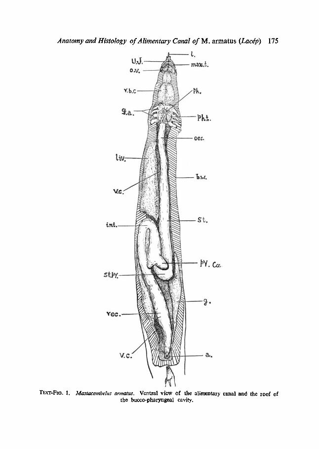
bounded by an upper and a lower labial fold. The upper jaw extends anterior- ly in front of the lower and participates in the formation of the trilobed snout, consisting of a median stiff, solid, and pointed process and two lateral soft, hollow and blunt projections (Text-Fig. 1). Immediately be- hind each jaw is a triangular oral valve.
Anatomy and Histology of Alimentary Canal of M. armatus (Lacdp) 175
~'.h
v,c
V' .C- I Y / / A ~ a,.
Tsx"r-Fm. 1. Mastacembelus armatus. Ventral view of the alimentary canal and the roof of the bucco-pharyngcal cavity.
176 Sahsm KANT NAGAR AND WALI MOttD. KtTAN
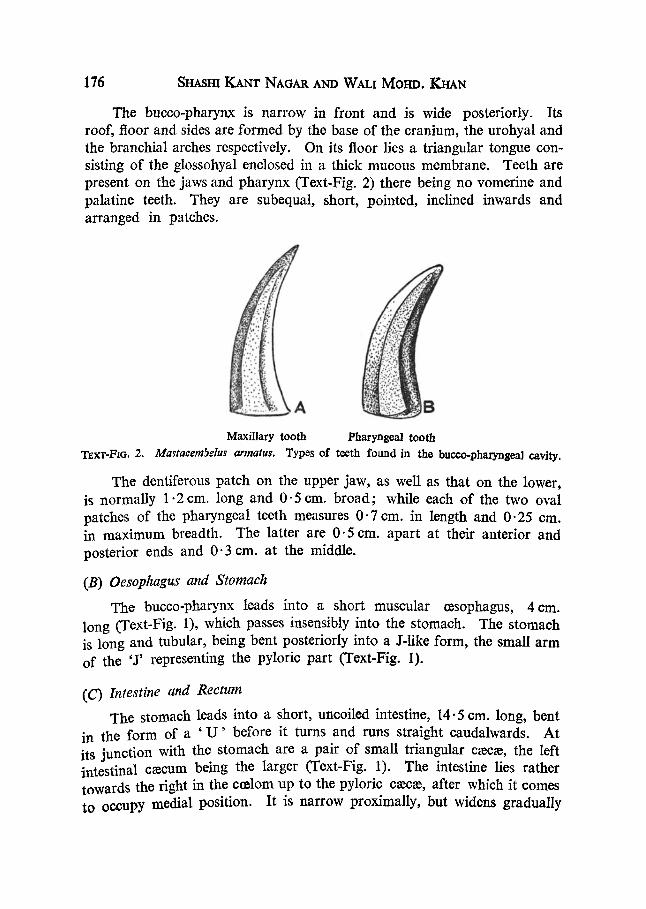
The bucco-pharynx is narrow in front and is wide posteriorly. Its roof, floor and sides are formed by the base of the cranium, the urohyal and the branchial arches respectively. On its floor lies a triangular tongue con- sisting of the glossohyal enclosed in a thick mucous membrane. Teeth are present on the jaws and pharynx (Text-Fig. 2) there being no vomerine and palatine teeth. They are subequal, short, pointed, inclined inwards and arranged in patches.
l Maxillary tooth Pharyngeal tooth
T~xr-FtG. 2. Mastaeera5elus armatus. Types of teeth found in the bucoo-pharyngeal cavity.
The dentiferous patch on the upper jaw, as well as that on the lower, is normally 1.2cm. long and 0-5cm. broad; while each of the two oval patches of the pharyngeal teeth measures 0-7 cm. in length and 0.25 era. in maximum breadth. The latter are 0.5 cm. apart at their anterior and posterior ends and 0"3 cm. at the middle.
(B) Oesophagus and Stomach The bucco-pharynx leads into a short muscular czsophagus, 4era.
long (Text-Fig. 1), which passes insensibly into the stomach. The stomach is long and tubular, being bent posteriorly into a J-like form, the small arm of the 'J' representing the pyloric part (Text-Fig. 1).
(C) Intestine and Rectum The stomach leads into a short, uncoiled intestine, 14.5 cm. long, bent
in the form of a ' U ' before it turns and runs straight caudalwards. At its junction with the stomach are a pair of small triangular c~ec~e, the left intestinal c~ecum being the larger (Text-Fig. 1). The intestine lies rather towards the right in the c~lom up to the pyloric c~ec~e, after which it comes to occupy medial position. It is narrow proximally, but widens gradually

Anatomy and Histology of Alimentary Canal of M. armatus (Lacdp) 177
towards its posterior end, where it passes into the rectum, which is demar- cated from it internally by an ileo-rectal valve as found by Dawes (192y) in Pleuronectes platessa and A1-Hussaini (1945) in Scarus sordidus. The rectum narrows slightly before it opens out through the anus.
(D) Folds of the Mucosa The alimentary canal in an average-sized specimen is 29 cm. from
pharynx to anus. Its various parts can be differentiated to a fair extent by the variation in the pattern. The lining in the buccal cavity is smooth and fold-less, but in the pharynx it is disposed in wavy folds arranged longitu- dinally. In the ee;ophagus and the stomach the folds are straight, thick and high. They narrow and increase in number on reaching the pyloric portion. In the intestine, rectum and pyloric c~eca~ their pattern is web- like ; while near the anus they are not very distinct, although similar to those in the rectum (Text-Fig. 3).
gg.~
tl
The mucosal folds of the stomach The mucosal folds of tke antericr region of intestine
TExr-FIo. 3. Mastacembelus armatus. Mucosal folds of the alimentary canal.
IV. HISTOLOGY OF THE ALIMENTARY CANAL
(,4) The Snout and the Upper Lip The lip is composed of two principal layers--the epidermis and the
dermis (Plate IX, Figs. 1-3). The epidermis is several layers thick, com- posed of polygonal cells with oval nuclei. The outer cells are flattened and the inner columnar, resting on a basement membrane. A major portion of the epidermis is occupied by numerous long saccular mucous cells with crescentic nuclei. Taste-buds are profusely present near the apex of the lip, each being borne on an elevation of the dermis.
The dermis consists of a rather loose connective-tissue in its outer part and a compact one inside and bears abundant zdipose coils and l:!ood capil-
178 SHasrn KANT NAGAR AND WALl Morro. KHAN
laries. Underneath the subepidermal basement membrane are numerous pigment cells; while a cartilaginous axis is present in the centre of the median process of the lip.
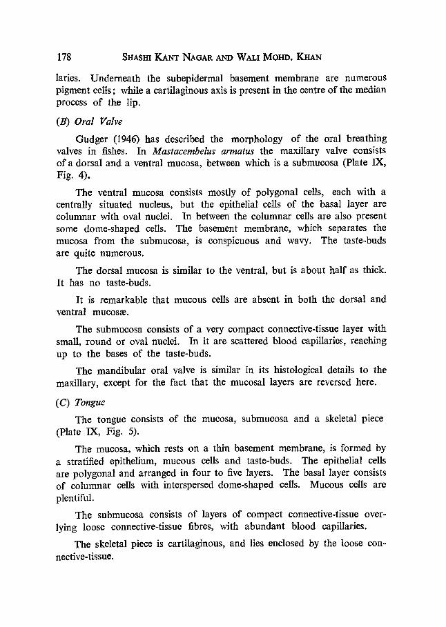
(B) Oral Valve Gudger (1946) has described the morphology of the oral breathing
valves in fishes. In Mastacembelus armatus the maxillary valve consists of a dorsal and a ventral mucosa, between which is a submucosa (Plate IX, Fig. 4).
The ventral mucosa consists mostly of polygonal cells, each with a centrally situated nucleus, but the epithelial cells of the basal layer are columnar with oval nuclei. In between the columnar cells are also present some dome-shaped cells. The basement membrane, which separates the mucosa from the submucosa, is conspicuous and wavy. The taste-buds are quite numerous.
The dorsal mucosa is similar to the ventral, but is about half as thick. It has no taste-buds.
It is remarkable that mucous cells are absent in both the dorsal and ventral mucos~e.
The submucosa consists of a very compact connective-tissue layer with small, round or oval nuclei. In it are scattered blood capillaries, reaching up to the bases of the taste-buds.
The mandibular oral valve is similar in its histological details to the maxillary, except for the fact that the mucosal layers are reversed here.
( C) Tongue The tongue consists of the mucosa, submucosa and a skeletal piece
(Plate IX, Fig. 5).
The mucosa, which rests on a thin basement membrane, is formed by a stratified epithelium, mucous cells and taste-buds. The epithelial cells are polygonal and arranged in four to five layers. The basal layer consists of columnar cells with interspersed dome-shaped cells. Mucous cells are plentiful.
The submucosa consists of layers of compact connective-tissue over- lying loose connective-tissue fibres, with abundant blood capillaries.
The skeletal piece is cartilaginous, and lies enclosed by the loose con- nective-tissue.
Anatomy and Histology of Alimentary Canal.of M. armatus (Lacdp) 179
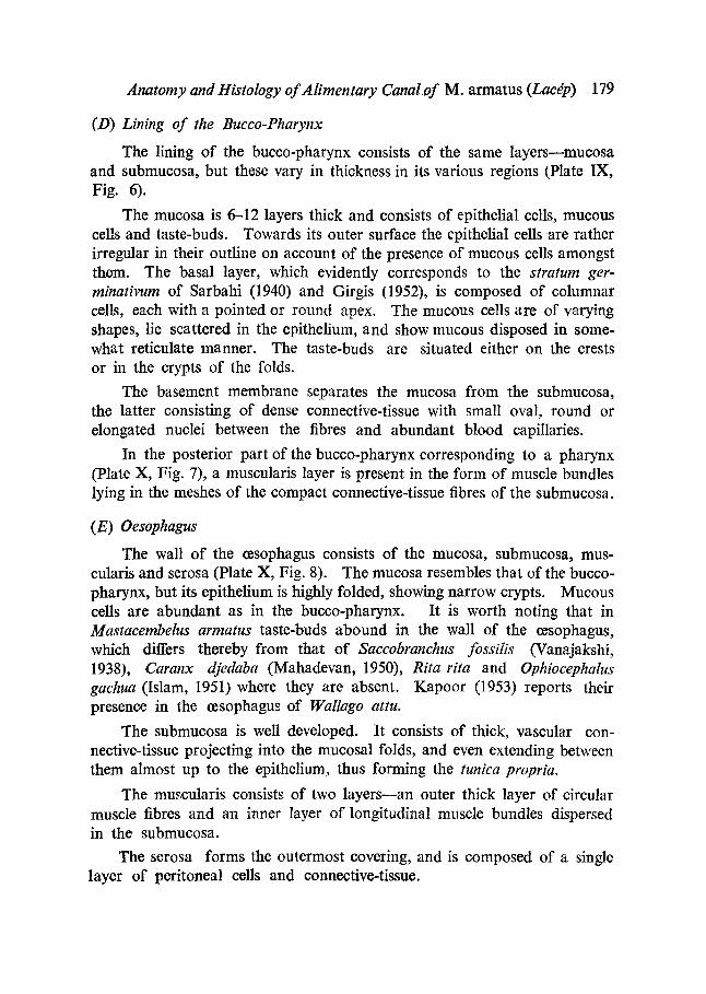
(D) Lining of the Bucco-Pharynx The lining of the bucco-pharynx consists of the same layers--mucosa
and submucosa, but these vary in thickness in its various regions (Plato IX, Fig. 6).
The mucosa is 6-12 layers thick and consists of epithelial cells, mucous cells and taste-buds. Towards its outer surface the epithelial cells are rather irregular in their outline on account of the presence of mucous cells amongst them. The basal layer, which evidently corresponds to the stratum ger- minativum of Sarbahi (1940) and Girgis (1952), is composed of columnar cells, each with a pointed or round apex. The mucous cells are of varying shapes, lie scattered in the epithelium, and show mucous disposed in some- what reticulate manner. The taste-buds are situated either on the crests or in the crypts of the folds.
The basement membrane separates the mucosa from the submucosa, the latter consisting of dense connective-tissue with small oval, round or elongated nuclei between the fibres and abundant blood capillaries.
In the posterior part of the bucco-pharynx corresponding to a pharynx (Plate X, Fig. 7), a muscularis layer is present in the form of muscle bundles lying in the meshes of the compact connective-tissue fibres of the submucosa.
(E) Oesophagus The wall of the oesophagus consists of the mucosa, submucosa, mus-
cularis and serosa (Plate X, Fig. 8). The mucosa resembles that of the bucco- pharynx, but its epithelium is highly folded, showing narrow crypts. Mucous cells are abundant as in the bucco-pharynx. It is worth noting that in Mastacembelus armatus taste-buds abound in the wall of the ~sophagus, which differs thereby from that of Saccobranchus fossilis (Vanajakshi, 1938), Caranx djedaba (Mahadevan, 1950), Rita rita and Ophiocephalus gachua (Islam, 1951)where they are absent. Kapoor (1953) reports their presence in the oesophagus of Wallago attu.
The submucosa is well developed. It consists of thick, vascular con- nective-tissue projecting into the mucosal folds, and even extending between them almost up to the epithelium, thus forming the tunica propria.
The muscularis consists of two layers--an outer thick layer of circular muscle fibres and an inner layer of longitudinal muscle bundles dispersed in the submucosa.
The serosa forms the outermost covering, and is composed of a single layer of peritoneal cells and connective-tissue.
180 SHASrn KANT NAGAR AND WALI MOHD. KHAN
(F) Stomach
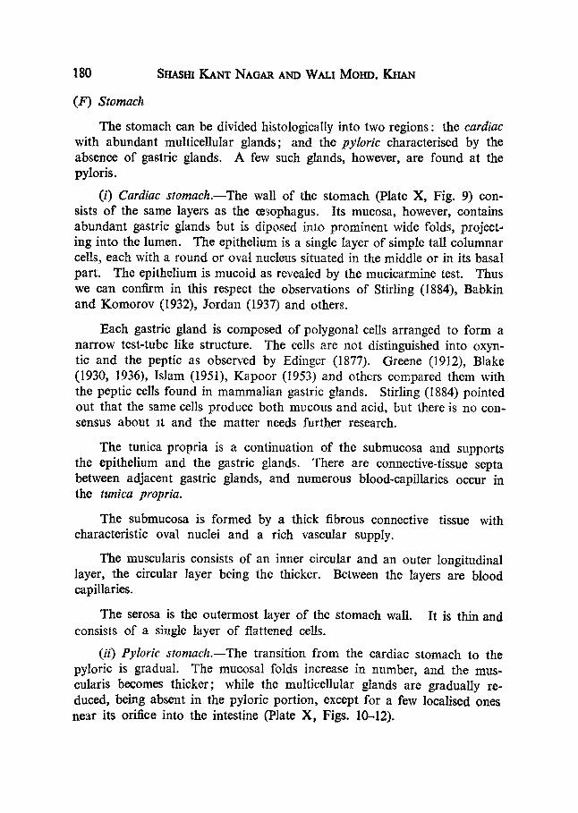
The stomach can be divided histologically into two regions: the cardiac with abundant multicellular glands; and the pyloric characterised by the absence of gastric glands. A few such glands, however, are found at the pyloris.
(i) Cardiac stomach.--The wall of the stomach (Plate X, Fig. 9) con- sists of the same layers as the t~sophagus. Its mucosa, however, contains abundant gastric glands but is diposed into prominent wide folds, project- ing into the lumen. The epithelium is a single layer of simple tall columnar cells, each with a round or oval nucleus situated in the middle or in its basal part. The epithelium is mucoid as revealed by the mucicarmine test. Thus we can confirm in this respect the observations of Stirling (1884), Babkin and Komorov (1932), Jordan (1937) and others.
Each gastric gland is composed of polygonal cells arranged to form a narrow test-tube like structure. The ceils are not distinguished into oxyn- tic and the peptic as observed by Edinger (1877). Greene (1912), Blake (1930, 1936), Islam (1951), Kapoor (1953) and others compared them with the peptic cells found in mammalian gastric glands. Stirling (1884) pointed out that the same cells produce both mucous and acid, but there is no con- sensus about at and lhe matter needs further research.
The tunica propria is a continuation of the submucosa and supports the epithelium and the gastric glands. There are connective-tissue septa between adjacent gastric glands, and numerous blood-capillaries occur in the tunica propria.
The submucosa is formed by a thick fibrous connective tissue with characteristic oval nuclei and a rich vascular supply.
The muscularis consists of an inner circular and an outer longitudinal layer, the circular layer being the thicker. Between the layers are blood capillaries.
The serosa is the outermost layer of the stomach wall. It is thin and consists of a single layer of flattened cells.
(ii) Pyloric stomach.--The transition from the cardiac stomach to the pyloric is gradual. The mucosal folds increase in number, and the mus- cularis b~omes thicker; while the multicellular glands are gradually re- duced, being absent in the pyloric portion, except for a few localised ones near its orifice into the intestine (Plate X, Figs. 10--12).
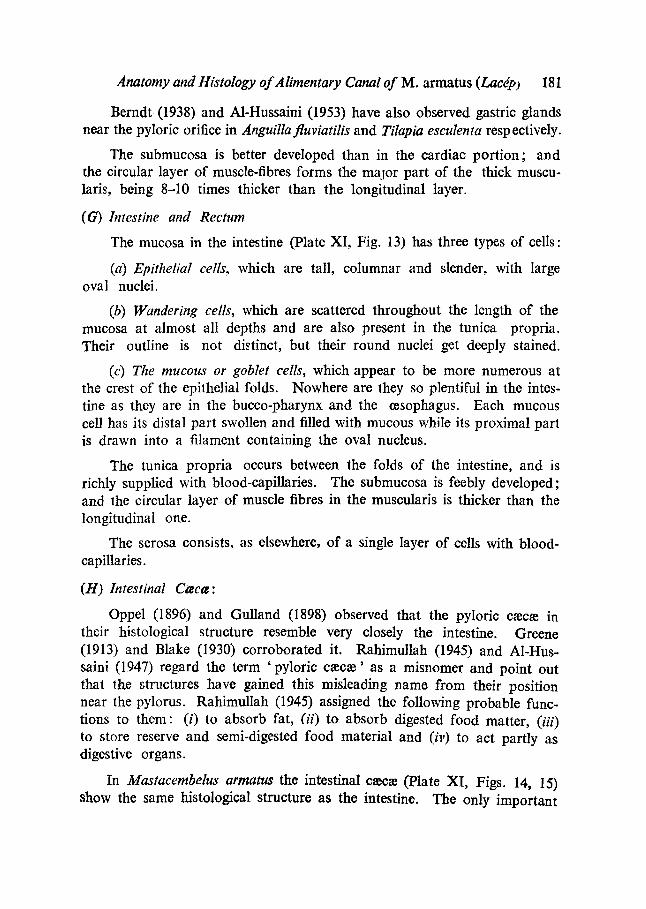
Anatomy and Histology of Alimentary Canal of M. armatus (Lacdp) I81
Berndt (1938) and A1-Hussaini (1953) have also observed gastric glands near the pyloric orifice in Anguillafluviatilis and Tilapia esculenta respeetively.
The submueosa is better developed than in the cardiac portion; and the circular layer of muscle-fibres forms the major part of the thick muscu- laris, being 8-10 times thicker than the longitudinal layer.
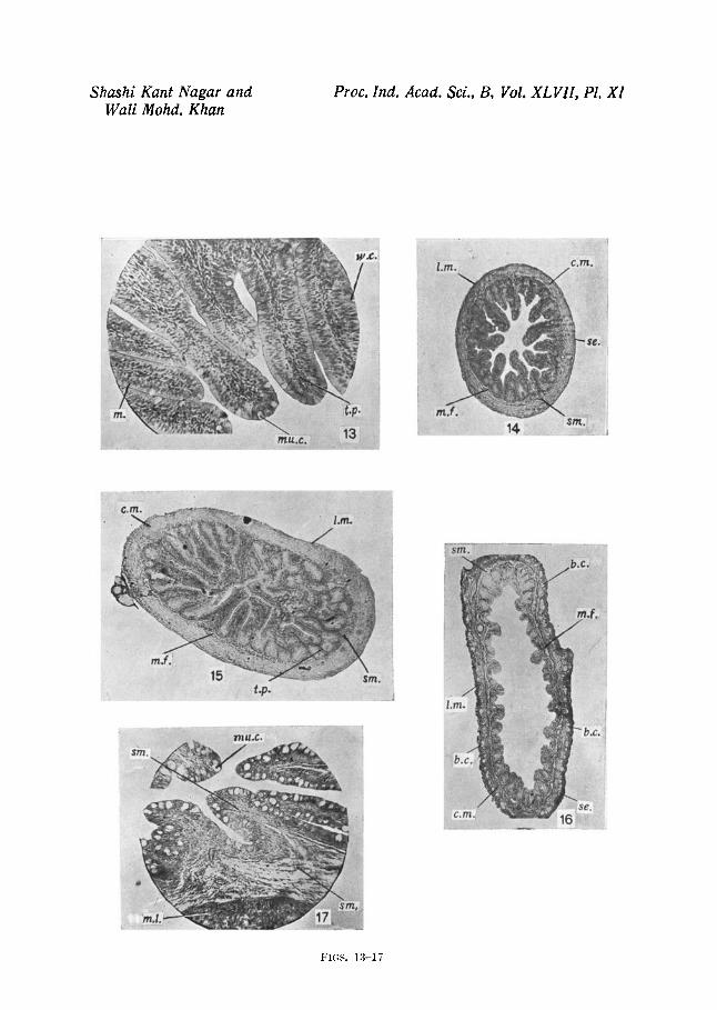
(G') Intestine and Rectum The mucosa in the intestine (Plate XI, Fig. 13) has three types of cells:
(a) Epithelial cells, which are tall, columnar and slender, with large oval nuclei.
(b) Wandering cells, which are scattered throughout the length of the mucosa at almost all depths and are also present in the tunica propria. Their outline is not distinct, but their round nuclei get deeply stained.
(c) The mucous or goblet cells, which appear to be more numerous at the crest of the epithelial folds. Nowhere are they so plentiful in the intes- tine as they are in the bucco-pharynx and the oesophagus. Each mucous cell has its distal part swollen and filled with mucous while its proximal part is drawn into a filament containing the oval nucleus.
The tunica propria occurs between the folds of the intestine, and is richly supplied with blood-capillaries. The submueosa is feebly developed; and the circular layer of muscle fibres in the muscularis is thicker than the longitudinal one.
The serosa consists, as elsewhere, of a single layer of cells with blood- capillaries.
(1-I) Intestinal C~ecce:
Oppel (1896) and Gulland (1898) observed that the pyloric c~ee~e in their histological structure resemble very closely the intestine. Greene (1913) and Blake (1930) corroborated it. Rahimullah (1945) and AI-Hus- saini (1947) regard the term 'pyloric c~ec~e' as a misnomer and point out that lhe structures have gained this misleading name from their position near the pylorus. Rahimullah (1945) assigned the following probable func- tions to them: (i) to absorb fat, (ii) to absorb digested food matter, (iii) to store reserve and semi-digested food material and (iv) to act partly as digestive organs.
In Mastaeembelus armatus the intestinal e~ec~e (Plate XI, Figs. 14, 15) show the same histological structure as the intestine, The only important
182 SI-IASHI KANT NAGAR AND WALI MOHD. KHAN
difference being the larger number of mucosal folds and their greater length, so that they seem to fill up almost the whole of the lumen.
The rectum (Hate XI, Fig. 16) resembles the intestine in its histological structure, except for the fact that the mucosal folds are extremely reduced in it and are rather distinct from each other. The vascular supply is richer.
Near the anus (Plate XI, Fig. 17) the mucosal epithelium becomes strati- fied, the mucous cells are numerous and muscularis is thickened.
SUMMARY
The authors have described in detail the anatomy and histology of the alimentary canal of the spiny eel Mastacembelus armatus (Lac6p). Amongst the more important features discovered by them might be mentioned the participation of the upper jaw in the formation of the trilobed snout, which bears a cartilaginous axis in its median lobe; the presence of both maxillary and mandibular valves in the bucco-pharynx and the absence of mucous ceils on them; the occurrence of taste-buds not only in the wall of the bucco- pharynx and on the tongue but also in the o~sophagus; the disposition of the gastric glands in the stomach and the non-differentiation of their ceils into oxyntic and peptic; the close histological similarity between the intes- tine, intestinal c~c~e and the presence in abundance of mucous cells near the anus.
ACKNOWLEDGEMENTS
The authors are extremely grateful to Dr. B. C. Mahendra, Professor of Zoology, Agra College, Agra, for the interest taken in going through the manuscript and for suggesting valuable changes. Thanks are also due to Dr. B. G. Kapoor, Research Investigator, Fisheries Section, Ministry of Agriculture, Government of India, for his continuous guidance and sugges- tions throughout the course of the present investigation; as well as to Prin- cipal, M. M. Begg of Delhi College, for the laboratory facilities provided to them.
1. A1-Hussaini, A. H.
2.
REFERENCES
.. "The anatomy and histology of the alimentary tract of the coral-feeding fish, Scarus sordidus (Klunz)," Bull. Inst. Egypte, 1945, 27, 349-77.
.. "The anatomy and histology of the alimentary tract of the bottom-feeder, Mulloides auriflamma," J. Morph., 1946, 75p 121-53.

Anatomy and Histology of Alimentary Canal of M. armatus (Laedp) 3. A1-Hussaini, A . H . ..
4.
5.
6.
7.
8. m - - m and Kholy, A. A . . .
9. Angelescu, V. and Gneri, F. S.
10. Babkin, B. P. and Komorov, S. A.
11. Barrington, E. J. W.
12. Berndt, O.
13. Blake, I. H.
14.
15. Bfihler, H.
16. Chan, V. M.
183
"The anatomy and histology of the alimentary tract of the plankton-feeder, Atherina forskali (Rupp.)," 1. Morph., 1947 a, 80, 251-86.
.. "The feeding habits and the morphology of the alimentary canal of some teleosts living in the neighbourhood of the Marine Biological Station, Ghardaqa, Red Sea," Publi- cations o f the Marine Biological Station, Gbardaqa (Red Sea), 1947 b, No. 5, 4-61.
.. "On the functional morphology of the alimentary tract of some fish in relation to differences in their feeding habits: Anatomy and Histology," Quart. Journ. Micr. Sci., 1949, 90 (2), 109-40.
.. "On the functional morphology of the alimentary tract of some fish in relation to differences in their feeding habits: Cytology and Physiology," Ibid., 1949, 90(4), 323-54.
.. "The feeding habits and guts of teleosts, especially of the Northern Red Sea," Revue de la Faculte des Sciences dd l' Universite d'Istanbul, 1951, 17(2)B, 121-29.
"On the functional morphology of the alimentary tract of some Omnivorous teleosts fish," Proc. Egyptian Academy o f Sciences, 1953, 9, 17-39.
.. "Adaptaciones del aparato digestivo al regimen alimenticio en algunos peees del Rio Uruguay ydel Rio de la plata," Revista del lr:stituto Nucional de lnvestigacion de las cien- cias Naturale~ Bernardino Rividavidz. Ciencias Zoologicas, 1949, No. 6, 161-272.
.. "The influence of gastric mucous on peptic digestion," Jour. Canad. Med. Ass., i932, 27, 463.
.. "Gastric digestion in the lower vertebrates," Biol. Rev., 1942, 17, 1-27.
.. "Morphologie und Histologie des Rumpfdarmes von Anguil- la fluviatilis und die Veranderungen desselben im Indivi- dualzyklus," Zool. Jahrb, Jena, 1938, 64.
.. "Studies on the comparative histology of the digestive tube of certain teleost fishes: A predaceous fish, the sea bass (Centropristes striatus),'" d. Morph. and Physiol., 1930, 50 (1), 39-70.
.. "Studies on the comparative histology of the digestive tube of certain teleost fishes--IIIr A bottom-feeding fish, the sea robin (Prionotus carolinus)," d. Morph., 1936, 60(1)~ 77-102.
.. "Die Verdauungsorgane der Stromateid~e (Teleost)," Zeitsch. Wiss. BioL Abt. A. Morph. u. OkoL Tiere, 1930, 19 (I), 89-115.
.. "The histology of the alimentary tract of the deep-water gurnard, Peristedion longispatha," Univ. Nebraska Stud., 1941, 41(I), 7-30.
184 Sm~.sm KANT NAGAR AND WALl Moire. KI-tAN
17. Curry, E.
18. Dawes, B.
19. Dharmarajan, M.
20. Edinger, L.
21. Ghazzawi, F. M.
22. Girgis, S.
23. Greene, C. W.
24. Gudger, E. W .
25. GuUand, G. L.
26. Islam, A. U.
27. Jacobsen, E. M.
28. Kapoor, B. G.
29. Mahadevan, S.
30.
31. Mohsin, S. M.
32.
33. Oppel, A.
.. "The histology of the digestive tube of the carp (Cyprinus carpio communis)," J. Morph., 1939, 65(1), 53-78.
.. "The histology of the alimentary, tract of the plaice (Fleurd- nectes platessa)," Quart..lourn. Micro. ScL, 1929, 73(2), 243-74.
.. "The anatomy and histology of the alimentary system of Otolithus tuber," Prec. Ind. Sci. Congr., 1936, 23(3), 354.
.. "Ober die Schleimhaut des Fischdarmes nebst Bemerkungen Zur Phylogenese der Drilsen des Darmrohres," Arch. fur. Mikros. Anat., 1877, 13, 651-92.
.. "The pharynx and intestinal tract of the Egyptian mullets: Mugil cephalus and Mugil capitomlI. On the morphology and histology of the alimentary canal in Mugil capito," Notes and Memoirs, No. 6, Fisheries Research Directorate, Cairo, Egypt, 1935, 1-31.
.. "On the anatomy and histology of the alimentary tract of an herbivorous bottom-feeding Cyprinoid fish, Labeo Horie (Cuvier)," o r. Morph., 1952, 90(2), 317-62.
.. "Anatomy and histology of the digestive tract of the king Salmon," Bull. U.S. Bur. Fish., 1912, 32, Document No. 777, 73-100.
.. "Oral Breathing valves in fishes," d. Morph., 1946, 79(2), 263-85.
.. "Minute Structure of the digestive tract of the Salmon and the changes which occur in it in freshwater," Anat. Anz., 1898, 14.
•. "The comparative histology of the alimentary canal of certain freshwater teleost fishes," Prec. Ind. Acad. Sci., 1951, 33 (6), 297-321.
.. "Anatomy and histology of the digestive tract of a deep- sea fish, Coelorhynchus carminatus," Univ. Stud. Nebraska, 1939, 39(1), 1-27.
. . "The anatomy and histology of the alimentary canal in rela- tion to its feeding habits of a siluroid fish, Wallago attu (BI. & Schn.)," J. Zool. Soc. India, 1953, 5(2), 191-210.
.. "The digestive system of Caranx djedaba (Forsk.) and Trichi- urushaumela (Forsk.)," J. Madras Univ., 1950, 21 B, 25--48.
.. "The digestive system of Mugil crenilabis (Forsk.)," 1bid., 1954, 24B, 143-60.
.. "The morphology and histology of the alimentary tract of Anabas testudineus," Journ. Os. Univ., (Science Faculty), 1944--46, 12, 66-75.
.. "Histology of the alimentary tract of a freshwater gob '¢ Glossogobtus giuris (Ham.)," Prec. Ind. ScL Congr., 1946,' 33.
.. "Lehrbuch der Vergleichenden mikroskopischen Anatomi~ der Wirbeltiere," Jena, 1896o
Anatomy and Histoiogy of Aiimentary Canal of M. armatus (Lacgp) 185
34. Pictet, A.
35. Pillay, T. V. R.
36. Rahimullah, M.
37.
38. Rogick, M. D.
39. Sarbahi, D. S.
40. Schacht, H.
41. Stifling, W.
42. Suyehiro, Y.
43. Tortones¢, E.
44. Vanajakshi, T. P.
45. Weinreb, E. L. and Bilstad, N. M.
. . "Contribution a l 'etude histologique du tube digestff des poissons Cyprinoides," Revue Suisse de Zoologie, 1909, 17, 1-78.
. . "Studies on the food, feeding habits and alimentary tract of the gray mullet, Mugil tade (Forskal)," Prec. Nat. Inst. SoL India, 1953, 19(6), 777-827.
.. "Contributions to our knowledge of the pyloric ceec~e in three families o f freshwater Indian fishes (Ophiocel~halid~e, Notopterid~ and Mastacemblid~) together with some remarks on their probable functions," 1'roe. lnd. Acad. Sci., 1943, 18(5)B, 85-96.
.. " A comparative study of the morphology, histology and probable functions of the pyloric c~ec~ in Indian fi~hes together with discussion on their homology," Ibid., 1945, 21 B, 1-37.
• ."Studies of the comparative histology of the digestive tulze of certain teloost fishesmlI. (Campostoma anomalum),- J. Morph. ar, d Physiol, 1931, 52(1)~ 1-25.
. . "The alimentary tract o f Labeo rohita (Ham.) ," Jaurn. Ray. Asi. Soc. Bengal, 1940, 5(2), 88-116.
.. "Obe r den Vorderdarm tier Cyprinodonten," Jahrb. Morph. u. Mikrosk. Anat. Abt.--II, Zeitsch. Mikrosk. Anat. Ges., 1931, 26, 534--46.
. . "'On the ferments or enzymes of the digestive tract in fishes," Journ. Anat. PhysioL, 1884, 18~ 426--35.
. . " A study on the digestive system and feeding habits of fish," Jap. Journ. ZeaL, 1942, 10(1), 1-303.
.. "Anatomia e istologia del tube digerente di Coris julis L. (Pisces Labriformes), in rapporto al regime alimentare," Archive ZaoL ltal., 1952, 37, 1-27.
. . "Histology of digest!ve tract of Saccobranehus fossilis and Macrones vitattus," Prec. Ind. Aead. Sei., 1938, 7B , 61-80.
. . "Histology of the digestive tract and adjacent structures of the rainbow trout, Salmo gairdneri irideus," Copeia, 1955, No. 3, 194-204.
ABBREVIATIONS USED 1N TEXT-FIGURES AND PLATES
d.m. . . dorsal mucosa, py.ca. ep. . . epidermis, r.b.c. g. . . gonads, rec. g.a. . . gill arches, so. g.g. . . gastric gland, sin. int. . . i n t e s t i n e , st. tnt.f. . . intestine fold. st.f.
. . intestinal c~c~.
. . roof of the buccal •. rectum.
• . s e r o 8 3 .
•. submucosa.
•. stomach.
•. stomach fold.
cavity.
I86
int.w.
I .
LL
1.m. l iv .
m .
max. t.
m . f .
a .
b.c.
b.w.
c a .
c.L
g ' .m .
d.
FIG. 1. FIG. 2.
FIG. 3.
FxG. 4.
FIe, 5.
FxG. 6.
FIe. 7.
FIG. 8.
F ie . 9.
FIG. 10.
SHASHI K A N T N A G A R AND W A L l M O H D . K H A N
.. intestine wall. st .py. . . pyloric stomach.
. . lip. st.w. . . stomach wall.
. . lateral lobes, t.b. . . taste-bud.
. . longitudinal muscles, t.p. . . tanica propria.
. . liver, u.j. . . upper jaw.
. . mucosa, v.c. . . visceral cavity (Clo~om).
. . maxillary teeth, v.m. . . ventral mucosa.
. . mucosal folds, w.c. . . wandering ceils.
•. anus• m.L . . mucosal layer.
. . blood capillaries, mu.c. . . mucous cells.
. . body wall. o.v. . . oval valve.
. . cartilage, oes. . . oesophagus.
. . central lobe. pg.c. . . pigment cells.
. . circular muscles, ph. . . pharynx.
. . dermis, ph.t . . . pharyngeal teeth.
FXPLANATION OF PLATES
PI~T~ IX
A transverse section of the tip of the lip, showing trilobed pattern.
A transverse section of the mid-region of the lip, showing the central cartilaginous and lateral hollow lobes.
A transverse section of the basal region of the lip under low power.
A portion of the transverse section of the maxillary oral valve under high power, showing the dorsal mucosa, the thicker ventral mucosa with a taste-bud and a common submucosa.
A portion of the transverse section of the tongue, under high power showing mucosa with a taste-bud, mucous cells and submucosa.
A portion of the transverse section of the buccal lining, under high power, showing stratified epithelium mucous cells, a taste-bud and the submucosa.
PLATe X
portion of t he transverse section of the anter ior paxt of the pharynx, under high power, showing stratified epithelium, mucous cells, taste-bud in the mucosa, and the submucosa.
A portion of the transverse section of the anterior part of the oesophagus, under high power, showing mucosa with stratified epithelium, mucous cells, taste-bud and the submucosa.
portion of the transverse section of the cardiac stomach, under high power, showing gastric glands in the mucosa.
transverse section of the pyloric stomach, under low power, showing mucosa composed of a single layer of columnar epithelium and a well-developed muscularis
A
A
A

shashi Kant Nagar and Wall Mohd. Khan
Proc. Ind. Acad. Sci., B, Vol. XLVII, Pl. IX
~ 7 = ~ i ~ : ' . , ... "" ' . " , - , :,.;,,:,,: . : : :,7 :II--.~:,.L,: ! -~':- ' ' " ' : " , , . = " " i i, ":i
ii:: . i e.i!?i,i!!~!i: ~ J .... ~I~]Kl~ .
" , . . . . . . - -?t.b-. . . . . . . . . . . . . . . . . . . . . . . . : ' ; , " " " . , : r ) 7 7 ~ ! ~ ) a ~ . - : . : ! , , - "
'~ {{ l; : "'/~:" Y'ag?:~::~'*'~"~>'r"
' 3 , : , - . . . . . . . . = ' > : '
i . n t .
!< : ':: ,t
F I G S . 1-6
, ¢ , • . ? ~

Shashi Kant Nagar and Wali Mohd, Khan
P.r, oc. Ind. Acad, Sci., B, Vol. XLVII, Pl. X
! :! s
s~2,,
i:
• , v
. s e ~
• " ~ ' ~ r'= ' "~ " , ~'~!!~ i~ ̧~: " '~
g.g,
m.
F I G S . 7-12
~Sm.
Shashi Kant Nagar and Wall Mohd. Khan
Proc. Ind. Acad. Sci., B, Vol. XLVII, Pl, XI
" , " , . t
w.c, L: i .m, elm,. :
: . . . 2
4
ii~iiil 14 ::" ~' ~m.
m
j i > / i
' ~ I ,~
top.,i :~,, S i n , •
_ _ J
' ;irr" ~ 1
/ C,n'L~
• . , : 1 6 "
F I ( ; S . 1 3 - 1 7

Anatomy and Histology of Alimentary Canal of M . a r m a t u s (Lacdp) Fio. 11.
FIo. 12.
FIo. 13.
Fie. 14. FIG. 15. I~o. 16.
Fro. 17.
187
A portion of the transverse section of the pyloric stomach, under high power, showing the mucosal folds without the gastric glands and the submucosa.
A portion of the transverse section of the pyloris, under high power, showing the presence of a few localised gastric glands.
PLATE XI
A portion of the transverse section of the intestine, under high power, showing mucosal folds with epithelial cells, mucous cells, wandering cells and the tunica propria.
A transverse section of the right pyloric c~ec~, under low power. A transverse section of the left pyloric cmc~, under low power. A transverse section of the rectum under low power, showing the mucosa, submucosa,
muscularis and highly vascularised serosa.
A portion of longitudinal section of the anus, under high power, showing abundance, of mucous cells in the stratified epithelium of mucosa.