Microscopic structure of the cotton fiber - Nvlpubs.nist.gov
21
U. S. DEPARTMENT OF COMMERCE NATIONAL BUREAU OF STANDARDS RESEARCH PAPER RP1362 Part of Journal of Research of the N.ational Bureau of Standards, Volume 26, February 1941 MICROSCOPIC STRUCTURE OF THE COTTON FIBER By Charles W. Hock, Robert C. Ramsay, and Milton Harris 1 ABSTRACT During the examination of cotton fibers that had received various chemical treatments, a number of observations pertaining to the structural details of the fiber were made. Those phenomena which appeared to be new were investi- gated in detail. In additi on, experiments described by earlier investigators were repeated in order to have a better composite picture of the structure of the fiber. The cell wall of a cotton fiber consists of a primary and a secondary wall. The latter, which comprises the bulk of the fiber, consists of innumerable spirally oriented cellulose fibrils enclosed by a winding which also makes a spiral, but in the opposite direction from the former. Both the winding and the fibrils reverse their direction at frequent int ervals along the axis of the fiber, their points of reversal being coincident. The secondary wall is enclosed by a thin primary wall. The latter is made up of fine crisscrossing strands of cellulose embedded in a membrane consisting principally of wax and pectic substance. The lumen also contains wax and pectic materials, plus various amounts of degenerated protoplasm. When cotton fibers are swollen under certain conditions, a lamellat e structure is discernible in the secondary wall. The numb er of these lamellae increases with the age of the fiber. On treatment of cotton fibers with cuprammonium hydroxide reagent, the cel- lulose dissolves, leav ing residues which vary in amount and in structure, depend- ing upon the extent of purification of the fibers. The residue from raw and from dewaxed fibers consists of fragments of the primary wall, and of a lesser amount of material from the lumen . The behavior of the fibers in the reagent depends in part on their maturity. Immature fibers containing only small amounts of cel- lulose swell relatively little in the reagent, and the undissolved wax and pectic materials maintain the original tubular shape of the primary wall. When older fibers are given the same treatment, they swell abruptly, thereby causing the primary wall to break in many places, giving rise to "balloons." Irregular swelling along the fiber axis, which results in the formation of bal- loons, appears to be dependent in part on the orientation of the fibrils, and in part on the constricting influences of the winding and of the primary wall. CONTENTS Page 1. Introduction___ ____ ______ __ _ ___ __ ______ ___ _ ___ __ ___ __ __ ___ ____ __ 94 II. Materials______ ____ ______ ______ __ ____ _____ _ ___ _ ____ ______ __ ___ __ 94 III. Experimental procedure and results____ _________________ ___________ 95 1. Primary walL___ ____ __ ____ ____ ________ ____ _________ __ __ __ 95 2. Single fibers in cuprammonium reagent__________ ____________ 96 3. Cross sections of fibers in cuprammonium reagent______ _____ __ 97 4. Lamellae and growth rings______________ ___________________ 98 5. Fibrillar structure ____________________ ____ _______ _________ _ 100 6. Balloon formation ____ ____________________________________ 102 IV. References ______ _____________________ ___________________________ 104 1 Research Associates at the National Bureau of Standards, representing the Textile Foundation. 93
-
Upload
khangminh22 -
Category
Documents
-
view
1 -
download
0
Transcript of Microscopic structure of the cotton fiber - Nvlpubs.nist.gov

U. S. DEPARTMENT OF COMMERCE NATIONAL BUREAU OF STANDARDS
RESEARCH PAPER RP1362
Part of Journal of Research of the N.ational Bureau of Standards, Volume 26, February 1941
MICROSCOPIC STRUCTURE OF THE COTTON FIBER
By Charles W. Hock, Robert C. Ramsay, and Milton Harris 1
ABSTRACT
During the examination of cotton fibers that had received various chemical treatments, a number of observations pertaining to the structural details of the fiber were made. Those phenomena which appeared to be new were investigated in detail. In addition, experiments described by earlier investigators were repeated in order to have a better composite picture of the structure of the fiber.
The cell wall of a cotton fiber consists of a primary and a secondary wall. The latter, which comprises the bulk of the fiber, consists of innumerable spirally oriented cellulose fibrils enclosed by a winding which also makes a spiral, but in the opposite direction from the former. Both the winding and the fibrils reverse their direction at frequent intervals along the axis of the fiber, their points of reversal being coincident. The secondary wall is enclosed by a thin primary wall. The latter is made up of fine crisscrossing strands of cellulose embedded in a membrane consisting principally of wax and pectic substance. The lumen also contains wax and pectic materials, plus various amounts of degenerated protoplasm.
When cotton fibers are swollen under certain conditions, a lamellate structure is discernible in the secondary wall. The number of these lamellae increases with the age of the fiber.
On treatment of cotton fibers with cuprammonium hydroxide reagent, the cellulose dissolves, leaving residues which vary in amount and in structure, depending upon the extent of purification of the fibers. The residue from raw and from dewaxed fibers consists of fragments of the primary wall, and of a lesser amount of material from the lumen. The behavior of the fibers in the reagent depends in part on their maturity. Immature fibers containing only small amounts of cellulose swell relatively little in the reagent, and the undissolved wax and pectic materials maintain the original tubular shape of the primary wall. When older fibers are given the same treatment, they swell abruptly, thereby causing the primary wall to break in many places, giving rise to "balloons."
Irregular swelling along the fiber axis, which results in the formation of balloons, appears to be dependent in part on the orientation of the fibrils, and in part on the constricting influences of the winding and of the primary wall.
CONTENTS Page
1. Introduction___ ____ _ _ _ _ _ _ __ _ ___ __ ______ ___ _ ___ _ _ ___ __ __ _ _ _ ____ _ _ 94 II. Materials______ ____ ______ ______ __ ____ _____ _ ___ _ ____ _ _____ _ _ ___ __ 94
III. Experimental procedure and results____ _ _ _ _ _ _ _ _ _ _ _ _ _ _ _ _ _ _ _ _ _ _ _ _ _ _ _ _ 95 1. Primary walL___ ____ __ ____ ____ ________ ____ _ _ _ _ _ _ _ _ _ __ __ _ _ 95 2. Single fibers in cuprammonium reagent__________ ____________ 96 3. Cross sections of fibers in cuprammonium reagent______ _____ __ 97 4. Lamellae and growth rings______________ ___________________ 98 5. Fibrillar structure ____________________ ____ _______ _________ _ 100 6. Balloon formation ____ ____________________________________ 102
IV. References ______ _____________________ ___________________________ 104
1 Research Associates at the National Bureau of Standards, representing the Textile Foundation.
93

94 Journal oj Research oj the National Bureau oj Standards [Vot. t6
1. INTRODUCTION
As a part of a program of research relating to the chemical and physical structure of textile fibers, microscopic examinations were made of cotton fibers which had received various chemical treatments. A number of observations pertaining to the structural details of the cell wall of the cotton fiber were made. Those phenomena which appeared to be new were investigated in considerable detail. In addition, experiments described by earlier investigators (Balls [1, 2, 3],2 Denham [4], Sakostschikoff and Korsheniovsky [5], Kerr [6], Anderson and Kerr [7], Farr [8], and Barrows [9]) were repeated, and are reported here for the sake of completeness.
II. MATERIALS
Two varieties of Gossypium hirsutum L., Missdel-7 and Mexican Big Boll, were used in the investigation. Samples of each were furnished by the Agricultural Marketing Service of the U. S. Department of Agriculture. Missdel-7 was grown at the Delta Experiment Station, Stoneville, Miss. Only fibers from open bolls of this variety were used. The samples of Mexican Big Boll variety, grown at the Experiment Station at Raleigh, N. 0., were from special case-historied material collected for joint fiber structural studies by the Bureau of Plant Industry and the Agricultural Marketing Service. The samples of the latter were principally young or immature fibers from bolls of known age. Fibers of different ages were obtained by tagging the flowers on the day of blooming and then collecting the bolls on successive dates so as to give material of the desired age. Two series of immature Mexican Big Boll fibers were used. One, collected during the summer of 1939, consisted of dried fibers in which deposition of secondary cellulose had begun. The other, collected during the summer of 1937, consisted of very young fibers with primary wall only. The second set of fibers was obtained from bolls, the locks of which, at the time of collection, had been placed in boiling water for a few minutes to untangle the fibers, and then stored in approximately 70-percent ethyl alcohol.
Those fibers which received no chemical treatments are termed "raw fibers." In the present paper the young Mexican Big Boll fibers which had been boiled in water and then placed in alcohol are also included in this category, although it is recognized that the treatment was not without effect on the composition of the fibers. The natural waxes were removed from some of the raw fibers by extraction with alcohol and ether for 24 hours each. These fibers are termed "dewaxed fibers." A portion of the dewaxed material was further purified by extraction with alkali according to the method recommended for the preparation of standard cellulose. The procedure was essentially the same as that described by Oorey and Gray [10], except that the apparatus of Worner and Mease [11] was employed. These fibers, from which both wax and pectic substance were removed [12], are termed "depectinized fibers."
Ouprammonium hydroxide solution was prepared according to the recommendation of Mease [13]. The solution contained 240 ±5 g of ammonia (NHa) , 15 ±0.1 g of copper, and 1.0 g of sucrose per liter. The concentration of nitrite was less than 0.5 percent. When
• Figures in brackets Indicate the literature references at the end of this paper.

Hock, Ramsav,] Harris Structure oj Cotton Fiber 95
not in actual use, the cuprammonium hydroxide was kept under nitrogen in tightly stoppered bottles in the refrigerator.
Trimethylbenzylammonium hydroxide was used in some of the experiments. The stock solution, which was 2.5 N, was diluted with water just before using.
For staining cellulose, iodine and sulfuric acid [14], zinc chlorideiodine [14], and an alkaline solution of Congo red (0.5 percent of dye in a 0.5-percent solution of sodium hydroxide) were used. Aqueous solutions of ruthenium red were used for staining pectic compounds [15]. Although ruthenium red is not a specific stain for pectic substance, native pectic compounds invariably take a red color in the presence of this dye [7].
III. EXPERIMENTAL PROCEDURE AND RESULTS
The cell wall of the cotton fiber is composed of a primary and a secondary wall. The primary wall, in accordance with the description of Anderson and Kerr [7], is considered to be the outer sheath of the fiber, or the portion of the wall formed as the fiber increases in length. Secondary wall then refers to that part of the wall which is laid down after the fibers cease to elongate appreciably. Only the thin primary wall encloses the protoplasm of the cotton hail' as the fiber elongates during the first 13 to 20 days after its origin. Thereafter, the thickness of the wall is increased by a deposition of cellulose which comprises the secondary wall. For convenience in discussion, these two distinct layers are considered separately.
1. PRIMARY WALL
Young fibers, 3, 5, 8, and 15 days old, were mounted in water and examined microscopically. With ordinary light there was no evidence of structure in the thin primary wall. The walls stained deeply and uniformly with ruthenium red. When treated with an alkaline solution of Congo red, zinc chloride-iodine, or iodine and sulfuric acid, the fibers gave a faint color reaction. In older primary walls, for example in a 15-day fiber, an ill-defined pattern was observable in the wall.
On examination with crossed nicols, unstained fibers showed practically no birefringence. Insertion of a selenite plate between the nicols produced faint colors indicative of a predominantly transverse orientation. Although these fibers showed little birefringence when unstained, they were clearly anisotropic after treatment with a dye such as Congo red. This increase in birefringence upon staining is due, apparently, to the double refraction of the preferentially oriented dye molecules [16, 17].
When wax and pectic substance were removed from 15-day fibers the material which remained still had the outline of the original fiber and stained with Congo red, zinc chloride- iodine, and iodine and sulfuric acid. Between crossed nicols, the stained depectinized fibers were birefringent and showed a pattern like that in the stained walls of raw fibers. When the purified fibers were placed in cuprammonium hydroxide solution, they appeared to dissolve completely in the reagent.
The stained walls of both the raw and the depectinized fibers were suitable for observing the cellulose 3 of the primary wall. In agree-
3 While the identity or this material has not heen definitely established. X-ray investigations, staining reactions, and behavior in cnprammonium hydroxide solutions indicate that it Is probably cellulosic in nature.

96 Journal oj Research oj the National Bureau oj Standards [Vol. so
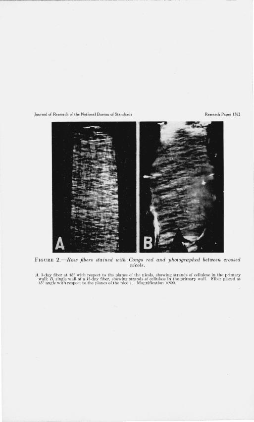
ment with the description by Balls [2, 3], and by Anderson and Kerr [7], the cellulose, as observed with crossed nicols, was found to be present as criss-crossing strands. At 45° with respect to the planes of the nicols, the fiber showed its maximum brightness and presented a reticulate appearance (fig. 1, A). When a fiber was rotated to the right or left of its position of maximum brightness, either one or the other of the systems of crisscrossing strands predominated the image (fig. 1, B and C). Measurements 4 of the angles which these birefringent strands made with the axis of the fiber were made on fibers of different ages. The results indicate that the angle of orientation decreases with increase in age of the fiber. In other words, the orientation was more nearly transverse in the younger than in the older ones. Likewise, the netlike structure of the cellulose, when the fibers were placed at 45°, appeared coarser (compare fig. 1, A, with fig. 2, A) and showed greater birefringence in the older fibers. The greater birefringence of the older fibers is not readily apparent from the photographs, as they were taken primarily to show the netlike structure. Measurements also indicated that the angle which the cellulose strands made with the fiber axis was greater at the tip than at the base. These differences were especially noticeable in 15-day fibers, where the growing cell had nearly attained its maximum length.
The above observations were confirmed by the examination of single walls obtained by cutting longitudinal sections of the fibers with the aid of a freezing microtome. Staining reactions and birefringence measurements gave essentially the same results as those obtained with intact fibers (fig. 2, B) .
After the initiation of secondary thickening, the predominantly transverse orientation (with respect to the fiber axis) of the cellulose in the primary wall was masked, after a few days, by the more nearly parallel orientation of the cellulose in the secondary wall. This change was evident upon examination of the fibers with crossed nicols and a selenite plate, as well as by observation with ordinary light, and is in agreement with the results of the X-ray work of Berkley [18].
2. SINGLE FIBERS IN CUPRAMMONIUM REAGENT
The bulk of the cellulose of a cotton fiber is in the secondary wall. For this reason the attention of most investigators has been directed mainly to this part of the fiber.
In a previous investigation [19] it was found that the cellulose dissolved during the treatment of cotton with cuprammonium hydroxide solutions, leaving residues which varied in amount and in structure, depending upon the extent of purification of the fibers. When raw and dewaxed mature fibers were placed on microscope slides and then treated with cuprammonium reagent, the fibers immediately began to swell and twist, often forming balloon-like structures (fig. 3, A). The residue which remained after dissolution of the cellulose consisted principally of fragments (fig. 3, B) of the primary wall and, to a lesser extent, of material from the lumen. These residues were isotropic, contained pectic substance [12], and stained deeply with ruthenium red. They were often clumped into accordion-like structures which could be stretched with the needles of a micromanipulator
I to form continuous tubes corresponding to the condition in the intact , The angle which the strands made with the axis of the fiber were measured at the center of the fiber by
means of a rotating stage marked in degrees.

Journal of Research of the National Bureau of Standards Research Paper 1362
FIGURE l. - Raw 15-day ccttonfiber stained with Congo red and photographed between crossed nicols.
A, Fiber at '150 with respect to planes of the nicols, showing criss·crossin g strand s of cell'!!ose in the primary wall; B , same fiber placed at about 150 to the plant' of light pass in g through the po lar izer, show ing oue system of s trands which predominate in t his position: C, same as B, with fiber placed at about 15" to the plane of light pass ing through the analyzer, ann showing the other set of ce llulose strands. l'vJagoifiration X890.
Journal of Research of the National Bureau of Standards Research Paper 1362
FIGURE 2.- Raw fiber s stained with Congo red and photographed between crossed nicols.
A, 3·day fiber a t 15' with respect to the planes of the nicols, show in g strands of cellulose in thl' primary wall; B, single wall of a J5·day fiber, showing strands of cellulose ill the p rimary wall . Fiber placed at 45° angle with respect to the planes of the n icols. Magnification XROO.

Journal of Research of the Nat ional Bureau of Standards Research Paper 1362
t~ ) < •
, .. • . 'C q
0 <::1 0 .J ,. .,
, . .. D ,.- 0> " - ,
c FIGU RE 3.- Raw cotton fibers in cupmmmoniu1n hydroxide solution.
A , Mature fibers swollen to form balloons, magnification X l1 5: R, resid ue which remains after the cellulose of raw mature fihers has dissol ved in the reagen t, magnification X 1l 5; C, typical res idue of a 22·clay raw fiber after treatment with cupramrnonium reagent,magnification X180; J), res idue which remains after treating cross sect ions of mature raw fibers with cuprammonium reage nt, magnifi cation X 11 5: E, raw cross sections of mature cotton fibers swollen slightly in di lute cuprammon ium hydroxide solution, showing deepl y s tained material in the lumen and at the periohery of the fibers. magn ification X II70.
All the fibers were stained with rutheniem red .
. _ - ---- -----

Hock, Ramsay,] Harris Structure oj Gotton Fiber 97
fibers. These tubes, which appeared to be soft and pliable, were readily torn by this treatment. Depectinized fibers dissolved in cuprammonium reagent, leaving only a negligible amount of isotropic residue which exhibited no definite cell structure.
When immature fibers in which only a small amount of secondary thickening had occurred were treated with cuprammonium reagent, relatively few visible changes took place. Few" balloons" were formed and, therefore, the primary wall was left practically intact. The mild reaction of young fib ers in cuprammonium hydroxide was in sharp contrast to the swelling, writhing, and twisting of mature fibers. Young fibers containing only small amounts of cellulose swelled little in the r eagent, and the undissolved wax and pectic materials maintained the original tubular shape (fig. 3, G) of the primary wall. By noting their behavior in cuprammonium hydroxide, it was thus possible to distinguish mature from immature fibers.
3. CROSS SECTIONS OF FIBERS IN CUPRAMMONIUM REAGENT
In order to study further the behavior of cotton fibers in cuprammonium reagent, cross sections of raw, dewaxed, and depectinized fibers were made. The usual procedure for making sections, whereby the fibers are embedded in paraffin, collodion, gelatin, or a similar material, could not be used, as it is necessary to remove the embedding materials from the sections after they are cut, and the solvents used for this purpose also take out some of the waxes or pectic substance from the untreated cotton fiber. Accordingly, cross sections were made by simpler methods, which may not give sections as uniform as those obtained with embedded material, but have the advantage of leaving the fibers unaltered in their chemical composition.
By means of the device of Hardy [20], fibers were cut into sections about 10 microns thick without embedding. The sections were placed on microscope slides and then examined, as cuprammonium hydroxide solution was drawn under the cover glass. The sections of raw fibers swelled in the reagent, and in a short time the cellulose appeared to be dissolved, leaving a residue in the form of rings and other fragments of material (fig. 3, D) which stained with ruthenium red. Most of the residue came from the surface of the fiber, a lesser amount from the lumen. When this behavior was observed with crossed nicols, it was noted that the birefringence of the sections was lost as the cellulose dissolved and that the residue was isotropic. When sections were swollen only slightly in dilute cuprammonium reagent and then placed in ruthenium red, deeply stained red bands were discernible at the circumference and in the lumen (fig. 3, E). Sections of dewaxedfibers behaved in essentially the same way as raw cotton. Dissolution of the cellulose took place rapidly, leaving an isotropic residue which stained with ruthenium red and was similar to that obtained from the raw fibers. When sections of depectinized fibers were treated with cuprammonium hydroxide they dissolved almost completely in the reagent.
Cuprammonium reagent dissolves the cellulose of single cotton fibers or of cross sections in a relatively short time. On the other hand, dilute solutions of the reagent swell the fibers but dissolution of the cellulose is greatly retarded. Swelling fibers in this way offers an excellent means of observing details of their structure. Cross sections about 10 microns thick were placed on microscope slides and then
98 Journal of Research of the National Bureau of Standards [Vol. $6
treated with cuprammonium hydroxide solution which had been diluted four to six times with concentrated ammonium hydroxide. The sections swelled to many times their original diameter, thereby revealing a lamellate structure (fig. 4, A and B). During the course of swelling the sections usually inverted, so that the region originally at the surface of the fiber occupied the former position of the lumen. Where inversion was incomplete, cone-shaped structures were formed (fig. 4, 0). Inversion was observed in sections of fibers from which the wax and pectic substance had been removed, as well as in raw fibers. Very thin sections of mature fibers sometimes swelled without inverting. It should be pointed out, however, that a slight tear at the periphery, such as might easily occur in making sections only a few microns in thickness, would allow the sections to expand without inverting. Cross sections of immature fibers with only a small deposit of cellulose usually did not turn inside out. Apparently in immature fibers the lumen is sufficiently large to allow for expansion of the relatively small amount of cellulose. The regularity with which inversion accompanies the swelling of sections of mature fibers indicates that structural features of the fiber itself may be responsible for this phenomenon. Since inversion occurs regularly, even in depectinized fibers, the insoluble wax and pectic substance in the primary wall cannot be primarily responsible for the observed behavior. It appears, therefore, that inversion may be due to a constricting influence, possibly of a layer or membrane which has not yet been identified, or what seems to be more probable, to a differential rate of swelling between the inner and outer layers of the cellulose of the secondary wall.
4. LAMELLAE AND GROWTH RINGS
Lamellae were not readily seen in untreated cross sections but were clearly visible in properly swollen material. When treated with cuprammonium reagent diluted four to six times with concentrated ammonium hydroxide, swelling of the cellulose took place in such a way as to reveal lamellae. Slight pressure on the cover glass helped to spread the swollen sections and thus render the lamellae more distinct, but this technique was not necessary for making these structures visible. When, for example, the cover glass was supported by bits of broken cover glass to prevent pressure on the sections, swelling took place as usual and lamellae were clearly discernible. Likewise, when the sections were swollen in a hanging drop,s lamellae were similady observed.
On treating the swollen sections with Congo red, alternating lamellae appeared to be deeply and lightly stained with the dye (fig. 4, A and B). Between crossed nicols these sections showed alternating layers of strong and of weakly birefringent material (fig. 4, D).
The above-described techniques were applied to Missdel-7 and to Mexican Big Boll cottons. In both varieties the structure of the fibers was essentially the same. The average number of lamellae in a mature fiber was between 45 and 50. Individual lamellae varied in thickness from 0.1 to 0.2 micron.6 The first lamella appeared to be deposited between the 18th and 20th day after flowering, the
• The sections were placed in a drop of dilute cuprammonillIn hydroxide solution on a cover glass, and the latter was inverted over a depression in a microscope slide.
6 The width of the lamellae was obtained by dividing the thickness of the wall of the unswollen fiber by the number of layers counted after swelling.

Journal of Research of the National Bureau of Standards Research Paper 1362
B
FIGURE 4.- C1'oss sections of depectinized cotton fibers swollen in cupmmmonium hydroxide solution to show lamellae.
A, Section of mature fiber, sho'Yi ng lamellae, magnification X400; R, section of immature (25-day) fiber to sho\v lam(l llae. magnification XH JO; C, section of mature ftberswollcn to show lamellae, and partly inverted to produce a co ne-sbaped structure ; magnification X400; D. section of 2ft-day fiber to show lamellae. Photographed between crossed nicols, magnification X670.
All the fi bers were stained with Congo red.
Journal of Research of the National Bureau of Standards Research Paper 1362
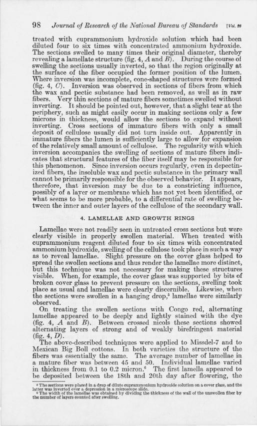
FIGURE 5.- Depectinized cotton fib ers swollen in cuprammonium hydroxide solution to show striations.
A , 51 -day fiber , showing striations, magnification X500; B, matme fiber, showing striations. Photograpbrd between crossed nicols, magnification X-420. All the fibers were stained with Congo red .

Hock, RamSav,] HaTTis Structure oj Cotton Fiber 99
number increasing thereafter as the fiber approached maturity. The increase in the number of lamellae with age can be seen by comparing figure 4 (A) with figure 4 (B).
The lamellae, so clearly observed in swollen cross sections, can also be seen in longitudinal view [1], when the microscope is focused midway between the upper and lower surfaces of a swollen fiber. Fibers from which both wax and pectic substance were removed were preferable for these observations, as the purified fibers swelled more uniformly than raw fibers . When observed in this way, the fibers showed striations 7 running parallel to the fiber axis and extending from the lumen to the outer surface (fig. 5, A). The striations were readily discernible in unstained mounts, but the addition of Congo red rendered the pattern more distinct. When examined with crossed nicols alternating stripes of strongly and of weakly birefringent material were observed (fig. 5, B). As the fiber matures, the thickness of the wall becomes greater and the number of striations is increased. The number of striations in Mexican Big Boll from 18 days to maturity was found to agree with the number of lamellae counted in cross sections.
According to Kerr [6], two lamellae, one compact and one porous, are deposited each day during the period of secondary-wall deposition. These two lamellae (or two adjacent striations) together constitute a daily growth ring. Using" growth ring" in this sense, the relationship between these rings and the age of the fiber is shown graphically in figure 6. It is readily seen that there is a close correlation between the counts of lamellae and of striations. For the first 10 to 15 days after the initiation of secondary deposition, one growth ring was laid down per day. Thereafter, until maturity, additional growth rings were formed, but the rate of one ring per day was apparently not maintained. However, closer examination of the data showed that the counts of lamellae and of striations varied considerably in fibers older than 35 days. This may mean that individual fibers continued to deposit one growth ring daily, but that the average for the group is lowered because of the early maturation of some of the fibers.
These results confirm the work of Balls [1] and Kerr [6]. They are not in agreement with the results of the investigations by Sakostchikoff and Korsheniovsky [5] and Barrows [9], who were unable to establish a consistent relationship between the number of lamellae and the age of the fibers.
Figure 7 shows the increase in width of the secondary wall with the age of the cotton fiber . After about the first 15 days of secondarywall formation there is only a slight increase in thiclmess as the fibers continue to mature. This is consistent with a recent investigation [21] in which it was demonstrated that the percentage of cellulose in a series of cotton samples of different ages increased rapidly until about the 35th day after flowering, and that thereafter there was only a small further increase with age. A comparison of the curves of figures 6 and 7 suggests that the initial growth rings are wider than those laid down after the 35th day.
7 In the present paper, the layers observed in cross section are called lamellae, whereas the layers observed in longitndinal view are called striations. Both lamellae and striations refer, however, to the same strnctures.

100 Journal oj Research oj the National Bureau oj Standards [Vol. f6
Lamellae, or striations, were also revealed when fibers were treated with reagents other than cuprammonium hydroxide. Trimethylbenzylammonium hydroxide had essentially the same effect as cuprammonium reagent. Combined treatment with sodium hydroxide and carbon disulfide, and treatment with phosphoric acid also
24
22
20
t'J 18 ~ n:: :c 16 I-3: o ffi 14
u.. o n:: 12 w m :2: ~ 10
8
6
4
2 18
o ..
•
22 26 30 34 38 42 46 50 54
DAYS AFTER FLOWERING
FIGURE 6.-Growth rings in relation to the age of the fiber. Open circles represent growth rings estimated from the number of lamellae counted in fiber cross sectious
(25 sections counted for each point of the curve). Solid circles represent growth rings estimated from the number of striations counted in longitudinal view (50 sections counted for each point of the curve).
revealed a lamellate structure in the fiber, but cuprammonium hydroxide and trimethylbenzylammonium hydroxide were preferable for this work.
5. FIBRILLAR STRUCTURE
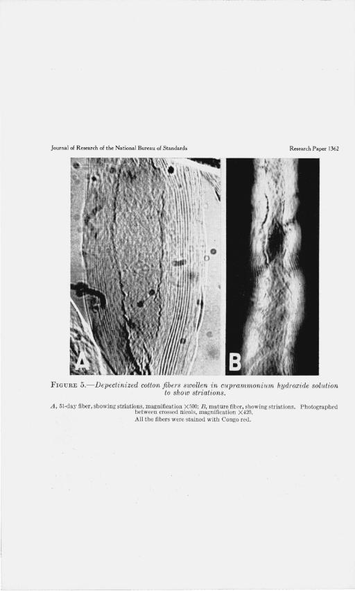
The investigation of the structural details of the cotton fiber was continued with single fibers. These were mounted in distilled water (fig. 8, A) and then examined with crossed nicols. When the fibers were placed approximately parallel to the plane of vibration of light through one of the prisms, the high birefringence of the fiber was interrupted by dark extinction bands at irregular intervals along its

IIoel" Ra7nSav,] Harris Structure oj Cotton Fiber 101
axis (fig. 8, B). The same phenomenon has previously been noted by Balls [2]. When a selenite plate was inserted between the nicols, contrasting colors, indicative of differences in orientation, appeared in regions adjacent to the extinction bands (fig. 8, C). The number of convolutions or twists of the fiber which occurred in a region
0 6.0
0 0
5.5
50
if) 0 z 4.5
0 0 Ct: C> ':fi 4.0 ~
-l -l 3.5 0 « :::: lL.
3.0 0
I r-0
2,5 §:
2.0
1.5
1.0 0
0 18 22 26 30 34 38 42 46 50 54
DAYS AFTER FLOWERING
FIGURE 7.-lncrease in width of the wall with increase in age of the fiber. Each point of the curve represents an average of 200 measurements.
between two bands of extinction varied considerably, depending apparently on the length of the region. However, in a region which showed uniform optical properties the convolutions were always in the same direction.
The optical variations just described were also correlated with differences in structure observed in depectinized 8 fibers swollen in dilute cuprammonium hydroxide or in solutions of trimethylbenzylammonium hydroxide. For this experiment the cuprammonium reagent was diluted three to four times with concentrated ammonia, and the trimethylbenzylammonium hydroxide was diluted with
8 Depectinized fibers were best suited for tbese experiments, since, after removal of the outer membrane of wax and pectic substance. irrcgular swelling along the axis could be minimized.

102 Journal oj Research oj the National Bureau oj Standards [Vol.£6
water to a concentration of 2.2 to 2.4 N. Upon swelling, the fibers clearly revealed a fibrillar structure. The bulk of a fiber was made up of innumerable fine fibrils oriented at an acute angle with respect to the axis of the fiber. The fibrils were enclosed by a winding 9
which appeared to be a little coarser than the fibrils themselves. In fibers which had been swollen only slightly, it was observed that
this outer winding was closely spaced and made a moderately steep spiral (fig. 9, A and B). The winding, like the bulk of the fiber, stained with Congo red, zinc chloride-iodine, and iodine and sulfuric acid, and appeared to dissolve in cuprammonium reagent. It made either an S or a Z twist 10 around the fiber, reversal of direction taking place (fig. 9, C) many times in a single fiber. Examination showed that both types of twist occur with about equal frequency. Invariably the orientation of the fibrils immediately beneath the outer winding was in the opposite direction. In other words, if the winding made an S twist, the fibrils immediately beneath made a Z twist. As far as could be determined, all of the fibrils under the winding at anyone place appeared to run in the same direction.
It became apparent that reversals of direction of the cellulose fibrils and of the winding were responsible for the optical differences observed with crossed nicols, and that the band of extinction was the place at which the reversals occurred. The number of optical reversals occurring in a single fiber or fragment invariably was the same as the number of fibril reversals. Likewise, the relationship between the fibrillar orientation and the convolutions became clear. It was found that the direction of a convolution usually was the same as the spiral direction of the winding. That is to say, when the winding made an S twist around the axis, the convolutions in . that region also showed an S twist.
The fibrillar structure of the secondary wall was studied to better advantage upon handling the fibers with microneedles. In some cases the outer winding was picked up by the needles and pulled away from the rest of the fiber or the winding was pulled along the axis of the fiber in such a way as to form a clump. Needles were brought into the innumerable fine fibrils comprising the bulk of the cellulose of the secondary wall only by careful manipulation. The difficulty in picking up these fibrils appeared to be due to lateral cohesion of the fibrils themselves. After insertion of the needles, however, the fibrils could be separated from one another (fig. 10, A and B) and otherwise subjected to micromanipulative methods.
6. BALLOON FORMATION
Depectinized fibers were also examined in cuprammonium hydroxide solutions of various concentrations. It soon became clear that there was a relationship between the optical differences and fibril reversals on the one hand, and balloon formation on the other. In concentrated cuprammonium hydroxide solutions (reagent diluted not more than three times with concentrated ammonia) dissolution of the cellulose of the fibers took place in a relatively short time. The fibers appeared to break into large chunks which became progressively smaller as dis-
• Although the winding closely resembles the fibrils which make up the bulk of the fiber, to facilitate discussion it will be considered separately.
10 According to ASTM Standards on Textile Materials [22]. a yarn or cord has an S twist if, when held in a vertical position, the spiral conforms in slope to the central portion of the letter S, and a Z twist if the spirals conform to the central portion of the letter Z. In the present paper, the same terminology will be applied to fibrils.
Journal of Research of the National Bureau of Standards Research Paper 1362
::::?f ; ::;
c FIGURE S.- Raw mature fiber mounted in water and photographed under various
lighting conditions.
A , Ordinary light, showing convolutions in the fiht~r ; B, bctwcen crossed nicols, showing bands or ext inction where t he fibrill ar orientation reverses ; C, between crossed nicols and with selenite p late, showing the effect due to ditl'crcncm: in color in n:>gioDs adjacent to th e extinction hands; magnification , A , R, and C, X 180.
I
I

Journal of Research of the National Bureau of Standards Research Paper 1362
FIG U R E 9.- Depectinized fibers showing the winding.
A, Fiber swollen slight ly in dilute cuprammonium hydroxide solution ; E, fiber treated with zinc chlori dciodine and photographed between crossed nicols; C, fiber, showi ng reversal, treated with zinc chlorideiodine and photographed between crossed nicols; magnification, A, B, and C, X420.

Journal of Research of the National Bureau of Standards Research P aper 1362
FIGURE lO .- Depectinized fibers swollen in dilute w prammoniwn hydroxi de solution and handled with micToneedles.
A, A single fiber bcing d issected with microneedles to show fibrillar structure; B, fibr ils bei ng pullcd away from the fiber. (The needles are not shown in the pho tomicrograph .) Magnification , A a ncl B , X500.
Journal of Research of the National Bureau of Standards Research Paper 1362
FIGURE n.-Cotton fibers swollen in cuprammonium hydroxide solution to produce balloons.
A, Depectinized tibers, showing balloons, photographed between crossed nicols, (selenite plate inserted), magnification X260; B, depectinized fibers, showing windi ng. The coarse appearance of the winding may have resulted from the coalescence of indi vidual fibrils during incipient balloon formation, magnification X420. C, typical string of balloons in a depectinized fiber, magnification X500; D, string of balloons in a raw fiber, showing the winding and fragments of the primary wall connecting the constrictions. Fiber stained with ru thenium red . M agnification X440.

Flock. Ramsav.] Harris Structure oj Gatton Fiber 103
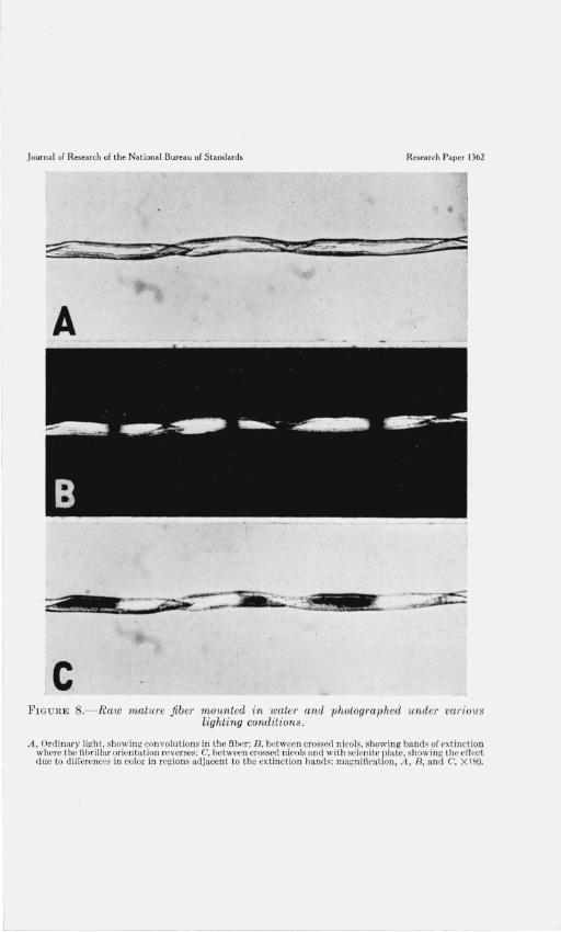
solution proceeded. Observed between crossed nicols, the highly birefringent fibers were seen to break up into fragments which steadily became less birefringent until the field of the microscope was black. In dilute solutions (diluted between 3 and 10 times) the fibers swelled greatly but complete dissolution generally did not occur. As swelling took place balloons were occasionally formed. When the behavior of the fibers was observed with crossed nicols, the fibers were seen to become progressively dimmer as the cellulose dissolved. The expanded part of a balloon was the first to lose its birefringence, since here the cellulose had become highly disorganized. On the other hand, the constrictions were slow in losing their anisotropy (fig. 11, A), since the reagent penetrated these regions slowly. When fibers which had lost much of their birefringence during swelling in cuprammonium solution were placed in water, some of the original birefringence of the fibers, as well as some of their fibrillar appearance, was restored.
The occurrence of balloons in depectinized fibers was somewhat surprising, since experiments by ourselves and by others indicated that the primary wall plays an important role in the formation of balloons. Accordingly, the phenomenon was studied further, especially in depectinized fibers from which the wax and pectic substance had been removed. The optimum concentration of cuprammonium hydroxide solution for observin~ balloons in these fibers was between 15 and 20 percent (1.5 to 2.0 m! of the reagent diluted to 10 ml by the addition of concentrated ammonia). When depectinized fibers were placed in these solutions they often swelled unevenly along the axis, thereby giving rise to balloons (fig. 11, B and C). The expanded portion of the balloons almost invariably occurred between the points at which the fibrils reversed, whereas the reversal points themselves formed the constrictions between adjacent balloons. Fragments of depectinized fibers which showed no extinction bands usually swelled without the formation of balloons, and the direction of the fibrils was found to be constant. Because of the orientation of the cellulose within the fiber, the greatest swelling occurs perpendicular to the fiber axis, while at the same time there is an actual decrease in fiber length. As these changes in length and in width take place, the winding assumes a more transverse position (fig. 11, B) and becomes clumped at the constrictions. However, the uniform lateral expansion of the fiber is hindered somewhat by this same winding which does not expand freely in that direction. Accordingly, the winding also appears to exert a constricting influence on the swelling of the fibers.
It has been recognized that the primary wall is responsible, at least in part, for the formation of balloons. The rupture of this wall into a series of constrictions which restrains the expanding cellulose has frequently been described. Furthermore, fragments of raw fibers which show uniform optical characteristics often produce many balloons as swelling occurs, whereas similar fragments of depectinized fibers do not produce balloons. When balloons are formed in raw fibers the cellulose fibrils may push through the winding and the primary wall, so that a constriction consists of a clump of the winding as well as of a portion of the primary wall (fig. 11, D). Thus the above experiments suggest that the irregular swelling along the fiber axis, which frequently results in balloon formation, is dependent in part on the orientation of the fibrils and in part on the constricting influences of the winding and of the primary wall.
104 Journal oj Research oj the National Bureau oj Standards [Vol. f8
IV. REFERENCES
[1] W. L. Balls, Proc. Roy. Soc. (London) [B] 90, 342 (1919). [2] W. L. Balls, Proc. Roy. Soc. (London) [B] 72, 72 (1923). [3] W. L. Balls, Studies of Quality in Cotton (Macmillan & Co. Ltd., London,
1928). [4] H. J. Denham, J. Textile Inst. 14, T86 (1923). [5] A. P. Sakostschikoff and G. A. Korsheniovsky, Faserforschung 9, 249 (1932). [6] T. Kerr, Protoplasma, 27, 229 (1937). [7] D. B. Anderson and T. Kerr, Ind. Eng. Chem. 30, 48 (1938). [8] W. Ie. Farr, Contrib. Boyce Thompson Inst. 10, 71 (1938). [9] F. L. Barrows, Contrib. Boyce Thompson Inst. 11, 161 (1940).
[10] A. B. Corey and II. L. Gray, Ind. Eng. Chem. 16, 853, 1130, (1924) . [11] R. Ie. Worner and R. T. Mease, J. Research NBS 21, 609 (1938) RP1146. [12] R. L. Whistler, A. R. Martin, and M. Harris, J. Research NBS 24, 555 (1940)
RP1299. [13] R. T. Mease, J. Research NBS 22, 271 (1939) RP1179. [14] C. Doree, The Methods of Cellulose Chemistry, D. Van Nostrand Co., Inc.,
New York, N. Y., (1933). [15] L. Mangin, Compt. rend. 116, 653 (1893). [16] D. R. Morey, Textile Research 3,325 (1933). [17] A. Frey-Wyssling, Die Stoffausscheidung der hOheren Pflanzen. Julius
Springer, Berlin (1935). [18] E. E. Berkley, Textile Research 9, 355 (1939). [19] C. W. Hock and M. Harris, J. Research NBS 24, 743 (1940) RP1309. [20] J. 1. Hardy, U. S. Dept. of Agriculture Circular 378 (1935). [21] R. L. Whistler, A. R. Martin, and 1. M. Conrad, J. Research NBS 25, 305
(1940) RP1326. [22] ASTM Standards on Textile Materials, Report of Committee D-13, p. 5
(1940) .
WASHINGTON, September 28, 1940.