Misinformation from Sputum Cultures Without Microscopic ...
10
JOURNAL OF CLINICAL MICROBIOLOGY, Nov. 1977, p. 518-527 Vol. 6, No. 5 Copyright © 1977 American Society for Microbiology Printed in U.S.A. Misinformation from Sputum Cultures Without Microscopic Examination HERBERT S. HEINEMAN,t* JAGJIT K. CHAWLA, AND WENDELL M. LOFTON Microbiology Laboratory, Philadelphia General Hospital, Philadelphia, Pennsylvania 19104 Received for publication 8 June 1977 Only 13 of 38 hospital laboratories surveyed include a Gram stain routinely in microbiological sputum examination. In a prospective three-hospital study, 60% of over 1,200 "sputum" specimens consisted predominantly of saliva, as judged by cell composition. Compared with the results of cultures in which microorga- nisms presumptively identified on sputum smears were specifically sought ("di- rected cultures"), cultures of the same specimens processed in the routine manner missed pneumococci 61%, haemophili 23%, and yeasts 44% of the time. The findings were similar in all three hospitals despite differences in administration, staffing, primary culture media, and workload. Unless microscopic examination is routinely included, half of all microbiological information rendered on sputum specimens is meaningless and subject to dangerous misapplication. Furthermore, culture must be guided by microscopic findings, or respiratory pathogens will frequently be missed. Finally, when routine culture and smear disagree, the culture cannot be assumed to be correct. Microscopic examination should be mandatory in sputum microbiology, both for specimen evaluation and as a guide to what to look for in culture. Microscopic examination of smears is a time- tested, but not particularly honored, part of spu- tum microbiology. In the belief that culture tells all, the Gram stain has been relegated to the status of an optional accessory. The information it provides is regarded as tentative at best, mis- leading at worst, and always to be superseded by culture. Shulman and co-workers (16), analyzing the errors and hazards in the diagnosis of bacterial pneumonias, cited the frequency with which the etiological agent is missed on Gram smears of sputum but did not mention culture as a source of error. Standard texts in laboratory diagnosis give varying amounts of space to discussion of the Gram stain, but none holds it to be essential. It has been said to be valuable in the diagnosis of staphylococcal and gram-negative pneumonia (4), misleading in pneumococcal pneumonia ex- cept in typical cases (1), and even misleading in staphylococcal pneumonia (15). Reference to microscopy for evaluation of specimens is excep- tional (7, 13), even though proper collection of specimens is emphasized and the frequent sub- mission of saliva as sputum is appreciated (17). A number of recent publications (3, 5, 7, 12) have addressed themselves to cellular analysis of sputum without, however, modifying the prevalent views regarding identification of mi- t Present address: Mercy Catholic Medical Center, Miser- icordia Division, Philadelphia, PA 19143. croorganisms. Despite earlier warnings (2, 14, 18), culture remains virtually the sole basis for microbiological diagnosis. Our experience in sputum microbiology has convinced us that microscopic examination is much too useful to be left to discretion. Overin- terpretation may indeed lead to false conclu- sions. However, without microscopy, culture re- sults are of unknown relevance and, because of the faith placed in them, may be dangerously misleading. In agreement with others, we find that a very substantial proportion of specimens submitted as sputum are so contaminated with saliva that the results of culture (often resistant gram-negative bacilli) cannot possibly be applied to the diagnosis of respiratory infection and that the only way to recognize this fact is to examine every specimen microscopically. In contradic- tion to widely held beliefs, we find that pre- sumptive identification of respiratory pathogens other than staphylococci and gram-negative ba- cilli, specifically pneumococci and haemophili, in Gram-stained smears is quite feasible. Finally, a survey of area hospital laboratories indicates that in two-thirds, the practice is to make Gram-stained smears only on request. Ev- idently, this simple and inexpensive test is not universally accepted, and repeated emphasis on its value is needed. (This work was originally presented at the 77th Annual Meeting of the American Society for Microbiology [H. S. Heineman and W. E. 518
-
Upload
khangminh22 -
Category
Documents
-
view
1 -
download
0
Transcript of Misinformation from Sputum Cultures Without Microscopic ...

JOURNAL OF CLINICAL MICROBIOLOGY, Nov. 1977, p. 518-527 Vol. 6, No. 5Copyright © 1977 American Society for Microbiology Printed in U.S.A.
Misinformation from Sputum Cultures WithoutMicroscopic Examination
HERBERT S. HEINEMAN,t* JAGJIT K. CHAWLA, AND WENDELL M. LOFTONMicrobiology Laboratory, Philadelphia General Hospital, Philadelphia, Pennsylvania 19104
Received for publication 8 June 1977
Only 13 of 38 hospital laboratories surveyed include a Gram stain routinely inmicrobiological sputum examination. In a prospective three-hospital study, 60%of over 1,200 "sputum" specimens consisted predominantly of saliva, as judgedby cell composition. Compared with the results of cultures in which microorga-nisms presumptively identified on sputum smears were specifically sought ("di-rected cultures"), cultures of the same specimens processed in the routine mannermissed pneumococci 61%, haemophili 23%, and yeasts 44% of the time. Thefindings were similar in all three hospitals despite differences in administration,staffing, primary culture media, and workload. Unless microscopic examinationis routinely included, half of all microbiological information rendered on sputumspecimens is meaningless and subject to dangerous misapplication. Furthermore,culture must be guided by microscopic findings, or respiratory pathogens willfrequently be missed. Finally, when routine culture and smear disagree, theculture cannot be assumed to be correct. Microscopic examination should bemandatory in sputum microbiology, both for specimen evaluation and as a guideto what to look for in culture.
Microscopic examination of smears is a time-tested, but not particularly honored, part of spu-tum microbiology. In the belief that culture tellsall, the Gram stain has been relegated to thestatus of an optional accessory. The informationit provides is regarded as tentative at best, mis-leading at worst, and always to be supersededby culture.Shulman and co-workers (16), analyzing the
errors and hazards in the diagnosis of bacterialpneumonias, cited the frequency with which theetiological agent is missed on Gram smears ofsputum but did not mention culture as a sourceof error.Standard texts in laboratory diagnosis give
varying amounts of space to discussion of theGram stain, but none holds it to be essential. Ithas been said to be valuable in the diagnosis ofstaphylococcal and gram-negative pneumonia(4), misleading in pneumococcal pneumonia ex-cept in typical cases (1), and even misleading instaphylococcal pneumonia (15). Reference tomicroscopy for evaluation of specimens is excep-tional (7, 13), even though proper collection ofspecimens is emphasized and the frequent sub-mission of saliva as sputum is appreciated (17).A number of recent publications (3, 5, 7, 12)
have addressed themselves to cellular analysisof sputum without, however, modifying theprevalent views regarding identification of mi-
t Present address: Mercy Catholic Medical Center, Miser-icordia Division, Philadelphia, PA 19143.
croorganisms. Despite earlier warnings (2, 14,18), culture remains virtually the sole basis formicrobiological diagnosis.Our experience in sputum microbiology has
convinced us that microscopic examination ismuch too useful to be left to discretion. Overin-terpretation may indeed lead to false conclu-sions. However, without microscopy, culture re-sults are of unknown relevance and, because ofthe faith placed in them, may be dangerouslymisleading. In agreement with others, we findthat a very substantial proportion of specimenssubmitted as sputum are so contaminated withsaliva that the results of culture (often resistantgram-negative bacilli) cannot possibly be appliedto the diagnosis of respiratory infection and thatthe only way to recognize this fact is to examineevery specimen microscopically. In contradic-tion to widely held beliefs, we find that pre-sumptive identification of respiratory pathogensother than staphylococci and gram-negative ba-cilli, specifically pneumococci and haemophili,in Gram-stained smears is quite feasible.
Finally, a survey of area hospital laboratoriesindicates that in two-thirds, the practice is tomake Gram-stained smears only on request. Ev-idently, this simple and inexpensive test is notuniversally accepted, and repeated emphasis onits value is needed.
(This work was originally presented at the77th Annual Meeting of the American Societyfor Microbiology [H. S. Heineman and W. E.
518

MISINFORMATION FROM SPUTUM CULTURES
Lofton, Abstr. Annu. Meet. Am. Soc. Microbiol.1977, C195, p. 68].)
MATERIALS AND METHODSAll sputum specimens were handled in the standard
manner, as described in the microbiology proceduremanual of Philadelphia General Hospital. Processingbegan within 30 min of receipt in the laboratory. Fromthe most purulent or mucoid portion of the specimen,a smear was made and plates of 5% sheep blood agar,chocolate agar, and MacConkey agar were inoculated.The smear was Gram stained and read by one of a
number of technicians, the interpretation being re-corded on the stat copy, which was immediately sep-arated from the remainder of the report and returnedto the patient area. An evaluation of the specimenwas entered on the chart copy, specifically, "purulentsecretions" (polymorphonuclear cells), "respiratory se-cretions" (ciliated cells), and/or "oral secretions"(squamous cells). Bacterial morphology was notedonly on the stat copy. The technician handling theculture plates on subsequent days was usually notguided by the microscopic findings.
Plates were incubated in 5% carbon dioxide over-night; if growth was too slight for isolation of colonies,incubation was continued for 24 h more. On the basisof gross visual inspection, suspect colonies were sub-cultured and subjected to standard procedures forbacterial identification. Pneumococci were identifiedas alpha-hemolytic streptococci inhibited by optochin;haemophili were identified as gram-negative coccoba-cili showing characteristic growth on chocolate agarand satellitism around a streak inoculum of Staphy-lococcus aureus on sheep blood agar.
Parts of study. (i) Evaluation of specimens.The criterion for specimen evaluation was the esti-mated proportion of saliva in the specimen. Sincesquamous and polymorphonuclear cells are the quan-tifiable markers of saliva and inflammatory secretions(operationally equivalent to sputum), respectively, anattempt was made to compute the proportions ofsaliva and sputum from the numbers of these cells.Using a calibrated loop (of the type designed forurinary colony counts), loopfuls of normal saliva fromthree volunteers were carefully placed on slides and
P- -- -- n._ _-
-9-_
allowed to dry without spreading. After staining, sev-eral oil immersion fields were scanned, and it wasdetermined that the average field contained 3 to 5squamous cells. The same procedure was carried outwith grossly purulent sputum, yielding 50 to 100 pol-ymorphonuclear cells per average oil immersion field.Since each field represented the dried residue of ap-proximately equal volumes of saliva and sputum, itwas determined for the purpose of specimen evalua-tion that a volume of purulent sputum contains ap-proximately 20 times as many polymorphonuclears asthere are squamous cells in an equal volume of saliva.Therefore, a smear showing more than 1 squamouscell per 20 polymorphonuclears, regardless of absolutenumbers, was regarded as consisting of more than 50%saliva.
Approximately 500 Gram-stained smears were ex-amined under low-power (xlOO) magnification, andnote was made of the relative proportion of cells.Smears of specimens estimated to contain more than50% saliva (Fig. 1, left) were not examined further.Smears computed to consist mostly of inflammatoryexudate (Fig. 1, right) were examined under oil (xl,-000) for microbial morphology.
(ii) Predictability of culture results fromsmears. The microorganisms were not only describedby morphology and staining reaction, but specific iden-tification was attempted for certain known and sus-pected respiratory pathogens, namely, pneumococci,staphylococci and haemophili (Fig. 2 through 4). Thepresence of disproportionate numbers of other gram-negative bacilli (Fig. 5) or of yeast cells (Fig. 6) waslikewise noted as abnormal. Gram-positive bacilli,long-chain streptococci, and gram-negative cocci, aswell as mixtures of many types of bacteria, were con-sidered grounds for predicting "normal flora" in cul-ture.
After the smear interpretations had been recorded,the culture results were retrieved from the laboratoryrecords.
(iii) Validity of culture results. Because of adisappointing lack of correlation between certain ap-parently diagnostic smear findings and culture results(see Table 1), the possibility was investigated that thesmear interpretation was correct and the culture resultwas incorrect. Consecutive smears were examined, and
r.
v. -
11*. .:sY,IV ;..
1
FIG. 1. Two specimens submitted as sputum. (left) Predominant squamous cells characteristic of saliva.(right) Abundance ofpolymorphonuclear cells characteristic of inflammatory exudate. More than half of the"sputum" specimens received in a microbiology laboratory may consist mostly of saliva.
VOL. 6, 1977 519

520 HEINEMAN, CHAWLA, AND LOFTON
WI*i
I_
L
FIG. 2. Typical sputum smear in pneumococcusinfection.
wk. 'Ia-41-
¾1.
Ad
FIG. 5. Typical sputum smear in gram-negativebacillary infection.
A*l it'...4 4 \4w
IIf
C
f 4f,sOL . .... .,,
,4
FIG. 3. Typical sputum smear in staphylococcus
infection.
1~~~~~~~~~~I-tzZ4^
FIG. 4. Typical sputum smear in haemophilus in-fection.
approximately 60 were selected on the basis that theyshowed either (i) yeast cells or (ii) a combination ofnumerous polymorphonuclear cells and a predomi-nance of bacteria resembling pneumococci or haemo-phili, as in Fig. 2, 4, and 6. In other words, they were
felt to be virtually diagnostic of infection or significantovergrowth. Cultures were handled in the routinemanner, but in addition, unknown to the technicians,duplicate subcultures were performed by one of theauthors (W.M.L.), who intercepted the primary cul-
FIG. 6. Excessive number ofyeast cells in sputumsmear.
ture plates at the time of discarding (generally 1,occasionally 2, days after the initial examination bythe technician) and reexamined them, particularly forthe organisms thought to be recognized on the smear.The criteria for bacterial identification were the sameas used in the routine culture. A comparison was thenmade of smear interpretation, results of routine cul-ture, and results of culture as directed by microscopicfindings ("directed culture").
(iv) Comparison of three hospital laborato-ries. Since routine culture at Philadelphia GeneralHospital frequently failed to yield potential pathogensthat could be found by directed search (see Table 2),the possibility of defective laboratory practices at thishospital was investigated by extending the study totwo other hospitals, namely, a 500-bed university hos-pital and a 250-bed community hospital. With thecooperation of the director of the microbiology labo-ratory and the bacteriology supervisor at each hospi-tal, all Gram-stained smears and primary cultureplates were saved. Again, seemingly diagnostic smearswere selected for directed culture, which was per-formed at Philadelphia General Hospital, and the re-sults were correlated with the results of culture at thehospital of origin.
(iv) Survey of hospital practices. Because ofthe important implications of our findings, as discussedbelow, a questionnaire (Fig. 7) was sent to the labo-
It
i
Ur .Att,~~
-A
J. CLIN. MICROBIOL.
*1
lowitt1-

MISINFORMATION FROM SPUTUM CULTURES
QUESTIONNAIRE ON SPUTUM GRAM STAINS
Name of hospitalNumber of specimens for bacterial culture per month
Gram stains are done on fresh sputum specimens (check one)routinelyon request
(a) total(b) sputum
I. Method of Reporting Gram StainA. Cells (check one)
1. Described interpretatively, e.g., "purulent"; "oral epithelium"; etc.2. Described morphologically, e.g., "polymorphonuclear"; "squamous"; etc.3. Not described in report
B. Microorganisms (check one)1. Described by suspected identity, e.g., "probable pneumococci"; "resembling staphylo-
cocci"; etc.2. Described only by stain reaction and morphology, e.g., "gram-positive cocci in pairs and
chains"; etc. __C. Evaluation of specimen (check one)
1. Report includes evaluation, e.g., "satisfactory specimen"; "mostly saliva"; etc.2. Report is purely descriptive
II. Laboratory Use of Gram StainA. As guide to culture (check one)
1. Technician uses Gram stain findings (i.e., on sputum smears) to make special search forparticular organisms in culture __
2. Culture is processed independently of Gram stain findingsB. As guide to disposition of specimen (check one)
1. IfGram stain indicates unsatisfactory specimen (e.g., mostly squamous cells with adherentmixed bacteria), culture is not done, pending request for a better specimen
2. All specimens are cultured regardless of Gram stain findings
FIG. 7. Questionnaire sent to Philadelphia area hospitals.
ratory directors of all hospitals in the Philadelphiaarea. The replies were analyzed to determine to whatextent the procedures that we now recommend arealready being used.
RESULTSEvaluation of specimens. Of 496 consecu-
tive specimens examined under low-power mag-nification, 198 had a polymorphonuclear-squa-mous cell ratio of 20:1 or greater, and 298 had aratio of less than 20:1. Thus, the proportion ofspecimens labeled "sputum" that consisted ofat least half saliva was 60% at PhiladelphiaGeneral Hospital. The proportions at the othertwo hospitals were remarkably similar, 232/400(58%) and 181/304 (60%). In at least half of theunacceptable specimens, squamous cells actuallyoutnumbered polymorphonuclears, indicatingthe virtual absence of any sputum at all (Fig. la).Predictability of culture results from
smears. Sixty-five smears were selected asbeing virtually diagnostic. The extent to whichculture results confirmed this impression isshown in Table 1. Only 39 of 65 predictions(58%) were correct overall, the best performancebeing recorded for staphylococci (11/15, or 73%)and gram-negative bacilli other than haemophili
TABLE 1. Predictability of routine culture resultsfrom Gram stain interpretation
Gram stain interpretation (no. of No. culturesamples) positive
Staphylococci (15) 11Pneumococci (10) 2Haemophili (4) 2Other gram-negative bacilli (31) 22Yeasts (5) .......................... 2
(22/31, or 71%). The smear predictions leastoften confirmed by culture were for pneumo-cocci (2/10, or 20%).Validity of culture results. Fifty-nine ad-
ditional smears interpreted as showing pneu-mococci, haemophili, or yeasts specifically wereselected because of the previous poor showingof the Gram stain in predicting their growth inculture (Table 1). As illustrated in Fig. 2, 4, and6, there was good reason to consider the orga-nisms as pathogenically significant, based ontheir preponderance and association with in-flammatory cells. Table 2 depicts the results. In10 of 30 cases (33%), the prediction of pneumo-cocci from the smear was borne out by routineculture. However, directed culture yielded posi-
VOL. 6, 1977 521
522 HEINEMAN, CHAWLA, AND LOFTON
tive results in 19 more, so that smear interpre-tation was actually confirmed 29 of 30 times(97%). For haemophili, undirected culture con-
firmed the smear 13 of 18 times (72%), whereasdirected culture confirmed 4 more, for a totalof 17 of 18 (94%). In the case of yeasts, all 15(100%) were confirmed by directed culture, incontrast to only 9 of 15 (60%) undirected cul-tures.Comparison of three hospital laborato-
ries. Table 3 compares the sensitivity of routinecultures at the three hospitals. Also given are
data regarding directorship, supervision, staff-ing, workload, and procedure. Despite the differ-ences, all three commonly reported cultures neg-
ative for pathogens that were identifiable on
smears.Survey of hospital practices. Completed
questionnaires were received from 37 hospitallaboratories, processing an aggregate of 5,300sputum specimens per month. The average num-
ber per month for all hospitals, including Phila-delphia General, was 145, with a range of 20 to470. An analysis of the replies is presented inTable 4. A number of findings deserve note.
TABLE 2. Sensitivity of routine culture and culturedirected by Gram stain interpretation
Gram stain interpretation No. culture positive(no. of samples)a Routineb Directed"
Pneumococci (30) 10 29Haemophili (18) 13 17Yeasts (15) 9 15
a On 4 of 59 Gram stains, both pneumococci andhaemophili were recognized. They are tabulated sep-arately.
Culture independent of Gram stain interpretation.Attention directed to organism identified on Gram
stain.
J. CLIN. MICROBIOL.
First, only one in three laboratories routinelymakes smears from sputum. Second, one in fourdoes not describe cellular composition. Third,only one in four reports out an evaluation ofthe specimen. Fourth, only one in three uses
microscopic findings to guide culture. Fifth, notone rejects a specimen because microscopic ex-
amination indicates that it is unsatisfactory.When grouped according to workload, 7 of
the 13 laboratories routinely making smears are
among the 11 processing more than 200 speci-mens per month. Also, specimen evaluation isoffered by 45, 28, and 15% of laboratories withmonthly workloads of more than 200, 100 to200, and fewer than 100 specimens, respectively.Thus, it appears that more use is made of micro-scopic examination in laboratories with largerworkloads. However, one laboratory processing300 sputum specimens per month reported thatsmears were made only on request, the cellswere not described, no evaluation was offered,and culture was performed independent of mi-croscopic findings.
DISCUSSIONOur findings support the viewpoint that any
attempt to diagnose respiratory infection by spu-tum culture without microscopic examinationinvites confusion and misinformation. Unfortu-nately, the limitations of the Gram stain seemto have received much more emphasis than itsvirtues. This bias is reflected both in the spottyuse of the Gram stain and in the noninterpreta-tive manner in which findings are usually re-
ported.As a means of judging the quality of a speci-
men, microscopic examination is unequalled. Incontrast to gross inspection, it permits unequiv-ocal identification of inflammatory and salivarycellular constituents. This application of sputum
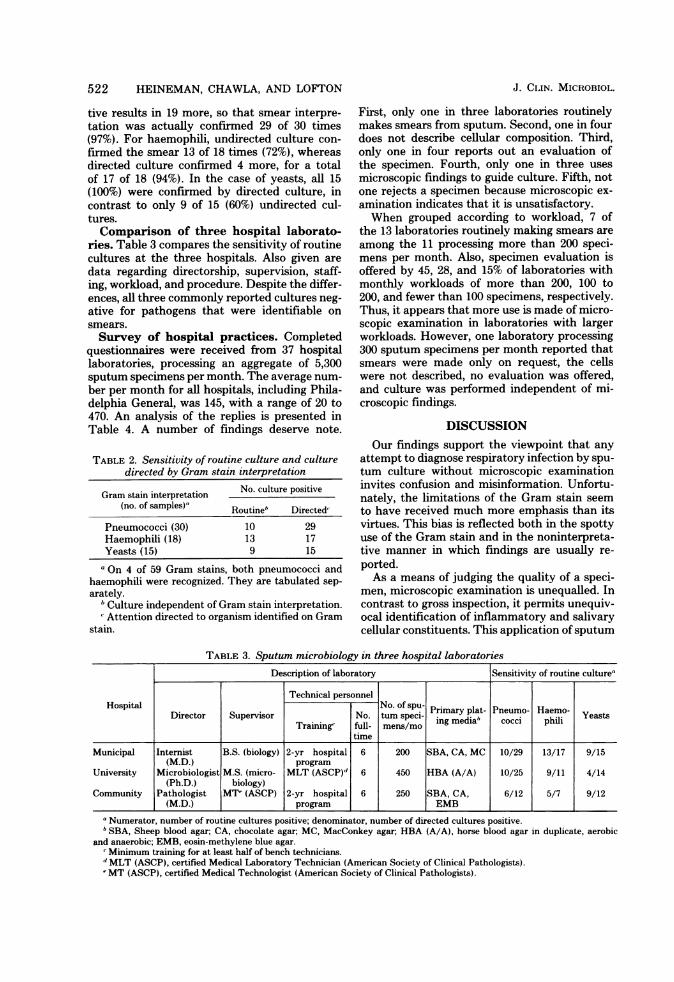
TABLE 3. Sputum microbiology in three hospital laboratories
Description of laboratory Sensitivity of routine culturea
Technical personnelHospital No. of spu- Primary plat- Pneumo- Haemo-
Director Supervisor No. turn speci-in media'h cocci phili Yeasts
Training" full- mens/mo ingtime
Municipal Internist B.S. (biology) 2-yr hospital 6 200 SBA, CA, MC 10/29 13/17 9/15(M.D.) program
University Microbiologist M.S. (micro- MLT (ASCP)' 6 450 HBA (A/A) 10/25 9/11 4/14(Ph.D.) biology)
Community Pathologist MT1 (ASCP) 2-yr hospital 6 250 SBA, CA, 6/12 5/7 9/12(M.D.) program EMB
a Numerator, number of routine cultures positive; denominator, number of directed cultures positive.b SBA, Sheep blood agar; CA, chocolate agar; MC, MacConkey agar; HBA (A/A), horse blood agar in duplicate, aerobic
and anaerobic; EMB, eosin-methylene blue agar.c Minimum training for at least half of bench technicians.d MLT (ASCP), certified Medical Laboratory Technician (American Society of Clinical Pathologists).e MT (ASCP), certified Medical Technologist (American Society of Clinical Pathologists).

MISINFORMATION FROM SPUTUM CULTURES
TABLE 4. Clinical laboratory practice in 38 Philadelphia area hospitals, processing a total of 5,500 sputumspecimens monthly
No. of laboratoriesaPractice
<100 100-200 >200
Performance of Gram stainRoutinely 4 (20) 2 (28) 7 (64)On request only 16 (80) 5 (72) 4 (36)
Method of reportingCells
Interpretative 2 (10) 1 (14) 2 (18)Morphological 11 (55) 4 (58) 8 (73)Not at all 7 (35) 2 (28) 1 (9)
MicroorganismsTentatively identified 1 (5) 1 (14) 1 (9)Morphological 19 (95) 6 (86) 10 (91)
Specimen evaluationIncluded 3 (15) 2 (28) 5 (45)Not included 17 (85) 5 (72) 6 (55)
Laboratory useAs guide to culture
Directed culture 8 (40) 3 (43) 3 (27)Independent culture 12 (60) 4 (57) 8 (73)
As guide to dispositionDiscard if unsatisfactory 0 (0) 0 (0) 0 (0)Culture all 20 (100) 7 (100) 11 (100)
a Laboratories divided into three groups, processing <100, 100 to 200, and >200month. Numbers in parentheses are percentages within each group.
microscopy has been recognized for a consider-able time (3, 5, 7, 12, 13), and it is remarkablethat the majority of laboratories continue toreport "sputum culture" results without qualifi-cation on specimens easily recognizable as con-sisting wholly or partly of saliva.
Less well appreciated is the feasibility of iden-tifying certain microorganisms on a Gram-stained smear. Quite reasonably, it is considereddangerous to attempt a definitive diagnosis inthis manner, and discrepancies between impres-sions gained from smear and from culture areusually settled in favor of the latter. This rule,however, needs to be reexamined. Before anaer-obic culture achieved widespread clinical appli-cation, the smear was often the only clue to thetrue nature of so-called sterile abscesses (8). Onemay safely assume that not only anaerobic, butalso aerobic and facultative, organisms some-times escape detection, especially in mixed cul-ture. Unfortunately, no expectorated specimenfails to yield at least some bacteria under allconditions of incubation, so that the stimulusto reconcile microscopic and cultural findingshas not been as strong as in the case of sterilespecimens. Consequently, it is usually assumedthat microscopic findings not borne out in cul-ture have simply been misinterpreted.However, the fallibility of cultures, as rou-
tinely done, should not be underestimated. Anextreme example is seen in Fig. 8, thanks to the
sputum specimens per
unmistakable microscopic appearance of theyeast. Less direct, but no less convincing, evi-dence was presented many years ago by studentsof pneumococcal infection, who frequently failedto recover the pathogen from the sputum ofpatients with bacteremic pneumonia (2, 14). Onthis basis, Barrett-Connor suggested that Gramstain examination was superior to culture, withwhich we would agree. Others also have reporteddiscrepancies between Gram stain and culturefindings both for pneumococci and for haemo-phili (10). In all these studies, culture was per-formed in a routine manner, in which colonialappearance was used to draw attention to thepossible presence of pathogens. Negative culturereports were apparently accepted as such andwere explained by inadequacy of the specimen,uneven distribution of microorganisms, delay inlaboratory processing, or competition from othermicroorganisms. To our knowledge, however,the question whether sputum cultures that ap-pear negative really are negative has not beensystematically studied. Our approach was tosearch specifically for whatever appeared un-mistakable on the smear. With each specimenacting as its own control, we found that as manyas half of perfectly typical smears (Fig. 2, 4, and6) were confirmed in culture only when attentionwas directed to the suspected organism. Theselection of typical smears is emphasized topoint out that the search was not directed to a
523VOL. 6, 1977

524 HEINEMAN, CHAWLA, AND LOFTON
FIG. 8. Overgrowth by gram-negative bacilli. (left) "Pure culture of Klebsiella." (right) Smear of originalspecimen showing abundance of yeasts, whose growth was completely buried by the bacteria. The outcomewas predictable from the smear, and a medium selective for yeasts should have been added.
rare or occasional organism but to the predom-inant flora, so that what was being missed mightwell have been clinically significant.
The examination of Gram-stained smears thuscontributed materially to diagnosis, not as a
source of tentative impression but as a means
of improving the performance of culture.In advocating mandatory microscopic exami-
nation of all sputum specimens, we would expectit to serve useful functions in specimen evalua-tion, culture direction, and clinical interpreta-tion.Specimen evaluation. Physicians them-
selves often do not collect sputum from theirpatients and may have no idea how adequate a
specimen is sent for culture. Therefore, theymust be told whether information derived there-from is likely to be relevant to the infection. Atthe very least, this means appending to theculture report a statement that, rather thanmerely listing the microscopic findings, clearlystates whether the specimen consists predomi-nantly of inflammatory exudate or saliva. Betterstill, specimens judged to consist mainly of salivashould not be cultured at all, as suggested byBartlett (3), since the results are meaninglessor, worse, misleading. Although Bartlett has alsosuggested holding an unsatisfactory specimen in
case a better one cannot be obtained, we wouldgo further, taking the stand that an unsatisfac-tory specimen has no diagnostic value, even ifit is the only one available, and discarding itwithout further processing. This approach hasample precedent. Many laboratories will notfully process contaminated specimens, whethercontamination is defined as submission in a non-
sterile container or (for urine) an implausiblemixture of three or more bacterial species. Thereare as yet no universally accepted criteria forsputum contamination. Our formula differs fromthose of Bartlett (3) and Murray and Washing-ton (12), and the best one possibly still remainsto be developed. Irrespective of detail, thescreening procedure is a quickly performed low-power microscopic examination, which will leadto considerable saving of laboratory time andexpenses as well as elimination of useless infor-mation.Culture direction. Competent technologists
are expected to examine culture plates and,knowing the source of the specimen, to isolate,quantitate, and identify possible pathogens. Ourdata, from three independent laboratories, indi-cate that everyday practice falls far short of thisgoal. Furthermore, we wonder whether the"nonvalue of sputum culture" described by Bar-rett-Connor (2) and similar experiences of others(10, 14) do not illustrate the shortcomings ofthe usual routine as much as the limited poten-tial of culture. Not surprisingly, we confirmedthat a careful search uncovers a good deal morethan a routine one. And one way to insure acareful search, without relying on the ever-fad-ing effects of education, admonition, and so on,is to know that a presumptive identification hasalready been made and awaits confirmation.This is the meaning of directed culture, and itrequires interpretation of the smear in termsnot of shapes and colors but of specific identifi-cation. Granting that such identification is notdefinitive until confirmed in culture, we wouldemphasize that it is intended primarily for inter-
J. CLIN. MICROBIOL.
MISINFORMATION FROM SPUTUM CULTURES
nal use by the laboratory. With adequate quali-fication, however, this information can be ex-
tremely helpful to the clinician. For example, a
predominance of gram-negative bacilli (Fig. 5),although not permitting even a tentative specificdiagnosis, does rule out gram-positive coccal in-fection. For staphylococci, the microscopic ap-
pearance is virtually diagnostic (Fig. 3); theirpresence on a smear should be reported imme-diately and by name (not as "gram-positive cocciin pairs and clusters") because of the crucialtherapeutic implications. Conversely, sincestaphylococcal infections are characterized bydense bacterial populations (19), failure to rec-
ognize them with ease in the smear should castsome doubt on the significance of a positiveculture.
Identification of pneumococci is also feasible,provided certain cautions are observed. Thepoor performance attributed to Gram-stainedsmears is probably due to at least two correcta-ble factors. First, as discussed above, the error
is frequently in the culture. Second, there isunnecessary confusion between pneumococciand viridans streptococci. A common mistake isto attach significance to the occasional gram-
positive diplococci in a mixed bacterial popula-tion, which, more often than not, are constitu-ents of normal flora. A typical picture, on theother hand, is unlikely to mislead the microscop-ist (1) and should be reported unhesitatingly as
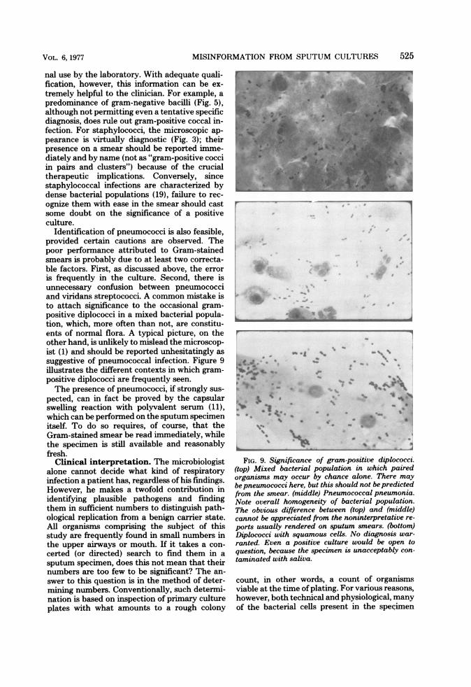
suggestive of pneumococcal infection. Figure 9illustrates the different contexts in which gram-
positive diplococci are frequently seen.
The presence of pneumococci, if strongly sus-
pected, can in fact be proved by the capsularswelling reaction with polyvalent serum (11),which can be performed on the sputum specimenitself. To do so requires, of course, that theGram-stained smear be read immediately, whilethe specimen is still available and reasonablyfresh.
Clinical interpretation. The microbiologistalone cannot decide what kind of respiratoryinfection a patient has, regardless of his findings.However, he makes a twofold contribution inidentifying plausible pathogens and findingthem in sufficient numbers to distinguish path-ological replication from a benign carrier state.All organisms comprising the subject of thisstudy are frequently found in small numbers inthe upper airways or mouth. If it takes a con-
certed (or directed) search to find them in a
sputum specimen, does this not mean that theirnumbers are too few to be significant? The an-
swer to this question is in the method of deter-mining numbers. Conventionally, such determi-nation is based on inspection of primary cultureplates with what amounts to a rough colony
-k.4..b * .''''
p 't W _ , s 4X, Z- r
FIG. 9. Significance of gram-positive diplococci.(top) Mixed bacterial population in which pairedorganisms may occur by chance alone. There maybepneumococci here, but this should not be predictedfrom the smear. (middle) Pneumococcal pneumonia.Note overall homogeneity of bacterial population.The obvious difference between (top) and (middle)cannot be appreciated from the noninterpretative re-ports usually rendered on sputum smears. (bottom)Diplococci with squamous cells. No diagnosis war-ranted. Even a positive culture would be open toquestion, because the specimen is unacceptably con-taminated with saliva.
count, in other words, a count of organismsviable at the time of plating. For various reasons,however, both technical and physiological, manyof the bacterial cells present in the specimen
525VOL. 6, 1977

526 HEINEMAN, CHAWLA, AND LOFTON
and easily recognized by direct visualization mayfail to reproduce in culture. Therefore, whenmicroscopic examination shows a predominanceof organisms so as to make a diagnosis virtuallycertain (a decision that can usually be made inless than 10 s) and the culture does not confirmthe expected proportions, the correctness of theculture may well be called into question. Specif-ically, if the sputum of a patient with a typicalclinical picture appears on smear as in Fig. 2and yields few or no pneumococci in culture, adiagnosis of pneumococcal pneumonia wouldnonetheless appear justified (2).
In certain clinical situations, even culturesideally performed yield results that are inher-ently uninformative, misleading, or likely to bequestioned by the clinician. One example is as-piration lung abscess, culture of whose contentsusually reveals normal oral flora. Furthermore,antibiotic therapy, prolonged hospitalization (9),or chronic alcoholism (6) may lead to upperrespiratory colonization with gram-negative ba-cili. If these are detected in culture, there is adanger of relating the pathology to these quasi-pathogens and misdiagnosing the true bacterialetiology. Only microscopic examination, asshown in Fig. 10, can tell the true story. Con-versely, the clinician may disregard a true path-ogen because it is rarely associated with respi-ratory infection. In one such example, coagulase-negative staphylococci were repeatedly isolatedfrom a patient with chronic bronchitis. Onlyafter inspection of the sputum smear (Fig. 11)could the possibility oflaboratory contaminationor undetected anaerobes be dismissed.
ACKNOWLEDGMENTSThis work was made possible through the generous coop-
eration of Donald D. Stieritz and David R. Davidson, Micro-biology Laboratory, Hahnemann Hospital, and Halvey E.Marx and Rita R. Radano, Microbiology Laboratory, Miseri-cordia Division, Mercy-Catholic Medical Center, Philadel-
v Sm
FIG. 10. Mixed bacteria characteristic of aspira-tion lung abscess.
J. CLIN. MICROBIOL.
-v t
FIG. 1 1.bronchitis.
Staphylococci in sputum from chronic
phia. Robert Austrian, University of Pennsylvania, mademany helpful suggestions in the preparation of the manuscript.
LITERATURE CITED1. Austrian, R. 1966. The role of the microbiological labo-
ratory in the management of bacterial infection. Med.Clin. North Am. 50:1419-1432.
2. Barrett-Connor, E. 1971. The nonvalue of sputum cul-ture in the diagnosis of pneumococcal pneumonia. Am.Rev. Respir. Dis. 103:845-848.
3. Bartlett, R. C. 1974. A plea for clinical relevance inmedical microbiology. Am. J. Clin. Pathol. 61:867-872.
4. Bauer, J. D., P. G. Ackermann, and G. Toro. 1974.Clinical laboratory methods, 8th ed., p. 655. C. V. MosbyCo., St. Louis.
5. Chodosh, S. 1970. Examination of sputum cells. N. Engl.J. Med. 282:854-857.
6. Fuxench-Lopez, Z., and C. Ramirez-Ronda. 1977. Pha-ryngeal flora in male alcoholic patients. A prospectivestudy. Clin. Res. 25:28A.
7. Gardner, P., and H. T. Provine. 1975. Manual of acutebacterial infections: early diagnosis and treatment, p.40, 45. Little, Brown & Co., Boston.
8. Heineman, H. S., and A. I. Braude. 1963. Anaerobicinfection of the brain. Observations on 18 consecutivecases of brain abscess. Am. J. Med. 35:682-697.
9. Johanson, W. G., A. K. Pierce, J. P. Sanford. 1969.Changing bacterial flora of hospitalized patients: emer-gence of Gram negative bacilli. N. Engl. J. Med.281:1137-1140.
10. Lepow, M. L., N. Balassanian, J. Emmerich, R. B.Roberts, M. S. Rosenthal, and E. Wolinsky. 1968.Interrelationships of viral, mycoplasmal, and bacterialagents in uncomplicated pneumonia. Am. Rev. Respir.Dis. 97:533-545.
11. Merrill, C. W., J. M. Gwaltney, J. 0. Hendley, andM. A. Sande. 1973. Rapid identification of pneumo-cocci. Gram stain vs the Quellung reaction. N. Engl. J.Med. 288:510-512.
12. Murray, P. R., and J. A. Washington, II. 1975. Micro-scopic and bacteriologic analysis of expectorated spu-tum. Mayo Clin. Proc. 50:339-344.
13. Niejadlik, D. C. 1974. The sputum, p. 1242. In I. Davidsonand J. B. Henry (ed.), Todd-Sanford clinical diagnosisby laboratory methods, 15th ed. The W. B. SaundersCo., Philadelphia.
14. Rathbun, H. K., and I. Govani. 1967. Mouse inoculationas a means of identifying pneumococci in the sputum.Johns Hopkins Med. J. 120:46-48.
15. Shaffer, J.G., and M. Goldin. 1974. Medical microbiol-ogy, p. 939. In I. Davidson and J. B. Henry (ed.), Todd-
.
I
.. Aw-AC.ae.' I
I

MISINFORMATION FROM SPUTUM CULTURES
Sanford clinical diagnosis by laboratory methods, 15thed. The W. B. Saunders Co., Philadelphia.
16. Shulman, J. A., L. A. Phillips, and R. G. Petersdorf.1965. Errors and hazards in the diagnosis and treatmentof bacterial pneumonias. Ann. Intern. Med. 62:41-58.
17. Sonnenwirth, A. C. 1970. Collection and culture of spec-imens and guides for bacterial identification, p. 1143.In S. Frankel, S. Reitman, and A. C. Sonnenwirth (ed.),
Gradwohl's clinical laboratory methods and diagnosis,7th ed. C. V. Mosby Co., St. Louis.
18. Tillotson, J. R., and M. Finland. 1969. Bacterial colo-nization and superinfection of the respiratory tract com-plicating antibiotic treatment of pneumonia. J. Infect.Dis. 119:597-624.
19. Wise, R. I. 1965. The staphylococcus-approach to ther-apy. Med. Clin. North Am. 49:1403-1404.
VOL. 6, 1977 527


![Microscopic and macroscopic creativity [Comment]](https://static.fdokumen.com/doc/165x107/63222cba63847156ac067f99/microscopic-and-macroscopic-creativity-comment.jpg)