NTP Laboratory Manual for Sputum Smear Microscopy.pdf
90
NTP Laboratory Manual for Sputum Smear Microscopy LABORATORY MANUAL FOR SPUTUM MICROSCOPY Government of Nepal Ministry of Health Department of Health Services National Tuberculosis Centre Fourth Edition Revised 2017
-
Upload
khangminh22 -
Category
Documents
-
view
0 -
download
0
Transcript of NTP Laboratory Manual for Sputum Smear Microscopy.pdf

NTP Laboratory Manual for Sputum Smear Microscopy
LABORATORY MANUAL FOR
SPUTUM MICROSCOPY
Government of Nepal
Ministry of Health Department of Health Services
National Tuberculosis Centre Fourth Edition Revised 2017

NTP Laboratory Manual for Sputum Smear Microscopy
g]kfn ;/sf/
:jf:Yo dGqfno
:jf:Yo ;]jf ljefu
/fli6«o Ifo/f]u s]Gb| l7dL, eQmk'/
dGtJo
Ifo/f]u Ps k|d'v hg:jf:Yo ;d:ofsf] ?kdf /xL cfPsf] 5 . Ifo/f]u lgoGq0fdf pknJw >f]t / ;fwgsf]
;d'lrt kl/rfng ul//x]sf] jt{dfg cj:yfdf g]kfnn] ljZj :jf:Yo ;+3n] tf]s]sf] ;'rfÍ eGbf al9 ;kmntf
xfl;n ul//]xsf] s'/f ;j{ljlbt} 5 . of] ;kmntf xfl;n ug{df g]kfn ;/sf/sf] sfo{qmd k|ltsf] k|lta4tf,
ljleGg bft[ ;+:yfx? h:t} M Unf]jn km08, ljZj :jf:Yo ;+3, gj]{lhog Pzff]l;P;g km/ x6{ P08 nË; Kof;]G6
nufPt ljleGg ;+3 ;+:yfsf] ;xof]u pNn]vlgo /x]sf] 5 . h;sf] kmn:j?k Ifo/f]u k|lt ;dfhdf /x]sf]
gsf/fTds wf/0ff kl/j{tg eO{ ;sf/fTds wf/0ff aGb} uPsf] 5 . Ifo/f]u lgoGq0f sfo{qmdsf] nflu eljiodf
klg lt bft[ ;+3 ;+:yfx?sf] ;xof]usf] ck]Iff ul/Psf] 5 .
g]kfn ;/sf/n] Ifo/f]u sfo{qmdnfO{ k|fyldstf k|fKt sfo{s|dsf] ?kdf /fv]sf] 5 . xfnsf lbgx?df Ifo/f]u
lgoGq0f sfo{qmddf 6LjL PrcfOeLsf] ;x ;+qmd0fsf] a9\bf] k|sf]k, ax'cf}iflw k|lt/f]wL Ifo/f]usf] a[l4 /
j;fO{;/fO{ h:tf r'gf}ltx? ;fdgf ug{ Ifo/f]u lgoGq0f sfo{qmdn] lautsf gLlt tyf sfo{qmdx?nfO{
;dofg's"n kl/dfh{g ub}{ ljZj :jf:Yo ;+u7gsf] /0fgLlt cg?k gofF /0fgLlt cjnDag u/]sf] 5 .
of] Laboratory Manual for Sputum Microscopy k'l:tsf /fli6o Ifo/f]u lgoGq0f sfo{qmddf ;+nUg ;/sf/L,
u}/ ;/sf/L, lghL If]qdf ;~rflnt :jf:Yo ;+:yfsf k|of]uzfnfdf sfo{/t sd{rf/Lx? ;a}sf] k|of]usf] nflu
tof/ kfl/Psf] xf] . o; sfo{ k'l:tsfsf] dfWodaf6 k|of]uzfnfsf] dfWodaf6 Ifo/f]uLsf] lgbfg / pkrf/df
plrt Joj:yfkg ug{ d2t k'Ug]5 . ljZj :jf:Yo ;+u7gn] l;kmfl/; u/]sf] kl/dflh{t gLlt cg';f/, Ifo/f]u
pkrf/sf] nflu cGt/fli6«o dfkb08, The Stop TB Strategy / Ifo/f]usf lj/fdLsf] a8fkq c+lusf/ u/L ljZj
:jf:Yo ;+u7gsf] kl/dflh{t lgb]{lzsf cg';f/ g]kfnsf] kl/k|]Iodf Laboratory Manual for Sputum
Microscopy k'l:tsf tof/ kfl/Psf] 5 .
o; rf}yf]+ ;+:s/0fnfO{ tof/ ug{sf] nfuL /f=If=s]Gb|sf tfnLd ;+of]hs ozf]bf /fhe08f/Lsf] ;+of]hsTj / Nofa
k|d'v uf]s{0f/fh lwdL/]n] g]t[Tj u/L Nof=6]=c /fdafj' >]i7, Nof=6]=c s[i0f clwsf/L d=k=If]=:jf=gL ;'v]{tsf
SjfnL6L sG6«f]n clws[t cf]d/fh cfrfo{, h]g]6k sfnLdf6Lsf dfO{s|f]jfof]nf]hLi6 eujfg dx{hg, cfO{cf]Pdsf
lkmN8 sf]l8{g]6/ nfnd0fL clwsf/L nufot Ifo/f]u lgoGq0f sfo{qmddf cfa4 laleGg ;/sf/L tyf u}/ ;/sf/L
;+3 ;+:yfsf k|ltlgwLx? tyf cGo ;/f]sf/jfnfx?sf] cys k|of; af6 tof/ u/LPsf] xf] . o;sf nfuL d
;a}nfO{ wGojfbsf lbg rfxG5' .
of] sfo{ k':tLsfsf] laifodf s]xL ;'emfjx? Pj+ ;Nnfx ePdf tnsf] 7]ufgfdf kqrf/ ug'{'x'g xflb{s cg'/f]w ub{5' .
========================
8f= s]bf/ g/l;x s] ;L
lgb]{zs
kf]=a=g+= (%!&
kmf] g+= M ^^#)&)^
^^#))##
km\ofS; M ^^#%(*^

NTP Laboratory Manual for Sputum Smear Microscopy
Contents
1. INTRODUCTION ............................................................................................................................... 1
1.1 PURPOSE OF THIS MANUAL...................................................................................................... 1
2. INTRODUCTION OF NATIONAL TUBERCULOSIS PROGRAM (NTP): ....................................... 2
2.1 EVOLUTION OF TB STRATEGY ................................................................................................. 2 2.1.1 THE DOTS STRATEGY (1995-2005) ..................................................................................... 2 2.1.2 STOP TB STRATEGY (2006-2015) ........................................................................................ 2 2.1.3 THE END TB STRATEGY (2016-2035) ................................................................................... 2 2.2 BURDEN OF TUBERCULOSIS IN NEPAL ..................................................................................... 4 2.3 HISTORY OF TUBERCULOSIS CONTROL IN NEPAL ..................................................................... 4 2.4 INTRODUCTION TO DIRECTLY OBSERVED TREATMENT SHORT-COURSE (DOTS) ........................ 5 2.4.1 POLITICAL COMMITMENT ...................................................................................................... 5 2.4.2 CASE DETECTION THROUGH QUALITY ASSURED BACTERIOLOGY ............................................. 6 2.4.3 STANDARDIZED TREATMENT WITH SUPERVISION AND PATIENT SUPPORT .................................. 6 2.4.4 AN EFFECTIVE DRUG SUPPLY AND MANAGEMENT SYSTEM....................................................... 6 2.4.5 MONITORING AND EVALUATION SYSTEM, AND IMPACT MEASUREMENT .................................... 6 2.5 MICROSCOPY NETWORK: ........................................................................................................ 6 2.5.1 ROLE OF LABORATORY PERSONALS IN MICROSCOPY CENTER: ................................................. 8 2.5.2 WHO ARE THE SUSPECTS FOR SPUTUM SMEAR EXAMINATION? ................................................ 8 2.5.3 WHY IS SPUTUM EXAMINATION IMPORTANT? .......................................................................... 8 2.5.4 FLOW CHART 1: DIAGNOSIS OF PULMONARY TB .................................................................... 9 2.5.5 WHAT IS A DIAGNOSIS SPUTUM EXAMINATION? ....................................................................... 9 2.5.6 WHAT IS A FOLLOW UP SPUTUM EXAMINATION? .................................................................... 10 3.1TRANSMISSION OF TUBERCULOSIS ......................................................................................... 11 3.2 COMMON SITE OF TUBERCULOSIS IN THE BODY: ..................................................................... 11 3.3 WHAT IS TUBERCLE BACILLUS? ............................................................................................ 12 3.3.1 PHYSICAL AND CHEMICAL CHARACTERISTICS ....................................................................... 12 3.3.2 NUTRITION AND GROWTH ................................................................................................... 13 3.4 WHAT IS DRUG RESISTANCE? ................................................................................................ 13 3.4.1 MULTI - DRUG RESISTANT TUBERCULOSIS (MDR TB): ......................................................... 13 3.4.2 EXTENSIVELY DRUG-RESISTANT TB (XDR-TB): ................................................................... 14 3.5 HOW DOES ACQUIRED DRUG RESISTANCE OCCUR? ................................................................. 14 3.6 WHAT ARE CULTURE AND DRUG SUSCEPTIBILITY TEST (DST)? ................................................ 14 3.7 LABORATORY DIAGNOSIS OF TB ........................................................................................... 14 3.7.1 CONVENTIONAL DIAGNOSTIC TECHNIQUES ........................................................................... 14 3.7.2 NEW DIAGNOSTIC TECHNIQUES: .......................................................................................... 15 3.8 DIAGNOSTIC ALGORITHM FOR GENERAL TB: ......................................................................... 18 3.10 HOW DOES TB SPREAD? .................................................................................................... 21 3.11 WHAT ARE THE SYMPTOMS OF PULMONARY TB? .................................................................. 21 3.12 HOW MANY TYPES OF TB ARE THERE? ................................................................................. 21 3.13 WHAT ARE THE COMMON EXTRA-PULMONARY TBS? ............................................................. 22 3.14 REGISTRATION CATEGORIES OF TB PATIENT ........................................................................ 22 3.14.1. NEW: ............................................................................................................................. 22 3.14.2. PREVIOUSLY TREATED .................................................................................................... 22 3.14.3 PREVIOUS TREATMENT HISTORY UNKNOWN ....................................................................... 22 3.15 WHAT ARE THE DRUG REGIMENS AND HOW DO WE USE THEM IN NEPAL? ............................... 23 3.16 INTRODUCTION TO HIV AND AIDS ................................................................................ 24 3.16.1 WHY IS TUBERCULOSIS SO COMMON IN PEOPLE WITH AIDS? .............................................. 24 3.16.2 EPIDEMIOLOGY OF HIV IN NEPAL ...................................................................................... 24 3.16.3 MODES OF TRANSMISSION ................................................................................................ 24 3.16.4 WINDOW PERIOD ............................................................................................................. 24 3.16.5 DIAGNOSING HIV ............................................................................................................. 25

NTP Laboratory Manual for Sputum Smear Microscopy
3.16.6 CARE OF HIV POSITIVE..................................................................................................... 25 3.16.7ANTIRETROVIRAL THERAPY ............................................................................................... 25 3.16.8 MONITORING ANTIRETROVIRAL THERAPY .......................................................................... 25 3.16.9 TB-HIV CO-INFECTION IN NEPAL ...................................................................................... 26 3.16.10 RECOMMENDED COLLABORATIVE TB/HIV ACTIVITIES ....................................................... 26
4. LABORATORY PREPARATION FOR SPUTUM SMEAR EXAMINATION.................................... 27
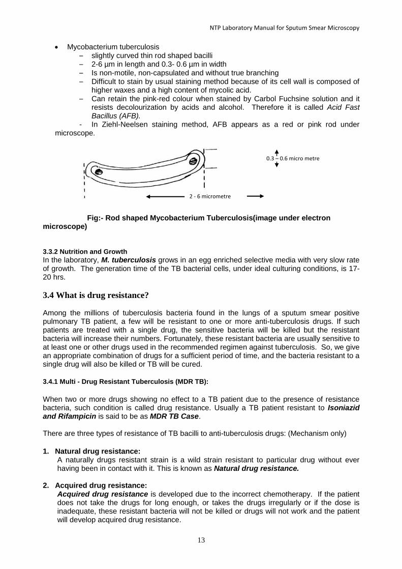
4.1 ROLE OF LABORATORY PERSONNEL ............................................................................ 27 4.2 SAFETY PRECAUTIONS ................................................................................................... 28 4.3 STERILISATION AND DISINFECTION ............................................................................... 28 4.4 USE OF DISINFECTANTS.................................................................................................. 29 4.5 LABORATORY MANAGEMENT ........................................................................................ 32 4.6 PREPARATION OF REAGENTS ........................................................................................ 33 4.7 MICROSCOPE (BRIGHT FIELD AND FLUORESCENCE) ................................................. 34 4.7.1 INTRODUCTION .................................................................................................................. 34 4.7.2 BASIC PRINCIPLE OF FLUORESCENCE STAINING .................................................................... 34 4.7.3 PREPARATION OF FLUORESCENCE STAINING SOLUTION (AURAMINE METHOD) ........................ 34 4.7.4 STAINING PROCEDURE: ...................................................................................................... 35 4.8 MAINTENANCE OF MICROSCOPE ................................................................................... 38 4.9 MICROSCOPE CLEANING ................................................................................................ 39
5. PROCEDURE FOR SPUTUM SMEAR EXAMINATION .................................................................. 41
5.1 SPUTUM COLLECTION, STORAGE AND TRANSPORTATION........................................ 42 5.2 INSTRUCTIONS TO THE PATIENT FOR SPUTUM COLLECTION .................................... 42 5.3 SPUTUM TRANSPORTATION ........................................................................................... 43 5.4 NUMBERING FOR EXAMINATION SLIDE ......................................................................... 44 5.5 INSTRUCTION FOR SLIDE NUMBERING: .................................................................................... 45 5.6 MAIN CAUSES OF FALSE POSITIVE/FALSE NEGATIVE SPUTUM SMEAR RESULTS .. 51 5.6.1 FALSE POSITIVE RESULT: ................................................................................................... 51 5.6.2 FALSE NEGATIVE RESULT: .................................................................................................. 51
6. RECORDING AND REPORTING ..................................................................................................... 52
6.1 SPUTUM SAMPLE EXAMINATION REQUEST AND REPORT HMIS 6.1 ........................................... 52 6.2 LABORATORY REGISTRATION BOOK HMIS 6.2 OR NTP LAB REGISTER): ................................. 52 6.3 TB TREATMENT CARD (HMIS 6.3) ......................................................................................... 52 6.4 PATIENT’S CARD (HMIS 6.4) ................................................................................................. 52 6.5 TB TREATMENT REGISTER (HMIS 6.5) .................................................................................. 52
7. QUALITY ASSURANCE FOR SPUTUM SMEAR MICROSCOPY ................................................. 57
7.1 INTRODUCTION ..................................................................................................................... 57 7.2 A QUALITY ASSURANCE PROGRAM HAS THREE MAIN COMPONENTS: ........................................ 57 7.3 INTERNAL QUALITY CONTROL ....................................................................................... 58 7.4 LABORATORY NETWORK: .............................................................................................. 58 7.4.1 PERIPHERAL ...................................................................................................................... 58 7.4.2 INTERMEDIATE ................................................................................................................... 58 7.4.3 NATIONAL REFERENCE OR CENTRAL ................................................................................... 58 7.5 METHOD OF EXTERNAL QUALITY ASSESSMENT (EQA) ......................................................... 60 7.6 SAMPLE SIZE DETERMINATION WITH EXAMPLES ........................................................ 63 7.7 ASSESSMENT POINTS OF STAINED SMEAR SLIDES .................................................... 66
ANNEX ................................................................................................................................................... 83

NTP Laboratory Manual for Sputum Smear Microscopy
1
Chapter I
1. INTRODUCTION
Tuberculosis is one of the most prevalent infectious disease and significant public health problem in Nepal and continues to pose serious threat to the health of the population and development of the country. Currently nearly 80,000 people have tuberculosis in Nepal, with more than 44,000 new cases arising every year. About half of these are infectious (sputum smear-positive) cases which continue the chain of transmission. The majority of TB patients belong to the economically active age groups of 15–45 years. Bacteriological examination of sputum is the reliable method for diagnosing infectious TB in Nepal. Moreover, people with sputum smear positive TB is much more infectious than people with sputum smear negative TB. Sputum microscopy is not only reliable; it is also very simple and cost effective. Sputum microscopy is therefore the highest priority in the National Tuberculosis Programme (NTP) for the diagnosis of TB. Besides microscopy, Culture and Molecular techniques are also available as diagnostic tools. This manual is prepared based on the guidelines of National Tuberculosis Programme. It should be followed by all government, nongovernment and any other organizations involved in TB control activities within the country.
1.1 Purpose of this manual This manual is prepared for the laboratory workers in Nepal who are involved in sputum smear examination.
It will help you to understand your role in the NTP.
It will show you how to process the sputum smear examination for TB.
It will show you how to use or maintain the NTP recording and reporting system.
It will inform you about quality control for the sputum smear examination.
It will help you how to prevent from laboratory infection and proper disposal system.
It will teach you about occupational safety.

NTP Laboratory Manual for Sputum Smear Microscopy
2
Chapter II THE NATIONAL TUBERCULOSIS PROGRAMME
2. Introduction of National Tuberculosis Program (NTP):
The National Tuberculosis Programme (NTP) is an approach within the national health system to prevention, control and management of TB. It has policies, plans and activities to achieve good case finding and treatment of tuberculosis patients. The NTP is covering countrywide, continuous, permanent and integrated with the general health services. It is relevant to the needs of the population. The NTP is a joint effort of the government and the community aimed at reducing, and in the long term eliminating, suffering due to TB.
2.1 Evolution of TB Strategy
2.1.1 The DOTS strategy (1995-2005)
a. Government commitment b. Case detection through passive case finding c. Standardised chemotherapy to all sputum smear positive TB cases of under proper
case management conditions d. Establishment of a system of regular supply of anti-TB drugs e. Establishment of a monitoring system, for programme supervision and evaluation.
2.1.2 Stop TB Strategy (2006-2015)
a. Pursue high quality DOTS expansion and enhancement b. Address TB/HIV,MDR-TB and other challenges c. Contributing to health system strengthening d. Engage all care provider e. Empower people with TB and communities f. Enable and promote research
2.1.3 The End TB Strategy (2016-2035)
In May 2014, the world health Assembly in its resolution WHA 67.1 adopted the global strategy and
targets for tuberculosis prevention, care and control after 2015 based on a bold vision of a world without
tuberculosis and targets of ending the global tuberculosis epidemic, elimination of associated catastrophic
cost for tuberculosis affected households. The three pillars of the strategy include-integrated, patient
centred care and prevention, bold policies and supportive systems; and intensified research and
innovation. The strategy is based on principles of government stewardship and accountability; with
monitoring and evaluation; strong coalition with civil society organizations and community; protection
and promotion of human rights, ethics and equity; and adaptation of the strategy and targets at the country
level, with global collaboration.

NTP Laboratory Manual for Sputum Smear Microscopy
3
Vision:
A world free of tuberculosis-zero deaths, disease and
suffering due to tuberculosis
Goal: End the global tuberculosis epidemic
INDICATORS MILESTONES TARGETS
2020 2015 SDG 2030
End TB
2035
Reduction in number
of TB deaths
compared with 2015
35% 75% 90% 95%
Reduction in TB
incidence rate
compared with 2015
(%)
20%
(<85/100000)
50%
(<55/100000)
80%
(<20/100000)
90%
(<10/100000)
TB-affected families
facing catastrophic
costs due to TB
0% 0% 0% 0%
Principles: 1. Government Stewardship and accountability, with monitoring and evaluation 2. Strong coalition with civil society organizations and communities 3. Protection and promotion of human rights , ethics and equity 4. Adaptation of the strategy and targets at country level with global collaboration
Pillars and components:
2.1.3.1 Integrated, patients centred TB care and prevention
A. Early diagnosis of TB including universal drug-susceptibility testing, and systematic screening of contacts and high-risk groups
B. Treatment of all people with TB including drug-resistant TB, and patient support C. Collaborative TB/HIV activities and management of comorbidities D. Preventive treatment of persons at high risk, and vaccination against TB

NTP Laboratory Manual for Sputum Smear Microscopy
4
2.1.3.2 Bold policies and supportive systems A. Political commitment with adequate resources for TB care and prevention
B. Engagement of communities, civil society organizations, and public and private care Providers C. Universal health coverage policy and regulatory frameworks for case notification, vital Registration, quality and rational use of medicines, and infection control D. Social protection, poverty alleviation and actions on other determinants of TB
2.1.3.3 Intensified research and innovation
A. Discovery, development and rapid uptake of new tools, interventions and strategies B. Research to optimize implementation and impact, and promote innovations
2.2 Burden of Tuberculosis in Nepal Tuberculosis is one of the most prevalent infectious disease and significant public health problem in Nepal and continues to pose serious threat to the health of the population and development of the country. Currently nearly 80,000 people have tuberculosis in Nepal, with more than 44,000 new cases arising every year. About half of these are infectious (sputum smear-positive) cases which continue the chain of transmission. Over 225,000 people will develop tuberculosis during next five years which is equivalent to inhabitants of a densely populated hill district of the country. The majority of TB patients belong to the economically active age groups of 15–45 years. Without appropriate TB treatment, nearly 94,992 people would die in Nepal over the next five years. Given that National TB Programme remains well functioning the number of deaths in the next five year period will be reduced by 75% to 24,770, with a saving of around 70,222 lives.
2.3 History of Tuberculosis Control in Nepal
1951
Tuberculosis Control Programme (TBCP) was launched by Government of Nepal.
1953
Tokha Sanatorium and Central Chest Clinic (CCC) came into existence offering
diagnosis and treatment services.
1955
Nepal Anti-TB Association (NATA) established
1965
NATA outpatient clinic became operational
1970 TBCP was reorganized with tripartite agreement between Government of Nepal,
WHO and UNICEF. TBCP provided nationwide TB control services in selected
districts. NATA Chest Hospital came in to operation
1989
National Tuberculosis Centre Thimi, Bhaktapur at the central level and Regional
Tuberculosis Centre (RTC) Pokhara were established with cooperation of Japan
International Cooperation Agency (JICA).
1993
Till this date unsupervised Short Course Chemotherapy (SCC) was provided in
selected districts with the support of INGOs and bilateral partners. Unfortunately
this resulted in high defaulter rate and resistant TB cases.
1994
Joint review by Government of Nepal, WHO and other International and National
partners recommended DOTS strategy for TB control in the country.

NTP Laboratory Manual for Sputum Smear Microscopy
5
1995
DOTS strategy was adopted by MOHP/NTP
1996 DOTS strategy based programme started in four pilot districts covering 1.7% of the
population.
2001
Nationwide DOTS coverage achieved. 315 Centres and 1,050 Sub-centres in all 75
districts of the country provided DOTS based services.
2005
MDR TB Management Programme started
2006
MOHP/NTP adopted new STOP TB Strategy
2007
PAL initiative launched in two pilot districts
2007
Fixed Dose Combination Adopted
2008
International Standards of TB Care (ISTC) endorsed and adopted by Nepal Medical
Association and Professional Societies
2008
DOTS programme services were expanded through 4,323 sites including 1,088
Treatment Centres, 3,147 Sub-Centres and 88 Urban DOTS centres covering all
health institutions in the country.
2009 TB HIV Co-infection Programme and Expansion
2012
Isoniazid Preventive Therapy (IPT) started.
2012
Gene-Xpert Technology started.
2013
Universal Coverage on TB treatment.
2014 HIV screening for all new TB cases
2015 Convert all Sub-Treatment centre to Treatment Centre
2016 Implement of Community DOTs
2.4 Introduction to Directly Observed Treatment Short-Course (DOTS) DOTS is a result oriented and effective strategy of NTP. Through DOTS, treatment of TB patients under the direct supervision of trained health personnel is ensured. The DOTS strategy is based on the five essential components. They are; 2.4.1 Political Commitment
Political Commitment denotes the commitment of all GoN, GOs/ NGOs, people and
community.

NTP Laboratory Manual for Sputum Smear Microscopy
6
2.4.2 Case detection through Quality Assured Bacteriology
Quality Assured Bacteriology refers to case detection through Gene-Xpert and culture technique. Laboratory service is the back-bone of NTP. Diagnosis and treatment follow-up of tuberculosis is done by sputum examination which should be easily available, simple, qualitative and sustainable.
2.4.3 Standardized treatment with supervision and patient support
The main aim of NTP is to provide high quality treatment services to patient. It applies Fixed Dose Combination (FDC) Drug Regimen as recommended by WHO.
2.4.4 An Effective drug supply and management system
Basic element of NTP is to provide regular and sustainable quality drugs supply.
2.4.5 Monitoring and Evaluation System, and Impact Measurement
There should be a regular monitoring, evaluation and Impact measurement system to make NTP sustainable and effective.
2.5 Microscopy Network: The microscopy network consists of multipurpose laboratories at hospitals and primary health centres (PHCs) throughout the country. They carry out microscopic examination of sputum smears stained by the Ziehl-Neelsen method. At the central level, NTC (National Tuberculosis Centre) collaborates with NPHL (National Public Health Laboratory) and NHTC (National Health Training Centre) for implementing Microscopy service in NTP. The Reference Laboratory for Tuberculosis at the NTC develops laboratory policies for the NTP. It also prepares training programme for Laboratory personals provides culture and drug susceptibility testing services, and carries out quality control of Regional Tuberculosis Quality Control Centres (RTQCC).
From the central level, the TB control programme is conducted through the RHD (Regional Health Directorate) and the DHO (District Health Office) to the Microscopy Centres and Treatment Centres for case finding, monitoring of treatment and Quality Control (QC) of sputum smear microscopy examination. The Regional Tuberculosis Quality Control Centres (RTQCC) provides training, supervision, logistic support and quality control for peripheral microscopy centres. The Microscopy Centres and Treatment Centres are linked each other by the process of the sputum smear examinations. Sputum smear microscopy remains the key tool for diagnosis of infectious tuberculosis. National Tuberculosis Control Programme operates a network of laboratories with permanent External Quality Assessment System. Central Laboratory at National Tuberculosis Centre is responsible for planning, training, monitoring, supervision and evaluation of the laboratory network in the country. Central Laboratory also provides quality control services for the Central Region.

NTP Laboratory Manual for Sputum Smear Microscopy
7
Five Regional Health Directorates are supposed to do EQA. Regional quality control centres are
now operating independently under NTC and Regional Health Directorates, these includes QC
Labs in the Western and Mid-Western Regions.
The microscopy network has two main functions in the NTP:
Centre
National TB Reference Laboratory
Planning, Monitoring, Supervision, Training, Logistics, EQA, Smear
Microscopy, Culture & Drug Susceptibility Test
Regional TB Quality Control Centre
Region FWDR MWDR WDR CDR EDR
Regional level planning, implementation, training, supervision, QC, logistic
and supply
DPHO / DHO
Smear Microscopy
District
Microscopy Centres
Smear microscopy
EQA for Smear Microscopy
National TB Center
Region
FWDR MWDR WDR CDR EDR
GoN GoN RTC NTC GoN
District
DPHO / DHO
Microscopy Centres
1. Diagnosis of new TB
2. Monitoring of the TB treatment

NTP Laboratory Manual for Sputum Smear Microscopy
8
2.5.1 Role of laboratory personals in microscopy center:
In Nepal, most of the bacteriological diagnosis of tuberculosis is carried out in peripheral or local laboratories whose major responsibility is to provide diagnostic microscopy for the NTP based on sputum smear examination by Ziehl-Neelsen (ZN) staining. So laboratory personals should be well cable of performing sputum smear microscopy. The responsibilities of the laboratory personals in microscopy centre are:
Collecting adequate number and good quality sputum specimens.
Preparing sputum smear according to Standard Operating Procedures (SOPs).
Performing microscopy examination promptly and accurately.
Recording and reporting correctly with all patient’s details and results in the laboratory register book.
Promptly sending microscopy results to the treatment centre.
Complete filling of all the laboratory forms needed for the NTP program evaluation.
Keeping all the examined slides serially and prepare them according to Lot Quality Assurance Sampling (LQAS) System for external quality assessment (EQA).
2.5.2 Who are the suspects for sputum smear examination?
Anyone who has had a cough for more than 2 weeks, fever, unexplained weight loss (more than 1.5 kg in one month) should have a sputum examination. Extra-pulmonary tuberculosis suspects should also have a sputum examination, as they may also have tuberculosis in their lungs. Two examinations from different sputum specimens of one TB suspects are recommended, because multiple sputum examination has higher sensitivity yield. If we only collect 1 specimen, we will miss some smear positive cases. A series of two examinations can detect almost all sputum positive cases.
Collect 2 Sputum Samples as follows:
1. Supervised spot sputum specimen at the first visit
Early morning sputum
specimen in the next day
First Spot Morning
2.5.3 Why is sputum examination important?
The sputum examination is much more reliable diagnostic tool than an X-ray. Because tuberculosis is an infectious disease caused by a type of bacteria, to identify bacteria is only the best method of diagnosing TB. It is also simple, cost effective and available at many medical facilities.

NTP Laboratory Manual for Sputum Smear Microscopy
9
People with sputum smear positive pulmonary TB are 10 times more infectious than people with sputum smear negative TB. People with sputum smear positive TB have a higher mortality rate without treatment than people with sputum negative TB, so to find out sputum smear positive pulmonary TB is the main strategy of NTP. 2.5.4 Flow chart 1: Diagnosis of Pulmonary TB
Tuberculosis case findings There are two ways of case findings:
1. Passive case finding: It means diagnosis of TB in Patients attending health facilities by themselves.
2. Active case finding: It means that health worker themselves seek out tuberculosis suspect eg. Collecting sputum from chest symptomatic in the community.
2.5.5 What is a diagnosis sputum examination?
The sputum specimens are examined to identify smear positive pulmonary TB before starting treatment are known as diagnosis sputum examinations. Sputum examination is the best way of diagnosing pulmonary TB.
Register as P(+) treat
for TB
Repeat the the
tests after 2
weeks.
tests after 2
weeks.
Cough for more
than 2 weeks
Give symptomatic
treatment and do
examination of
2 sputum smears
If smear is negative then
1 course antibiotic better
to advice.
1 or more positive
smears
At least one slide
positive.
If negative result
Refer to Physician

NTP Laboratory Manual for Sputum Smear Microscopy
10
If smear examination failed to detect, then it should be performed by Gene Xpert. We should examine the sputum of anyone who has a cough for more than 2 weeks. We must examine the sputum of those suspected to have extra-pulmonary tuberculosis, as they may also have tuberculosis in their lungs. We collect 2 specimens of sputum from a chest symptomatic suspect. If we collect 1 specimen only, we will miss some smear positive cases. 2.5.6 What is a follow up sputum examination?
Follow up sputum specimens are examined are during the treatment for checking the effectiveness of the treatment. The patient must have the sputum examinations after 2(3*), 5 and last months of treatment.
T r e a t m e n t
Treatment duration No. of sputum examination Type of sputum sample
0 month (Diagnosis)
2
-spot -morning
2 (3*) months
(follow-up)
1
-spot
5 months (follow-up)
1
-spot
Last month (follow-up)
1
-spot
Diagnosis examination
Follow-up examination
(3*; Category 2 regimens patient) (For follow up examination, early morning sample is best preferable)

NTP Laboratory Manual for Sputum Smear Microscopy
11
Chapter III GENERAL INFORMATION ON TUBERCULOSIS
3. Tuberculosis (TB): Tuberculosis (TB) is an air borne infectious disease caused by Mycobacterium tuberculosis. Mycobacterium tuberculosis is also called acid fast bacilli (AFB). Tuberculosis is most commonly transmitted by inhalation of infected droplet nuclei which are discharged in the air when a sputum smear positive TB patient coughs or sneezes. Only about 5%–10% of infected persons (primary infection) develop active tuberculosis disease. Among the remaining 90% to 95 % of infected persons, initial infection usually goes without further consequences. Transmission of TB infection occurs almost exclusively through the respiratory route. The infection may then spread from the primary lung lesion to any part of the body via the blood stream, lymphatic and bronchial systems. Pulmonary TB, sputum smear is positive, is highly infectious and should receive topmost priority for treatment. Sputum smear-negative cases are much less infectious than those who are smear-positive. Extra-pulmonary TB can affect the lymph nodes, pleura, bones and joints, the genito-urinary tract, the nervous system (meningitis), intestines, etc. If untreated, TB leads to death within 5 years in at least half the patients. About 20 to 25% will naturally heal and 25 to 30% remain positive and continue to spread the disease in the community.
3.1Transmission of Tuberculosis
The bacteria that cause tuberculosis spread from the lungs of people with TB when they cough sneezes of spit. When a person inhales these bacteria it invades the lung. Macrophages surround and take up the bacteria. Immune cells try to kill the bacteria and cause an area of local inflammation in the lung , This is called a ‘primary focus’, The bacteria may also spread to hailer, lymph, glands, causing enlargement . The combination of a primary focus and the affected lymph nodes is called a ‘primary complex’. Not every, who is infected with the bacteria gets TB. Only 10% of people who are infected get TB disease. If the infected person has good immunity disease may not occur, however if the immunity is weak (e.g. in malnutrition or people with HIV infection) then TB disease can develop soon after infection.
3.2 Common site of tuberculosis in the body: The most common site of tuberculosis infection in the body is lungs and secondly in lymph node. In addition to that TB can infect any part of body except nail and hair (Ref.- John Crofton). Some common sites for tuberculosis are shown in the diagram below:

NTP Laboratory Manual for Sputum Smear Microscopy
12
3.3 What is Tubercle Bacillus? Tubercle bacillus in sputum stained by Ziehl-Neelsen method at the magnification of x1000 is shown on page no. 98 which is also known as Acid Fast Bacilli. Mycobacterium tuberculosis was discovered by German Scientist Robert KOCH on 24th March 1882. 3.3.1 Physical and chemical characteristics
Mycobacterium tuberculosis is generally a slightly curved, thin rod, measuring 2-6 micro metre (µm) in length and 0.3-0.6 micro metre (µm) in thickness (1 µm = 1/1000 mm). This organism is non-motile, non-capsulated and having no true branching. It is difficult to stain this organism by usual staining method because the cell wall of M. tuberculosis is composed of higher waxes and a high content of mycolic acid. It can retain the pink-red colour when stained by Carbol Fuchsine solution, and it resists decolourization by acids and alcohol. Therefore it is called Acid Fast Bacillus (AFB).In a sputum smear stained by Ziehl-Neelsen method, AFB appears as a red or pink rod under microscope.
Nervous system
Lymph node
Lungs
Kidney and urinary tract
Pleura
Bone or joint
Abdomen
NTP Laboratory Manual for Sputum Smear Microscopy
13
Mycobacterium tuberculosis – slightly curved thin rod shaped bacilli – 2-6 µm in length and 0.3- 0.6 µm in width – Is non-motile, non-capsulated and without true branching – Difficult to stain by usual staining method because of its cell wall is composed of
higher waxes and a high content of mycolic acid. – Can retain the pink-red colour when stained by Carbol Fuchsine solution and it
resists decolourization by acids and alcohol. Therefore it is called Acid Fast Bacillus (AFB).
- In Ziehl-Neelsen staining method, AFB appears as a red or pink rod under microscope.
Fig:- Rod shaped Mycobacterium Tuberculosis(image under electron microscope) 3.3.2 Nutrition and Growth
In the laboratory, M. tuberculosis grows in an egg enriched selective media with very slow rate of growth. The generation time of the TB bacterial cells, under ideal culturing conditions, is 17-20 hrs.
3.4 What is drug resistance? Among the millions of tuberculosis bacteria found in the lungs of a sputum smear positive pulmonary TB patient, a few will be resistant to one or more anti-tuberculosis drugs. If such patients are treated with a single drug, the sensitive bacteria will be killed but the resistant bacteria will increase their numbers. Fortunately, these resistant bacteria are usually sensitive to at least one or other drugs used in the recommended regimen against tuberculosis. So, we give an appropriate combination of drugs for a sufficient period of time, and the bacteria resistant to a single drug will also be killed or TB will be cured.
3.4.1 Multi - Drug Resistant Tuberculosis (MDR TB):
When two or more drugs showing no effect to a TB patient due to the presence of resistance bacteria, such condition is called drug resistance. Usually a TB patient resistant to Isoniazid and Rifampicin is said to be as MDR TB Case. There are three types of resistance of TB bacilli to anti-tuberculosis drugs: (Mechanism only)
1. Natural drug resistance:
A naturally drugs resistant strain is a wild strain resistant to particular drug without ever having been in contact with it. This is known as Natural drug resistance.
2. Acquired drug resistance:
Acquired drug resistance is developed due to the incorrect chemotherapy. If the patient does not take the drugs for long enough, or takes the drugs irregularly or if the dose is inadequate, these resistant bacteria will not be killed or drugs will not work and the patient will develop acquired drug resistance.
2 - 6 micrometre
0.3 – 0.6 micro metre

NTP Laboratory Manual for Sputum Smear Microscopy
14
3. Primary drug resistance:
If a patient with acquired resistance infects another person, that person will develop primary drug resistance, even though he has not taken anti-tuberculosis drugs before.
3.4.2 Extensively drug-resistant TB (XDR-TB):
It is defined as MDR-TB that is resistant as well as to any one of the fluoroquinolones (ciprofloxacin, ofloxacin, levofloxacin etc) and to at least one of three injectable second-line drugs aminoglycosides (Kanamycin, Capreomycin or Amikacin)
3.5 How does acquired drug resistance occur? Drug resistance could develop due to the following reasons:
Inadequate treatment
Inappropriate treatment regiment
Inadequately formulated drugs
TB treatment not following DOTS
Defaults drug taking
Irregular drug taking
3.6 What are culture and drug susceptibility test (DST)? Sputum culture examination is a bacteriological method of detecting live TB Bacilli. Culture is the process of growing the numbers of organism in artificial media. It can be done by using different types of culture media. Most common culture media is Lowenstein Jensen (LJ), which is egg based solid enriched media. Another media is liquid media (eg. Middle brook). Culture is the gold standard method in tuberculosis diagnosis. Result turn out time is longer in solid media (8weeks) than liquid media (4weeks). Colony characters are observed in solid media and other bio-chemical tests are used in liquid media. TB bacilli grow very slowly in an egg enriched selective media (Ogawa, Lowenstein-Jensen media). It takes four to eight weeks before the colonies of bacteria can be seen with the naked eyes. The drug susceptibility test is done to know the characteristics of TB bacilli whether they are susceptible or resistant to anti TB drugs. Drug susceptibility test is important not only to choose the most effective drug regimen for the treatment of individual patient but also for the epidemiological purpose to assess the efficiency of the treatment service in NTP. The laboratories performing Mycobacterial culture and drug susceptibility tests in Nepal are as follows;
The National Tuberculosis Centre, National Tuberculosis Reference Laboratory, Thimi, Bhaktapur
German Nepal TB Project (GENETUP), National Tuberculosis Reference Laboratory, Kalimati, Kathmandu
3.7 Laboratory diagnosis of TB Laboratory diagnosis can classify into two types.
1. Conventional diagnostic techniques 2. New diagnostic techniques
3.7.1 Conventional diagnostic techniques
Conventional diagnostic techniques consists microscopy examination and solid culture.
3.7.1.1 Microscopy Examination:- It detects the character of Acid fastness of bacilli. Ziehl Neelsen technique is the widely used technique for microscopy.

NTP Laboratory Manual for Sputum Smear Microscopy
15
3.7.1.2 Culture Technique:- This method detects the viable TB bacilli in culture media. It is a process of growing the numbers of microorganism in artificial media. There are different types of solid culture media. Lowenstein and Jensen (L-J) media is the most commonly used solid culture for growing TB bacilli. It is a confirmative diagnostic technique but slow technique. Usually TB bacilli grow in three to four weeks. 3.7.2 New diagnostic techniques:
LED Fluorescence Microscopy
Liquid culture (Bactec MGIT-960)
Molecular techniques (PCR) :- Gene Xpert, MTBDRplus Line Probe Assay (LPA), Loop Mediated Isothermal Amplification (LAMP)
Lipoarabinomannan Glycoprotein (LAM) Antigen in urine
IGRA:- Interferon Gama Release Assay, it detects latent TB in adults and children.
3.7.2.1 LED Fluorescence Microscopy: A fluorescence microscope is an optical microscope used to study properties of organic or inorganic substances using the phenomena of fluorescence and phosphorescence instead of, or in addition to, reflection and absorption.
3.7.2.2 Liquid Culture MGIT-960:- The BACTEC MGIT 960 System is an in vitro diagnostic instrument for rapid detection of Mycobacteria in clinical specimens other than blood. This system is simple, efficient, safe to use and occupies small laboratory space. The MGIT 7ml tube contains modified middle brook 7H9 broth. Culture tubes contain a fluorescent sensor at the bottom which responds to the concentration of oxygen Initial concentration of dissolved oxygen quenches the emission from the compound, and little fluorescence can be detected. Actively respiring microorganisms consume the oxygen which allows the compound to fluorescence and detect as a positive. It is completely automatic system. In liquid culture, TB bacilli usually grow in 7-8 days and DST takes about one more week.
3.7.2.3 Gene-Xpert MTB/RIF:
Gene–Xpert MTB/RIF is novel technology for the diagnosis of tuberculosis. It is a real time multiplex PCR technology with integrated and closed fluid transfer system. It is a single hands free step technology.

NTP Laboratory Manual for Sputum Smear Microscopy
16
It detects mycobacterium tuberculosis complex and its resistance to rifampicin by PCR amplification of the rpoB gene. Rifampicin is the most effective and powerful first line anti-tuberculosis drug. Most of the rifampicin resistant patients are also resistant to isoniazid but isoniazid resistant patient may not resistance to rifampicin. The sensitivity and specificity is much higher than sputum microscopy. It gives result within two hours, so it had also called as Rapid Diagnostic Test (RDT). It is Point of Care Test (POC).
Sample reagent Cartridge Computer Gene-Xpert machine 3.7.2.4 Molecular Line Probe Assay (LPA): LPA is a multiplex PCR with reverse hybridization technique. It detects genetic identification of the M. tuberculosis complex (MTBC) and its resistance to rifampicin, isoniazid, quinolone and second line injectable drugs from cultivated samples and from direct patient sample. Genotype MTBDRplus and MTBDRsl are the two commonly used LPA techniques for diagnosis of drug resistant tuberculosis. The Genotype MTBDRplus line probe assay was designed for the rapid detection of resistance to rifampicin and isoniazide by detecting mutation in rpoB, katG and inhA gene and MTBDRsl detect resistance to quinolone (ciprofloxacin, ofloxacin, moxifloxacin) dy detecting mutation in quinolone associated gene gyrA and second line injectable drugs (kanamycin, capreomycin, amikacine) by detecting mutation in rrs gene. MDR-TB and XDR-TB can be diagnosed within 1 or 2 days by this method from direct sputum sample.
DNA amplification Hybridyzation Result evaluation
3.7.2.5 Loop mediated isothermal amplification (LAMP): LAMP is a single tube technique for the amplification of DNA. It is an isothermal nucleic acid amplification technique. In LAMP PCR, isothermal amplification is carried out at a constant temperature, and does not require a thermal cycler. In LAMP, the target sequence is amplified at a constant temperature of 60 - 65 °C using either two or three sets of primers and a polymerase with high strand displacement activity in addition to a replication activity. Typically, 4 different primers are used to identify 6 distinct regions on the target gene, which adds highly to the specificity. An additional pair of "loop primers" can further accelerate the reaction. Amplification of DNA takes one hour.

NTP Laboratory Manual for Sputum Smear Microscopy
17
3.7.2.6 Lipoarabinomannan (LAM):
LAM test is based on the detection of mycobacterial lipoarabinomannan (LAM) antigen in urine have emerged as potential point-of-care tests for tuberculosis (TB). LAM antigen is a lipopolysaccharide present in mycobacterial cell walls, which is released from metabolically active or degenerating bacterial cells and appears to be present only in people with active TB disease. Urine-based testing would have advantages over sputum-based testing because urine is easy to collect and store, and lacks the infection control risks associated with sputum collection.
3.7.2.7 Interferon-gamma assay (IGRA):
Interferon-gamma release assays (IGRAs) is an important advance in the diagnosis of latent
tuberculosis infection (LTBI). IGRA is in vitro blood tests of cell-mediated immune response;
they measure T cell release of interferon (IFN)-gamma following stimulation by antigens unique
to Mycobacterium tuberculosis.
The goal of testing for latent TB infection is to identify individuals who are at increased risk for
the development of tuberculosis (TB) and therefore who would benefit from treatment of latent
TB infection. Only those who would benefit from treatment should be tested, so a decision to
test presupposes a decision to treat if the test is positive.

NTP Laboratory Manual for Sputum Smear Microscopy
18
3.8 Diagnostic Algorithm for General TB:
Algorithm 1:
Persons of presumptive TB1
Priority patient2 for Xpert MTB/RIF testing Other patient categories
- Collect sputum and test forXpert or refer
1 sputum for Xpert to nearby Xpert Centre.
-Collect 2 sputum samples
-Perform 2 sputum smears
-Review
treatment based
on Xpert
MTB/RIFresult
-Review treatment
based on Xpert
MTB/RIF result
One or both smear
positive
Both smear
negative
-Treat with first line
regimen.
-Review treatment
based on Xpert
MTB/RIF result
-Re-evaluate the
patient clinically.
- Review clinical
decisions based on
Xpert MTB/RIF
result

NTP Laboratory Manual for Sputum Smear Microscopy
19
1Persons being evaluated for TB include all persons with signs or symptoms suggestive of TB or persons with a chest X-ray with abnormalities suggestive with TB. This algorithm may also be used for persons being evaluated for extrapulmonary TB.
2 Priority patients include PLHIV, contacts of RR/MDR-TB, lost to follow-up, relapse, failure, non converters (smear positive at end of the intensive phase of treatment), children, patient live in close (congregate)setting, sample collected through courier system, diabetic, patient live near to Xpert Centre.
Interpretation of Xpert test result:
No result,
Error, or
Invalid test
MTB Detected,
Rifampicin
Indeterminate
MTB Detected,
Rifampicin
Resistance
MTB not
Detected
• Repeat
Xpert
MTB/RIF
• Follow 1 to
interpret
MTB Detected,
Rifampicin
\Sensitive
• Treat with
first line
regimen
• Re-evaluate
the patient
clinically
• Consider
repeat Xpert
MTB/RIF test
• Use clinical
judgment for
treatment
decisions
Re-treatment,
non converter
and contacts of
MDR-TB
If New TB
or MDR-
TB (never
been
treated)
• Perform DST of Second Line
Drugs and Isoniazisd DST
• Treat with second line regimen
• Repeat Xpert
MTB/RIF
• Follow to
interpret with
result
MTB detected,
Rifampicin
Resistance
• Repeat Xpert MTB/RIF
MTB detected,
Rifampicin
Sensitive
MTB not
detected
• Re-evaluate
the patient
clinically
• Consider
repeat Xpert
MTB/RIF
testing
decisions
• Treat with
first line
regimen

NTP Laboratory Manual for Sputum Smear Microscopy
20
3.9 Algorithm 2: Algorithm for testing for second-line drugresistance among rifampicin-resistant (RR) TB or MDR-TB patients.
All patients with RR TB or MDR-TB
Refer a specimen for SL-LPA Version 2.0 test
SL-
LPA:Resistance
to Second Line
Injectable Drugs (SLID) only
SL-LPA:
Resistance to
both FQ and
SLID
SL-
LPA:Resistanc
e notdetected
to both FQ and
SLI
SL-LPA:
Resistance to
Fluroquinolone
(FQ) only
SL-LPA:
Indeterminate
• Initiate XDR
TB Resistance
treatment based
on national
guidelines.
• Initiate Pre-
XDR TB SLID
Resistance
treatment based
on national
guidelines.
• Initiate Pre-
XDR TB FQ
Resistance
treatment based
on national
guidelines.
• Initiate
shorter MDR-
TB treatment
regimenif
patient meets
criteria1
Repeat SL
LPA and
manage
treatment based
on repeated
result.

NTP Laboratory Manual for Sputum Smear Microscopy
21
3.10 How does TB Spread? TB bacilli spread from the lungs of people with TB when they cough, sneeze, speak or spit. Another person inhales the droplets containing TB bacilli through respiration and may become infected with TB. It is actually not spread from food, smoking or drinking alcohol. Neither is it spread by insects and parasites nor by heredity. Not everyone who is infected with TB bacilli gets TB. If the infected person has good immunity, the disease may not develop. However if their immunity is weak (e.g., due to malnutrition, HIV infection) then TB can develop soon after infection.
By cough About 1.5 m
By sneeze About 3 m
Not by food
Fig:-by close contact with tuberculosis patient
3.11 What are the symptoms of pulmonary TB?
Cough for the duration of 2 week or more (the commonest symptom of pulmonary TB)
Fever (Often in the evening or at night)
Shortness of breath
Chest pain
Haemoptysis (blood stain sputum)
Loss of appetite
Weight loss
Night sweats
Fatigue (feeling tired)
Sputum production
3.12 How many types of TB are there? The type of tuberculosis is defined by the site of the disease in the body. 80 % of tuberculosis occurs in the lungs and is called pulmonary TB. The occurrence of TB on the sites other than lungs is called extra-pulmonary tuberculosis.
Type of Tuberculosis on the basis of organs involves:
1. Pulmonary Tuberculosis:
a. Pulmonary bacteriological confirmed (PBC)
b. Pulmonary clinically diagnosed (PCD)

NTP Laboratory Manual for Sputum Smear Microscopy
22
2. Extra pulmonary Tuberculosis
a. Extra Pulmonary bacteriological confirmed
b. Extra Pulmonary clinically diagnosed
3.13 What are the common extra-pulmonary TBs? Gland TB Intestinal TB Bone and Joint TB Urogenital TB TB Meningitis Skin TB Abdominal TB Eye TB etc. Miliary TB Genitourinary TB Laryngeal TB Pleurisy TB
3.14 Registration categories of TB patient After diagnosis, the patient must be registered and should be given the treatment. The registration categories of TB patients designed by the NTP will help the health workers to decide which category of treatment is appropriate to the patient.
3.14.1. New:
A patients who has received no or less than one month of anti-tuberculosis treatment
3.14.2. Previously treated
3.14.2.1 Relapse: A patient whose most recent treatment outcome was “cured” or “treatment completed”, subsequently diagnosed with bacteriologically positive TB by sputum smear microscopy or culture
3.14.2.2 Treatment after failure: A patient who has received treatment for TB and in whom treatment has failed. Failure is defined as sputum smear positive at five or end of treatment.
3.14.2.3 Treatment after lost to follow up: A patient who returns to treatment, bacteriologically positive by sputum smear microscopy or culture, following interruption of treatment for two or more consecutive months.
3.14.2.4 Other previously treated patient: Patients who have completed TB treatment but not evaluated or have no evaluation document available. The TB patient registered in NTP and transferred from any treatment centre to another centre. 3.14.3 Previous Treatment history unknown
Case that does not fulfil the above criteria. This is usually a patient who has been taking anti-tuberculosis drugs for more than 4 weeks but has not been registered within the NTP.

NTP Laboratory Manual for Sputum Smear Microscopy
23
3.15 What are the drug regimens and how do we use them in Nepal ?
Treatment Regimen
TB Treatment Regimen Regi
men Follow up Examination
Category I
- New pulmonary TB cases
(PBC+PCD)
2
HRZ
E + 4
HR
Months 2, 5, and End of Treatment Sputum Smear
Test
If still +ve at 2 months
• go for Rapid DST while continuing the treatment
• if Xpert +veRiF sensitive
• extend intensive phase for 1 more month.
If smear is still +ve at 5 months or end of Treatment
• declare as Treatment Failure
• switch to category II and screen for DR TB
- New EP-TB cases
(BC+CD)
2
HRZ
E + 4
HRE
For EP TB 2 months follow up with sputum smear
regardless of chest symptoms.
If smear +ve
• label as Treatment Failure and manage
accordingly.
At the end of continuation phase, based on the clinical
judgement (Improving) by treating physician, the
continuation phase can be extended up-to 3 more
months.
Category II
- All Pulmonary Retreatment Cases
At-least RIF sensitive
- All (Mild form) EP TB Retreatment
cases
**For exceptional (severe form)
Pulmonary TB cases (New or
Relapse)
- Milliary TB
3
HRZ
E + 6
HRE
Months 3, 5 and End of Treatment Sputum Smear Test
If Smear +ve at 3 months:
• Go for Rapid DST while continuing the treatment
• Extend intensive phase for 1 more month.
If Smear +ve at 5 months
• Repeat Rapid DST while continuing the treatment
If RiF Sensitive
• continue ATT while waiting for report of Culture
DST and manage accordingly.
Culture, FLDST and SLDST
If Smear +ve at end of Treatment
Repeat Rapid DST while continuing the treatment if
RiF Sensitive continue ATT for 3 more months
At the same time send for Culture, FLDST and SLDST
manage based on the result.
**For exceptional (severe form) EP
TB cases (New or Relapse) Milliary
TB CNS, Musculo & Skeletal TB
3
HRZ
E + 6
HRE
At the end of continuation phase, based on the clinical
judgement (Improving) by treating physician, the
continuation phase can be extended up-to 3 more
months.
Use of Streptomycin:
Streptomycin is in the process of being phased out.
Streptomycin can still be used when:
o Other drugs have to be replaced cause of toxicity, especially Ethambutol.
o Can be used in CNS TB based on the judgement by treating physician.
(Continuation phase can go up to additional of 3 more months at 5 months follow up with sputum smear regardless of chest symptoms). *Use of Streptomycin:
ᵜ Streptomycin from previously known category II is now phased out. ᵜ Streptomycin can still be used when: ᵜ Other drugs have to be replaced cause of toxicity, especially Ethambutol. For exceptional complicated EP TB cases
ᵜ CNS TB ᵜ Miliary TB (pulmonary TB) ᵜ Musculo-skeletal TB 3 HRZE + 6 HRE

NTP Laboratory Manual for Sputum Smear Microscopy
24
3.16 INTRODUCTION TO HIV AND AIDS
HIV stands for "Human Immunodeficiency Virus", which is the causative organism of the HIV infection. HIV virus infects the human immune cells called CD4 cells and weakens the immune system of the body leading to a condition called "Acquired Immunodeficiency Syndrome (AIDS)". During the course of infection HIV destroys the CD4 cells making human body prone to many infections which are not common in the absence of HIV. Such infections are termed as opportunistic infections (OI).
3.16.1 Why is tuberculosis so common in people with AIDS?
The ability of a person to restrict and contain tuberculosis infection depends on its cellular immunity. Because HIV weakens cellular immunity, TB bacilli can grow more easily, and tuberculosis disease develops. HIV infection can therefore cause latent tuberculosis infection to progress to tuberculosis disease. The aim of the tuberculosis control programme, in countries where HIV infection occurs, must be to quickly diagnose and cure the largest possible number of newly occurring tuberculosis cases, especially those who are smear-positive. This will reduce the risk of increased transmission of tuberculosis infection to the general population.
3.16.2 Epidemiology of HIV in Nepal The human immunodeficiency virus (HIV) pandemic presents a massive challenge to the control of tuberculosis (TB). According to UNAIDS estimates 33 million people in the world are living with HIV and every year 2.5 million new people get infected with the virus (UNAIDS 2007). In Nepal according to recent estimates about 70,000 people are living with HIV making 0.49% prevalence in the general adult population. Nepal is categorized as a country with the concentrated HIV epidemic with less than one percent in general adult population and over 5% in certain risk groups, mainly Intravenous Drug Users (IDU). The recent prevalence of HIV in IDU is 20.7% in Kathmandu, 3.4% in Pokhara, 8.1% in Eastern Terai districts and 8.0 in Western Terai districts (IBBS 2009).In other groups HIV prevalence is lower than 5% except in the group of male sex workers (a sub group of “Men who have Sex with Men”) prevalence of HIV is 5.2%(IBBS 2009). According to sentential Surveillance of Nepal 2011/12, the prevalence of HIV positive among TB patient is 2.4%. Moreover, the progression of TB among HIV positive individuals, decreased by about 5-15%.
3.16.3 Modes of transmission
HIV is found in all body secretion of the HIV infected person. The amount of HIV is adequate enough for transmission only in blood, semen, vaginal secretion and breast milk. Any contact with HIV infected blood, semen, vaginal secretions and breast milk with broken skin or broken mucous membrane of previously not infected person causes infection. Any way which facilitates such contact is the mode of transmission of HIV. The modes of HIV transmission are:
- Sexual contact without using condom-facilitating the mixing of infected semen with blood (through micro trauma of the vaginal mucosa) or infected vaginal secretion with blood (through micro trauma of penile skin) or infected blood with blood (micro trauma of penile skin and mucous membrane of anal mucosa- in case of anal sexual contact). Among the sexual contact anal sex has got highest risk of HIV transmission as chances of trauma are comparatively higher during this sexual act.
- Needle sharing between HIV infected persons and not infected with HIV- - Transfusion of HIV infected blood or transplant of HIV infected organ - Mother to child transmission during pregnancy, labor and breast feeding: highest risk is
during labor. - During medical procedures if universal precautions are not followed
3.16.4 Window period
The window period represents the stage immediately after becoming infected but before body creates antibodies. In most people, it takes the body 3-4 weeks to make enough antibodies to

NTP Laboratory Manual for Sputum Smear Microscopy
25
be detected by laboratory tests. It may take up to 3 months for laboratory tests to detect HIV antibodies in a person’s body.
If people are tested during the window period, they test negative even though they are infected because the body does not produce enough antibodies to trigger a positive test result. The virus can pass from one person to another during the window period.
3.16.5 Diagnosing HIV
There are two ways of diagnosing HIV – by identification of the viral particles in the body of infected person or by identification of the antibodies developed by the body against the viral particles. The first method is called virological method and is not commonly available in Nepal. DNA PCR is the nucleic acid based qualitative test for identification of viral particles. The diagnosis of HIV has traditionally been based on the detection of antibodies against HIV. Wide ranges of different HIV antibody tests are available including ELISA (Enzyme linked Immunosorbent Assays), rapid HIV tests and Western Blot. Rapid HIV tests are widely used in Nepal and are supplied through National HIV logistic system of the country. More than one rapid test kits should be used in order to come with a definitive diagnosis. This order is known as testing algorithm or testing strategy. Serial testing strategy for HIV testing is used in Nepal. Three test kits are used in order following this strategy. Using the rapid test kits, the result of HIV test is provided within same day. A system of quality assurance of the rapid test is necessary for using rapid tests. 3.16.6 Care of HIV positive
As soon as the person is identified positive for HIV, he should be enrolled into the system of Continuum of Care for HIV positive. The first task of the HIV clinician is to do the clinical staging or CD4 testing. Depending on the result of the clinical staging or CD4 test the person is entitled for Cotrimoxazole prophylaxis, or Anti-retroviral therapy (ART). All HIV positive clients are investigated for Tuberculosis immediately after diagnosis and Isoniazid preventive therapy (IPT) is started if indicated.
3.16.7 Antiretroviral Therapy
Anti-retroviral drugs are the drugs used against HIV. These drugs inhibit the replication of HIV. When antiretroviral drugs are given in combination, HIV replication and immune deterioration can be delayed, and survival and quality of life improved. There are five classes of antiretroviral drugs available in use: Fusion inhibitors, Nucleoside reverse transcriptase inhibitors, Nucleotide reverse transcriptase inhibitors, Non-nucleoside reverse transcriptase inhibitor and protease inhibitors. Fusion inhibitors are not currently available in Nepal. The drugs available in Nepal for the first line regimen through Government supply are- Zidovudine (AZT)/ Stavudine (d4T) ; Lamivudine (3TC), Nevirapine (NVP) /Efavirenz(EFV). At least three ARV should be used in combination for effective treatment. Among these ARV, Nevirapine is hepato-toxic and should be used with caution in patient taking Rifampicin. Reduction of HIV related morbidity and mortality, maximal and durable suppression of viral load, restoration and/or preservation of immunologic function and Improvement of quality of life are the goals of Anti-Retroviral Therapy (ART)
3.16.8 Monitoring Antiretroviral Therapy
CD4 and Viral loads test are done for monitoring ART. CD4 cells are the cells of the immune system, which are targeted by HIV in the body. Number of CD4 decrease as HIV progresses in the body. Normal range of CD4 in human body ranges from 500 to 1500/microliter. If the CD4 count decreases even with ART, this is considered as immunological failure of antiretroviral therapy.
The viral load test is a quantitative measurement of HIV nucleic acid (RNA) that provides important information that is used in conjunction with the CD4 cell count:

NTP Laboratory Manual for Sputum Smear Microscopy
26
to monitor the status of HIV disease, to guide recommendations for therapy, and to predict the future course of HIV.
Routine blood test for Hb and liver function are indicated in regular interval for all taking ART. 3.16.9 TB-HIV Co-infection in Nepal
National TB program has conducted periodic sentinel site HIV prevalence among TB patient since 1993/94. According to these surveys the prevalence of HIV among TB patients is rising. Table 1 : Results of HIV prevalence among TB patients 1993/94 – 2006/07
Year of sentinel survey % of HIV among TB patients
1993/94 0.00
1995/96 0.60
1998/99 1.88
1999/2000 1.39
2001/2002 2.44
2006/2007 2.40
2011/2012 2.40
Source: NTP report 2012
3.16.10 Recommended collaborative TB/HIV activities
Collaborative activities should address the interface of the Tuberculosis and the HIV/AIDS epidemics for which joint programs should be carried out as part of health sector responding to the intersecting Tuberculosis and HIV epidemics.

NTP Laboratory Manual for Sputum Smear Microscopy
27
Chapter IV LABORATORY PREPARATION FOR SPUTUM SMEAR EXAMINATION 4. LABORATORY PREPARATION FOR SPUTUM SMEAR EXAMINATION
4.1 ROLE OF LABORATORY PERSONNEL
NTP Laboratory personnel should persuade the following tasks to keep well - managed
laboratory;
Safety
Keep the laboratory neat and clean.
Forbid smoking, eating and drinking in the laboratory.
Destroy examined sputum and contaminated materials appropriately.
Regularly disinfect working bench, towels and laboratory gowns.
Proper hand washing.
Facility
Keep the laboratory neat and clean.
Keep the microscope in good condition.
Order equipment, reagent and other materials on time.
Sputum collection
Explain the reason for the sputum examination to the patients.
Explain the importance of follow-up sputum examination to the patients.
Give instructions to patients for quality sputum collection.
Label the patient's serial number on the side of sputum container, not on the cap.
Check the quality of the sputum.
Examination
Register the patient's vital information.
Make the smear and fix it.
Stain the smear by Ziehl-Neelsen method.
Examine the smear by microscopy.
Recording & Reporting
Maintain the NTP laboratory register daily.
Send the results of examination to the treatment centre or give to the patient.
Submit a monthly report to the DHO/DPHO.
Fill up the LQAS 1 form well.
Quality control
Keep all examined slides in serial order for LQAS so that supervisor can select the sample slides easily.
Send the collected slides with LQAS-1 forms to Regional TB Quality Control Centre/Assessor.
Receive and file the LQAS-3 feedback report.
Maintain a high quality of examination.
Planning.
For the health workers of Non-Microscopy Centre The worker should send the sputum smear slides after fixation with the examination request
form to the DHO laboratory or the nearest microscopy centre for sputum smear examination.

NTP Laboratory Manual for Sputum Smear Microscopy
28
4.2 SAFETY PRECAUTIONS Safety Tuberculosis is transmitted through air; therefore laboratory workers must avoid the occurrence of aerosols from sputum samples to keep themselves safe from TB infection. The laboratory worker should keep the following rules:
Keep the laboratory environment as clean as possible and minimise the movement of people.
Wear an apron, gloves and masks in the laboratory.
Wash hands with soap and water after making each smears and before leaving the laboratory.
Sterilise all contaminated materials by burning, boiling or soaking in disinfectant.
Do not eat, drink and smoke in the laboratory.
Do not use the same desk for smear making and microscopy work.
Do not flame the wet smear. , it can create aerosols.
Do not handle sputum specimen roughly to avoid production of aerosols.
Do not mouth pipette in the laboratory Handle specimens carefully especially, - When you open or close the specimen container - When you pick up the sputum - When you smear the sputum
4.3 STERILISATION AND DISINFECTION
Sterilisation is to make an article free of any micro-organisms.
Disinfection is to make an article free from vegetative form micro-organisms.
TB bacilli can be killed by the following methods:
Method Duration Materials which can be sterilised in this way.
Burning
Few Seconds Bamboo sticks
Cottons
Papers
Containers
Sputum
Boiling
10-20 minutes Forceps
Sunlight 2-7 hours Clothes

NTP Laboratory Manual for Sputum Smear Microscopy
29
5 % Lysol
Few seconds - 12 hours Hand
Clothes
Working bench
Floor
5 % Phenol
1-24 ours Floor
Working bench
Clothes
By autoclaving
121°Cfor 15 minutes at 15
lbs pressure
Sputum container
Test tube,
Forceps etc.
4.4 USE OF DISINFECTANTS The high lipid content of cell wall of mycobacteria confers resistance to classical disinfectants. Quaternary ammonium compounds inhibit tubercle bacilli but do not kill them and they are also resistant to acid and alkali. The efficient disinfectants suitable for use in tuberculosis laboratories are those containing phenols, hypochlorite, alcohol, formaldehyde, iodophor or glutaraldehyde. These are usually selected according to the material to be disinfected. Sweet-smelling “antiseptics” should not be used. Disinfectant solutions should be prepared fresh every day and should not be stored in diluted form because their activity will diminish. Work surfaces should be decontaminated at least once a day with an appropriate disinfectant ad immediately after any accidental contamination with infectious materials. Laboratory personnel should disinfect their hands after manipulating the infectious materials and after removing the gloves and before leaving the laboratory.

NTP Laboratory Manual for Sputum Smear Microscopy
30
Phenol should be used at a concentration of 0.5% and contact time should be 15-30 minutes, depending on the type and volume of material to be disinfected. Hypochlorite should be used at a concentration of 5%, with a contact time of 15-30 minutes, depending on the type or volume of material to be disinfected. Hypochlorite solutions are useful for the disinfecting of material containing organic debris because of their digestive action. Glutaraldehyde does not require dilution but an activator (provided separately by the manufacturer) must be added. Glutaraldehyde is usually supplied as a 2% solution, while the activator is a bicarbonate compound. Glutaraldehyde is useful for decontaminating bench surfaces and glassware. The activated solution should be used within two weeks and discarded if turbid. Alcohols, usually 70% ethanol (methylated spirits) or propanol is used for decontaminating benches and surfaces. It should also be used instead of water to balance centrifuge tubes. When hands become contaminated, a rinse with 70% isopropanol followed by thorough washing with soap and water is effective. Iodine and Iodophores preparations should be used at a concentration of 3% to 5% and contact time should be 15-30 minutes. Iodophors are useful for mopping up spills and for hand washing. Formaldehyde vapours can be used to disinfect biosafety cabinets and laboratory. All of the above disinfectants are toxic and undue exposure may result in respiratory distress, skin rashes or conjunctivitis. However, used normally and according to the manufacturers’ instructions, they are safe and effective. Source: SAARC Training Module For AFB Smear Microscopy and Quality Assurance in AFB Smear Microscopy, 2011 4.5 Disposal system Do not leave any contaminated material without sterilisation after examination. It may be a source of disease transmission to people. Therefore, all the contaminated materials must be disposed of properly.

NTP Laboratory Manual for Sputum Smear Microscopy
31
By burying
Dig a deep hole (depth about one meter). Cover the hole with wood or a plate.
Fill the hole when contaminated material have filled half of the hole.
By burning Incinerator Autoclaving
X Contaminated materials should not be discarded in the following places or ways;
X Not on the ground X Not in to the river or pond X Do not leave the hole
unfilled X Not in the municipality containers

NTP Laboratory Manual for Sputum Smear Microscopy
32
4.5 LABORATORY MANAGEMENT Materials needed
Laboratory register book Lab result form Sputum container *1 Glass slide Diamond pencil *3
Staining solutions Staining pan & bridge Bamboo stick *2 Slide stand Anisole *4
Spirit Lamp Steel rod with cotton Forceps Microscope Slide Box
Waste disposal container
Note:
*1. Transparent and leak proof plastic container *2. Wooden or bamboo sticks can be used. Length of stick should be approximately
10 cm *3. Ampoule cutter can be used instead of diamond pen for writing the slide number
on the glass slide. A marking pen should never be used. Frosted slide can be used instead.
*4. Anisole can be used instead of immersion oil. Anisole can be removed by water therefore it is no need to use xylene
Register Book and Forms It will be according to HMIS forms. (HMIS-6.1, 6.2)
WASTE
25%

NTP Laboratory Manual for Sputum Smear Microscopy
33
4.6 PREPARATION OF REAGENTS
High quality reagents should be prepared
Staining solutions must be prepared in a proper way
Make sure the balance is in proper level before chemicals are weighed
Read the bottom of meniscus when you measure liquid
Use clean equipment x Never let any chemical mix with other chemicals
Label the name of the reagent, concentration and date of preparation on the bottle
Store the chemicals in a safe place and keep for the specified period
Store the reagents in a dark place to avoid exposure to light.
Store the reagent in dark amber colour bottle. Formulas to prepare the reagents for Ziehl-Neelsen staining method: 1) Stock carbol fuchsine
Fuchsine (basic) 10 g
Ethanol (95 %) 100 ml Dissolve the basic fuchsine in ethanol.
2) 5 % Phenol solution
Phenol melted 5 ml Distilled water 95 ml Add the melted phenol slowly to distilled water while stirring.
* Gently Warm the bottle with pure phenol crystals to liquefy and measure it with a warm pipette.
Be careful that the phenol is corrosive so Avoid mouth pipetting.
3) Ziehl's solution (Working carbol fuchsine solution)
Stock alcoholic fuchsine 10 ml 5 % phenol solution 90 ml Mix the stock alcoholic fuchsine with 5 % phenol while stirring.
Filter solution before use for remove fuchsine crystals or particles.
Preparation of working solution (carbol fuchsine solution) directly
Basic fuchsine powder 1gm
Ethanol (95 %) 10 ml Phenol melted 5 ml Distilled water 85 ml Dissolve the basic fuchsine powder in ethanol
Add distilled water in alcoholic fuchsine with melted phenol while stirring
Filter solution before use to remove fuchsine crystals or particles.
4) 25% Sulphuric acid solution
Sulphuric acid (conc. H2SO4) 25ml
Distilled water 75 ml Add concentrate sulphuric acid slowly to distilled water using a safety pipette. Never add water to sulphuric acid.
Avoid mouth pipetting.
5) 0.1 % Methylene blue solution
Methylene blue 0.1 g
Distilled water 100 ml Dissolve the Methylene blue in distilled water.
Filter solution before use.

NTP Laboratory Manual for Sputum Smear Microscopy
34
4.7 MICROSCOPE (BRIGHT FIELD AND FLUORESCENCE) 4.7.1 Introduction
Bright field microscopy is the simplest of all the optical microscopy illumination techniques. Sample illumination is transmitted (i.e., illuminated from below and observed from above) white light and contrast in the sample is caused by absorbance of some of the transmitted light in dense areas of the sample. Bright field microscopy is the simplest of a range of techniques used for illumination of samples in light microscopes and its simplicity makes it a popular technique. The typical appearance of a bright field microscopy image is a dark sample on a bright background, hence the name.
A fluorescence microscope is an optical microscope used to study properties of organic or inorganic substances using the phenomena of fluorescence and phosphorescence instead of, or in addition to, reflection and absorption. The term "fluorescence microscope" is colloquially synonymous with epifluorescence microscope, but also refers to microscope designs such as the confocal microscope which also use fluorescence to generate the image.
4.7.2 Basic principle of fluorescence staining
Mycobacteria retain the primary stain even after exposure to decolorizing with acid alcohol, hence term AFB. A counter stain is employed to highlight the stained organisms for easier recognition. Potassium permanganate is used as counter stain and it helps prevent non-specific fluorescence. With auramine staining, the bacilli appear as slender bright yellow luminous rods. The identification of the mycbacteria with auramine O is due to the affinity of the mycolic acid in the cell walls for the fluorochromes. In fluorescent microscopy, light rays of shorter wave length pass through smear stained with fluorescent dye, such as auramine O, which have the property of absorbing light rays of shorter wave length and emitting light rays of longer wave length. A mercury vapour lamp is used as a source of light and by means of suitable filter only light rays of shorter wave lengths are allowed to emerge and these rays are used for microscopy. The condenser of the microscope is made of quartz which will not absorb UV rays.
Auramine O, also called Basic yellow 2, aizen auramine, Pyoktanin Yellow, Canary Yellow, is a diarylmethane dye used as a fluorescent stain. In its pure form, Auramine O appears as yellow needle crystals. It is very soluble in water and soluble in ethanol. Auramine O can be used to stain acid-fast bacteria (e.g. Mycobacterium, where it binds to the mycolic acid in its cell wall) in a way similar to Ziehl-Neelsen stain.
4.7.3 Preparation of fluorescence staining solution (auramine method)
1. Primary stain: Auramine O ……………………………………………………………………………………..…………1.0 g Ethano…………………………………………………………………………………………………..100.0 ml Phenol (melted)….……………………………………………………………………………………….30.0 ml Distilled water …………………………………………………………………………………………..870.0 ml Dissolve auramine O powder in ethanol, mix liquid phenol in distilled water and mix both solutions. Keep in a dark bottle with label.

NTP Laboratory Manual for Sputum Smear Microscopy
35
2. Decolourizer: Hydrochloric acid (conc.) …………………………………………………………………………………10 ml Ethanol 95% ……………………………………………………………………………………………..990 ml Add acid slowly in ethanol. 3. Counter stain: Potassium permanganate (KMnO4) ………………………………………………………………………….0.5 g Distilled water ……………………………………………………………………………………………...100 ml Dissolve KMnO4 in distilled water. KMnO4 is explosive, therefore, avoid from combustible materials. 4.7.4 Staining procedure:
1. Place the smeared slide on the staining rack over a sink, leaving some distance between
individual slides. 2. Pour auramine O solution (freshly prepared) over the slides so that the smears are
completely covered. 3. Leave it for 15 to 20 minutes. 4. Wash with clean water and cover the slides well with acid alcohol solution and leave for
2-3 minutes. 5. Rinse with clean water 6. Flood the slides with counter stain and wait for about 1 minute. 7. Dry it in room temperature.
Note: In practice; decolourizer and counter stain same to Z-N can be used satisfactorily Table 2 : Fluorescence Microscopy AFB Quantification Scales
No. of AFB (200 x) Result No. of AFB (400 x) Result
0 Negative 0 Negative
1-29 / 100 F Scanty (exact number)
1-19 / 100 F Scanty (exact number)
30-299 / 100 F 1+ 20-199 / 100 F 1+
10-100 / F 2+ 5-50 / F 2+
> 100 / F 3+ >50 / F 3+
Source: The Union. Priorities for TB bacteriology Services in Low-Income Countries. Second edition, 2007.

NTP Laboratory Manual for Sputum Smear Microscopy
36
PARTS OF BINOCULAR MICROSCOPE
Eye Piece
Arm
Slide clip
Coarse Adjustment Knob
Fine Adjustment Knob
Slide moving knob
Base
Tube
Revolving Nose Piece
Objective lens
Mechanical Stage
Iris Diaphragm
Condenser
Mirror

NTP Laboratory Manual for Sputum Smear Microscopy
37
USE OF BINOCULAR MICROSCOPE
Fig. 1 Fig. 2 Fig. 3
Fig. 4 Fig. 5 Fig. 6
Fig. 7
Fig. 8
Fig. 9 Fig. 1 : Adjust the mirror. Fig. 2 : Place the specimen slide on the stage. Fig. 3 : Raise the condenser as high as possible. Focus the specimen with x 10 objective by turning the coarse focusing knob. Fig. 4 : Adjust the distance between the eyepieces until both right and left images Become one. Fig. 5 : Focus the image with the right eye looking into the right eyepiece by turning the
focusing knob. Fig. 6 : Focus the image with the left eye looking into the left eyepiece by turning the diopter
ring. Fig. 7 : Put the Anisole on the smear slide. Fig. 8 : Remove the eyepiece, and looking at the exit pupil of the objective through the empty
eyepiece tube. Open the condenser diaphragm iris to 70 - 80 % of the aperture angle of an objective.
Fig. 9 : Change the objective to x100 objective and focus it using fine adjustment knob.

NTP Laboratory Manual for Sputum Smear Microscopy
38
4.8 MAINTENANCE OF MICROSCOPE A microscope is a precision instrument therefore it should be maintained carefully.
Handle the microscope with care
Store in a cabinet or box - In a dry place - Dust free place - Vibration free place
Keep lenses clean - Use a soft cloth or lens paper moistened with pure alcohol - Do not rub the surface of a lens roughly or hard - Wipe away immersion oil or Anisole from the surface of an objective every day after
use X Never disassemble the microscope for repairing or for cleaning by yourself
Avoid unstable positions
Protect from dust
Handle with care
Protect from moisture or
direct
Keep away from a heater or
direct sunlight
Put vinyl cover on or keep in
a box
NTP Laboratory Manual for Sputum Smear Microscopy
39
4.9 MICROSCOPE CLEANING When using a microscope, always start with removing dust in the optical system. It is recommended that, as a rule, laboratory staff always clean the points indicated below before and after using a microscope. Dust and Grease cleaning points
Note: for wiping lenses and filters, the basic
procedure is to wipe the object from the centre, winding a spiral to the periphery.
Eye Piece
Never clean inside Tube
Objective lens
Mechanical Stage
Condenser
Mirror

NTP Laboratory Manual for Sputum Smear Microscopy
40
COMMON TROUBLES WITH MICROSCOPE AND POSSIBLE SOLUTIONS
Trouble/Problem Possible Cause ! Action/ Solution!
The brightness of view
field is poor or illuminated
irregularly.
* Condenser is too low. ! Raise the condenser to correct
its position.
* The mirror is not
properly adjusted.
* Low light source
! Adjust the mirror correctly or
use the concave side.
! Replace the light bulb
* Condenser iris
diaphragm is
Closed.
! Open the diaphragm properly.
There are dark shadows in
the field which move, as
you turn around the
eyepiece.
* The surface of the
eyepiece has scratches.
! A new eyepiece may be
needed.
* The eyepiece is dirty. ! Clean the eyepiece.
The image is not as clear as
it should be.
* The slide is upside
down.
! Turn over the slide.
Nothing can be seen
through the oil immersion
objectives.
* There is an air bubble in
the oil.
! Move the 100x lens quickly
from side to side to remove
bubbles.
* There is dirt on the
objective.
! Clean the lens.
* The oil is too sticky. ! Use thinner immersion oil or
specified immersion oil.
The view field through the
low power objective is not
clear as it should be.
* There is a layer of dust
on the upper surface of
the objective.
! Clean the lens.
* There is oil on the lens.
! Clean the lens.
* Objective has been
broken or is out of
order.
! A new objective may be
needed.

NTP Laboratory Manual for Sputum Smear Microscopy
41
Chapter V 5. PROCEDURE FOR SPUTUM SMEAR
EXAMINATION 5. PROCEDURE FOR SPUTUM SMEAR EXAMINATION
Flow chart 2 : Sputum smear examination Procedure
Spreading
Drying
Fixation
Non-Microscopy Centre Microscopy Centre
Packing in slide box Staining
Sending Washing
Decolourization
Washing
Counterstaining
Washing
Drying
Reading
Reporting
Sputum Collection & labelling
Registration
Labelling &Smearing
Transportation Staining
Staining & Examination at microscopy centre
Microscopy
Recording Recording
Reporting Reporting
External Quality Assessment

NTP Laboratory Manual for Sputum Smear Microscopy
42
5.1 SPUTUM COLLECTION, STORAGE AND TRANSPORTATION
Sputum collection: Sputum should be collected properly. Good quality and quantity of sputum are required as a specimen; otherwise it might give a false result and lead to wrong diagnosis. Good sputum sample:
The thick sputum that comes from deep inside the lung
Includes purulent (yellowish) parts and mucoid (sticky) parts
Collect sufficient amount of sputum sample (3 ml - 5 ml) # Not only saliva, nasal mucus or blood stained parts. Good container:
Disposable, clean, unbreakable, leak proof, wide mouthed plastic container.
Plastic container SHOULD BE TRANSPARENT
Newspaper and a used vial of streptomycin must not be used for the sample of culture and drug susceptibility test
Every specimen container must be labelled with a serial number Good sputum collection place:
Well-ventilated room or outside in an open place
As far from other people as possible
Sputum storage:
Sputum sample must be kept in the cool place to be protected from insects or mice
The sputum should not be left at room temperature
If not kept under cool & dry conditions, the sputum sample has limited validity, and can be stored no longer.
If the sample is for culture and DST store it in 2-8°c as soon as possible. Collect 2 Sputum Samples as follows:
1. Supervised spot sputum specimen at the first visit. 2. Early morning sputum specimen on the next day.
5.2 INSTRUCTIONS TO THE PATIENT FOR SPUTUM COLLECTION
- Explain to the suspects (respiratory symptomatic patient) about the reason for
sputum examination - Explain how many sputum samples are needed - Give instructions on how to collect the sputum

NTP Laboratory Manual for Sputum Smear Microscopy
43
Instructions should be as follows to get sufficient sputum: - Gargle to remove any thing remained inside mouth - Continue to take breaths as deep as possible. (Fig. a) - Drink a glass of warm water, or black tea with pepper or ginger. (Fig. b) - Let the head down over the side of the bed and cough up. - Get someone else to pat gently on the back of the patient with his fists. (Fig. c) - Cough should come from as deep down in the chest as possible. (Fig. d) x Do not give saliva or nasal mucus.
The early morning sputum must be collected before eating anything.
The patients who are taking anti-tuberculosis drugs do not need to stop taking drugs before collection, but they must gargle well to remove the food and anti-tuberculosis drugs remaining in the mouth just before collection of sputum for culture and susceptibility test.
a) drink warm salt water b) take deep breaths
c) pat gently on back d) cough up
5.3 SPUTUM TRANSPORTATION Sputum sample for culture and drug susceptibility test:
1. Sputum should be collected in a sterile container. It should be kept in a refrigerator until transported, and should be reached to the laboratory within 48 hours of collection.
2. Sputum should be protected from excessive heat and direct sun light. 3. Send sputum to the NTC or the nearest facility where these tests are performed.
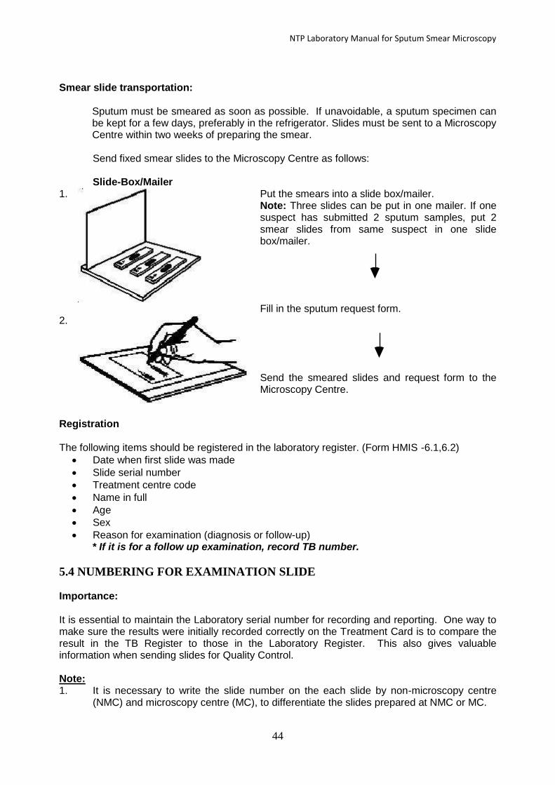
NTP Laboratory Manual for Sputum Smear Microscopy
44
Smear slide transportation: Sputum must be smeared as soon as possible. If unavoidable, a sputum specimen can
be kept for a few days, preferably in the refrigerator. Slides must be sent to a Microscopy Centre within two weeks of preparing the smear.
Send fixed smear slides to the Microscopy Centre as follows: Slide-Box/Mailer 1.
Put the smears into a slide box/mailer. Note: Three slides can be put in one mailer. If one suspect has submitted 2 sputum samples, put 2 smear slides from same suspect in one slide box/mailer.
Fill in the sputum request form. 2.
Send the smeared slides and request form to the Microscopy Centre.
Registration The following items should be registered in the laboratory register. (Form HMIS -6.1,6.2)
Date when first slide was made
Slide serial number
Treatment centre code
Name in full
Age
Sex
Reason for examination (diagnosis or follow-up) * If it is for a follow up examination, record TB number.
5.4 NUMBERING FOR EXAMINATION SLIDE Importance: It is essential to maintain the Laboratory serial number for recording and reporting. One way to make sure the results were initially recorded correctly on the Treatment Card is to compare the result in the TB Register to those in the Laboratory Register. This also gives valuable information when sending slides for Quality Control. Note: 1. It is necessary to write the slide number on the each slide by non-microscopy centre
(NMC) and microscopy centre (MC), to differentiate the slides prepared at NMC or MC.

NTP Laboratory Manual for Sputum Smear Microscopy
45
2. When microscopy centres write the slide No. on each slide, either only Nepali or English should be used.
3. For follow up slides, write the slide No. and then mention 'F' and the month of treatment. (eg. Slide No.-F- treatment month, HI Code, date)
The preferable size of the Microscopic glass slide is 75mm X 25mm X 1.3mm approximately.
5.5 Instruction for slide numbering: 1. Slide prepared by Microscopy Centre MC Lab. serial No./HI code No. and Date 2. Slide prepared by
Non-Microscopy Centre Slide received at Microscopy Centre NMC slide No./HI code No./ Date + MC Lab. serial No./HI code No.
EXAMPLE: This is the slide sent by NMC After getting the slide to the MC
12
3 B
/03
10
5A
/06
20
68
/05
/07
10
5A
/06
20
68
/05
/07
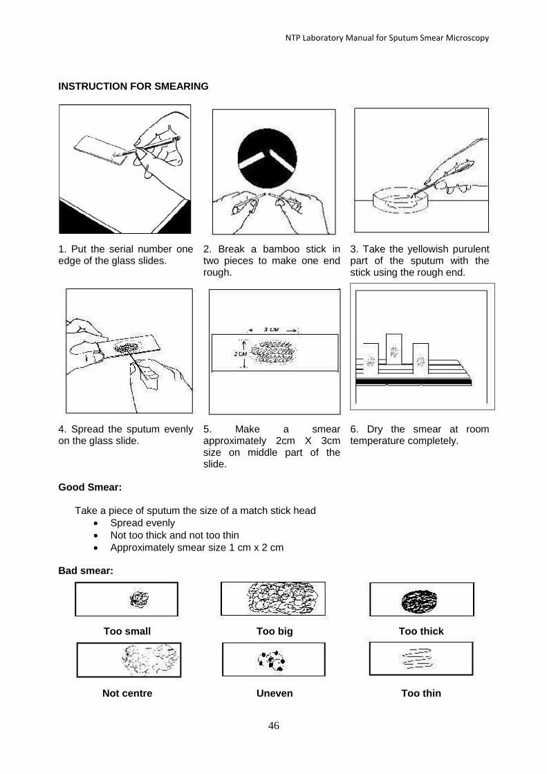
NTP Laboratory Manual for Sputum Smear Microscopy
46
INSTRUCTION FOR SMEARING
1. Put the serial number one edge of the glass slides.
2. Break a bamboo stick in two pieces to make one end rough.
3. Take the yellowish purulent part of the sputum with the stick using the rough end.
4. Spread the sputum evenly on the glass slide.
5. Make a smear approximately 2cm X 3cm size on middle part of the slide.
6. Dry the smear at room temperature completely.
Good Smear: Take a piece of sputum the size of a match stick head
Spread evenly
Not too thick and not too thin
Approximately smear size 1 cm x 2 cm Bad smear:
Too small Too big Too thick
Not centre Uneven Too thin

NTP Laboratory Manual for Sputum Smear Microscopy
47
Fixation x Do not put slide over the flame before smear is completely dried.
Fix the completely dried smear by passing it through the flame 2-3 times, about 5 seconds each time.
If the slide is put in the flame before smear is completely dried, aerosols of infectious bacteria might be formed.
STAINING (ZIEHL - NEELSEN METHOD)
1) Place the slide on the staining bridge.
2) Cover the whole surface of the slide with Ziehl's solution (Carbol
fuchsin solution).
3) Heat the slide until vapour rises from the stain. Leave it for 5
minutes. Do not boil.
4) Tip off excess staining
solution. 5) Wash off the slide with water. 6) Tip off excess water.
7) Decolorize with 25% Sulphuric Acid until no more stain remains.
(about 5 - 10 minutes).
8) Wash off the slide with water. If stain still comes off, repeat No.
7) again.
9) Tip off excess water.

NTP Laboratory Manual for Sputum Smear Microscopy
48
10) Counter-stain with 0.1 % Methylene Blue on the slide for
10 - 20 seconds.
11) Tip off excess staining solution.
12) Wash the slide with water.
13) Dry the stained slide at room temperature.
MICROSCOPY EXAMINATION
1) Put one drop of Anisole on the stained smear. Do not touch the smear with the Dropper.
2) Screen the smear with x 10 eyepiece and x 100 objective. Examine 100 visual fields.
3) Record the result on register book and report form.
4) Clean the objective with Lens/tissue paper or soft cloth.
5) Cover the microscope with vinyl dust cover or soft cloth and put in the box.
6) Wash Anisole off the smear by water and let it dry then keep it in slide box.

NTP Laboratory Manual for Sputum Smear Microscopy
49
Smear screening
For NEGATIVE result read at least 100 F (fields)
For 1-9 AFB, read at least 100 F (fields)
For 1+ result, read at least 100 F (fields)
For 2+ result, read at least 50 F (fields)
For 3+ result AFB, read at least 20 F (fields) Note: If you screen the smear with the size of 1 cm x 2 cm, two horizontal lines or three vertical lines give 100 V F. * F (One Field): the area which is seen at one time through the eyepiece Two lines (Horizontal) Three lines (Vertical)
or
Note: keep the examined slides on serial order then send it according to the LQAS system for EQA
REPORTING SCALE The number of AFB (Acid fast bacillus) found in an examination of 100 fields, according to the WHO/IUATLD grading scale, is very important information because it is related to the degree of infectivity and clinical severity of the patient. x Do not report it as "Mycobacterium tuberculosis" instead of "AFB" AFB means any bacteria which stain red or pink by Ziehl-Neelsen staining. Several kinds of AFB are present in nature. Table 3: Scale (Based on WHO/IUATLD grading scale)
No. of AFB Reporting scale No AFB found per 100 VF* Negative
1 to 9 AFB found per 100 VF Record exact no. of AFB per 100 VF 10 to 99 AFB found per 100 VF 1+
1 to 10 AFB found per VF 2+
More than 10 AFB found per VF 3+
*VF – Visual field (the area which is seen at one time through the eyepiece) Note: WHO (World Health Organization) IUATLD (International Union Against TB and Lung Diseases/ The Union)

NTP Laboratory Manual for Sputum Smear Microscopy
50
OBJECTS WHICH CAN BE SEEN ON SPUTUM SMEAR SLIDE
Various shapes of AFB
Rod type Thin and long type
Clumped type Coccus type

NTP Laboratory Manual for Sputum Smear Microscopy
51
5.6 MAIN CAUSES OF FALSE POSITIVE/FALSE NEGATIVE SPUTUM SMEAR
RESULTS 5.6.1 False Positive Result:
Although the result is Positive, the sample itself is actually AFB negative 5.6.2 False Negative Result:
Although the result is Negative, the sample itself is actually AFB positive
Note:
False positive (F+) results can cause not only unnecessary treatment for non-TB patients but also incorrect change of regimen to category II treatment after 5 months of drug taking.
False negative (F-) results can cause not only no treatment for smear positive patients but also incorrect continuation of the first regimen to failure cases.
Step Causes False ( - )
False ( + )
Specimen collection
- Poor sputum in quality & quantity (Improper collection of sputum) - Inadequate volume of sputum - Using reusable container - Artefact such as Nocardia species, spores of
bacillus species, pine pollen, food particles
* *
* *
Smear preparation
- Using scratched slides - Poor selection of sputum for smear making - Improper preparation of the smear *too thin / too thick / uneven / too little material - Insufficient fixation - Contamination from another positive sputum
* * *
* * *
Smear staining
- Over-heated staining - Precipitated stain (Fuchsin crystal) - Too short staining time with Ziehl’s solution - Improper decolourization - Intensive counter staining - Using low quality of chemicals - Using expired solution - Transfer of positive smear particle to another
* * * * * *
* * * *
Microscopic reading
- Insufficient scanning - Physical problem (colour blindness, other visual
disturbances) of examiners. - Erratic attitude
* * *
*
Recording Reporting
- Misidentification of patient and specimen - Mis-transcription of the recording & reporting - Mislabelling of specimen
* * *
* * *

NTP Laboratory Manual for Sputum Smear Microscopy
52
Chapter VI RECORDING AND REPORTING 6. RECORDING AND REPORTING
Recording: The practice of capturing data on patients’ management over time and across clinical sites and writing information either directly on paper forms and/or entering into a computer. Reporting: The routine tracking (monitoring) of priority program management information and its intended aggregated patient outcome data (evaluation) at the facility, County, State and National Level. Forms and reports: The laboratory workers should fill out the following forms and reports on time as needed.
6.1 Sputum sample examination request and report HMIS 6.1 This request form is used for sending the sputum to be examined from OPD or
treatment centre and also this is the sputum examination result form that is sent from the laboratory or microscopy centre to treatment centre or OPD.
6.2 Laboratory registration book HMIS 6.2 or NTP Lab Register): This registration book is kept at the microscopy centre.
This is the register, which contains details of all the sputum smear examinations carried out at the microscopy centre.
The laboratory serial number comes from the laboratory register. It starts from 01 at the beginning of every reporting year (1st Shrawan). The Laboratory staffs write this number on the Smear Request Form, on the Sputum Container, and on the Smeared Slide.
Every tuberculosis suspect has a separate line in the Laboratory Register. Remember to collect three specimens from each tuberculosis suspect and record the result in the register. Remember to write the suspect’s address in full, so that you can trace him if the result is positive and he does not come back.
Result of follow up smear examinations are also recorded in the register. For General Information:
6.3 TB Treatment Card (HMIS 6.3) This card is the record of TB patient under treatment. We make one for each patient and keep it at the treatment centre.
6.4 Patient’s Card (HMIS 6.4) This card contains similar information to the TB Treatment Card. This card belongs to the patient and is filled in by the treatment supervisor.
6.5 TB Treatment Register (HMIS 6.5) This register contains details of every TB patient registered at the treatment centre. The DTLO/A transfers information from the Tuberculosis Treatment Cards into this register when he visits the treatment centre each month. Four-monthly case finding, sputum conversion and treatment outcome reports are prepared by consulting this register. * HMIS- Health Management Information System

NTP Laboratory Manual for Sputum Smear Microscopy
53
HMIS 6.1: Ifo/f]u vsf/ kl/If0f cg'/f]w kmf/fd
(Tuberculosis Specimen Collection Form) Ifo/f]usf] ;Defljt la/fdLsf] lgbfg tyf pkrf/df /x]sf Ifo/f]usf la/fdLsf] cg'udg vsf/ kl/If0f ug{' kg]{ x'Fbf vsf/
kl/If0fsf nflu la/fdLnfO{ cg'/f]w u/L k|of]uzfnfdf kl/If0fsf nflu k7fpmg o; kmf/dsf] k|of]u ul/G5 .
o; kmf/ddf b'O efux? 5g, klxnf] efudf vsf/ kl/If0f cg'/f]w ug{sf nflu cfjZos laj/0f :jf:YosdL{n] el/ ;f] ;]jf
pknAw x'g] k|of]uzfnfdf k7fOG5, bf];|f] efudf la/fdLsf] vsf/ kl/If0f -:k'6d dfOqmf]:sf]kL jf lhg]PS;k6{_ u/L ;f] sf] glthf
glthf kmf/ddf pNn]v u/L cg'/f]w ug]{ :jf:Yo ;+:yfdf k7fpmg' kb{5 .
NTP Laboratory Manual for Sputum Smear Microscopy
54
sf8{ eg]{ tl/sf: l;=g+= zLif{s lgb]{zg
! Ifo/f]u pkrf/ s]G› pkrf/ ug]{ :jf:Yo ;+:yfsf] gfd o; dxndf n]Vg' kb{5 .
@ cf]=kL=8L= g+= Ifo/f]usf ;Defljt la/fdLnfO{ vsf/ kl/If0fsf] nflu k7fp+bf b}lgs la/fdL
;]jf /lhi6/sf] cf]kL8L btf{ g+= n]Vg' kb{5 .
# Ifo/f]u btf{ g+= pkrf/df /x]sf Ifo/f]usf la/fdLnfO{ cg'udg(Follow-up) vsf/
kl/If0fsf] nflu k7fpFbf Ifo/f]u pkrf/ /lhi6/af6 Ifo/f]u btf{ gDa/ n]Vg'
kb{5 .
$ ldlt vsf/ kl/Ifgsf nflu k7fOPsf] ldlt -ut],dlxgf / ;fndf_ n]Vg' kb{5 .
% la/fdLsf] gfd y/ ;]jf lng cfPsf JolQmsf] gfd y/ n]Vg' kb{5 .
^ pd]/ ;]jf lng cfpg] JolQmsf] k'/f u/]sf] pd]/ -jif{df_ n]Vg' kb{5 .
& ln+u ;]jf lng] JolQm dlxnf eP dlxnf / k'?if eP k'?if n]Vg' kb{5 .
* 7]ufgf: lhNnf, uf=la=;=, g=kf=,
jf8{ g+=, 6f]n
la/fdL a;f]af; ug]{ 7]ufgf lhNnf, uf=la=;=/g=kf=,j8f g+=/ 6f]n ;d]t v'Ng]
u/L n]Vg' kb{5 .
( cleefjssf] gfd la/fdLsf] cleefjssf] gfd n]Vg' kb{5 .
!) ;Dks{ g+=: la/fdLsf] jf la/fdLsf] kl/jf/sf] 3/sf] kmf]g g+= jf df]afOn g+= n]Vg' kb{5 .
!! kl/If0f
u/fpg' sf]
sf/0f
/f]u lgbfg la/fdLsf] /f]u lgbfgsf nflu vsf/ kl/If0f u/]sf] eP /f]u lgbfgdf -√_ lrGx
nufpg' kb{5 / RR TB/MDR TB lgbfgsf] nflu kl/If0f ul/g] xf] eg]
;f] ;d]t pNn]v ug{'kb{5 . cg'udg pkrf/df /x]sf Ifo/f]usf] la/fdL sf] cg'ejdf vsf/ kl/If0f ul/g] eP
cg'udgdf -√_ nufO{ pkrf/sf] s'g dlxgf -@/#,% / pkrf/sf] cGTodf_ s] xf]
;f] dlxgf n]Vg' kb{5
!@ Pr=cfO{=eL= ;+qmd0f la/fdL ;+u ePsf] k|df0f sf] cfwf/df Pr=cfO{=eL= ;+qmd0f eP gePsf] Plsg
u/L PrcfO{eL ;+qmd0f ;DalGw 5 5}g jf yfxf 5}g dWo] ;+qmd0f ePsf] eP
sf]8 !, geP sf]8 @ / yfxf gePsf] sf]8 # n]Vg' k5{
-PrcfO{eL ;+qmd0fsf] cj:yf ;DalGw ;f]w vf]h jf cGo s'g} klg lqmofsnfk ubf{
;]jfu|fxLsf] uf]kgLotfnfO dWogh/ ub}{ /fli6«o P8\; tyf of}g /f]u lgoGq0f s]G›n]
hfl/ u/]sf gLlt lgod tyf lgb]l{\zsfsf] kfngf clgjfo{ ?kdf ug{' k5 { . )
!# Ifo/f]usf] pkrf/ -klxn] lnPsf],
glnPsf], yfxf gePsf]_
la/fdL ;+u Plsg u/]/ Ifo/f]u pkrf/ klxnf lnPsf] glnPsf] Plsg u/L
pko'Qm Ps sf]7fdf -√_ lrGx nufpg' kb{5 .

NTP Laboratory Manual for Sputum Smear Microscopy
55
!$ cg'/f]w ul/Psf] kl/If0f:-
dfOqmf]:sf]kL, lhgPS;k6{, cGo_
kl/If0fsf] nflu cg'/f]w ul/Psf] dfOqmf]:sf]kL jf lhgPS;k6{ h'g xf] ;f] sf]
sf]7fdf -√_ lrGx nufpg' k5{ cGo kl/If0f -sNr/,8L=P;=l6 jf Pn=kL=P=_ eP
cGodf -√_ lrGx nufO{ ljj/0f v'nfpg' kb{5
!% cg'/f]w ug]{ JolQmsf] gfd,y/ tyf
x:tIf/
vsf/ kl/If0f sf] nflu cg'/f]w u/L k7fpg] :jf:Yosld{sf] gfd / y/ :ki6
v'nfO{ x:tfIf/ ug{'kb{5
glthf kmf/d:
dfOqmf]:sf]kL kl/If0f glthf -Nofaf]/]6/Ldf eg{' kg]{_
:jf:Yo ;+:yfaf6 k|fKt cg'/f]w kmf/d cg';f/ vsf/ kl/If0f ul/ ;f] sf] glthf o; kmf/ddf e/L h'g :jf:Yo ;+:yfaf6 cg'/f]w
kmf/d el/Psf] xf] ;f]lx ;+:yfdf dfOqmf]:sf]kL glthf k7fpg' kb{5 .
Nof=l;=g+=: To; ;+:yfsf] Nofa /lh:6/df pNn]lvt qmd ;+Vof cg';f/ n]Vg' kb{5 = x/]s cf=j=df= 1 b]lv gofF qmd;+Vof n]Vg'
kb{5 .
gd'gf ;+sng ldlt: o; dxndf k|of]uzfnfdf vsf/ gd'gf ;+sng ul/Psf] ldlt -ut],dlxgf / ;fndf_ pNn]v ug{'kb{5
gd'gf: Nofa kl/If0fsf] nflu lbPsf] gd'gf klxnf] eP A bf]>f] eP B df -√_ lrGx nufpg' kb{5 =
lsl;d: s:tf] k|sf/sf] vsf/ gd'gf ;+sng ePsf] xf] ;f] lgwf{/0f u/L /ut ldl;Psf] eP B, kx]+nf] / l9Ssf k/]sf] eP M /
/of{n dfq} eP S df uf]nf] 3]/f nufpg' kb{5 .
:d]o/ dfOqmf]:sf]lks glthf
dfOqmf]:sf]kL kl/If0f k5L k|fKt glthf cg';f/ % j6f glthf dxn dB] s'g} Pp6f dxndf dfq -√_ lrGx nufpg' kb{5 .
Scale
Bright Field Ziehl Neelseen stain
1000×Magnification (1 line =2cm=100 field)
Florescence (Auramine stain)
200×magnification (1line=2cm=20 field)
Florescence (Auramine Stain)
400×mmagnification (1line=2cm=40 field)
Negative No AFB No AFB No AFB
Scanty (actual number)
1-9 AFB/line 1-29/ 100F 1-19/ 100 F
1+ 10-99/ field 30-299/ 100 F 20-199/ 100 F
2+ 1-10 AFB/ field 10-100/ F 5-50/ F
3+ >10 AFB/ field >100/ F >50 AFB/ field
Bright Field Ziehl Neelseen stain को लागि तल दिईएको अनुसार िनुु पिछु l
AFB gb]lvPdf -Neg_ sf] sf]7fdf -√_ lrGx nufpg' kb{5 .
1-9 AFB/100 HPF (Scanty) b]lvPdf Extra number n]Vg' kb{5 .
10-99 AFB/100 HPF b]lvPdf !+ sf] sf]7fdf -√_ lrGx nufpg' kb{5 .
1-10 AFB/HPF b]lvPdf @ + sf] sf]7fdf -√_lrGx nufpg' kb{5 .
10AFB/HPF b]lvPdf #+ sf] sf]7fdf -√_ lrGx nufpg' kb{5 .
o;}u/L Florescence Auramine stain sf] klg ;DalGwt column sf] glthf cg';f/ (Neg) df -√_ lrGx nufpg]
n]Vg] 1+ ,2++ jf 3+ sf] dxn dB] Psdf -√_ lrGx nufpg' kb{5 .
kl/If0f ug]{ JolQmsf] b:tvt: o; sf]7fdf vsf/ kl/If0f ug]{ JolQmn] b:tvt ug{' kb{5 .
glthf cfPsf] ldlt: dfOqmf]:sf]kL glthf lg:s]sf] lbgsf] ldlt -ut,] dlxgf / ;fndf _ o; sf]7fdf pNn]v ug{'kb{5 .
lhgPS;k6{ (Gene Xpert) glthf:
gd'gf ;+sng u/]sf] ldlt: o; dxndf vsf/ gd'gf ;+sng ul/Psf] ldlt -ut],dlxgf,/ ;fndf _pNn]v ug{' kb{5 .
vsf/ Ifo/f]u sf] AofS6]l/of eP/gePsf](M. Tuberculosis Detected/not Detected-Y/N): vsf/ kl/If0fdf
Tuberculosis Bacteria b]lvPdf sf]8 g+= ! df / Bacteria gb]lvPdf sf]8 g+= @ df uf]nf] 3]/f nufpg' k5{
cGo(Invalid/No result/Error-I): vsf/ gd'gfsf] u'0f:t/hGo gePsf] sf/0fn] kl/If0fsf] glthfdf Plsg ug{ g;s]sf]
cj:yfdf cGo(Invalid/No result/Error-I) df -√_ lrGx nufpg' kb{5 .

NTP Laboratory Manual for Sputum Smear Microscopy
56
vsf/df Ifo/f]u Bacteria kQf nfu]df(M. Tuberculosis Detected): vsf/df Ifo/f]u sf] Bacteria ePdf tLg lsl;dsf]
glthf dWo] s'g glthf cfPsf] 5 ;f] glthf sf] sf]8 g+= df uf]nf] lrGx nufpg' kb{5 . h:t} l/kmflDkl;g
;]G;Ll6e(Rifampicin Sensitive-T) ePdf sf]8 g+=! Dff, l/kmflDkl;g ;]G;Ll6e(Rifampicin Sensitive-RR) ePdf sf]8 g+=
@ df / Og8L6/ldg]6 (Interminatr-TI_ ePdf sf]8 g+= # df uf]nf] 3]/f nufpg' kb{5 .
glthf cfPsf] ldlt: lhgPS;k6{ kl/If0f kZrft glthf k|fKt ePsf] ldlt -ut],dlxgf / ;fndf _ vsf/ kl/If0f ug]{ Nofassf
sd{rf/Ln] eg{' kb{5 .
kl/If0f ug]{sf] gfd tyf x:tfIf/: lhgPS;k6{ kl/If0f ug]{ Nofa sd{rf/Lsf] gfd / y/ :ki6 v'nfO{ x:tIf/ ug{' kb{5 .
k|dfl0ft u/]sf] gfd tyf x:tfIf/: lhgPS;k6{ kl/If0f ug]{ Nofa sd{rf/Lsf] ;'k/efOh/ jf Nofa k|d'vsf] gfd, y/ / x:tfIf/
ug{' kb{5 .
HMIS 6.2: क्षयरोग प्रयोगशाला रजिष्टर
(Tuberculosis Laboratory Register Specimen) क्षयरोग प्रयोगशाला रजिष्टर
ल्याब
सि.नं.
नमुना
िंकलन
समसि
सिरामीको जासि
कोड
असििािक
को नाम थर
िम्पकक नं. उमेर सिरामीको ठेगाना अनुरोध गने
उपचार केन्द्रको
नाम
ग म िा नाम थर म पु सजल्ला गा.सि.ि./न.पा. िडा नं.
१ २ ३ ४ ५ ६ ७ ८ ९ १० ११ १२ १३ १४ १५
क्षयरोग प्रयोगशाला रजिष्टर
क्षयरोग सनदानका लासग पे्रषण क्षयरोग
ओपी डी
दिाक नं.
एचआईिी िंक्रमण क्षयरोग को पसिला उपचार िए
निएको
पररक्षणको प्रकार
सनजी
स्वास्थ्य
िंस्था (P)
िमुदाय
(c)
िम्पकक
पररक्षण(T
)
िएको निएको थािा
निएको
िएको निएको थािा
निएको
सनदान अनुगमन
(मसिना लेखे्न
)
१६ १७ १८ १९ २० २१ २२ २३ २४ २५ २६ २७
पररक्षण नजििा जिजि कैसियि
सजन एक्सपर्क से्मयर माइक्रोस्कोपी
A B
नसिजा * ग म िा नसिजा* ग म िा नसिजा * ग म िा
२८ २९ ३० ३१ ३२ ३३ ३४ ३५ ३६ ३७ ३८ ३९ ४०

NTP Laboratory Manual for Sputum Smear Microscopy
57
Chapter VII QUALITY Assurance FOR SPUTUM SMEAR
Microscopy
7. QUALITY ASSURANCE FOR SPUTUM SMEAR MICROSCOPY
7.1 Introduction Quality assurance of sputum microscopy is an indispensable part of an effective TB control program. It encompasses the whole process of sputum collection, smear preparation, smear staining, microscopy, recording and reporting. The purpose of quality assurance programs is the improvement of the efficiency and reliability of smear microscopy services. In many countries with a high prevalence of tuberculosis (TB, direct sputum smear microscopy remains them most cost effective tool for diagnosing patients with infectious tuberculosis and for monitoring their progress on treatment. The World Health Organization strategy for tuberculosis control (DOTS) relies on a network of laboratories that provide acid fast bacilli (AFB) sputum smear microscopy. The establishment of a broad network of well-Functioning peripheral laboratories within the context of the health system and readily accessible to the population is a high priority for any tuberculosis control program. If the laboratory diagnosis is unreliable, all other activities will be affected. However, the quality of laboratory services often may not be considered a high priority of the National Tuberculosis Program (NTP). Microscopy errors are likely to result in failure to detect persons with infectious TB who will then continue to spread infection in the community, or unnecessary treatment for "non-cases". Errors in reading follow up smears can result in patients being placed on prolonged treatment or re-treatment, or in treatment discontinued prematurely. Therefore, quality assurance of laboratory services, including AFB sputum smear microscopy, is essential. Both the availability and quality of AFB smear microscopy are dependent on national programs that support, train, and monitor the testing performance of individual laboratories.
7.2 A quality assurance program has three main components: a. Quality Control (QC): Quality control is a process of effective and systematic internal
monitoring which aims to detect the frequency of errors against established limits of acceptable test performance. Although it is not usually feasible to determine error frequencies accurately, it is nevertheless a mechanism by which tuberculosis laboratories can at least validate the competency of their diagnostic services
b. Proficiency testing: Also known as External Quality Assessment (EQA), this is a program
designed to allow participant laboratories to assess their capabilities by comparing their results with those obtained with the same specimens in other laboratories of the network, e.g., Regional and National Reference Laboratories. EQA might be on-site evaluation of the laboratory for review QC and on-site rereading of smears.
c. Quality Improvement (QI): A process by which the components of smear microscopy diagnostic services are analyzed with the aim of looking for ways to permanently remove obstacles to success. Data collection, data analysis, and creative problem solving are the key components of this process. It involves continued monitoring, identifying defects, follows by remedial action including retraining when needed, to prevent recurrence of problems. QI often relies on effective on-site evaluation visits.

NTP Laboratory Manual for Sputum Smear Microscopy
58
7.3 INTERNAL QUALITY CONTROL
1. Poor quality sample gives false result. Always ask for the good quality sample. Mucopurulent sample is recommended.
2. Too thick or too thin smear can give false result. The thickness of smear should be such that we can partly read newspaper kept back of the slide.
3. Too old reagent may give false result. Carbol fuschin and methylene blue should be filtered before use.
4. All newly prepared staining reagents should be checked for its quality by staining a known positive and negative slide. If the known positive slide gives the negative result, that staining reagent must not be used.
5. All the steps of staining procedure should be strictly followed according to SOP (Standard operating procedure). Any alteration in the procedure can give false result.
6. Distilled water should be used in staining instead of Tap water. (tap water may give false positive result.)
7. High rate of positivity among the suspected patient may be due to false positive and low rate of positivity may be due to error in reagent, microscope and staining procedure.
7.4 LABORATORY NETWORK: It is very important to know the level of various laboratories network:
7.4.1 Peripheral
Laboratories located at primary health centers or district hospitals. Staff have technical proficiency to perform sputum smear microscopy utilizing Ziehl-Neelsen (ZN) staining. Peripheral laboratories must be visited on a regular basis by a district supervisor, who has been adequately trained to evaluate the basic functions of the microscopy.
7.4.2 Intermediate
Regional or provincial laboratories existing in larger hospitals or cities. Staff have technical proficiency to perform ZN microscopy, and may have capacity to perform fluorescence microscopy if volume is high. Intermediate laboratories should be capable of providing supervision, monitoring, training, and quality assurance to peripheral laboratories, including rechecking of smears.
7.4.3 National Reference or Central
May exist as part of the central public health laboratory, a research laboratory, or as an upgraded laboratory in the country's principal tuberculosis institution. Serves as the national reference laboratory for the TB program, with competence in direct ZN microscopy and, where appropriate, fluorescence microscopy. The national TB reference laboratory plays an essential role in the organization and maintenance of the network in terms of developing guidelines, ensuring high quality and standardized smear microscopy, and therefore must have the capacity to provide training and External Quality Assessment, including providing panel testing and rechecking to intermediate and peripheral laboratories.
What is an External Quality Assessment System (EQAS) for sputum microscopy? External Quality Assessment (EQA) system is defined as a system to ensure high quality laboratory services to detect AFB in sputum smear microscopic examinations. It consists of adequate sputum collection and handling, proper sputum smear preparation, proper staining, correct microscopic examinations of the slide and appropriate recording & reporting. EQA or Proficiency testing is a system of retrospectively and objectively compared results from different laboratories by external agency or Reference laboratory. There are three methods of EQA:
• On-site Evaluation • Panel Testing • Blinded Rechecking

NTP Laboratory Manual for Sputum Smear Microscopy
59
On-site Evaluation A field visit is the best method to obtain a realistic picture of the conditions and practices in the laboratory; therefore, on-site evaluation of peripheral laboratories is an essential component of a meaningful EQA programme.
Panel Testing Panel testing is a method of EQA that is used to determine whether a laboratory technician can adequately perform AFB smear microscopy A panel of 10 sputum slides (5 stained & 5 unstained) with known result form NRL to IRL, IRL to PL Random Blinded Rechecking (RBR) Blinded rechecking or rereading a sample of routine smears from the peripheral sites and intermediate labs by controllers at a higher level laboratory is considered the best method for evaluating performance and providing motivation to staff for improvement. A countrywide program for blinded rechecking of slides at regular intervals should be the long-term goal for optimal EQA.
Till the date Random Blinded Rechecking is implemented as a method of EQA for sputum smear microscopy throughout the country.
Blinded rechecking or rereading a sample of routine smears from the peripheral sites and intermediate labs by controllers at a higher level laboratory is considered the best method for evaluating performance and providing motivation to staff for improvement. A countrywide program for blinded rechecking of slides at regular intervals should be the long-term goal for optimal EQA.
Function of Regional TB Quality Control Centre NTP has established Regional TB Quality Control Centres (RTQCC) in all the five regions in July 1996 (2053/54).
Function of Regional TB Quality Control Centre NTP has established Regional TB Quality Control Centres (RTQCC) in all the five regions in July 1996 (2053/54). The functions of regional quality control centre are as follows;
1. Quality check centre for examined slides, and feedback those results to the microscopy
centres. 2. Regional training centre for TB microscopy. 3. Reagent preparation and distribution centre in the region. 4. Regional quality control assessors, who are working at the regional TB quality control
centre, are mainly technical supervisor for the microscopy centres.

NTP Laboratory Manual for Sputum Smear Microscopy
60
7.5 Method of External Quality Assessment (EQA)
EQA is carried out on a four month basis.
1. Microscopy Centre keeps all examined sputum slides serially as per lab register, so that
supervisor {District Laboratory Supervisor (DLS)/DTLA (O)} can selects the slides according to LQAS System for EQA.
2. Microscopy Centre sends those selected slides with results filled in LQAS-1 to Regional TB Quality Control Centre/Assessor through DLS or DTLA/O.
3. Assessor re-exams those smear slide using LQAS – 2A, If discrepancy arise LQAS-2B will be filled and send to NQCC.
4. Assessor sends Quality Check /Feedback Report (LQAS- 3) to Microscopy Centre through {District Laboratory Supervisor (DLS)/DTLA (O)}
5. If the quality of TB slide is not satisfactory, or any technical problem identified as per DLS or DTLA/O's supervision report, Regional Quality Control Assessor (RQCA) visits the Microscopy Centre and supervises as soon as possible. Also the RQCA, in co-ordination with RTLA/O (Regional TB/Leprosy Assistant/Officer), reports to the training centre about the necessity of refresher training to that Microscopy Centre, so that the training could be organized by central or regional level.
LOT QUALITY ASSURANCE SYSTEM (LQAS): LQAS is a method to determine an optimum
sample size which when applied properly, yields statistically acceptable samples to assess quality of work, in this case, the work of the laboratory technicians. This method was originally designed for manufacturing processes where an efficient statistical model was necessary in order to keep sampling costs to a minimum. This method has been applied in health care systems to determine whether a population meets a certain standard. A number of variables are used to determine sample size using LQAS.
Lot (N) : Total number of negative slides prepared in a specified period of time (one months, one quarter, one year). It is an operational quantity used to determine the sample size. Example: Lot = 5000/yr, 1250/quarter, 417/month. It is important to choose an interval of time that produces a Lot size that results in an economical and statistically valid sample. If the Lot size is too small, this may not be possible. Acceptance Number (d): The maximum number of false negative errors allowed in the sample after which the NTP/NRL can no longer be certain that the expected performance has been achieved. The value chosen for "d" has a direct impact on sample size, the larger the acceptance number, the larger the sample size required. In order to achieve the smallest, most efficient sample size, a value of d=0 is recommended.

NTP Laboratory Manual for Sputum Smear Microscopy
61
Slide Positivity Rate (SPR): This is the proportion of positive smears among all slides (diagnostic and monitoring) in the laboratory from which the sample is being taken. This number is estimated using the laboratory registers from the previous year or the preceding four quarters. Sample sizes can be set using the average positivity rate for a laboratory, region or country. Sensitivity: This is the expected performance in detecting positives, as compared to the controllers. Acceptable sensitivity should be determined by the NTP and NRL. The sensitivity, as defined here, is the detection of all positives, including low positives (1-9 AFB/100). Therefore, an overall sensitivity of 75-85% is recommended. Specificity: This is the expected performance in detecting negatives as compared to the controllers, which is set at 100%. Confidence Interval (CI): All of the sample sizes have been developed to determine if the laboratory has met the expected sensitivity within a 95% confidence level. Therefore, if the d=0 and there are no false negatives detected within the sample then the NRL can determine with a 95% confidence level that the peripheral laboratory is performing at or above the acceptable sensitivity.
Slides collection/sampling for EQA To make the slide rechecking programme valid representation of routine laboratory performance, the sample collected should be random and representative of all the smears examined by lab staff. The technical requirements for sampling are outlined here; Slide storage: store all the cleaned slides in the slide boxes in the same order as they
are listed in lab register. (Shown in the figure)
Slide collection/selection: DTLA/O in coordination with Lab staff will collect/select the slides during quarterly visit to MC. The selected slides will be collected by DTLA/O using the lab register for EQA.
7/100
2+
3+

NTP Laboratory Manual for Sputum Smear Microscopy
62
* Method of selecting slides for Blinded Rechecking from TB Lab Register. Circles indicate
slides to be collected:
Lab. S. No
Date
Name
Sex M/F
Name of Treatment Unit
Address New Patients
Reason for Examination
Result Sign
Rem
ark
s
Diagnosis
Follow up
A B
Neg
Neg Neg
Neg Neg
Neg
Neg Neg
Neg Neg
Neg Neg
Neg Neg
Neg
Neg Neg
Neg Neg
Neg Neg
Neg Neg
Neg Neg
Neg Neg
Neg (2+)
Neg
Neg Neg
Neg Neg
Neg
Neg Neg
Neg Neg

NTP Laboratory Manual for Sputum Smear Microscopy
63
7.6 SAMPLE SIZE DETERMINATION WITH EXAMPLES
There are some important steps for sample size determination;
Step 1 From the lab register of the previous year: Calculate the total slides examined/year = 1250 Total No. of positive slides/year = 125 Total No. of negative slides/year =1125
Step 2 Calculate slide positivity rate (SPR) = total No. of positive slides per year / total No. of slide *100 (round off to the nearest % i.e. 10 %. (SPR is valid for one yr only or new SPR should be calculated for each year)
Step 3 Decide on acceptable limits of performance in your lab Relative sensitivity (ability of lab staff to detect AFB relative to the controller) = 75% Acceptance No. {(Maximum number of errors (d)} = 0
Step 4 Select appropriate sample size table If a sensitivity of 75 % and d = 0 is chosen, look at the left side of the table to find the annual negative slide volume = 1000 and SPR =7.5%, then sample size for this year = 96
Step 5 Decide on a convenient interval to select the slide Recommended 3 times per year i.e. quarterly. Therefore, 96/3 = 32 slides to be collected every quarter (for current fiscal year)
Step 6 Systematically collect the slides using the laboratory register
Suppose 250 slides have been processed during last quarter (the quarter participating in
EQA), 32 slides need to be collected for the current quarter, therefore: 250/32 = 7.8
Collect every 7th slide.
Step 7 For the first or starting slide; use the last digit of a bank note (repeat for digit 0 or 1)
The annual sample size should be fixed beforehand by the Reference Level using LQAS Table 4 and 5.
Recommended annual sample sizes: Table 4 and 5 shows the number of slides recommended for blinded rechecking at various workloads and slide positivity rates. The table has been compiled on the basis of a sensitivity of 75% and specificity of 100% (for negative slides); acceptance number for errors equal to zero (d=0); and confidence level of 95%. The number of slides to be selected (sample size) should be fixed beforehand by the NTP. Determining sample size should not be left to the supervisor collecting the slides or to the technicians.

NTP Laboratory Manual for Sputum Smear Microscopy
64
Table 4: Sensitivity Relative to the Controllers at 75%
Negatives Examined Annually
Acceptance Number
Slide Positivity Rate (SPR)
Total Sample Required
2.5% 5.0% 7.5% 10.0% 13.0% 15.0% 18.0%
100 d=0 78 63 54 47 40 36 32
200 d=0 123 91 71 59 48 42 37
300 d=0 154 105 80 64 52 45 38
400 d=0 175 115 85 68 53 47 39
500 d=0 192 121 89 69 54 47 40
700 d=0 215 129 93 72 55 48 40
1000 d=0 236 136 96 73 56 49 41
2000 d=0 267 145 102 77 59 51 41
5000 d=0 289 152 104 78 59 51 43
10000 d=0 297 154 105 78 60 51 43
20000 d=0 302 155 106 79 60 52 43
50000 d=0 305 156 106 79 60 52 43
100 d=1 103 96 85 74 64 59 52
200 d=1 187 143 116 96 79 71 61
300 d=1 241 169 132 106 85 75 65
400 d=1 280 186 141 111 89 78 66
500 d=1 309 198 147 114 91 80 67
700 d=1 349 213 155 119 93 81 68
1000 d=1 386 224 160 123 95 82 70
2000 d=1 439 241 169 128 98 85 71
5000 d=1 478 252 173 130 99 86 72
10000 d=1 493 256 175 131 100 86 72
20000 d=1 501 258 176 131 100 86 72
50000 d=1 506 259 176 132 100 87 72

NTP Laboratory Manual for Sputum Smear Microscopy
65
Table 5: Sensitivity Relative to the Controllers at 75% Negatives Examined Annually
Acceptance Number
Slide Positivity Rate (SPR)
Total Sample Required
20.0%
23.0%
25.0% 28.0% 30.0% 33.0% 35.0%
100 d=0 30 26 24 22 21 19 18
200 d=0 34 29 27 24 23 19 18
300 d=0 35 30 28 25 23 21 18
400 d=0 35 31 28 25 23 21 18
500 d=0 36 31 28 25 23 21 20
700 d=0 36 31 29 25 23 21 20
1000 d=0 38 31 29 25 23 21 20
2000 d=0 38 32 29 25 24 21 20
5000 d=0 38 32 29 26 24 21 20
10000 d=0 38 32 29 26 24 21 20
20000 d=0 38 32 29 26 24 21 20
50000 d=0 38 32 29 26 24 21 20
100 d=1 49 44 41 38 36 33 31
200 d=1 56 49 45 40 39 34 32
300 d=1 59 51 47 42 39 36 34
400 d=1 60 52 48 43 40 36 34
500 d=1 61 52 48 43 40 36 34
700 d=1 61 53 49 43 40 36 34
1000 d=1 63 53 49 44 41 37 34
2000 d=1 64 55 51 44 41 37 34
5000 d=1 64 55 51 44 41 37 34
10000 d=1 64 55 51 44 41 37 35
20000 d=1 65 55 51 44 41 37 35
50000 d=1 65 55 51 44 41 37 35
The volume of annual sample size is calculated for sensitivity relative to the controllers at 75%; specificity 100% and acceptance number = 0 Note :
• MCs < 5% SPR should analyze the reasons
• ANSV(Annual Negative Sample Volume) < the indicated Annual Sample Size; - MC should submit all the slides

NTP Laboratory Manual for Sputum Smear Microscopy
66
7.7 ASSESSMENT POINTS OF STAINED SMEAR SLIDES
Sputum quality: by microscopic observation
1) The presence of dust cells (macrophages) and leukocytes in the sputum smear is considered as sputum, which is evidence that it is not saliva. The presence of more than 25 leukocytes and less than10 epithelial cells per field at the total magnification of x 100 is considered proper sputum specimen.
Smear making technique : by macroscopic observation
2) Size of smear Acceptable size of smear should be approximately 1 x 2 cm. Two horizontal lines or
three vertical lines of reading would cover about 100 fields.
3) Thickness of smear Acceptable thickness of smear can be checked by looking at printed letters thorough
the smear holding the smeared glass slide 4 - 5 cm over a printed paper. If letters cannot be read, it is too thick.
4) Evenness of smear Sputum should be spread evenly on the glass slide. Not too thick and not too thin.
Staining technique: by microscopic observation
5) Condition of decolourization of the smear stained by Ziehl-Neelsen method AFB and background of the smear must be clearly distinguished by decolourization.
AFB in faint red colour is over-decolourization. Under-decolourization is observed by remaining of Fuchsine colour on the background or remaining of Methylene blue on the unsmeared parts.
6) Smear cleanness Stained smear must be free from stain deposits, dirt, crystals produced by overheating
of staining, debris, etc. Microscopic reading technique
Assessor reads smear slides and compares his/her results with the result of Microscopy Centre. Analysis and calculation of overall agreement rate and false (+)/ (-) result can be done by using correlation table.
7) Agreement:
Number of Negative (-ve) and positive (+ve) result consistencies within range by grading. 8) Disagreement:
False (+) ve : Although the result of microscopy centre is Positive, the sample itself (result of assessor) is AFB negative
False (-) ve : Although the result of microscopy centre is Negative, the sample itself (result of assessor) is AFB positive

NTP Laboratory Manual for Sputum Smear Microscopy
67
Table 6: Correlation table
Example:
RReessuulltt
bbyy
AAsssseessssoorr
RReeppoorrtt ffrroomm MMiiccrroossccooppyy CCeennttrree
NNeegg 11--99//110000FF 11++ 22++ 33++ TToottaall
NNeegg 1100 1100
11--99//110000FF 22 22
11++ 33 11 44
22++ 11 11 22
33++ 22 22
TToottaall 1155 11 11 11 22 2200
Sputum Smear Results Result of Test Laboratory (Microscopy Centre)
Negative 1-9/100VF
1+ 2+ 3+ Total
Result of
Controlling
Laboratory
(RQCA/O)
Negative Negative consistency
False (+ve)
Assessor’s
total results
1-9 /100VFf
False (-ve)
Positive
1+
Consis Tency
2+
3+
within Range
Total
MC’s total results Grand total

NTP Laboratory Manual for Sputum Smear Microscopy
68
Number of (-ve) and (+ve) consistencies Within range by grading
Overall Agreement Rate = --------------------------------------------------------- x 100 (%) Grand Total Sensitivity = Total No. of +ve result agreed between MC and QC ----------------------------------------------------------------- X 100 Total +ve result of RQCA Example; 5 x100 = 50% (Se. 50%) 10 Total No. of –ve result agreed between MC and QC Specificity = --------------------------------------------------------------------- X 100 Total –ve result of RQCA Example; 10 x100 = 100% (Sp. 100%) 10 Total F -ve results False negative = ---------------------------- x 100 Total –ve results of MC Example; 5 x100 = 33% (F –ve = 33%) 15 Total F +ve results False positive = ------------------------------- x 100 Total +ve results of MC Example; 0 x100 = 0% (F –ve = 0%) 10
"Good Quality Smear Examination makes A Good Quality TB Control Programme "

NTP Laboratory Manual for Sputum Smear Microscopy
69
Instructions for filling in the Smear result sheet for EQA [LQAS 1] for sputum smear examination
1. Introduction:- This form is used to collect the examined slide with their results and
to collect the activity information from Microscopy Centres. 2. Necessity:- This form is important because the result obtained by Quality
Control Assessor after re-checking the slide of Microscopy Centres should be compared with their original results to prepare the Quality Check Feedback Report [LQAS 3] which should be sent to each Microscopy Centres.
3. How to fill in this form:- Microscopy Centre:- Write the name of Microscopy Centre. District:- Write the name of District. Month:- Write the month of slide collection for EQA purpose. e.g. from Shrawan to Kartik, 2054 Quarter/Year:- Mention the quarter (4 monthly) and the year. e.g. 1st quarter of 2054/55 Date of record:- Write the date when the results have been recorded in the form. Lab. In-charge:- Write the name of Laboratory In-charge. Recorded by:- Write the name of staff, who has recorded the information in this
form. MC Report of Previous Year (20 /20 ) Report of Microscopy Centre from the Lab Register of Previous
Year Total Examined Slides Write down from the Lab Register of Previous Year Total Negative Slides Only Negative Slides from the Lab Register of Previous Year Total Positive Slides Only Positive Slides from the Lab Register of Previous Year Slide Positivity Rate Calculate from the Lab Register of Previous Year (SPR = Total
+ve/Grand Total*100) Process of Slide Selection A process of determination sample size of LQAS System Annual Sample Size Sample Size to be chosen for current Fiscal Year (According to
prescribed LQAS table) Quarterly Sample Size Sample size to be chosen for participating quarter in EQA (Annual
sample size/3) Total Slides Examined During last Quarter Total slides examined during the Quarter participating in EQA
(Total (-ve & +ve) Slide interval……th Interval of Selected slide (collect every ……th slide) MC Report of current F/Y (,..Qtr 20 / 20 ) Details of MC for participating quarter in current Fiscal Year (,,,,Qtr
/Year……) S. No. of First slide selected Serial Number of the First Slide selected as per the last digit of a
Bank Note Total Neg. Slides sent for EQA Total Number of Negative Slides sent for EQA Total Pos. Slides sent for EQA Total Number of Positive Slides sent for EQA Total Slides Sent for EQA Total Number of selected Slides (Positive and Negative) sent for
EQA Slide No.:- Write the Slide No. (given by the Laboratory) in a serial order as per
Lab Register. Result:- Write the result of individual slide separately in the result column. e.g. Neg., 1 – 9 /100VF, 1+, 2+, 3+
Suspected person: Total no of suspected person, screening during defined Quarter/ year
Slide A: No of slide from spot sputum sample.
Slide B: No of slide from Early morning sputum sample
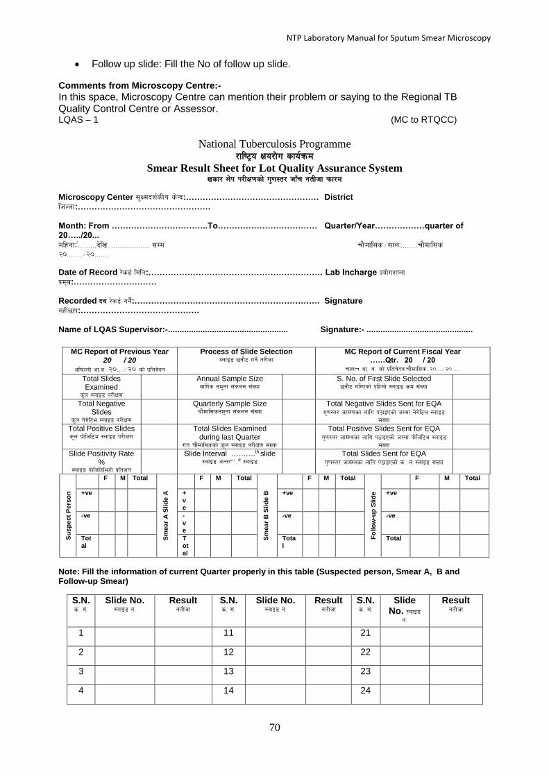
NTP Laboratory Manual for Sputum Smear Microscopy
70
Follow up slide: Fill the No of follow up slide. Comments from Microscopy Centre:-
In this space, Microscopy Centre can mention their problem or saying to the Regional TB Quality Control Centre or Assessor. LQAS – 1 (MC to RTQCC)
National Tuberculosis Programme /fli6«o Ifo/f]u sfo{qmd
Smear Result Sheet for Lot Quality Assurance System vsf/ n]k k/LIf0fsf] u'0f:t/ hfFr gtLhf kmf/d
Microscopy Center ;"Idbz{sLo s]Gb:………………………………………… District lhNnf:………………………………………… Month: From ……………………………..To……………………………… Quarter/Year………………quarter of 20…../20... dlxgfMM++=========b]lv==================== ;Dd rf}dfl;s÷;fnM========rf}dfl;s
@)=======÷@)=======
Date of Record /]s8{ ldlt:……………………………………………………… Lab Incharge k|of]uzfnf
k|d'v:………………………… Recorded by /]s8{ ug]{:…………………………………………………………. Signature ;xL5fk:……………………………………. Name of LQAS Supervisor:-.................................................... Signature:- ..............................................
MC Report of Previous Year
20 / 20
cl3Nnf] cf=a= @)===÷@) sf] k|ltj]bg
Process of Slide Selection :nfO8 5gf}6 ug]{ t/Lsf
MC Report of Current Fiscal Year ……Qtr. 20 / 20
rfn¬ cf= a= sf] k|ltj]bgMrf}dfl;s==@)===÷@)=====
Total Slides Examined
s"n :nfO8 k/LIf0f
Annual Sample Size jflif{s gd"gf ;+sng ;+Vof
S. No. of First Slide Selected 5gf}6 ul/Psf] klxnf] :nfO8 s|d ;+Vof
Total Negative Slides
s"n g]u]l6e :nfO8 k/LIf0f
Quarterly Sample Size rf}dfl;sgd"gf ;+sng ;+Vof
Total Negative Slides Sent for EQA u'0f:t/ hf¤rsf nflu k7fOPsf] hDdf g]u]l6e :nfO8
;+Vof
Total Positive Slides s"n kf]lhl6e :nfO8 k/LIf0f
Total Slides Examined during last Quarter
ut rf}dfl;ssf] s"n :nfO8 k/LIf0f ;+Vof
Total Positive Slides Sent for EQA u'0f:t/ hf¤rsf nflu k7fOPsf] hDdf kf]lhl6e :nfO8
;+Vof
Slide Positivity Rate %
:nfO8 kf]lhl6le6L k|ltzt
Slide Interval ……….th slide :nfO8 cGt/………
cf}++ :nfO8
Total Slides Sent for EQA u'0f:t/ hf¤rsf nflu k7fOPsf] sÒn :nfO8 ;+Vof
Su
sp
ect
Pers
on
F M Total
Sm
ear
A S
lid
e A
F M Total
Sm
ear
B S
lid
e B
F M Total
Fo
llo
w-u
p S
lid
e
F M Total
+ve +ve
+ve +ve
-ve -ve
-ve -ve
Total
Total
Total
Total
Note: Fill the information of current Quarter properly in this table (Suspected person, Smear A, B and Follow-up Smear)
S.N. s|= ;+=
Slide No. :nfO8 g+=
Result gtLhf
S.N. s|= ;+=
Slide No. :nfO8 g+=
Result gtLhf
S.N. s|= ;+=
Slide No. :nfO8
g+=
Result gtLhf
1 11 21
2 12 22
3 13 23
4 14 24

NTP Laboratory Manual for Sputum Smear Microscopy
71
Instructions for filling in the Smear result sheet for EQA [LQAS 2] for sputum smear examination
1. Introduction:- This form is used by Regional TB Laboratory Coordinator (RTLC) to provide the examined slides without their results to the RTQCC.
2. Necessity:- This form is important because the result obtained by RTQCAs after re-checking the slides of Microscopy Centres in a Blinded manner, which should be compared with their original results to prepare the Quality Check Feedback Report [LQAS 3], which should be sent to each Microscopy Centre.
3. How to fill in this form:- Microscopy Centre:- Write the name of Microscopy Centre. District:- Write the name of District. Month:- Write the month of slide collection for EQA purpose. e.g. from Shrawan to Kartik, 2054 Quarter/Year:- Mention the quarter (4 monthly) and the year. e.g. 1st quarter of 2054/55 Slide No.:- Write the Slide No. in a serial order as per LQAS 1. Result:- Write the result of RTQCA for individual slide in the Assessor’s
result column. e.g. Neg., 1 – 9 /100VF, 1+, 2+, 3+ Specimen quality Mention the quality of sputum specimen (good or poor) after
observing slide under Microscope Size B/S Mention the size of the smear whether it is Bigger or Smaller than
the recommended size Thickness K/N Mention the thickness of the smear whether it is Thick (K) or Thin
(N) than usual thickness Evenness Mention the evenness of the smear whether it is evenly spread (good) or
not (poor)
Staining U/O Mention the staining quality of the smear whether stained properly (good) or Under- decolorized / Over decolorized (poor)
Cleanness Mention the cleanness of the smear whether it is clean (good) or dirty
(poor)
Memo In this space, RTQCA can make a note, if found anything remarkable
during slide rechecking
5 15 25
6 16 26
7
17 27
8 18 28
9 19 29
10 20 30

NTP Laboratory Manual for Sputum Smear Microscopy
72
LQAS -2A (RTQCC)
National Tuberculosis Programme
Quality Control for Sputum Smear Examination (worksheet / )
Microscopy Centre: ………………………………….District: …………………………………… Month: …………… to ………………………………Quarter/Year: …Qtr……..20…./20…….. SN Slide
No. AFB
Result by
Specimen quality
Size B/S
Thickness K/N
Evenness Staining U/O
Cleanness
MC Assessor Good Poor Good Poor Good Poor Good Poor Good Poor Good Poor
1
2
3
4
5
6
7
8
9
10
11
12
13
14
15
16
17
18
19
20
21
22
23
24
25
26
27
28
29
30
Total
Memo:
Date of Exam……………………
Assessed by………………..

NTP Laboratory Manual for Sputum Smear Microscopy
73
Total Mcs in the Region ……………………. Total MCs Participated in the EQA………...….…………..
Total Slide screened in the Region … …… (Total Negative slide ………. Total Positive slide …..…….)
Total Slide send for QC………………...….. (Total Negative slide ………..Total Positive slide …………)
Total slide found False Report …..………. (False Negative ……………. False Positive …….………..)
Report Name of QCA Report Name of QCA
QC 1 QC 2Final Report RemarksS.No. Districts Microscopy Centre MC's Report
QC 2B National Tuberculosis Programme /fli6«o Ifo/f]u sfo{qmd
Smear Result Sheet for Discrepant QC Slides
Month: From ……………………………..To……………………………… Quarter/Year………………quarter of 20…../20 Regional Quality Control Center:………………………………………… Region…………………………………………

NTP Laboratory Manual for Sputum Smear Microscopy
74
Instructions for filling in the Smear result sheet for EQA [LQAS 3] for sputum smear examination
1. Introduction:- This form is used by RTQCC to prepare the final report of Quality
Check Report of Smear Microscopy. 2. Necessity:- This form is important because the final results of RTQCAs and
original results of Microscopy Centres will be compared and the summarized data with appropriate suggestions computed/prepared as Quality Check Feedback Report, which will be sent back to each Microscopy Centre. This report must be available at each Microscopy Centre.
3. How to fill in this form:- Microscopy Centre:- Write the name of Microscopy Centre. District:- Write the name of District. Month:- Write the months of slide collection for EQA purpose. e.g. from Shrawan to Kartik, 2054 Quarter/Year:- Mention the quarter (4 monthly) and the year. e.g. 1st quarter of 2054/55 Sputum Smear Result:- Write the compared results of RTQCA and MC in the result
columns of the correlation table appropriately, which gives a clear picture of RTQCA’s and MC’s total results.
e.g. Neg., 1 – 9 /100VF, 1+, 2+, 3+ along with their individual additional total.
Assessment for Reading Ability This portion is used to calculate overall agreement rate
(the compared results lying inside the highlighted area of the correlation table), MC’s Sensitivity relative to RTQCA, MC’s Specificity relative to RTQCA and disagreement between RTQCA and MC (Total Minor and/or Major errors with the correct Slide No.).
Specimen quality Mention the quality of total sputum specimens adequate (good) or
inadequate (poor) after observing slide under microscope as per recommendation.
Size B/S Mention the size of the total smears whether it is adequate (the recommended size) or inadequate (Bigger or Smaller)
Thickness K/N Mention the thickness of the total smears whether it is adequate usual (thickness) or inadequate (Thick or Thin)
Evenness Mention the evenness of the total smears whether it is adequate (good) or
uneven (poor)
Staining U/O Mention the staining quality of the total smears whether stained properly (good) or Under- decolorized / Over decolorized (poor)
Cleanness Mention the cleanness of the total smears whether it is adequate or dirty Comments: Overall assessment for total sputum smears examination; in this space,
RTQCA can write important suggestion to MC regarding quality
improvement
Reporting Date: Date of form completion
Signature of Assessor Signature of the RTQCA

NTP Laboratory Manual for Sputum Smear Microscopy
75
LQAS-3 (RTQCC to
MC)
National Tuberculosis Programme /fli6«o Ifo/f]u sfo{qmd
Quality Check Report on Sputum Smear Examination vsf/ n]k k/LIf0fsf] u'0f:t/ hf‘r k|ltj]bg
Microscopy Center ;"Idbz{sLo s]Gb|M ………………………………… District lhNnf: ……………………………….. Month: ……………………….............. to ………………………… Quarter/Year: …Qtr……..20…./20…….. dlxgfMM ===================++++++++++++++++++++++++++b]lv==================== ;Dd rf}dfl;s÷;fnM======rf}dfl;s ======@)=====÷@)========
Sputum Smear Results vsf/ n]k gtLhf
Report from Microscopy Center ;"Idbz{sLo s]Gb|sf] gtLhf
Negative g]u]l6e
1-9/100 VF ! – ( PPkmaL
1+ 1+
2+
@+
3+ 3+
Total hDdf
Result by Assessor
hfFrsLsf]
gtLhf
Negative
1-9/100 VF ! – ( PPkmaL
1+ 1 +
2+ 2+
3+ 3+
Total hDdf
Assessment for Reading Ability n]k k/LIf0f Ifdtfsf] d"Nof+sg: Overall agreement Rate Psd'i6 ;dfgtf b/: ……….% ( / ) Sensitivity ;]lG;l6lel6: ……....%, Specificity :k]l;lkml;l6:……….% Disagreement: c;dfgtf
No. of False Result unt gtLhfsf] ;+Vof
Total hDdf
Slide No. :nfO8 g+=
False (-ve) kmN; g]u]l6e
LFN
HFN
False (+ve) kmN; kf]lhl6e
LFP
HFP
Errors uNtL sdhf]/L: Minor Errors ;fwf/0f uNtL (LFN+LFP+QE) = Major Errors c;fwf/0f uNtL (HFN+HFP) =
Assessment for Smear Preparation Ability n]k agfpg] Ifdtfsf] d"Nof+++sg:
Specimen Quality gd"gf :t/: Adequate pko'Qm ( ) Inadequate cg'ko'Qm ( ) Size cfsf/: Adequate pko'Qm ( ) Small ;fgf] ( ) Big 7"nf ( ) Thickness afSnf]kg: Adequate pko'Qm ( ) Thin kftnf ]( ) Thick afSnf}
( ) Evenness Psgf;kg: Adequate pko'Qm ( ) Uneven Psgf; gePsf} ( ) Staining /+ufpg]sfo{: Adequate pko'Qm ( ) Under decolourization sdl8sn/fOh]zg ( ) Over decolourization a9L l8sn/fOh]zg ( ) Cleanness ;kmfkg: Adequate pko'Qm ( ) Dirty kmf]x/L ( ) Comments l6sf l6Kk0fL: Overall assessment for sputum smears examination vsf/ k/LIf0fsf] Psd'i7 d"NofÍg: Reporting Date k|ltj]bg tof/L ldlt: ……………………………………… Signature of Assessor P;];/sf] ;xLM …
Note: RTQCC should prepare five copies of this record. Report to MC, DHO (DTLA/O), RHD (RTLA/O/RTLC), NTC & one copy at
RTQCC.

NTP Laboratory Manual for Sputum Smear Microscopy
76
CLASSIFICATION OF ERRORS Choosing ‘0’ error means that one can be 95% certain that a laboratory has met the performance goal if no error is reported. For the purpose of EQA, the types of errors are classified on the basis of expected laboratory performances, not on the potential impact of patient management. The terms of errors widely used in LQAS are briefly mentioned here; Minor error: For the purpose of evaluating laboratory performance, this type of error is less serious and the frequency of minor errors may indicate technical deficiencies. But in clinical practice, these errors may have some impact on patient management. The types of minor error are given below;
Quantification error (QE): Difference of more than one grade in reading a positive slide between examinee and controller.
Low False Negative (LFN): Previously called a scanty false negative. A low positive (1-9AFB/100VF) smear that is misread as negative.
Low False Positive (LFP): Previously called a scanty false positive. A negative smear that is misread as low positive (1-9AFB/100VF).
Major error: For the purpose of evaluating laboratory performance, this type of error is more serious and the frequency of major errors may indicate technical deficiencies and indicat both HFP and HFN errors. In clinical practice, these errors may have serious impact on patient management. The types of major error are given below;
High False Negative (HFN): A 1+ to 3+ positive smear that is misread as negative.
High False Positive (HFP): A negative smear that is misread as 1+ to 3+ positive.
Table 7: Rechecking is not a method for validating individual patient diagnosis.
Result of Controlling
Laboratory(RQCA)
Result of Test Laboratory (Microscopy Centre)
Negative 1-9 AFB/100f 1+ 2+ 3+
Negative Correct LFP HFP HFP HFP
1-9 AFB/100f LFN Correct Correct QE QE
1+ HFN Correct Correct Correct QE
2+ HFN QE Correct Correct Correct
3+ HFN QE QE Correct Correct
Correct: No errors Minor error: Quantification error (QE)
Low False Negative (LFN) Low False Positive (LFP)
Major error: High False Negative (HFN)
High False Positive (HFP) Classification of Microscopy Centre according to Performance
1. Excellent = No. False Result (+ve/-ve) and more than 95% overall agreement rate. 2. Satisfactory= Less than 5% false report(+ve/-ve) and more than 95% overall agreement
rate. 3. Poor = More than 5% false report (+ve/-ve) with less than 95% over all agreement rate.

NTP Laboratory Manual for Sputum Smear Microscopy
77
Flow Chart 3: External Quality Assessment System (EQAS) in Nepal
Microscopy Centre Process & Responsible Person Forms
Daily
- All smear slides Laboratory staff
Monthly/Quarterly Smear Result sheet As per LQAS using lab register LQAS form 1
DLS/DTLA (O)
Quarterly
Regional QC centre RTQCC Working Sheet
EQA slides will be given To RTLA/O, EQA co-ordinator, LQAS form 2 RQCA without result NTC supervisor or RQCA
-Smear evaluation & Blinded rechecking
1st RQCA -If discrepancy of cross check
2nd RQCA
Re-staining of false positive or doubtful slides
NQCA
-Specific and suggestive Advice for improvement Feed-back Sheet
RQCA/O, RTLA/O, DLS and DTLA/O LQAS form 3
Microscopy Centre -Problem analysis Supervision
Check List -On job training
RQCA/O, RTLA/O DLS and DTLA/O
-Evaluation of supervision Supervision Report -Action taken
RQCA/O, RTLA/O DLS and DTLA/O
2 Slide sampling & Information Collection
3 Slide selection & Arrangement
4 Cross check
5 Counter check
6 Confirmation
7 Assessment & Feed-back
8 Supervision & Action taken
9 Follow-up
1 Slide storage

NTP Laboratory Manual for Sputum Smear Microscopy
78
Attachment (;+nUg_
Mycobacterium tuberculosis in sputum stained by Ziehl-Neelsen method at magnification x 1000
hLn–gLN;g t/Lsf4f/f /+ufOPsf] vsf/df !))) u'0ff DofUgLlkms];gaf6 x]bf{
b]lvg] Pl;8 kmf:6 Aofl;nfO
Severe Positive (oy]i7 kf]lhl6e)

NTP Laboratory Manual for Sputum Smear Microscopy
79
Clump of AFB (y'k|fsf/ P Pkm aL)
Coccus type of AFB (sf]s; h:t} P Pkm aL)

NTP Laboratory Manual for Sputum Smear Microscopy
80
Bacteria other than AFB (P Pkm aL afx]s cGo AofS6]l/of)
Crystals (s|Li6nx?)

NTP Laboratory Manual for Sputum Smear Microscopy
81
Epithelial Cells (Plky]lnPn sf]ifx?)
Dust cell (kmf]S;f]df kfOg] 8:6 sf]if)

NTP Laboratory Manual for Sputum Smear Microscopy
82
Auramine O stained AFB at x 250 magnification
(cf}/fldg cf] af6 /+ufO{ @%) u'0ff DoflUglkms];g u/L b]lvPs]f P Pkm aL)

NTP Laboratory Manual for Sputum Smear Microscopy
83
ANNEX
List of Lab reagents
EQA Guidelines standard for reagents and equipment
1) Standards for Reagents a. Specifications:
i. Basic fuchsin 1. The chemical name : Pararosaniline hydrochloride 2. The chemical structure : C19H18N3C1 3. Molecular Wt : 323.8 4. Colour : Metallic green 5. Dye content: Should be available on the container. Approximately 85% -
88% (to calculate the required amount of Basic fuchsin, divide the actual amount by dye content. For example: Dye content = 85%, actual amount = 10 gms, required amount = 10/0.85 = 11.76 gms.)
ii. Carbolic acid : 1. The chemical name : Phenol 2. The chemical structure : C6H5OH 3. Molecular Wt : 94.11 4. Melting point : 40°C±2 5. Purity : 99.5% 6. Please note : the critical concentration of Phenol in Carbol fuchsin is 5%. 7. Phenol is highly corrosive, handle with extreme care.
iii. Methylated Spirit 1. Chemical name : Ethanol denatured – 5% Isopropyl alcohol – 5%
Methanol 2. Molecular structure : C2H5OH 3. Molecular wt : 46.07 4. Purity : 90%
iv Sulphuric acid : 1. Chemical structure : H2SO4 2. Molecular wt : 98.08 3. Purity : 95-97% 4. Colour : Clear
v. Methylene blue: 1. The chemical name: Methylthionine chloride 2. The chemical structure: C16H18C1N3S 3. Molecular Wt: 319.9 4. Dye content: Should be available on the container. Approximately 82% (to
calculate the required amount of Methyl blude, divide the actual amount by dye content. For example: Dye content = 82%, actual amount = 1 gms, required amount = 1/0.82 = 1.22 gms.)
b. Immersion oil :
i. Immersion oil supplied by the manufacturer of microscope with refractive index closer to that of Glass or 1.515
ii. Liquid paraffin (heavy), refractive index of 1.48, a colourless, odourless, transparent, free from fluorescence in day light with relative density of 0.827 to 0.890, viscosity of 110 to 230 mPa 5, specific gravity of 0.6-0.78 at 15.5°C.

NTP Laboratory Manual for Sputum Smear Microscopy
84
2) Shelf life of prepared reagents: Carbol fuchsin, sulphuric acid, methylene blue reagents
may be kept for a maximum period of 4 months. 3) Identification: All reagents should have a label with name of the reagent, name of the
TU, name of MC, the date of preparation and the expiry date. The containers of Carbol fuchsin, Sulphuric acid, Methylene blue reagents should in addition have the name of the person preparing the reagent. Freshly prepared reagents should not be mixed with old stock.
4) Equipments : a. Slides :
i. Size : 75 mm x 25 mm, (approx.) ii. Thickness: 1.3 mm iii. Edges : Polished iv. Sealed in a moisture absorbing desiccant pack
b. Balance : i. Type of Electronic or Analytical balance
1. Electronic balance : a. General purpose table top laboratory balance, 220-230 V, stainless
steel platform, keypad auto calibration function, auto off, prolonged battery life, overload and under load, low battery LCD indicator.
b. Range : Wide range, 0.01 – 120 gms, (two digit decimal) c. Resolution : 0.01 gm
2. Analytical balance : a. Enclosed in a glass box with shutters, dimensions of the box in cms :
46 x 34 x 20 b. Oscillator type of balance, with leveling screws, two aluminium pans,
plumb line for adjusting horizontal level c. Weighing capacity: 1 mg to 200 gms, with fractional weight and
regular weight in boxes including rider and forceps to handle weights. c. Binocular microscopes: i. Specifications: As per Expert Committee recommendations.
Specification for Auramine O and KMnO4
Auramine O: Auramine hydrochloride; (1,1 –bis(p-dimethylaminophenyl) methylenimine hydrocholoride) Formula : C17H21N3HCL, H2O) Mol Wt. 321.85 Appearance: Yellow to brown powder Potency (Dye content): approximately 85.0% Absorbance: 435nm Auramine O is a yellow fluorescent dye; very soluble in water, soluble in ethanol use to stain acid-fast bacteria in sputum or in paraffin sections of infected tissue.
Potassium Permanganate :
Formula : KMnO4 Mol Wt : 158.04 Potency: > 99% Appearance: Purple solid, dissolves in water to give deep purple solutions.
Source: RNTCP Lab Network Guidelines, India

NTP Laboratory Manual for Sputum Smear Microscopy
85
cg';"rL
kf7s|d lasf; tyf kl/dfh{gdf of]ubfg k'¥ofpg] dxfg'efjx?sf] gfdfjnL M
s|.; Gffdy/ kb sfo{/t ;+:yf
! 8f= lasfz nfld5fg] lgb]{zs /f=If=s]=
@ 8f=z/b rGb| jdf{ lgb]{zs Zffs{ l6=aL= ;]G6/
# clgn yfkf k|d'v of]=c=tyf d"= zfvf /f=If=s]=
$ 8f=df]xg s'df/ k|;fO{ a=5ftL /f]u laZf]if1 /f=If=s]=
% ;fu/ l3ld/] Afl/i6 h=:Jff=k|= :jf=;]=lj=
^ Gff/fo0f k|;fb bfxfn n]vf -pk;lrj_ /f=If=s]]=
& 8f=k'ikf dNn 5ftL /f]u laZf]if1 cNsf c:ktfn
* 8f=/d]z sfGt clwsf/L Affn /f]u laZf]if1 s[i6 c:ktfn
( 8f=a|h]z >Ljf:ta 5ftL /f]u laZf]if1 la/]Gb| ;}lgs c:ktfn
!) 8f=efjgf >]i7 Kf|f]u|fd d]g]h/ h]g]6k sflndfl6
!! 8f=clifd >]i7 n]Sr/n Wf'lnv]n c:ktfn
!@ 8f=glag k|sfz ;fx 5ftL /f]u laZf]if1 /f=If=s]=
!# 8f=lgxfn l;+x d]l8sn clws[t laZj :jf:Yo ;3+
!$ 8f=cflzif >]i7 sG;N6]6 laZj :jf:Yo ;3+
!% 8f=z'e]z s'df/ >]i7 6LaL Pr=cfO{=le la1 ;]e b lrN8]g
!^ 8f=k|df]b e6/fO{ 8L cf/ 6LaL d] c 8fldg kmfpG8];g
!& 8f=/fh]Gb| v8sf d]=c= /f=If=s]=
!* 8f=/fw] Zofd s] ;L Pg=8An'=x]Ny clkm;/ cfO{=cf] Pd=
!( Hof]tL cfrfo{ dfO{s|f]afof]nf]lhi6 Pg kL Pr Pn
@) Uf]fs0f{ l3dL/] dfO{s|f]afof]nf]lhi6 /f=If=s]=
@! ozf]bf /fhe08f/L Tfflnd ;+of]hs -k=x]=g=c=;ftf}+ /f=If=s]=
@@ Efujfg dxh{g dfO{s|f]afof]nf]lhi6 h]g]6k sfnLdf6L
@# lawfg cfrfo{ Effiff la1 l6o" lslt{k'/
@$ Hffg' lwtfn Pd=8L= SAIPZ Sanepa, Lalitpur.
@% ;+ho bxfn Hf= :jf= c= /f=:jf=lz=;"=tyf ;+rf/
s]Gb|, 6]s'
@^ lrq hu+ zfxL Ifos'i6 clws[t lh :jf sf sf7df08f}+
@& laZj gfy Gof}kfg] Ifos'i6 clws[t lh :jf sf kfNkf
@* /fd u'nfd s0f{ Ifos'i6 clws[t lh :jf sf wg'iff
@( Kf'ikf If]qL Kf=x]=g=c= /f=If=s]= #) Kf'ik /fh hf]zL Tf=c= /f=If=s]= #! Uf]fs'n ld> Pn=PRf=Pn= kmf]sn k;{g /f=If=s]=÷ Pn=PRf=Pn=cfO{ #@ sdnf jfUn] Kf=x]=g=c= /f=If=s]= ## /fh]z s'df/ ld> sDk'6/ clws[t /f=If=s]= #$ l8 Pg zdf{ sN;nG6]G6 /f=If=s]= #% 7fs'/ k|;fb clwsf/L Gff;' /f=If=s]= #^ sdn 9'u]+n Gff;' /f=If=s]= #& lai0f' >]i7 nfO{a|]l/ogclws[t /f=If=s]= #* /fh]Gb| a:g]t k|f]u|fd d]g]h/ /f=If=s]=÷P;=;L=cfO{=
#( /fd afa' >]i7 Nof 6] clws[t 5}7f}+ /f=If=s]= $) s[i0f clwsf/L Nof= 6]= /f=If=s]= $! ;ld:7f l;x+ >]i7 k|f]u|fd cl;:6]06 8An' Pr cf]

NTP Laboratory Manual for Sputum Smear Microscopy
86
$@ x/L axfb'/ s'j+/ Tfflnd ;+of]hs /f=If=s]=÷P;=;L=cfO{=
$# /Tg >]i7 N]fvfkfn /f=If=s]=
$$ /Tg e6/fO{ Pd=P08 O{ l:k]l;nL:6 /f=If=s]]= ;]e
$% ceo >]i7 cfO{6L clws[t /f=If=s]=÷P;=;L=cfO{=
$^ g]qf a'9f If]qL lk|Eofn]G; ;e]{ ;]qm]6/L /f=If=s]=÷P;=;L=cfO{=
$& s[ltsf ltdN;Lgf clkm; ;]s]|6/L /f=If=s]=÷P;=;L=cfO{=
$* Kffj{tL yfkf lkmN8 ;'k/efO{h/ JANTRA
$( /ljg >]i7 Pd P08 O{ clws[t HERD
%) cHo"b]o>]i7 lk=P;=Pd= ;+of]hs /f=If=s]=÷P;=;L=cfO{=
%! t';f/ sfGtL /fo P8efO{h/ /f=If=s]=÷s]=Pg=;L=eL=
%@ ;hgf >]i7 lhNnf ;+of]hs la= Pg= Pd= l6
%# Nffn dgL clwsf/L Pkm l; cfO{ cf] Pd=
%$ /lag vltj8f vl/bf/ /f=If=s]= %% e/t vltj8f sf ; /f=If=s]= %^ d's'Gb ;''a]bL ; rfns /f=If=s]= %& laZj x/L v8sf ; rfns /f=If=s]= %* >L k|;fb cfrfo{ ; rfns /f=If=s]= %( l;tf /fd ;fksf]6f ; rfns /f=If=s]= ^) l6sf /fpt ; rfns /f=If=s]= ^! s[i0f a:g]t sf ; /f=If=s]= ^# z/0f uf]kfnL PSHo"l6e 8fO/]S6/ JANTRA
^$ cf]d cfrfo{ Nof 6] clws[t ;'v]{t If]= :jf= lg=
^% gjn lszf]/ >]i7 cfO{6L clws[t /f=If=s]=÷P;=;L=cfO{=