Metabolic Imaging Allows Early Prediction of Response to Vandetanib
10
Metabolic Imaging Allows Early Prediction of Response to Vandetanib Martin A. Walter 1,2 , Matthias R. Benz 2 , Isabel J. Hildebrandt 2 , Rachel E. Laing 2 , Verena Hartung 3 , Robert D. Damoiseaux 4 , Andreas Bockisch 3 , Michael E. Phelps 2 , Johannes Czernin 2 , and Wolfgang A. Weber 2,5 1 Institute of Nuclear Medicine, University Hospital, Bern, Switzerland; 2 Department of Molecular and Medical Pharmacology, David Geffen School of Medicine, UCLA, Los Angeles, California; 3 Institute of Nuclear Medicine, University Hospital, Essen, Germany; 4 Molecular Shared Screening Resources, UCLA, Los Angeles, California; and 5 Department of Nuclear Medicine, University Hospital, Freiburg, Germany The RET (rearranged-during-transfection protein) protoonco- gene triggers multiple intracellular signaling cascades regulat- ing cell cycle progression and cellular metabolism. We therefore hypothesized that metabolic imaging could allow noninvasive detection of response to the RET inhibitor vandetanib in vivo. Methods: The effects of vandetanib treatment on the full- genome expression and the metabolic profile were analyzed in the human medullary thyroid cancer cell line TT. In vitro, tran- scriptional changes of pathways regulating cell cycle progres- sion and glucose, dopa, and thymidine metabolism were correlated to the results of cell cycle analysis and the uptake of 3 H-deoxyglucose, 3 H-3,4-dihydroxy-L-phenylalanine, and 3 H-thymidine under vandetanib treatment. In vivo, the tumor metabolism under vandetanib was monitored by small-animal PET of tumor-bearing mice. Results: Vandetanib treatment resulted in the transcriptional downregulation of various effector pathways with consecutive downregulation of cyclin expression and a G 0 /G 1 arrest. In vitro, vandetanib treatment resulted in the decreased expression of genes regulating glucose, 3,4-dihy- droxy-L-phenylalanine, and thymidine metabolism, with a sub- sequent reduction in the functional activity of the corresponding pathways. In vivo, metabolic imaging with PET was able to assess changes in the tumoral glucose metabolism profile as early as 3 d after initiation of vandetanib treatment. Conclusion: We describe a metabolic imaging approach for the noninvasive detection of successful vandetanib treatment. Our results suggest that PET may be useful for identifying patients who respond to vandetanib early in the course of treatment. Key Words: animal imaging; endocrinology; oncology; early treatment monitoring; medullary thyroid cancer; positron emission tomography; tyrosine kinase inhibitor; vandetanib J Nucl Med 2011; 52:231–240 DOI: 10.2967/jnumed.110.081745 The RET (rearranged-during-transfection protein) proto- oncogene, located on chromosome 10q11.2, encodes for a tyrosine kinase of the cadherin superfamily that activates multiple intracellular signaling cascades regulating cell sur- vival, differentiation, proliferation, migration, and chemo- taxis (1). Gain-of-function mutations in the RET gene result in uncontrolled growth and cause human cancers and cancer syndromes, such as Hu ¨rthle cell cancer, sporadic papillary thyroid carcinoma, familial medullary thyroid carcinoma, and multiple endocrine neoplasia types 2A and 2B (2). The signaling pathways downstream of RET are activated by autophosphorylation of several tyrosine and serine res- idues (Fig. 1A) and regulate several genes involved in tumor progression (3). Among other effects, activated Rac (Ras-related C3 botulinum toxin substrate) (4), STAT3 (signal transducer and activator of transcription 3) (5), Src (v-src sarcoma [Schmidt-Ruppin A-2] viral oncogene homolog) (5), Ras (v-Ha-ras Harvey rat sarcoma viral oncogene homolog) (6), and PI3K (phosphatidylinositol- 3-kinase) (7) each results in the upregulation of cyclin activity, which consequently leads to accelerated cell cycle progression (8). Growth factor receptor–bound proteins 7 and 10 (Grb7 and Grb10) presumably contribute to the invasive cell potential (9), whereas phospholipase Cg (PLCg) plays a central role in calcium signaling and cell survival (10). With its signaling network leading to neoplastic trans- formation, the RET protooncogene is a highly attractive tar- get for selective cancer therapy. Vandetanib (zd6474; Fig. 1B) is an orally bioavailable small-molecule inhibitor of the RET tyrosine kinase. It effectively inhibits the increased activity of ret as a consequence of RET/PTC3 mutations in papillary thyroid cancer and M918T RET mutations occur- ring in MEN2B-associated and sporadic medullary thyroid cancers (MTCs) (11). The pharmacokinetics of vandetanib allow once-daily oral administration, and grade 3 or 4 tox- icity is found in about 33% of patients (12). The efficacy of vandetanib in medullary thyroid cancer is currently being evaluated in several phase II trials (13). These trials use Received Jul. 27, 2010; revision accepted Nov. 3, 2010. For correspondence or reprints contact: Martin A. Walter, Department of Molecular and Medical Pharmacology, David Geffen School of Medicine, 10833 Le Conte Ave., Los Angeles, CA 90095-6942. E-mail: [email protected] Guest Editor: Norbert Avril, Queen Mary, University of London COPYRIGHT ª 2011 by the Society of Nuclear Medicine, Inc. PET IN V ANDETANIB TREATMENT • Walter et al. 231
Transcript of Metabolic Imaging Allows Early Prediction of Response to Vandetanib

Metabolic Imaging Allows Early Prediction of Responseto Vandetanib
Martin A. Walter1,2, Matthias R. Benz2, Isabel J. Hildebrandt2, Rachel E. Laing2, Verena Hartung3, Robert D. Damoiseaux4,Andreas Bockisch3, Michael E. Phelps2, Johannes Czernin2, and Wolfgang A. Weber2,5
1Institute of Nuclear Medicine, University Hospital, Bern, Switzerland; 2Department of Molecular and Medical Pharmacology, DavidGeffen School of Medicine, UCLA, Los Angeles, California; 3Institute of Nuclear Medicine, University Hospital, Essen, Germany;4Molecular Shared Screening Resources, UCLA, Los Angeles, California; and 5Department of Nuclear Medicine, University Hospital,Freiburg, Germany
The RET (rearranged-during-transfection protein) protoonco-gene triggers multiple intracellular signaling cascades regulat-ing cell cycle progression and cellular metabolism. We thereforehypothesized that metabolic imaging could allow noninvasivedetection of response to the RET inhibitor vandetanib in vivo.Methods: The effects of vandetanib treatment on the full-genome expression and the metabolic profile were analyzedin the human medullary thyroid cancer cell line TT. In vitro, tran-scriptional changes of pathways regulating cell cycle progres-sion and glucose, dopa, and thymidine metabolism werecorrelated to the results of cell cycle analysis and the uptakeof 3H-deoxyglucose, 3H-3,4-dihydroxy-L-phenylalanine, and3H-thymidine under vandetanib treatment. In vivo, the tumormetabolism under vandetanib was monitored by small-animalPET of tumor-bearing mice. Results: Vandetanib treatmentresulted in the transcriptional downregulation of various effectorpathways with consecutive downregulation of cyclin expressionand a G0/G1 arrest. In vitro, vandetanib treatment resulted in thedecreased expression of genes regulating glucose, 3,4-dihy-droxy-L-phenylalanine, and thymidine metabolism, with a sub-sequent reduction in the functional activity of the correspondingpathways. In vivo, metabolic imaging with PET was able toassess changes in the tumoral glucose metabolism profile asearly as 3 d after initiation of vandetanib treatment. Conclusion:We describe a metabolic imaging approach for the noninvasivedetection of successful vandetanib treatment. Our resultssuggest that PET may be useful for identifying patients whorespond to vandetanib early in the course of treatment.
Key Words: animal imaging; endocrinology; oncology; earlytreatment monitoring; medullary thyroid cancer; positronemission tomography; tyrosine kinase inhibitor; vandetanib
J Nucl Med 2011; 52:231–240DOI: 10.2967/jnumed.110.081745
The RET (rearranged-during-transfection protein) proto-oncogene, located on chromosome 10q11.2, encodes fora tyrosine kinase of the cadherin superfamily that activatesmultiple intracellular signaling cascades regulating cell sur-vival, differentiation, proliferation, migration, and chemo-taxis (1). Gain-of-function mutations in the RET gene resultin uncontrolled growth and cause human cancers andcancer syndromes, such as Hurthle cell cancer, sporadicpapillary thyroid carcinoma, familial medullary thyroidcarcinoma, and multiple endocrine neoplasia types 2Aand 2B (2).
The signaling pathways downstream of RET are activatedby autophosphorylation of several tyrosine and serine res-idues (Fig. 1A) and regulate several genes involved intumor progression (3). Among other effects, activated Rac(Ras-related C3 botulinum toxin substrate) (4), STAT3(signal transducer and activator of transcription 3) (5),Src (v-src sarcoma [Schmidt-Ruppin A-2] viral oncogenehomolog) (5), Ras (v-Ha-ras Harvey rat sarcoma viraloncogene homolog) (6), and PI3K (phosphatidylinositol-3-kinase) (7) each results in the upregulation of cyclinactivity, which consequently leads to accelerated cell cycleprogression (8). Growth factor receptor–bound proteins 7 and10 (Grb7 and Grb10) presumably contribute to the invasivecell potential (9), whereas phospholipase Cg (PLCg) plays acentral role in calcium signaling and cell survival (10).
With its signaling network leading to neoplastic trans-formation, the RET protooncogene is a highly attractive tar-get for selective cancer therapy. Vandetanib (zd6474; Fig.1B) is an orally bioavailable small-molecule inhibitor ofthe RET tyrosine kinase. It effectively inhibits the increasedactivity of ret as a consequence of RET/PTC3 mutations inpapillary thyroid cancer and M918T RET mutations occur-ring in MEN2B-associated and sporadic medullary thyroidcancers (MTCs) (11). The pharmacokinetics of vandetaniballow once-daily oral administration, and grade 3 or 4 tox-icity is found in about 33% of patients (12). The efficacy ofvandetanib in medullary thyroid cancer is currently beingevaluated in several phase II trials (13). These trials use
Received Jul. 27, 2010; revision accepted Nov. 3, 2010.For correspondence or reprints contact: Martin A. Walter, Department of
Molecular and Medical Pharmacology, David Geffen School of Medicine,10833 Le Conte Ave., Los Angeles, CA 90095-6942.E-mail: [email protected] Editor: Norbert Avril, Queen Mary, University of LondonCOPYRIGHT ª 2011 by the Society of Nuclear Medicine, Inc.
PET IN VANDETANIB TREATMENT • Walter et al. 231

conventional tumor size criteria (World Health Organizationor Response Evaluation Criteria in Solid Tumors (14)),which are limited in assessing responses because cytostaticdrugs might not, or might only slowly, induce tumor shrink-age. Thus, there is a need for more sensitive readouts ofsuccessful RET kinase inhibition.Accelerated metabolism of glucose is a characteristic
feature of most cancer cells, including MTC, and is causedby increased expression activity of glucose transporters(GLUTs) as well as various glycolytic enzymes (15). Cur-rent literature data suggest that the activated RET protoon-cogene can modulate glucose metabolism via 5 pathways:PI3K (16) and PLCg (17) are physiologic mediators ofinsulin and increase glucose metabolism by upregulationof GLUT expression and hexokinase activity. ActivatedSrc results in increased transcription of GLUT1 leadingto an increase in cellular glucose uptake (18). IncreasedRas GTPase expression leads to increased glucose transport,
mainly because of increased GLUT transcription (19,20).Finally, activation of STAT3 significantly induces GLUTexpression and glucose uptake (21,22). This possible rela-tionship between RET signaling and tumor metabolic activityis of clinical interest because tumor glucose metabolism canbe assessed quantitatively and noninvasively by PETwith theglucose analog 18F-FDG. Several studies have indicated that18F-FDG uptake is increased in subgroups of metastaticMTC and that 18F-FDG PET can be used for restaging ofpatients with rising tumor markers. Using this clinical expe-rience and the experimental data discussed, we hypothesizedthat 18F-FDG PET may be used to monitor treatment withthe RET inhibitor vandetanib in patients with MTC.
In addition to 18F-FDG PET, PET with radiolabeled 3,4-dihydroxy-L-phenylalanine (DOPA) has been successfullyused for staging of MTC. DOPA is taken up by the L-typeamino acid transport system and is then metabolized todopamine by DOPA decarboxylase. PET with radiolabeled
FIGURE 1. (A) RET receptor and vandeta-
nib. Single-pass transmembranous RET
receptor plays central role in intracellular
signaling cascades that regulate cellularsurvival, differentiation, proliferation, migra-
tion, and chemotaxis. (B) Structure of RET
inhibitor vandetanib and network of RET-
mediated signaling events. RET autophos-phorylation sites are shown with their direct
targets. (C) In TT cell model, vandetanib dis-
played IC50 of 5.0 mM. (D) In untreatedTT cells, no expression of genes involved
in Src and Shank3 pathways was present,
whereas 3 d of treatment with vandetanib
resulted in downregulation of genesinvolved mainly in STAT3 and Grb 7 and
Grb10 pathways.
232 THE JOURNAL OF NUCLEAR MEDICINE • Vol. 52 • No. 2 • February 2011

DOPA may thus represent an alternative approach for imag-ing the biologic consequences of vandetanib treatment inMTC. Finally, mutated RET has been shown to trigger cellproliferation and increase cellular thymidine uptake (23).This process can be imaged clinically by PET with thethymidine analog fluorothymidine and may be anotheralternative for monitoring the effects of RET inhibition inMTC patients.Thus, experimental and clinical data suggest that PET
with radiolabeled 18F-FDG, DOPA, or fluorothymidine maybe used to assess response to vandetanib treatment in vivo.However, it is clear that glucose, DOPA, and thymidinemetabolism are regulated by a complex network of severalsignaling pathways. To determine whether these metabolicprocesses are suitable for in vivo treatment monitoring, wesystematically assessed the effects of vandetanib treatmenton the transcription of transporters and enzymes involved inthe metabolism of glucose, DOPA, and thymidine. We thenstudied the functional consequences of vandetanib treat-ment in vitro using radiolabeled 18F-FDG, DOPA, andfluorothymidine. On the basis of these data, we selected18F-FDG for further in vivo testing.We report that vandetanib treatment downregulates
cyclin activity with a consecutive G0/G1 arrest; that in vitrovandetanib treatment results in a decrease of glucose,DOPA, and thymidine metabolism; and that in vivo 18F-FDG PET can assess response to vandetanib as early as 3 dafter initiation. These results suggest that PET may be usefulfor identifying patients who respond to vandetanib early inthe course of treatment.
MATERIALS AND METHODS
In Vitro ModelThe human medullary thyroid cancer cell line TT (American
Type Culture Collection) was established from an untreated 77-y-old Caucasian woman. TT cells are a heterozygous carrier of ac634w (MEN2A-type) mutation in the cysteine-rich region of theextracellular domain of the RET gene. Cells were maintained inRPMI 1640 medium (Life Technologies, Inc.) supplemented with16% fetal calf serum, penicillin (100 IU/mL), and streptomycin(10 mg/mL; Life Technologies, Inc.). Cells were grown at 37�C inan atmosphere of 5% CO2.
Viability AssayThe antiproliferative activity of vandetanib was measured using
the ATPlite luminescence assay (Perkin-Elmer). Cells were platedin 384-well plates (3 · 103 cells in 50 mL of medium) using aMultidrop 384 (Thermo Fisher) and allowed to settle for 24 h.Serial 2· drug dilutions were prepared with a final volume of 60mL and final concentrations ranging from 1024 to 10210 M. Thediluted compounds were added using a 500-nL custom 384-wellpin tool (V&P Scientific). Cells were incubated for 72 h at 37�C,5% CO2, and then underwent an ATPlite assay to determine cellviability. The plates were measured on the Victor-3V using topread and an integration time of 0.5 s. The results are presented aspercentage of cell viability, compared with the dimethyl sulfoxide(DMSO) control. Inhibitory concentration of 50% (IC50) valueswere determined with curve-fitting analysis (nonlinear regression
curve, sigmoidal dose–response), using an algorithm provided byPrism, version 5 (GraphPad Software).
Cell Cycle AnalysesCells were plated in 6-well plates (1 · 105 cells in 1 mL of
medium). On the basis of the IC50 experiments, vandetanib at afinal concentration of 5 mM was added after 24 h. For controls,vehicle (DMSO) without vandetanib was added. Cells were incu-bated at 37�C, 5% CO2. At varying time points (24, 48, and 72 h),cells were trypsinized, washed, and resuspended in phosphate-buffered saline (PBS). Cell suspensions were stained with propi-dium iodide (Calbiochem) and RNase A (Sigma) in a hypotonicbuffer. The DNA content was then analyzed by a FACScan flowcytometer (Becton Dickinson) using the CellQuest 3.1 software(Becton Dickinson) for acquisition and the ModFit LT 2.0 soft-ware (Verity) for analysis.
Growth Curve AnalysesCells were plated in 6-well plates (Costar) at 1 · 105 per well.
After 24 h, vandetanib at a final concentration of 5 mM was added.Control experiments were performed by adding vehicle (DMSO)without vandetanib. The number of viable cells was determinedfrom triplicate wells after 24, 48, and 72 h by trypan blue exclu-sion using the Vi-Cell XR instrument (Beckman Coulter).
MicroscopySerial TT images of the same cell populations with and without
vandetanib treatment were obtained with an inverted microscope(TE2000S; Nikon), an automated x,y,z stage (Applied ScientificInstrumentation Inc.), 2 automated shutters (Vincent Associates),and a charge-coupled device camera (CascadeII; Photometrics).
RNA Extraction and Quality ControlTT cell cultures were treated with 5 mM vandetanib or vehicle
in pentuplicate. After 72 h of treatment, cells were rinsed oncewith ice-cold PBS and lysed. Total RNA was isolated as previousdescribed (24). Samples were analyzed on both a spectrophotom-eter and a 2100 Bioanalyzer (Agilent Technologies). For eachcondition, 3 samples with 28S/18S ratios of more than 1.5 andno evidence of ribosomal peak degradation were included forDNA microarray hybridization.
DNA Microarray Probe Preparation and HybridizationDNA microarray hybridization was performed in cooperation
with the UCLA DNA Microarray Core. Total RNA purified fromeach sample was used to synthesize biotinylated complementaryRNA (cRNA) for the Affymetrix GeneChip Array (HumanGenome U133 Plus 2.0 oligonucleotide microarray). Therefore,5 mg of total RNA were used to start cDNA synthesis using pol-ydT-24 primer containing a T7 RNA promoter. Biotin-labeledcRNA was synthesized using an Affymetrix RNA transcriptionlabeling kit. The labeled cRNA was purified with RNeasy MiniKit spin columns (Qiagen). The cRNA then was hybridized for16 h with the Human Genome U133 Plus 2.0 array, which inter-rogates 38,500 well-characterized human genes (Affymetrix Gen-eChip Technology). After hybridization, the chips were washedand stained with R-phycoerythrin-streptavidin, and the signal wasamplified with antistreptavidin and scanned with a GeneChipScanner 3000 (Affymetrix). The intensity of each feature of thearray was captured with an Affymetrix GeneChip Operating Sys-tem, according to standard Affymetrix procedures. The messengerRNA abundance was determined using the average of the differ-
PET IN VANDETANIB TREATMENT • Walter et al. 233

ences between perfect match and intentional mismatch intensitiesfor each probe set using the Affymetrix Microarray Suite 5.0(MAS 5.0; Affymetrix) algorithm.
Microarray Data AnalysisThe cel files generated by the GeneChip Operating System and
DChip software (Harvard School of Public Health) were used toinspect all array images for defects and quality as well as for datasummarization, normalization, filtering, and statistical analysis.
Directed Data AnalysesFirst, baseline expression and transcriptional changes under
vandetanib treatment in the TT cell model were analyzed: in themain pathways downstream of RET (Rac/Ras-related C3 botuli-num toxin substrate, STAT3, Grb7 and Grb10, Src, PLCg, andRas/Raf [V-raf-1 murine leukemia viral oncogene homolog 1]/MEK [mitogen-activated protein kinase kinase 1]/MAPK [mito-gen-activated protein kinase], Enigma, Shank3, PI3K/AKT [v-aktmurine thymoma viral oncogene homolog 1]); in the main regu-lators of the cell cycle (cyclins, cyclin-dependent kinases (8)),and the relevant transporters and intracellular enzymes in glucose(class I GLUTs (25) and glycolysis pathway), DOPA (LAT1 andLAT2 of the SLC7 family of amino acid transporters (26): DDC[dopa decarboxylase], COMT [catechol-O-methyltransferase],and MAO [monoamine oxidase]), and thymidine metabolism(SLC29 family of nucleoside transporter family (27): thymidinekinase, deoxythymidylate kinase [DTYMK], and nucleoside-diphosphate kinase [NDPK]). For this analysis of a priori–definedtranscripts, the t test was used to assess significant changes (P ,0.05) in expression under RET inhibition, whereas the Bonferroniadjustment was used in cases of multiple probes for 1 transcript.
Undirected Data AnalysesTo identify further differentially expressed genes regulated by
vandetanib treatment, the probe sets flagged absent were firstfiltered out. Differentially expressed probe sets were defined asthose showing at least a 5-fold change in either direction with astatistically significant difference (t test, P, 0.05), compared withthe control group. All fold changes derived from the parametricmodel are based on means. To identify biologically relevantgroups of genes affected by the treatment with vandetanib, weperformed gene functional annotation and cluster analysis usingthe Database for Annotation, Visualization and Integrated Discov-ery Bioinformatics Resources 2008 from the National Institute ofAllergy and Infectious Disease.
Radiotracer Uptake StudiesGlucose uptake was measured using [1,2-3H(N)]-2-deoxy-D-
glucose (3H-2DG; specific activity, 2.22 GBq/mmol [60 Ci/mmol];Sigma Aldrich), DOPA uptake was measured using L-[ring-2,5,6-3H]-3,4-dihydroxyphenylalanine (3H-DOPA; specific activ-ity, 2.22 GBq/mmol [60 Ci/mmol]; Moravek Biochemicals), andthymidine uptake was assessed using [methyl-3H]39-fluoro-39-deoxythymidine (3H-fluorothymidine; specific activity, 2.22GBq/mmol [60 Ci/mmol]; Moravek Biochemicals). Cells wereplated in 6-well plates (Costar) at 1 · 105 per well. After 24 h,vandetanib at a final concentration of 5 mM was added for periodsof 24, 48, and 72 h. Control experiments were performed by addingvehicle without vandetanib. After different treatment periods, freshcell culture medium containing 3H-2DG, 3H-fluorothymidine, and3H-DOPA at 37 kBq/mL (1 mCi/mL) were added to each well.Glucose-free medium was used for 2DG uptake studies. Cells were
incubated at 37�C for 60 min. Cells were then rinsed twice in ice-cold PBS and lysed with 0.1% sodium dodecyl sulfate. The celllysates were collected, and scintillation fluid (ICN) was added todetermine cell-associated 3H radioactivity using a b-counter (5600;Packard). Radioactivity uptake values were normalized to the num-ber of viable (trypan blue–negative) cells and expressed relative tountreated controls.
In Vivo ModelSevere combined immunodeficient (SCID/SCID) mice were
purchased from The Jackson Laboratory. TT cells (1 · 106/mouse)growing exponentially in culture were resuspended in PBS andMatrigel (BD Biosciences) and injected subcutaneously at theright shoulder of the animal. Experiments were performed whenthe tumor diameter had reached more than 5 mm3 (;4 wk aftertumor cell injection).
To assess the effect of vandetanib treatment on tumor growth,mice were paired according to the size of the TT cell xenograftand randomized into groups of 6 mice for intraperitoneal treatmenteither with vehicle or with vandetanib (250 mg/kg/d dissolved inPBS containing 0.5% v/v polysorbate 80). Tumor size wasmeasured by calipers, and tumor volumes were calculated usingthe following equation: tumor volume 5 0.5 · (greatest diameter)· (shortest diameter)2. All animal manipulations were conductedwith sterile techniques following the guidelines of the Universityof California at Los Angeles Animal Research Committee.
Histology and Mitotic IndicesTumors of mice randomized for treatment and vehicle were
excised after 7 d of treatment and fixed in 4% buffered formalin,processed, and embedded in paraffin. The tissue-embeddedparaffin blocks were cut, and slides were stained with Harris’hematoxylin and eosin. The mitotic index was based on the mostmitotically active areas. Five fields were counted, and the meannumber of mitotic figures per 40· magnification fields determined.
In Vivo Small-Animal PET/CTSmall-animal PET/CT scans were obtained in mice randomized
for vandetanib treatment or vehicle using the microPET Focus 220PET scanner (Siemens Preclinical Solutions) and MicroCAT II CTscanner (Siemens Preclinical Solutions). Mice were fasted for 12 hbefore 18F-FDG injection and placed on a heating pad (30�C)starting at 30 min before 18F-FDG injection. For tracer injectionand imaging, mice were anesthetized using 1.5%–2% isoflurane.Mice were imaged in a chamber that minimized positioning errorsbetween PET and CT to less than 1 mm. Imaging was started at60 min after an intraperitoneal injection of 7.4 MBq (200 mCi) of18F-FDG. Image acquisition time was 10 min. Images were recon-structed by filtered backprojection, using a ramp filter with a cut-off frequency of 0.5 Nyquist. Image counts per pixel per secondwere calibrated to activity concentrations (Bq/mL) by measuring a3.5-cm cylinder phantom filled with a known concentration of18F-FDG. Immediately after the PET scan, the mice underwenta 7-min micro-CT scan using routine image acquisition variables(70 kVp, 90 mAs, with 2-mm aluminum filters).
The AMIDE software was used to display PET and CT imagesand to define volumes of interest. The activity concentrationswere expressed as percentage of the decay-corrected injectedactivity per gram of tissue. Spheric regions of interest (2-mmdiameter) were placed on baseline and follow-up scans in thearea of the tumor with the highest 18F-FDG uptake. The meanstandardized uptake value (SUV) was defined by the average
234 THE JOURNAL OF NUCLEAR MEDICINE • Vol. 52 • No. 2 • February 2011

pixel value within this volume of interest. Regions of interestwere defined on fused PET/CT images generated by the AMIDEsoftware to ensure reproducible positioning of the regions ofinterest.
Statistical AnalysesDiscrete variables are summarized by counts and percentages,
and continuous variables by their median and range, unless statedotherwise. Tumor sizes (mm) and SUVs (g/mL) were comparedusing the Kruskal–Wallis 1-way ANOVA. To exclude any biasfrom varying tumor volumes in the in vivo model, we additionallyused a binary regression model to test the correlation between thecontinuous variable SUV changes and the binary variable vande-tanib treatment, with the volume changes as cofactor. All P valueswere 2-sided, and P values of less than 0.05 were considered to bestatistically significant. Data were analyzed using Statistica (ver-sion 6.0; Statsoft) software for Windows (Microsoft).
RESULTS
Vandetanib Downregulates Various EffectorPathways with Consecutive G0/G1 Arrest
Vandetanib displayed a sigmoidal dose–response curvein the ATPlite luminescence assay with an IC50 of 5.0 mM(Fig. 1C). This concentration was subsequently used for allin vitro experiments, including the DNA microarray stud-ies. In untreated TT cells, 26,338 of 54,613 probes (48.2%)were present, including the transcripts for all major path-ways driven by RET (Fig. 1D). Genome-wide screeningrevealed that vandetanib treatment resulted in significanttranscriptional changes (.5-fold change, P , 0.05) in103 genes, including 11 upregulated and 92 downregulated
transcripts (Supplemental Fig. 1; supplemental materialsare available online only at http://jnm.snmjournals.org).Functional annotations were available for 77 of these 92downregulated transcripts, revealing the most significantchanges in genes regulating cell cycle processes (27/77genes, 35.1%, P 5 5.8E215). Specifically, vandetanibtreatment resulted in significant downregulation of tran-scripts including STAT3, Grb7, and Grb10 as well as ofgenes involved in the Ras/Raf/MEK/MAPK pathway(MEK and MAPK1) and the PI3K/AKT pathway (Srchomology 2 domain containing transforming protein 1,Src homology 2 domain containing transforming protein2, Src homology 2 domain containing transforming protein4, RAI1, IRS2, SOS1, SOS2, and GRB2; Fig. 1D).
Furthermore, vandetanib treatment resulted in significantdownregulation of cyclins active in the G1 phase (cyclinD1), during G1- to S-phase transition (cyclin E2), duringthe S and G2 phases (cyclin A1, cyclin A2), and duringmitosis (cyclin B1, cyclin B2, Fig. 2A). In contrast, no tran-scriptional changes were found for any cyclin-dependentkinases. In separate experiments we examined the effectof vandetanib on the cell cycle by flow cytometry. Com-pared with vehicle, vandetanib treatment effectively reducedthe S-phase fraction as early as 24 h after the start of treat-ment, with a correlating increase of the G0/G1 phase frac-tion. No increase in apoptosis was noted, suggesting cellcycle arrest in the G0/G1 phase before entering the S phase(Fig. 2B). Proliferation of TT cell cultures was assessed over72 h in the presence or absence of vandetanib. Untreated TT
FIGURE 2. In vitro effects of vandetanib
on cell cycle and cell growth. Treatment with
vandetanib resulted in general downregula-tion of cyclins, including type D, E, and A
cyclins that regulate progression from G1
to S phase (A). Accordingly, vandetanibinduced arrest in G1 phase (B). Treatment
effects of vandetanib on cell number (C)
and morphology (D) in vitro are shown.
PET IN VANDETANIB TREATMENT • Walter et al. 235

cells displayed an exponential growth (r2 5 0.90), with adoubling time of 122 h, whereas vandetanib induced a stat-istically significant treatment effect as early as 24 h after thestart of treatment (P 5 0.029, Fig. 2C). Furthermore, serialmicroscopy revealed phenotypic treatment effects on mor-phology in all cells (Fig. 2D).
Vandetanib Treatment Results in Decrease ofGlucose, DOPA, and Thymidine Metabolism
In untreated TT cells, only GLUT1 and GLUT3 of the class
I GLUTs were expressed. In addition, transcripts were present
also for class II (GLUT11) and class III (GLUT8 and
GLUT12) GLUTs. Vandetanib treatment resulted in the
transcriptional downregulation of class I GLUTs but not of
any class II or class III GLUTs. Additionally, vandetanib
treatment resulted in the downregulation of all genes involved
in the glycolytic pathway, except glyceraldehyde-3-phosphate
dehydrogenase, phosphoglycerate mutase 2, and pyruvate
dehydrogenase beta. In accordance with the transcriptional
changes, the functional assay revealed a significant reduction
of 2DG uptake to 37.4% of the baseline uptake after 3 d of
vandetanib treatment (P 5 0.002, Fig. 3A).At baseline, the TT cells expressed 2 members of the solute
carrier family 7: LAT1 (SLC7A5 and SLC7A7) and LAT2(SLC7A6 and SLC7A8). Vandetanib treatment induced adownregulation of the transcripts for both transporters. Incontrast, vandetanib treatment resulted in upregulation oftranscripts for the intracellular enzymes of DOPA metabo-lism, DDC, COMT, and monoamine oxidase A. On thefunctional level, a reduction in DOPA uptake to 28.8% of
baseline was found after 72 h of vandetanib treatment (P 50.012, Fig. 3B).
Transcripts for all members of the equilibrative nucleo-side transporter family (SLC29A1, SLC29A2, SLC29A3,and SLC29A4) were present in the naıve TT cell line. After72 h of vandetanib treatment, significant upregulation ofSLC29A1, SLC29A2, and SLC29A3 was found. In con-trast, vandetanib treatment was associated with downregu-lation of transcripts for the intracellular enzymes ofthymidine metabolism: TK1, DTYMK, and NDPK. Parallelto the downregulation of these enzymes, a significant reduc-tion in thymidine uptake to 28.7% of baseline appearedafter 72 h of vandetanib treatment (P 5 0.012, Fig. 3C)
Vandetanib Has Significant Antitumoral Effects in InVivo TT Cell Xenotransplant Model
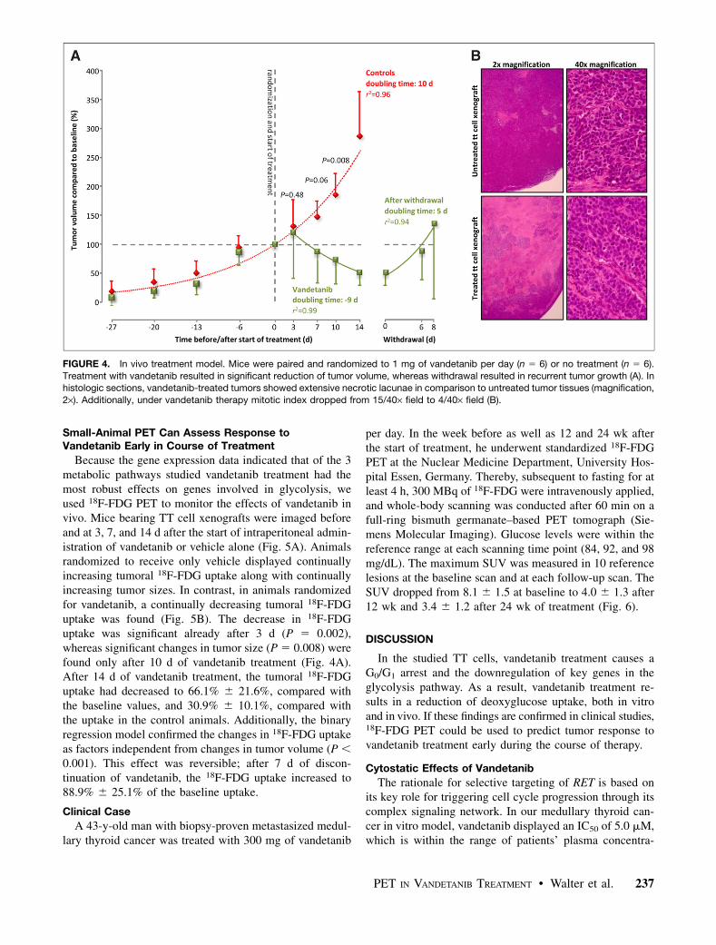
TT cell xenografts in mice receiving only vehicle showedan exponential growth curve with a doubling time of 10.0 d(r2 5 0.96, Fig. 4A). In contrast, xenografts in micerandomized for vandetanib treatment showed a volumereduction to 50% within 9.0 d. Significant treatment effects(P 5 0.008) on the tumor volume were established after 10d of vandetanib treatment. After withdrawal of vandetanib,all TT cell xenografts again displayed exponential growth(r2 5 0.94), with a mean doubling time of 5.1 d.
In naıve TT cell xenografts, microscopy demonstratedfew necrotic lagoons (magnification, 2·) with a mitoticindex of 15.0 6 6.4/40· field (magnification, 40·). In con-trast, after 14 d of vandetanib treatment, extensive necroticlagoons were found along with a significant (P 5 0.008)decrease of mitotic figures to 3.4 6 1.8/40· field (Fig. 3B).
FIGURE 3. In vitro effects of vandetanib on
tumor cell metabolism. In vitro treatment with
vandetanib resulted in downregulation of glu-cose carriers and glycolytic enzymes with
consecutively reduced 2DG uptake (A), down-
regulation of glycoprotein-associated aminoacid transporters with consecutively reduced
DOPA uptake (B), and downregulation of
enzymes of thymidine pathway with consecu-
tively reduced thymidine uptake (C) (represent-ing mean value of 2 separate experiments
conducted in triplicate). FLT 5 fluorothymidine.
236 THE JOURNAL OF NUCLEAR MEDICINE • Vol. 52 • No. 2 • February 2011
Small-Animal PET Can Assess Response toVandetanib Early in Course of Treatment
Because the gene expression data indicated that of the 3metabolic pathways studied vandetanib treatment had themost robust effects on genes involved in glycolysis, weused 18F-FDG PET to monitor the effects of vandetanib invivo. Mice bearing TT cell xenografts were imaged beforeand at 3, 7, and 14 d after the start of intraperitoneal admin-istration of vandetanib or vehicle alone (Fig. 5A). Animalsrandomized to receive only vehicle displayed continuallyincreasing tumoral 18F-FDG uptake along with continuallyincreasing tumor sizes. In contrast, in animals randomizedfor vandetanib, a continually decreasing tumoral 18F-FDGuptake was found (Fig. 5B). The decrease in 18F-FDGuptake was significant already after 3 d (P 5 0.002),whereas significant changes in tumor size (P 5 0.008) werefound only after 10 d of vandetanib treatment (Fig. 4A).After 14 d of vandetanib treatment, the tumoral 18F-FDGuptake had decreased to 66.1% 6 21.6%, compared withthe baseline values, and 30.9% 6 10.1%, compared withthe uptake in the control animals. Additionally, the binaryregression model confirmed the changes in 18F-FDG uptakeas factors independent from changes in tumor volume (P ,0.001). This effect was reversible; after 7 d of discon-tinuation of vandetanib, the 18F-FDG uptake increased to88.9% 6 25.1% of the baseline uptake.
Clinical Case
A 43-y-old man with biopsy-proven metastasized medul-lary thyroid cancer was treated with 300 mg of vandetanib
per day. In the week before as well as 12 and 24 wk afterthe start of treatment, he underwent standardized 18F-FDGPET at the Nuclear Medicine Department, University Hos-pital Essen, Germany. Thereby, subsequent to fasting for atleast 4 h, 300 MBq of 18F-FDG were intravenously applied,and whole-body scanning was conducted after 60 min on afull-ring bismuth germanate–based PET tomograph (Sie-mens Molecular Imaging). Glucose levels were within thereference range at each scanning time point (84, 92, and 98mg/dL). The maximum SUV was measured in 10 referencelesions at the baseline scan and at each follow-up scan. TheSUV dropped from 8.1 6 1.5 at baseline to 4.0 6 1.3 after12 wk and 3.4 6 1.2 after 24 wk of treatment (Fig. 6).
DISCUSSION
In the studied TT cells, vandetanib treatment causes aG0/G1 arrest and the downregulation of key genes in theglycolysis pathway. As a result, vandetanib treatment re-sults in a reduction of deoxyglucose uptake, both in vitroand in vivo. If these findings are confirmed in clinical studies,18F-FDG PET could be used to predict tumor response tovandetanib treatment early during the course of therapy.
Cytostatic Effects of Vandetanib
The rationale for selective targeting of RET is based onits key role for triggering cell cycle progression through itscomplex signaling network. In our medullary thyroid can-cer in vitro model, vandetanib displayed an IC50 of 5.0 mM,which is within the range of patients’ plasma concentra-
FIGURE 4. In vivo treatment model. Mice were paired and randomized to 1 mg of vandetanib per day (n 5 6) or no treatment (n 5 6).
Treatment with vandetanib resulted in significant reduction of tumor volume, whereas withdrawal resulted in recurrent tumor growth (A). Inhistologic sections, vandetanib-treated tumors showed extensive necrotic lacunae in comparison to untreated tumor tissues (magnification,
2·). Additionally, under vandetanib therapy mitotic index dropped from 15/40· field to 4/40· field (B).
PET IN VANDETANIB TREATMENT • Walter et al. 237

tions after oral administration (28). At this concentration,vandetanib treatment resulted in the transcriptional down-regulation of various effector pathways in TT cells, espe-cially the Rac, STAT3, and Grb7 and Grb10 pathways.Furthermore, interruption of RET signaling through thedownstream pathways effectively resulted in the downregu-lation of cyclin expression and prevented cell proliferation.
Metabolic Effects of Vandetanib
Consistent with our hypothesis, vandetanib also signifi-cantly affected the metabolic profile after only 3 d oftreatment. Gene expression profiling revealed transcrip-tional downregulation of key genes in glucose, DOPA, andthymidine metabolism. Among the RET-dependent path-ways, transcriptional downregulation was mainly found inmembers of the Grb7 and Grb10 and STAT3 pathways.Grb10 has previously been implicated in the regulation ofinsulin receptor signaling (29). Overexpression of Grb10 inL6 myocytes and 3T3-L1 adipocytes has been shown toenhance insulin-induced glucose transport, glycogen syn-thesis, and lipogenesis, whereas expression of dominant-
negative constructs, particularly the Grb10 SH2 domain,inhibited the insulin response (30). Interestingly, theGrb7/10/14 family was initially discovered through theirbinding to autophosphorylated epidermal growth factorreceptors (EGFRs) (29), suggesting a potential role of theGrb7 and Grb10 pathways also in the modulation of 18F-FDG uptake found after EGFR inhibition (31). Similarly,also interleukin-induced activation of STAT3 has beenshown to be essential for the expression of GLUTs, glucoseuptake, and activation of the glycolytic pathway (21). Con-versely, STAT3 inhibitors have been shown to blockGLUT2 expression and 2DG uptake (22). Consistent withthese observations, we found decreasing tumoral 18F-FDGuptake after vandetanib-dependent downregulation ofSTAT3 and the Grb7 and Grb10 pathways.
In this study, the glycolytic pathway was the only meta-bolic pathway with downregulation of both transporters aswell as intracellular enzymes. Furthermore, 18F-FDG hasthe best clinical availability and established value in medul-lary thyroid cancer imaging (32); therefore, it was selectedfor the in vivo imaging studies. In these in vivo studies, 18F-
FIGURE 5. In vivo treatment monitoring.
Metabolic changes under vandetanib
treatment could be visualized using 18F
small-animal PET (A). TT cell tumors ofvandetanib-treated mice showed signifi-
cantly lower 18F-FDG uptake already at 3 d
after initiation of treatment (B).
238 THE JOURNAL OF NUCLEAR MEDICINE • Vol. 52 • No. 2 • February 2011

FDG PET was able to reproduce the in vitro findings afteronly 3 d of vandetanib treatment. Importantly, the observeddecrease in 18F-FDG uptake after 3 d of treatment is notexplained by cell death because there was no increase inapoptotic cells in vitro at this time and no decrease in tumorsize in vivo. These results are consistent with clinical trials,in which tumor shrinkage in response to vandetanib hasbeen rare, even when patient survival was significantlyimproved (12).
Early Prediction of Treatment Response withCytostatic Drugs
The ability to predict tumor response early in the courseof therapy offers the opportunity to intensify treatment inpatients who are likely to respond to therapy. Conversely,treatment can be modified in patients who are unlikely torespond.In a recent phase II clinical trial, the disease control rate,
defined as complete response, partial response, or stabledisease by morphologic CT criteria (Response EvaluationCriteria in Solid Tumors), was similar in the arm receivingvandetanib versus the control arm (57% vs. 58%). In contrast,the vandetanib arm had a significantly longer progression-freesurvival (hazard ratio, 0.76) (12). These results demonstratethe poor prognostic value of morphologic responses to van-detanib treatment.
18F-FDG PET has been validated as an early indicatorof therapeutic responses in lymphoma (33) and a varietyof solid tumors, such as gastrointestinal stromal tumors(GIST), soft-tissue sarcomas (34), ovarian cancer (35), smallcell lung cancer (36), and adenocarcinoma of the oesopha-gogastric junction (37). In GIST tumors responding to thetyrosine kinase inhibitor imatinib (Gleevec), 18F-FDG PETdetects changes in tumor metabolism as early as 8 d aftertreatment initiation (38). Furthermore, 18F-FDG PET pre-dicts response as early as after 4 wk of therapy with themultitargeted tyrosine kinase inhibitor sunitinib in GIST(39). Vandetanib is a multitargeted tyrosine kinase inhibitor
against EGFR, vascular endothelial growth factor receptor,and RET. RET activation is the main oncogenic event inmedullary thyroid cancer (40). However, activity of otherreceptor tyrosine kinases, including vascular endothelialgrowth factor receptor and EGFR, are likely to contributeto tumor growth. Accordingly, 18F-FDG PET representsreadout of these combined effects of vandetanib treatment.The changes in the metabolic profile under vandetanib treat-ment found in this study provide a strong rationale to eval-uate 18F-FDG PET for the prediction of tumor response toRET kinase inhibitors in clinical trials.
CONCLUSION
A variety of novel approaches that target RET directly orindirectly have recently emerged, and an increasing numberare currently being assessed in clinical trials (40). Addition-ally, a variety of alternative small-molecule inhibitors arecurrently being evaluated (13). With the increasing numberof available treatment options, careful patient selection isnecessary to ensure targeted therapy is administered tothose most likely to gain clinical benefit. Therefore, theidentification of surrogate markers of treatment efficacy isa key factor for the success of these novel treatmentapproaches. If validated in clinical trials, metabolic imag-ing may significantly affect selection and response readoutof RET inhibition therapy.
ACKNOWLEDGMENTS
This study was supported in part by funds from theUniversity Basel and the Swiss National Science Foundation.
REFERENCES
1. de Groot JW, Links TP, Plukker JT, Lips CJ, Hofstra RM. RET as a diagnostic
and therapeutic target in sporadic and hereditary endocrine tumors. Endocr Rev.
2006;27:535–560.
2. Kouvaraki MA, Shapiro SE, Perrier ND, et al. RET proto-oncogene: a review
and update of genotype-phenotype correlations in hereditary medullary thyroid
cancer and associated endocrine tumors. Thyroid. 2005;15:531–544.
FIGURE 6. Clinical treatment monitoring.
Serial 18F-FDG PET scans of 43-y-old man
with metastasized medullary thyroid cancer
before (A) as well as at 12 (B) and 24 (C)weeks after start of treatment with 300 mg
of vandetanib per day. Maximum SUV in 10
reference lesions dropped from 8.1 6 1.5 at
baseline to 4.0 6 1.3 after 12 wk and 3.4 61.2 after 24 wk of treatment.
PET IN VANDETANIB TREATMENT • Walter et al. 239

3. Watanabe T, Ichihara M, Hashimoto M, et al. Characterization of gene expres-
sion induced by RET with MEN2A or MEN2B mutation. Am J Pathol.
2002;161:249–256.
4. Mettouchi A, Klein S, Guo W, et al. Integrin-specific activation of Rac controls
progression through the G1 phase of the cell cycle. Mol Cell. 2001;8:115–127.
5. Sinibaldi D, Wharton W, Turkson J, Bowman T, Pledger WJ, Jove R. Induction
of p21WAF1/CIP1 and cyclin D1 expression by the Src oncoprotein in mouse
fibroblasts: role of activated STAT3 signaling. Oncogene. 2000;19:5419–5427.
6. Winston JT, Coats SR, Wang YZ, Pledger WJ. Regulation of the cell cycle
machinery by oncogenic ras. Oncogene. 1996;12:127–134.
7. D’Amico M, Hulit J, Amanatullah DF, et al. The integrin-linked kinase regulates
the cyclin D1 gene through glycogen synthase kinase 3beta and cAMP-respon-
sive element-binding protein-dependent pathways. J Biol Chem. 2000;275:
32649–32657.
8. Malumbres M, Barbacid M. Cell cycle, CDKs and cancer: a changing paradigm.
Nat Rev Cancer. 2009;9:153–166.
9. Pero SC, Daly RJ, Krag DN. Grb7-based molecular therapeutics in cancer. Ex-
pert Rev Mol Med. 2003;5:1–11.
10. Patterson RL, van Rossum DB, Nikolaidis N, Gill DL, Snyder SH. Phospholipase
C-gamma: diverse roles in receptor-mediated calcium signaling. Trends Biochem
Sci. 2005;30:688–697.
11. Carlomagno F, Vitagliano D, Guida T, et al. ZD6474, an orally available inhib-
itor of KDR tyrosine kinase activity, efficiently blocks oncogenic RET kinases.
Cancer Res. 2002;62:7284–7290.
12. Heymach JV, Paz-Ares L, De Braud F, et al. Randomized phase II study of
vandetanib alone or with paclitaxel and carboplatin as first-line treatment for
advanced non-small-cell lung cancer. J Clin Oncol. 2008;26:5407–5415.
13. Sherman SI. Early clinical studies of novel therapies for thyroid cancers. Endo-
crinol Metab Clin North Am. 2008;37:511–524.
14. Therasse P, Arbuck SG, Eisenhauer EA, et al. New guidelines to evaluate the
response to treatment in solid tumors. European Organization for Research and
Treatment of Cancer, National Cancer Institute of the United States, National
Cancer Institute of Canada. J Natl Cancer Inst. 2000;92:205–216.
15. Macheda ML, Rogers S, Best JD. Molecular and cellular regulation of glucose
transporter (GLUT) proteins in cancer. J Cell Physiol. 2005;202:654–662.
16. Samih N, Hovsepian S, Aouani A, Lombardo D, Fayet G. Glut-1 translocation in
FRTL-5 thyroid cells: role of phosphatidylinositol 3-kinase and N-glycosylation.
Endocrinology. 2000;141:4146–4155.
17. Wright DC, Craig BW, Fick CA, Lim KI. The effects of phospholipase C in-
hibition on insulin-stimulated glucose transport in skeletal muscle. Metabolism.
2002;51:271–273.
18. Salter DW, Baldwin SA, Lienhard GE, Weber MJ. Proteins antigenically related
to the human erythrocyte glucose transporter in normal and Rous sarcoma virus-
transformed chicken embryo fibroblasts. Proc Natl Acad Sci USA. 1982;79:
1540–1544.
19. Hiraki Y, Rosen OM, Birnbaum MJ. Growth factors rapidly induce expression of
the glucose transporter gene. J Biol Chem. 1988;263:13655–13662.
20. Rollins BJ, Morrison ED, Usher P, Flier JS. Platelet-derived growth factor reg-
ulates glucose transporter expression. J Biol Chem. 1988;263:16523–16526.
21. Guillet-Deniau I, Pichard AL, Kone A, et al. Glucose induces de novo lipo-
genesis in rat muscle satellite cells through a sterol-regulatory-element-bind-
ing-protein-1c-dependent pathway. J Cell Sci. 2004;117:1937–1944.
22. Suh HN, Lee YJ, Han HJ. Interleukin-6 promotes 2-deoxyglucose uptake
through p44/42 MAPKs activation via Ca21/PKC and EGF receptor in primary
cultured chicken hepatocytes. J Cell Physiol. 2009;218:643–652.
23. Hennige AM, Lammers R, Hoppner W, et al. Inhibition of Ret oncogene ac-
tivity by the protein tyrosine phosphatase SHP1. Endocrinology. 2001;142:
4441–4447.
24. Walter MA, Seboek D, Demougin P, et al. Extraction of high-integrity RNA
suitable for microarray gene expression analysis from long-term stored human
thyroid tissues. Pathology. 2006;38:249–253.
25. Joost HG, Bell GI, Best JD, et al. Nomenclature of the GLUT/SLC2A family of
sugar/polyol transport facilitators. Am J Physiol. 2002;282:E974–E976.
26. Verrey F, Closs EI, Wagner CA, Palacin M, Endou H, Kanai Y. CATs and
HATs: the SLC7 family of amino acid transporters. Pflugers Arch. 2004;
447:532–542.
27. Baldwin SA, Beal PR, Yao SY, King AE, Cass CE, Young JD. The equilibrative
nucleoside transporter family, SLC29. Pflugers Arch. 2004;447:735–743.
28. Kovacs MJ, Reece DE, Marcellus D, et al. A phase II study of ZD6474 (Zactima), a
selective inhibitor of VEGFR and EGFR tyrosine kinase in patients with relapsed
multiple myeloma: NCIC CTG IND.145. Invest New Drugs. 2006;24:529–535.
29. Holt LJ, Siddle K. Grb10 and Grb14: enigmatic regulators of insulin action–and
more? Biochem J. 2005;388:393–406.
30. Deng Y, Bhattacharya S, Swamy OR, et al. Growth factor receptor-binding
protein 10 (Grb10) as a partner of phosphatidylinositol 3-kinase in metabolic
insulin action. J Biol Chem. 2003;278:39311–39322.
31. Su H, Bodenstein C, Dumont RA, et al. Monitoring tumor glucose utilization by
positron emission tomography for the prediction of treatment response to epidermal
growth factor receptor kinase inhibitors. Clin Cancer Res. 2006;12:5659–5667.
32. Khan N, Oriuchi N, Higuchi T, Endo K. Review of fluorine-18-2-fluoro-2-
deoxy-D-glucose positron emission tomography (FDG-PET) in the follow-up
of medullary and anaplastic thyroid carcinomas. Cancer Control. 2005;12:
254–260.
33. Terasawa T, Nihashi T, Hotta T, Nagai H. 18F-FDG PET for posttherapy assess-
ment of Hodgkin’s disease and aggressive Non-Hodgkin’s lymphoma: a system-
atic review. J Nucl Med. 2008;49:13–21.
34. Benz MR, Czernin J, Allen-Auerbach MS, et al. FDG-PET/CT imaging predicts
histopathologic treatment responses after the initial cycle of neoadjuvant che-
motherapy in high-grade soft-tissue sarcomas. Clin Cancer Res. 2009;15:2856–
2863.
35. Avril N, Sassen S, Schmalfeldt B, et al. Prediction of response to neoadjuvant
chemotherapy by sequential F-18-fluorodeoxyglucose positron emission tomog-
raphy in patients with advanced-stage ovarian cancer. J Clin Oncol. 2005;23:
7445–7453.
36. Weber WA, Petersen V, Schmidt B, et al. Positron emission tomography in non-
small-cell lung cancer: prediction of response to chemotherapy by quantitative
assessment of glucose use. J Clin Oncol. 2003;21:2651–2657.
37. Lordick F, Ott K, Krause BJ, et al. PET to assess early metabolic response and to
guide treatment of adenocarcinoma of the oesophagogastric junction: the MU-
NICON phase II trial. Lancet Oncol. 2007;8:797–805.
38. Stroobants S, Goeminne J, Seegers M, et al. 18FDG-Positron emission tomog-
raphy for the early prediction of response in advanced soft tissue sarcoma treated
with imatinib mesylate (Glivec). Eur J Cancer. 2003;39:2012–2020.
39. Prior JO, Montemurro M, Orcurto MV, et al. Early prediction of response to
sunitinib after imatinib failure by 18F-fluorodeoxyglucose positron emission to-
mography in patients with gastrointestinal stromal tumor. J Clin Oncol. 2009;27:
439–445.
40. Drosten M, Putzer BM. Mechanisms of disease: cancer targeting and the impact
of oncogenic RET for medullary thyroid carcinoma therapy. Nat Clin Pract
Oncol. 2006;3:564-74.
240 THE JOURNAL OF NUCLEAR MEDICINE • Vol. 52 • No. 2 • February 2011