Amide-to-Ester Substitution Allows Fine-Tuning of the Cyclopeptide Conformational Ensemble
6
Bioorganic Chemistry DOI: 10.1002/anie.200907274 Amide-to-Ester Substitution Allows Fine-Tuning of the Cyclopeptide Conformational Ensemble** Tommaso Cupido,* Jan Spengler, Javier Ruiz-Rodriguez, Jaume Adan, Francesc Mitjans, Jaume Piulats, and Fernando Albericio* Natural or designed peptide ligands rarely bind to cognate receptors in their most stable conformation when in solu- tion. [1] The receptor, through reciprocal induced fitting, [2] applies pressure on the conformational ensemble of the peptide to select its complementary conformation. Given the flexible nature of the peptides’ modular architecture, a bioactive sequence must often be primed for recognition of its target to achieve high binding affinity or specificity. For rational peptide design, cyclization or structure-inducing residues have been successfully used to accomplish ligand preorganization. [3] However, deconvolution of the averaged NMR spectra of strained peptides or other macrocycles had shown that the bound-state conformations are poorly popu- lated in the free uncomplexed state. [4] In small cyclic peptides, the stereochemistry of the backbone, rather than interactions with or among side chains, determines the conformational ensemble by establishing a defined pattern of local torsional preferences. [5] Intramolecular H bonds act on the equilibrium distribution of the conformational ensemble and favor specific conformations. Thus, we envisioned amide-to-ester substitution or “Ester Scan” as an interesting modification for the peptide back- bone, which influences the conformational ensemble as well as its equilibrium distribution; this influence is achieved through modulation of the backbone torsional preferences and H-bonding pattern, respectively. [6] As a model we choose cilengitide (CIL), which is an Arg- Gly-Asp (RGD) peptide of sequence cyclo[RGDfNMeV] (see Scheme 1), with well-characterized biological and con- formational properties. [7] CIL displays nanomolar inhibition of vitronectin binding to the isolated avb3 and avb5 integrin receptors, and it blocks integrin-dependent adhesion of tumor and endothelial cells to immobilized extracellular matrix (ECM) proteins and reduces angiogenesis and tumor growth in vivo. [8] Modulation of the internal H-bond pattern of nonmethylated CIL precursors—achieved by changing the flexibility or the chirality of the backbone—was found to influence the antagonist activity on the vitronectin (VN) and fibrinogen (FB) receptors, and has been proposed to control laminin P1 vs. vitronectin receptor specificity. [9] We synthesized all five depsi-analogues of the depsipep- tide CIL (D1–D5) by stepwise assembling of the linear precursors on 2-CTC resin (Fmoc/tBu strategy) and cycliza- tion in solution. For the introduction of the a-hydroxy acid residues onto the growing peptide chain, their HFA-acti- vated/protected derivatives were used [10] (Scheme 1). For the acylation of the free hydroxy group, DIPDCI/DMAP-activa- tion was used. [11] For conventional peptide cyclization, the optimal site for macrocycle formation is between the Gly (acting as the C terminus) and Asp (acting as the N terminus) residues because Gly cannot epimerize. This strategy was used for the synthesis of parent CIL, D1, and D2 depsipep- tides with PyBOP/HOAt. [12] The macrolactonization required for the preparation of D3 was successful with MSNT activation and NMI as the base. For the depsipeptides D4 and D5, certain particularities of the ester bond had to be taken into account. Our attempts to synthesize the linear precursor of D4 (OGly analogue) starting from OGly as the C terminus, resulted in low yields, and was likely because of cleavage of the ester bond that was mediated by base during repeated treatment with piperidine. Therefore, NMeVal was chosen as the C terminus. For the preparation of D5, the cyclization was performed at the (apparently) less attractive position between ( d)Phe and NMeVal. Macrolactamization at less hindered sites was impeded by intramolecular nucleo- philic attack on the ester bond, which occurred during peptide chain elongation, and eliminated the (D)Phe–NMeVal couple as dioxopiperazine. [13] Given the increased steric demand of the N-terminal NMeVal, we chose the more reactive PyAOP as the coupling reagent and HOAt as the additive. [14] After cleavage from the solid support, all linear peptide and depsipeptide precursors were obtained in over 85 % yield. Head-to-tail cyclization was performed in solution and gave good yields in all cases and, finally, the protecting groups on the side chains were removed using [Pd(PPh 3 ) 4 ]/phenylsilane [*] T. Cupido, Dr. J. Spengler, Dr. J. Ruiz-Rodriguez, Prof. F. Albericio Institute for Research in Biomedicine (IRB), Barcelona Science Park (PCB) and CIBER-BBN Networking Centre on Bioengineering, Biomaterials and Nanomedicine PCB Baldiri Reixac 10, 08028 Barcelona (Spain) Fax: (+ 34) 93-403-7126 E-mail: [email protected] [email protected] Prof. F. Albericio Department of Organic Chemistry, University of Barcelona Martȶ i Franquŕs 1–11, 08028 Barcelona (Spain) Dr. J. Adan, Dr. F. Mitjans, Dr. J. Piulats Biomed Division, Leitat Technological Center Institution, Barcelona Science Park, Baldiri Reixach 15–21, 08028 Barcelona (Spain) [**] This work was partially supported by CICYT (CTQ2009-07758 and CTQ2008-02856/BQU). The Generalitat de Catalunya (2009SGR 1024, 2009SGR-1472), the Institute for Research in Biomedicine, and the Barcelona Science Park are also acknowledged for support. Supporting information for this article (experimental details of cyclodepsipeptide syntheses, cellular assays, structure analysis and calculations, characterization of compounds, and MD calculations) is available on the WWW under http://dx.doi.org/10.1002/anie. 200907274. Communications 2732 # 2010 Wiley-VCH Verlag GmbH & Co. KGaA, Weinheim Angew. Chem. Int. Ed. 2010, 49, 2732 –2737
Transcript of Amide-to-Ester Substitution Allows Fine-Tuning of the Cyclopeptide Conformational Ensemble

Bioorganic ChemistryDOI: 10.1002/anie.200907274
Amide-to-Ester Substitution Allows Fine-Tuning of the CyclopeptideConformational Ensemble**Tommaso Cupido,* Jan Spengler, Javier Ruiz-Rodriguez, Jaume Adan, Francesc Mitjans,Jaume Piulats, and Fernando Albericio*
Natural or designed peptide ligands rarely bind to cognatereceptors in their most stable conformation when in solu-tion.[1] The receptor, through reciprocal induced fitting,[2]
applies pressure on the conformational ensemble of thepeptide to select its complementary conformation. Given theflexible nature of the peptides’ modular architecture, abioactive sequence must often be primed for recognition ofits target to achieve high binding affinity or specificity. Forrational peptide design, cyclization or structure-inducingresidues have been successfully used to accomplish ligandpreorganization.[3] However, deconvolution of the averagedNMR spectra of strained peptides or other macrocycles hadshown that the bound-state conformations are poorly popu-lated in the free uncomplexed state.[4] In small cyclic peptides,the stereochemistry of the backbone, rather than interactionswith or among side chains, determines the conformationalensemble by establishing a defined pattern of local torsionalpreferences.[5] Intramolecular H bonds act on the equilibriumdistribution of the conformational ensemble and favorspecific conformations.
Thus, we envisioned amide-to-ester substitution or “EsterScan” as an interesting modification for the peptide back-bone, which influences the conformational ensemble as wellas its equilibrium distribution; this influence is achievedthrough modulation of the backbone torsional preferencesand H-bonding pattern, respectively.[6]
As a model we choose cilengitide (CIL), which is an Arg-Gly-Asp (RGD) peptide of sequence cyclo[RGDfNMeV](see Scheme 1), with well-characterized biological and con-formational properties.[7] CIL displays nanomolar inhibitionof vitronectin binding to the isolated avb3 and avb5 integrinreceptors, and it blocks integrin-dependent adhesion of tumorand endothelial cells to immobilized extracellular matrix(ECM) proteins and reduces angiogenesis and tumor growthin vivo.[8] Modulation of the internal H-bond pattern ofnonmethylated CIL precursors—achieved by changing theflexibility or the chirality of the backbone—was found toinfluence the antagonist activity on the vitronectin (VN) andfibrinogen (FB) receptors, and has been proposed to controllaminin P1 vs. vitronectin receptor specificity.[9]
We synthesized all five depsi-analogues of the depsipep-tide CIL (D1–D5) by stepwise assembling of the linearprecursors on 2-CTC resin (Fmoc/tBu strategy) and cycliza-tion in solution. For the introduction of the a-hydroxy acidresidues onto the growing peptide chain, their HFA-acti-vated/protected derivatives were used[10] (Scheme 1). For theacylation of the free hydroxy group, DIPDCI/DMAP-activa-tion was used.[11] For conventional peptide cyclization, theoptimal site for macrocycle formation is between the Gly(acting as the C terminus) and Asp (acting as the N terminus)residues because Gly cannot epimerize. This strategy wasused for the synthesis of parent CIL, D1, and D2 depsipep-tides with PyBOP/HOAt.[12] The macrolactonization requiredfor the preparation of D3 was successful with MSNTactivation and NMI as the base. For the depsipeptides D4and D5, certain particularities of the ester bond had to betaken into account. Our attempts to synthesize the linearprecursor of D4 (OGly analogue) starting from OGly as theC terminus, resulted in low yields, and was likely because ofcleavage of the ester bond that was mediated by base duringrepeated treatment with piperidine. Therefore, NMeVal waschosen as the C terminus. For the preparation of D5, thecyclization was performed at the (apparently) less attractiveposition between (d)Phe and NMeVal. Macrolactamization atless hindered sites was impeded by intramolecular nucleo-philic attack on the ester bond, which occurred during peptidechain elongation, and eliminated the (D)Phe–NMeVal coupleas dioxopiperazine.[13] Given the increased steric demand ofthe N-terminal NMeVal, we chose the more reactive PyAOPas the coupling reagent and HOAt as the additive.[14]
After cleavage from the solid support, all linear peptideand depsipeptide precursors were obtained in over 85% yield.Head-to-tail cyclization was performed in solution and gavegood yields in all cases and, finally, the protecting groups onthe side chains were removed using [Pd(PPh3)4]/phenylsilane
[*] T. Cupido, Dr. J. Spengler, Dr. J. Ruiz-Rodriguez, Prof. F. AlbericioInstitute for Research in Biomedicine (IRB), Barcelona Science Park(PCB) and CIBER-BBN Networking Centre on Bioengineering,Biomaterials and NanomedicinePCB Baldiri Reixac 10, 08028 Barcelona (Spain)Fax: (+ 34)93-403-7126E-mail: [email protected]
Prof. F. AlbericioDepartment of Organic Chemistry, University of BarcelonaMart� i Franqu�s 1–11, 08028 Barcelona (Spain)
Dr. J. Adan, Dr. F. Mitjans, Dr. J. PiulatsBiomed Division, Leitat Technological Center Institution, BarcelonaScience Park, Baldiri Reixach 15–21, 08028 Barcelona (Spain)
[**] This work was partially supported by CICYT (CTQ2009-07758 andCTQ2008-02856/BQU). The Generalitat de Catalunya (2009SGR1024, 2009SGR-1472), the Institute for Research in Biomedicine,and the Barcelona Science Park are also acknowledged for support.
Supporting information for this article (experimental details ofcyclodepsipeptide syntheses, cellular assays, structure analysis andcalculations, characterization of compounds, and MD calculations)is available on the WWW under http://dx.doi.org/10.1002/anie.200907274.
Communications
2732 � 2010 Wiley-VCH Verlag GmbH & Co. KGaA, Weinheim Angew. Chem. Int. Ed. 2010, 49, 2732 –2737

for the allyl group of D3, and TFA/TIS/H2O for the tBu groupof all other pepitides. Finally, purification by HPLC providedthe macrocycles CIL and D1–D5 in 95 % purity.
For the synthesized compounds, their antagonist activitytowards distinct integrin subtypes was tested indirectly as thecapacity to inhibit the initial adhesion of a set of cellular linesto the appropriate ECM protein substrate. Adhesion inhib-ition assays of integrin avb3 and avb5 were carried out byperforming a double-dependent adhesion study of HUVECendothelial and M21 melanoma cells on VN.[15] Inhibition byCIL and D4 was detected in the high nM range; D5 was atleast three times more active; D1 showed a two-fold reductionin potency; D2 was one order of magnitude less potent thancilengitide; D3 was totally inactive at concentration as high as30 mm (Table 1). To ascertain whether these differences inpotency were a result of the preferential binding of the
RGD cyclodepsipeptides to either ofthe avb5 or avb3 integrin receptors, weperformed an adhesion assay withM21 cells plated on FB (avb3-depen-dent adhesion only), and another withHT29 colon adenocarcinoma cellsplated on VN (avb5-dependent adhe-sion only). The same pattern of inhib-itory potency described above wasobserved in these assays. HT29 cellsalso adhere to fibronectin (FN)through the closely related avb6 inte-grin subtype; neither CIL nor depsi-peptides D1–D4 inhibited the adhesionof HT29 cells to this substrate (data notshown), in contrast, D5 showed mod-erate inhibitory activity (IC50 value:8.64 mm). As expected, neither CIL norany of the depsipeptides inhibited theadhesion of M21 cells to collagen or FN(a2b1- and a3b1-dependent adhe-sion[16]), thus indicating that they donot antagonize non-av-containingintegrins (data not shown).
Ligand-occupied avb3 and avb5 re-ceptors down-regulate apoptotic sig-nals from unattached integrins andpromote the survival of differentiatedendothelial cells.[17] CIL antagonizesoccupied avb3 and avb5 integrin andinhibits the proliferation of HUVECcells plated on purified VN (Table 2).Analogue D5 displayed an IC50 valueten-fold lower than that of CIL. Ana-logues D4 and D1 showed inhibitoryactivity similar to that of CIL, whereasD2 was more than ten-fold less potent.Again D3 showed no inhibitory activ-ity. As a positive control, we used theanti-av-integrin monoclonal antibodyMab 17E6.[18]
The conformation of the synthe-sized compounds in solution was stud-
ied by 1D and 2D 1H NMR spectroscopy. The high temper-ature coefficient of the NH-Arg group, the narrow range onwhich the 3JHAHN coupling constants were distributed, and a
Scheme 1. Synthetic scheme and chemical structures of final RGD cyclodepsipeptides andcilengitide. 2-CTC= 2-chlorotrityl chloride, DIPDCI=1,3-diisopropyldicarboimide, DMAP=
4-dimethylaminopyridine, Fmoc =9-fluorenylmethyloxycarbonyl, HFA = hexafluoroacetone,HOAt = 1-hydroxy-7-azabenzotriazole, MSNT= 1-(2-mesitylenesulfonyl)-3-nitro-1,2,4-triazole,NMI = N-methylimidazole, PyAOP= 7-azabenzotriazol-1-yloxy)tripyrrolidinophosphonium hexa-fluorophosphate, PyBOP= 1-benzotriazolyloxytris(pyrollidino)phosphonium, SPPS = solid-phasepeptide synthesis.
Table 1: Adhesion inhibition assays of CIL and D1–D5. IC50 values aregiven in mm and the standard deviations are in parentheses.
Peptides avb3 + avb5 avb3 avb5HUVEC on VN M21 on VN M21 on FB HT29 on VN
CIL 0.46 (0.10) 0.21 (0.10) 0.32 (0.10) 0.20 (0.04)D1 0.84 (0.15) 0.45 (0.21) 0.37 (0.10) 0.41 (0.16)D2 4.32 (0.82) 5.35 (0.75) 5.67 (0.90) 20.51 (3.3)D3 >30 >30 >30 >30D4 0.27 (0.05) 0.19 (0.06) 0.20 (0.10) 0.58 (0.13)D5 0.18 (0.06) 0.08 (0.02) 0.10 (0.02) 0.06 (0.03)Mab 17E6 0.001 0.001 0.001 0.0001
AngewandteChemie
2733Angew. Chem. Int. Ed. 2010, 49, 2732 –2737 � 2010 Wiley-VCH Verlag GmbH & Co. KGaA, Weinheim www.angewandte.org

conserved pattern of conformationally relevant NOE inter-actions (see the Supporting Information for details) suggestthat the cyclodepsipeptide analogues of cilengitide retain theoverall conformation and side chains topology of the parentcyclopeptide. The time- and ensemble-averaged 3D struc-tures of the synthesized compounds, obtained by simulatedannealing (SA) calculations, were generally very similar—with most variability limited to the (d)Phe F angle and theAsp Y angle (i.e. the tilt angle of the Asp-(d)Phe amidegroup; see Figure 1 and the Supporting Information). Theonly significant violation of the NMR constraints was found inthe lowest-energy conformation of D4, in which the amideplane of Asp-(d)Phe was flipped about 1308 with respect to itsrestrained position, thus suggesting that a conformationalequilibrium is occurring in this cyclodepsipeptide analogue.Despite the likeness of their lowest-energy NMR structures,the antagonist potencies of the cyclodepsipeptides on theintegrin receptors differed by more than two orders ofmagnitude.
The observation that the solution (Figure 1; picture to theleft of each structure) and the integrin-bound cilengitidestructures[19] show different arrangements of the RGD se-quence led us to the following hypothesis: divergence in theIC50 values for cyclodepsipeptide analogues of cilengitide mayresult from a differential population of the RGD conforma-tion competent for receptor binding (see the SupportingInformation).
We then attempted molecular dynamics (MD) calcula-tions to gain insight into the distributions that make up the
averages of the conformational properties, as determined byNMR spectroscopy. Unrestrained simulation trajectories of22 ns in water were generated using a polarizable forcefield.[20] The lowest-energy SA conformations were used asstarting structures (see the Supporting Information). Clusteranalysis of the trajectories obtained by the MD calculationsconfirmed that CIL and RGD cyclodepsipeptides D1–D5populate similar conformational ensembles. These ensemblesare composed of several conformers that differed by thetwisting and flipping angles of the planes for amide and esterbonds.
The residence time of single conformers (mostly 10�8–10�9 s) and the dynamics of the conformational changes,which are associated with the ester or amide plane flipping,are governed by the torsional preferences of the ester bondsand by the fast (10�11 s) formation and disruption of back-bone-to-backbone H-bonds. Such an ensemble approachcorrelates well with the observed NOE interactions andother NMR measurements, and furnishes an explanation forthe bioactivity of RGD cyclodepsipeptides. In CIL, fastflipping of the amide plane for the Arg-Gly bond definestwo major families of conformers, A (including the SAstructure) and B (including the integrin bound, and thepreviously published cilengitide solution structure[7]), whichare populated 55% and 35% of the simulation time,respectively (Figure 2a). While in the A or B conformer,CIL oscillates between several closely related conformers infast equilibrium among their torsional forms. Analogue D2can be described by an equilibrium of two closely related Aand A’ conformers, in which the amide groups of the bioactiveRGD sequence are oriented in the same direction (Fig-ure 2a). The two conformers differed by the twist angle of theester group.
In analogy with CIL, the flip of the plane for the Arg-Glyamide group, which converts conformer A into B, was alsoobserved during the MD calculations of D2. Conformer B ofD2, however, appeared less stable and with a shorterresidence time than that of its all-amide counterpart (com-
Table 2: Proliferation inhibition assay of CIL and D1–D5. IC50 values aregiven in mm and the standard deviations are in parentheses.
Peptide HUVEC cells Peptide HUVEC cells
CIL 0.36 (0.10) D4 0.54 (0.09)D1 0.34 (0.20) D5 0.04 (0.01)D2 6.43 (2.4) Mab 17E6 0.00018D3 >30
Figure 1. CIL and D1–D5 3D structures obtained by SA. For each macrocycle, the full SA lowest-energy structure is shown, and to its left fivebackbone conformations representing SA solutions within 3 kcalmol�1 are shown (see the Supporting Information for details).
Communications
2734 www.angewandte.org � 2010 Wiley-VCH Verlag GmbH & Co. KGaA, Weinheim Angew. Chem. Int. Ed. 2010, 49, 2732 –2737
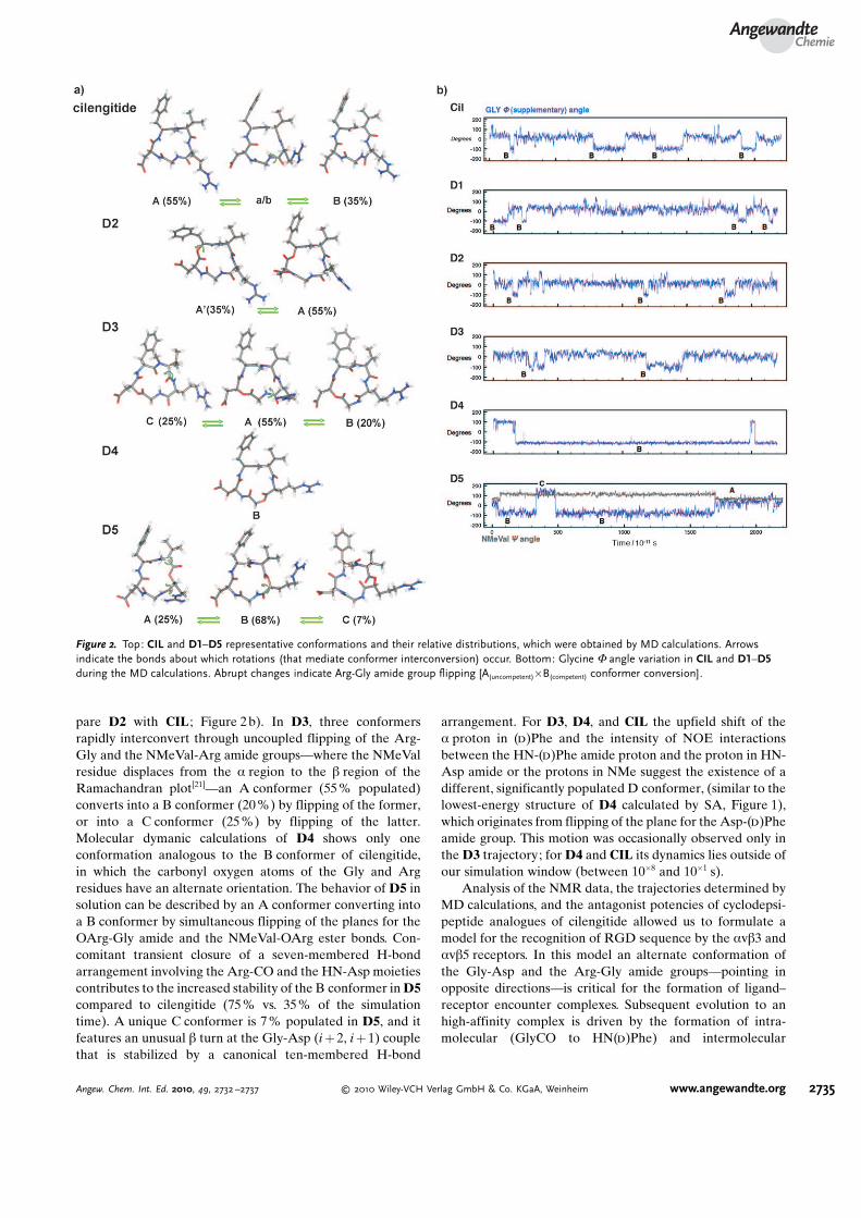
pare D2 with CIL ; Figure 2 b). In D3, three conformersrapidly interconvert through uncoupled flipping of the Arg-Gly and the NMeVal-Arg amide groups—where the NMeValresidue displaces from the a region to the b region of theRamachandran plot[21]—an A conformer (55 % populated)converts into a B conformer (20 %) by flipping of the former,or into a C conformer (25%) by flipping of the latter.Molecular dymanic calculations of D4 shows only oneconformation analogous to the B conformer of cilengitide,in which the carbonyl oxygen atoms of the Gly and Argresidues have an alternate orientation. The behavior of D5 insolution can be described by an A conformer converting intoa B conformer by simultaneous flipping of the planes for theOArg-Gly amide and the NMeVal-OArg ester bonds. Con-comitant transient closure of a seven-membered H-bondarrangement involving the Arg-CO and the HN-Asp moietiescontributes to the increased stability of the B conformer in D5compared to cilengitide (75 % vs. 35% of the simulationtime). A unique C conformer is 7% populated in D5, and itfeatures an unusual b turn at the Gly-Asp (i + 2, i + 1) couplethat is stabilized by a canonical ten-membered H-bond
arrangement. For D3, D4, and CIL the upfield shift of thea proton in (d)Phe and the intensity of NOE interactionsbetween the HN-(d)Phe amide proton and the proton in HN-Asp amide or the protons in NMe suggest the existence of adifferent, significantly populated D conformer, (similar to thelowest-energy structure of D4 calculated by SA, Figure 1),which originates from flipping of the plane for the Asp-(d)Pheamide group. This motion was occasionally observed only inthe D3 trajectory; for D4 and CIL its dynamics lies outside ofour simulation window (between 10�8 and 10�1 s).
Analysis of the NMR data, the trajectories determined byMD calculations, and the antagonist potencies of cyclodepsi-peptide analogues of cilengitide allowed us to formulate amodel for the recognition of RGD sequence by the avb3 andavb5 receptors. In this model an alternate conformation ofthe Gly-Asp and the Arg-Gly amide groups—pointing inopposite directions—is critical for the formation of ligand–receptor encounter complexes. Subsequent evolution to anhigh-affinity complex is driven by the formation of intra-molecular (GlyCO to HN(d)Phe) and intermolecular
Figure 2. Top: CIL and D1–D5 representative conformations and their relative distributions, which were obtained by MD calculations. Arrowsindicate the bonds about which rotations (that mediate conformer interconversion) occur. Bottom: Glycine F angle variation in CIL and D1–D5during the MD calculations. Abrupt changes indicate Arg-Gly amide group flipping [A(uncompetent)�B(competent) conformer conversion].
AngewandteChemie
2735Angew. Chem. Int. Ed. 2010, 49, 2732 –2737 � 2010 Wiley-VCH Verlag GmbH & Co. KGaA, Weinheim www.angewandte.org
(HNAspcilengitide-(b)Arg-216avb3 integrin) H-bonds[22] (see the Sup-porting Information).
In this scenario, the dynamics of the plane flip of the Arg-Gly amide group and the stability of the alternate conforma-tion are key determinants of the binding affinity of RGD cy-clopeptides and cyclodepsipeptides to the av integrins. Theconformational preference of the nanomolar antagonistcilengitide is still dominated by an unproductive A conformerthat represents the most stable conformation in solution,while the alternate B conformer is only 35 % populated(Figure 2a). Accordingly, D5 had improved potency becausethe alternate conformation is promoted by the concomitantflip of the plane for the ester bond and stabilized by theformation of a transient backbone H-bond. Analogue D3,despite significantly populating the B “alternate” conformer,has no H-bond donor capability at the Asp residue becausethe NH group is substituted by an oxygen atom, and it lackedany antagonistic activity. Analogue D2 showed severelydiminished antagonist character because it poorly populatesthe B conformer (Figure 2a).
So far, the use of MD calculations to quantify conforma-tion–activity relationships have been severely limited by thetime resolution of state-of-art MD calculations, which isrestricted to the ms range, and therefore inadequate to capturesome aspects of the conformational flexibility of peptides andproteins. In this study, such an analysis is further complicatedby the general inaccuracy of current force fields for thedescription of nonstandard residues. Indeed, our 22 ns MDcalculations failed to completely reproduce the conforma-tional behavior of all RGD macrocycles. As gauged byMD calculations, D4 mostly adopts the complementary B-receptor conformer, whereas the NMR data showed evidenceof a significantly populated D conformer (which remainedunexplored during the dynamics experiments). Then, MD cal-culations led to an overestimation of the population for theB conformer and the antagonist potency of D4. According tothe presence of the D conformer, D4 displayed an antagonistpotency comparable to that of CIL and inferior to that ofanalogue D5.
Taken together these results demonstrate that the tar-geted application of amide-to-ester substitution or “EsterScan” in the design of cyclic peptides can be used to fine-tunethe backbone conformation and to select a bioactive confor-mer from the conformational pool. Remarkably, D5 hadthree- to ten-fold increased potency with respect to the parentcompound, depending on the assay. To the best of ourknowledge, D5 is the most potent small-molecule dual avb3and avb5 antagonist discovered to date. Preorganization offlexible peptides into their bioactive conformations has beenoften achieved by adding local conformational constraintsthat reduce the allowed backbone F and Y angles. Herein, wehave described a complementary approach which relies onrelieving specific conformational constraints by amidereplacement with ester groups, thus leading to a targetedreorganization of the internal equilibrium of the conforma-tional ensemble.
Received: December 24, 2009Published online: March 8, 2010
.Keywords: bioorganic chemistry · conformation analysis ·depsipeptides · molecular dynamics · NMR spectroscopy
[1] E. Perola, S. P. Charifson, J. Med. Chem. 2004, 47, 2499.[2] G. H. Loew, S. K. Burt, Proc. Natl. Acad. Sci. USA 1978, 75, 7 –
11.[3] a) J. D. Tyndall, B. Pfeiffer, G. Abbenante, D. P. Fairlie, Chem.
Rev. 2005, 105, 793; b) R. M. Freidinger, D. F. Veber, D. S.Perlow, J. R. Brooks, R. Sapestein, Science 1980, 210, 4470;c) R. M. Freidinger, et al., J. Med. Chem. 1990, 33, 1843 (see theSupporting Information for full citation).
[4] a) S. Horne, C. A. Olsen, J. M. Beierle, A. Montero, M. R.Ghadiri, Angew. Chem. 2009, 121, 4812; Angew. Chem. Int. Ed.2009, 48, 4718; b) J. Jim�nez-Barbero, A. Canales, P. T. North-cote, R. M. Buey, J. M. Andreu, J. F. Diaz, J. Am. Chem. Soc.2006, 128, 8757.
[5] R. Haubner, R. Gratias, B. Diefenbach, S. L. Goodman, A.Jonczyk, H. Kessler, J. Am. Chem. Soc. 1996, 118, 7461.
[6] a) E. T. Powers, S. Deechongkit, J. W. Kelly, Adv. Protein Chem.2005, 72, 39; b) J. A. Scheike, C. Baldauf, J. Spengler, F.Albericio, M. T. Pisabarro, B. Koksch, Angew. Chem. 2007,119, 7912; Angew. Chem. Int. Ed. 2007, 46, 7766.
[7] M. A. Dechantsreiter, E. Planker, B. Matha, E. Lohof, G.Holzemann, A. Jonczyk, S. L. Goodman, H. Kessler, J. Med.Chem. 1999, 42, 3033.
[8] a) G. C. Alghisi, L. Ponsonnet, C. Ruegg, PLoS One 2009, 4,e4449; b) S. M. Weis, D. G. Stupack, D. A. Cheresh, Cancer Cell2009, 15, 359; c) Recent results shown that low nM concen-trations of cilengitide can promote tumour angiogenesis byagonizing instead of antagonizing the avb3 integrin. A. R.Reynolds, et al., Nat. Med. 2009, 15, 392. (see the SupportingInformation for full citation).
[9] a) A. Geyer, G. M�ller, H. Kessler, J. Am. Chem. Soc. 1994, 116,7735; b) G. M�ller, M. Gurrath, H. Kessler, R. Timpl, Angew.Chem. 1992, 104, 341; Angew. Chem. Int. Ed. Engl. 1992, 31, 326.
[10] a) F. Albericio, K. Burger, J. Ruiz-Rodriguez, J. Spengler, Org.Lett. 2005, 7, 597; b) Typically, this coupling step requires 4 hoursin THF, whereas the coupling of HFA-(d)OPhe to the N-terminal NMeVal required 48 hours and a higher number ofmolar equivalents. Glycolic acid (OGly), phenylacetic acid[(d)OPhe], and hydroxyvalerianic acid (OVal) used for thesubstitution of the Gly, (d)Phe, and NMeVal are commerciallyavailable. Appropriately protected malic (OAsp) and argininicacid (OArg) were obtained as described; for OAsp, see: K.Pumpor, E. Windeisen, J. Spengler, F. Albericio, K. Burger,Monatsh. Chem. 2004, 135, 1427; for OArg, see: T. Cupido, J.Spengler, K. Burger, F. Albericio, Tetrahedron Lett. 2005, 46,6733.
[11] O. Kuisle, E. Quino�, R. J. Riguera, Org. Chem. 1999, 64, 8063.[12] F. Albericio, J. M. Bofill, A. El-Faham, S. A. Kates, J. Org. Chem.
1998, 63, 9678.[13] a) B. F. Gisin, R. B. Merrifield, J. Am. Chem. Soc. 1972, 94, 3102;
b) S. Capasso, A. Vergara L. Mazzarella, J. Am. Chem. Soc. 1998,120, 1990.
[14] a) Phosphonium salts are preferred ahead of the uronium/aminium salts, because they do not provoke capping of theN terminal function as the latter do, see: F. Albericio, M. Cases,J. Alsina, S. A. Triolo, L. A. Carpino, S. A. Kates, TetrahedronLett. 1997, 38, 4853.
[15] a) D. G. Stupack, X. S. Puente, S. Boutsaboualoy, C. M. Stor-gard, D. A. Cheresh, J. Cell Biol. 2001, 155, 459; b) S. Maubant,D. Saint-Dizier, M. Boutillon, F. Perron-Sierra, P. J. Casara, J. A.Hickman, G. C. Tucker, E. Van Obberghen-Schilling, Blood2006, 108, 3035.
[16] C. Li, J. B. McCarthy, L. T. Furcht, G. B. Fields, Biochemistry1997, 36, 15404.
Communications
2736 www.angewandte.org � 2010 Wiley-VCH Verlag GmbH & Co. KGaA, Weinheim Angew. Chem. Int. Ed. 2010, 49, 2732 –2737
[17] D. G. Stupack, D. A. Cheresh, Oncogene 2003, 22, 9022.[18] F. Mitjans, D. Sander, J. Adan, A. Sutter, J. M. Martinez, C. S.
Jaggle, J. M. Moyano, H. G. Kreysch, J. Piulats, S. L. Goodman, J.Cell Sci. 1995, 108, 2825.
[19] J. Xiong, T. Stehle, R. Zhang, A. Joachimiak, M. Frech, S. L.Goodman, M. A. Arnaout, Science 2002, 296, 151.
[20] a) R. W. Dixon, P. A. Kollman, J. Comput. Chem. 1997, 18, 1632;b) V. Babin, J. Baucom, T. A. Darden, C. Sagui, J. Phys. Chem. B2006, 110, 11571.
[21] C. M. Wilmot, J. M. Thornton Protein Engineering, Design andSelection 1990, 3, 479.
[22] L. Marinelli, A. Lavecchia, K. E. Gottschalk, E. Novellino, H.Kessler, J. Med. Chem. 2003, 46, 4393.
AngewandteChemie
2737Angew. Chem. Int. Ed. 2010, 49, 2732 –2737 � 2010 Wiley-VCH Verlag GmbH & Co. KGaA, Weinheim www.angewandte.org