
An amide based dipodal Zn2+ complex for multications recognition: Nanomolar detection
6
An amide based dipodal Zn 2 þ complex for multications recognition: Nanomolar detection Umesh Fegade a,c , Hemant Sharma b , Narinder Singh b , Sopan Ingle c , Sanjay Attarde c , Anil Kuwar a,n a School of Chemical Sciences, North Maharashtra University, Jalgaon 425001, Maharashtra, India b Department of Chemistry, Indian Institute of Technology, Ropar, Rupanagar, Punjab, India c School of Environmental and Earth Sciences, North Maharashtra University, Jalgaon 425001, Maharashtra, India article info Article history: Received 9 October 2013 Received in revised form 14 November 2013 Accepted 13 January 2014 Available online 22 January 2014 Keywords: Zinc receptor Nanomolar detection Density functional theory abstract An imine-linked dipodal Zn 2 þ complex has been synthesized and elevated its binding affinity towards library of metal ions (Cr 3 þ , Mn 2 þ , Fe 3 þ , Co 2 þ , Ni 2 þ , Cu 2 þ , Zn 2 þ , Cd 2 þ , Hg 2 þ , Pb 2 þ and Bi 3 þ ). The recognition properties of receptor 4 were performed in DMSO/H 2 O (50:50; v/v) and showed quenching upon interaction with Fe 3 þ , Co 2 þ and Cu 2 þ ions. A photoinduced electron transfer (PET) is potential mechanism behind quenching. The coordination of such cations with the receptor 4 at amide part of carbonyl oxygen hindered the PET process and accordingly Turn-OFF the fluorescence of the receptor 4. The photophysical properties (fluorescence and absorption spectra) and density functional theory (DFT) clearly explains the binding mode between the receptor 4 and the detected cations. The selectivity and sensitivity of the receptor 4 for Fe 3 þ , Co 2 þ and Cu 2 þ ions were acceptable and achieving a detection limit at the nano-molar level. & 2014 Elsevier B.V. All rights reserved. 1. Introduction The determination of metal ions has continually gained remarkable interest in last few decades due to its importance in chemistry and biology [1]. Among them, biologically important transition metal ions play a key role in many biological and environmental processes [2]. Hence, it is compulsory to establish accurate and simple methods for the quick determination of transition metal ions due to wide utility. Until now, a lot of methods have been used to determinate transition metals including inductively coupled plasma mass (ICP-MS) [3], atomic absorption spectroscopy [4], colorimetry [5], flow injection [6] and electrochemical methods [7]. On the other hand, fluorescence approaches have increased greater awareness due to their simple equipment, rapid comeback, sky-scraping sensitivity and straightfor- ward action. Based on their availability, low cost, long-wavelength emission, with high molar absorption coefficient and quantum yield, malonohydrazide based receptor has already been used as fluorescent probes to determinate transition metal ion recently [8]. Most of the iron present in biological systems is tightly associated with enzymes and specialized transport and storage proteins e.g. in haemoglobin, the red pigment in the erythrocytes and rest of stored in ferretin. It plays a vital role in oxygen transfer processes in DNA and RNA synthesis [9]. The deficiency of Fe 3 þ causes anaemia, hemochromatosis, liver damage [10] and Parkin- son's disease [11]. Cobalt(II) is at the heart of B 12 vitamins [12].The structure of this is based on the corrin ring. It is used to treat anemia because it stimulates the production of erythropoietin which makes red blood cells. Like any other element, a high concentration of cobalt is harmful to the human body [13]. Excess intake of cobalt can result in vomiting, nausea, vision problems, heart problems and thyroid damage [14,15]. Copper is the third most abundant important transition metal ion in the human body [16]. Many proteins use copper ions as a cofactor for electron transport, or as a catalyst in oxido-reduction reactions. Copper distribution in the human body is highly con- trolled, because of its cellular toxicity. An excess of copper ions in living cells can catalyze the production of reactive oxygen species (ROS), which that can damage lipids, nucleic acids, and proteins. Several serious diseases, including Alzheimer's disease [17], Indian childhood cirrhosis (ICC) [18], prion disease [19], and Menkes and Wilson diseases [20] have been associated with the cellular toxicity of copper ions. Due to its extensive applications in our daily lives, copper is also a common metal pollutant. The limit for copper in drinking water, as set by the US Environmental Protec- tion Agency (EPA) is 1.3 ppm, which is a molar concentration of roughly 20 μM. In our paper, we prepared malonohydrazide based amides having carbonyl groups and have been utilized as the metal ion receptor in Contents lists available at ScienceDirect journal homepage: www.elsevier.com/locate/jlumin Journal of Luminescence 0022-2313/$ - see front matter & 2014 Elsevier B.V. All rights reserved. http://dx.doi.org/10.1016/j.jlumin.2014.01.035 n Corresponding author. E-mail address: [email protected] (A. Kuwar). Journal of Luminescence 149 (2014) 190–195
Transcript of An amide based dipodal Zn2+ complex for multications recognition: Nanomolar detection
An amide based dipodal Zn2þ complex for multicationsrecognition: Nanomolar detection
Umesh Fegade a,c, Hemant Sharma b, Narinder Singh b, Sopan Ingle c,Sanjay Attarde c, Anil Kuwar a,n
a School of Chemical Sciences, North Maharashtra University, Jalgaon 425001, Maharashtra, Indiab Department of Chemistry, Indian Institute of Technology, Ropar, Rupanagar, Punjab, Indiac School of Environmental and Earth Sciences, North Maharashtra University, Jalgaon 425001, Maharashtra, India
a r t i c l e i n f o
Article history:Received 9 October 2013Received in revised form14 November 2013Accepted 13 January 2014Available online 22 January 2014
Keywords:Zinc receptorNanomolar detectionDensity functional theory
a b s t r a c t
An imine-linked dipodal Zn2þ complex has been synthesized and elevated its binding affinity towardslibrary of metal ions (Cr3þ , Mn2þ , Fe3þ , Co2þ , Ni2þ , Cu2þ , Zn2þ , Cd2þ , Hg2þ , Pb2þ and Bi3þ). Therecognition properties of receptor 4 were performed in DMSO/H2O (50:50; v/v) and showed quenchingupon interaction with Fe3þ , Co2þ and Cu2þ ions. A photoinduced electron transfer (PET) is potentialmechanism behind quenching. The coordination of such cations with the receptor 4 at amide part ofcarbonyl oxygen hindered the PET process and accordingly Turn-OFF the fluorescence of the receptor 4.The photophysical properties (fluorescence and absorption spectra) and density functional theory (DFT)clearly explains the binding mode between the receptor 4 and the detected cations. The selectivity andsensitivity of the receptor 4 for Fe3þ , Co2þ and Cu2þ ions were acceptable and achieving a detectionlimit at the nano-molar level.
& 2014 Elsevier B.V. All rights reserved.
1. Introduction
The determination of metal ions has continually gained remarkableinterest in last few decades due to its importance in chemistry andbiology [1]. Among them, biologically important transition metal ionsplay a key role in many biological and environmental processes [2].Hence, it is compulsory to establish accurate and simple methods forthe quick determination of transition metal ions due to wide utility.Until now, a lot of methods have been used to determinate transitionmetals including inductively coupled plasma mass (ICP-MS) [3],atomic absorption spectroscopy [4], colorimetry [5], flow injection[6] and electrochemical methods [7]. On the other hand, fluorescenceapproaches have increased greater awareness due to their simpleequipment, rapid comeback, sky-scraping sensitivity and straightfor-ward action. Based on their availability, low cost, long-wavelengthemission, with high molar absorption coefficient and quantum yield,malonohydrazide based receptor has already been used as fluorescentprobes to determinate transition metal ion recently [8].
Most of the iron present in biological systems is tightlyassociated with enzymes and specialized transport and storageproteins e.g. in haemoglobin, the red pigment in the erythrocytesand rest of stored in ferretin. It plays a vital role in oxygen transfer
processes in DNA and RNA synthesis [9]. The deficiency of Fe3þ
causes anaemia, hemochromatosis, liver damage [10] and Parkin-son's disease [11]. Cobalt(II) is at the heart of B12 vitamins [12].Thestructure of this is based on the corrin ring. It is used to treatanemia because it stimulates the production of erythropoietinwhich makes red blood cells. Like any other element, a highconcentration of cobalt is harmful to the human body [13]. Excessintake of cobalt can result in vomiting, nausea, vision problems,heart problems and thyroid damage [14,15].
Copper is the third most abundant important transition metalion in the human body [16]. Many proteins use copper ions as acofactor for electron transport, or as a catalyst in oxido-reductionreactions. Copper distribution in the human body is highly con-trolled, because of its cellular toxicity. An excess of copper ions inliving cells can catalyze the production of reactive oxygen species(ROS), which that can damage lipids, nucleic acids, and proteins.Several serious diseases, including Alzheimer's disease [17], Indianchildhood cirrhosis (ICC) [18], prion disease [19], and Menkes andWilson diseases [20] have been associated with the cellulartoxicity of copper ions. Due to its extensive applications in ourdaily lives, copper is also a common metal pollutant. The limit forcopper in drinking water, as set by the US Environmental Protec-tion Agency (EPA) is 1.3 ppm, which is a molar concentration ofroughly 20 μM.
In our paper, we prepared malonohydrazide based amides havingcarbonyl groups and have been utilized as the metal ion receptor in
Contents lists available at ScienceDirect
journal homepage: www.elsevier.com/locate/jlumin
Journal of Luminescence
0022-2313/$ - see front matter & 2014 Elsevier B.V. All rights reserved.http://dx.doi.org/10.1016/j.jlumin.2014.01.035
n Corresponding author.E-mail address: [email protected] (A. Kuwar).
Journal of Luminescence 149 (2014) 190–195
photoinduced electron transfer (PET) sensors because the lone pairelectrons of the carbonyl group in the moiety can act as an electrondonor to quench the emission of cations. Protonation or participationof the lone pair electrons on the carbonyl group upon metal ioncomplexation diminishes the PET effect, causing a quenching in thefluorophore's emission intensity (fluorescence turn-off) [21,22]. Thecombination of these two effects could be used as a concept to designmolecular probes for sensing three metal ions. In present work, wesynthesized receptor 4 and its binding ability has been tested towardsvarious cations (Cr3þ , Mn2þ , Fe3þ , Co2þ , Ni2þ , Cu2þ , Zn2þ , Cd2þ ,Hg2þ , Pb2þ and Bi3þ) in DMSO/H2O (50:50; v/v) solvent and havingappropriate cavity for Fe3þ , Co2þ and Cu2þ ions.
2. Experimental
2.1. Chemicals and instruments
All spectroscopic grade chemicals and solvents were usedwithout further purification. The fluorescence and UV–visiblespectra were recorded on Fluoromax-4 Spectrofluorometer andShimadzu UV-24500 with 5 nm slit width. The metal nitrate saltsare used in study. Ultrapure water with a millipore purificationsystem (Milli-Q water) was used throughout the analytical experi-ments. 1H-NMR spectra were recorded on a Varian NMR mercurySystem 300 spectrometer operating at 300 MHz in DMSO-d6.
2.2. Sample preparation
A stock solution of receptor 4 (1�10�3 M) in DMSO/H2O(50:50; v/v) solution was prepared (receptor 4 is freely solublein DMSO at 25 1C) and the corresponding working solutions(c¼1�10�5 M) were simply prepared by diluting with DMSO/H2O (50:50; v/v). All stock and working solution were prepared inultrapure water and spectroscopic grade DMSO. Stock solution ofcations (1�10�2 M) was prepared with DMSO/H2O (50:50; v/v)solution and the corresponding working solutions (1�10�4 M)were simply prepared by diluting with DMSO/H2O (50:50; v/v).
2.3. UV–visible analysis
The UV–visible spectrophotometer experiments were carriedout with Shimadzu UV-24500 spectrophotometer in DMSO/H2Osolvent system at room temperatures (298 K) with the aim ofdetermining the selectivity among the cations (Cr3þ , Mn2þ , Fe3þ ,
Co2þ , Ni2þ , Cu2þ , Zn2þ , Cd2þ , Hg2þ , Pb2þ and Bi3þ) in DMSO/H2O (50:50; v/v) with receptor 4. The titration experiment wasperformed to showing satisfactory linear relationship betweenconcentrations and absorbance intensity and for correlation coef-ficient. These titration experiments were accomplished through astepwise addition of metal salt solutions (0.02 ml, 2�10�4 M,guest) to a solution of receptor 4 (2 ml, 1�10�5 M) in DMSO/H2O(50:50; v/v) in the cell. The absorbance was recorded at200–600 nm alongside a reagent blank.
2.4. Fluorescence analysis
The fluorescence titration experiments were carried out with aFluoromax-4 spectrofluorometer in DMSO/H2O (50:50; v/v) sol-vent system at room temperatures (298 K) with the aim ofdetermining the association constant (K) for receptor 4-cationsin this solvent system. These titration experiments were accom-plished through a stepwise addition of metal salt solutions(0.03 ml, 1�10�4 M, guest) to a solution of receptor 4 (2 ml,1�10�5 M) in DMSO/H2O (50:50; v/v) in the cell. The fluores-cence intensity was recorded at λex/λem¼320/470 nm alongside areagent blank. The excitation and emission slits were both set to5.0 nm. This was followed by a 5 min interval (in which thereaction cell was left under continuous stirring to ensure mixing).Then, the fluorescence data were collected and processed usingthe Benesi-Hildebrand Plot to calculate the association constant(K) of the appropriate cations complexes.
2.5. Synthesis of receptor 3 (N01-(2-hydroxybenzylidene)-N03-[(E)-(2-hydroxyphenyl)methylidene] propanedihydrazide)
Receptor 3 was synthesized by reaction of one mole of diethylmalonate (0.32 g, 0.2 mmol) with two moles of hydrazine hydrate(0.20 g, 0.4 mmol) without solvent stirring for 30 min, further itwas reacted with O-hydroxy benzaldehyde (0.49 g, 0.004 M) inethanol (50 ml) for refluxing 1.5 h. Receptor 3 was obtained withquantitative yield and appearance of white powder. Yield–88%,Solubility in DMSO, mp-198–200 1C. 1H-NMR (300 MHz, DMSO-d6,ppm) δ¼3.9 (s, 2H, CH2), 6.5–7.0 (m, 6H, Ar–H), 7.7–8.1 (m, 4H,Ar–H), 11.4 (s, 2H, NH), 13.2 (s, 2H, Ar–OH). 13C NMR, (75 MHz,DMSO-d6, ppm) δ¼40.5, 116.5, 118.6, 120.0, 131.3, 133.7, 159.0,163.25 IR (KBr, cm�1) υ¼598, 750, 778, 894, 893, 1101, 1307, 1610,1652, 3049, 3205. MS (ESI):m/z requires C17H16N4O4: 340.11, found340.89.
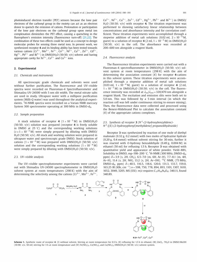
Scheme 1. Synthetic route of receptor 4; (I) without solvent, Stirring at room temperature for 0.5 h, (II) refluxing for 1.5 h in ethanol, (III) ZnCl2 �7H2O in DMSO:MeOH(10:90; v/v, 50 ml) stirring for 3 h at room temperature and (IV) Fe(NO3)3, Co(NO3)2 and Cu(NO3)2 DMSO/H2O (50:50; v/v) solvent system.
U. Fegade et al. / Journal of Luminescence 149 (2014) 190–195 191

2.6. Synthesis of receptor 4
Receptor 4 was synthesized by reaction of one mole of receptor3 (0.68 g, 0.2 mmol) with one moles of ZnCl2 �7H2O (0.52 g,0.2 mmol) in DMSO: MeOH (10:90; v/v, 50 ml) stirring for 3 h atroom temperature. The precipitation was collect by filtration atroom temperature and dried in vacuum. Further it was washedwith water then ethanol followed by petroleum ether. Yield- 84%,IR (KBr, cm�1) υ¼682, 750, 783, 893, 1197, 1379, 1572, 1620, 2923.MS (ESI): m/z requires C17H14N4O4Zn: 403.71, found 404.83.
3. Result and discussion
3.1. Synthesis of receptor 4
Scheme 1 shows the synthesis of the ligand and its zinccomplex. The novel dipodal receptor 3 was obtained by thereaction of diethyl malonate with hydrazine hydrate to obtainmalonohydrazide, further it was reacted with salicyladehyde inethanol with refluxing for 1.5 h. Complexation between thereceptor 3 and the zinc chloride in DMSO: MeOH solvent wascarried out at room temperature. After 3 h, solid was precipitated,and then the precipitates were collected and washed with thesmall amount of cooled methanol. ESI-MS measurement of anacetonitrile solution of the obtained residue showed peak at m/z404.83, [receptor 4] corresponding to a mononuclear complexcomposed of Ligand/Zn¼1:1 (Fig. S10, Supporting information(SI)). IR spectrum of the zinc complex indicated the structuralinformation, in which the hydroxyl groups take part in thecomplexation with zinc since the band responsible for hydroxylprotons become broad (Figs. S11 and S12, Supporting information(SI)). All compounds were fully characterized via spectroscopic andanalytical methods.
3.2. Recognition studies of receptor 4
The emission profiles of receptor 4 consist of band at 470 nm,upon excitation at 320 nm. The cations binding ability of receptor4 was evaluated by addition various metal nitrates (Cr3þ , Mn2þ ,Fe3þ , Co2þ , Ni2þ , Cu2þ , Zn2þ , Cd2þ , Hg2þ , Pb2þ and Bi3þ) inDMSO/H2O (50:50, v/v) solvent system. It was noticed that most ofcations did not cause any significant change in emission spectrumof receptor 4 expect Fe3þ , Co2þ , Cu2þ ions have quenched (Fig. 1).The quenching is due to photoinduced electron transfer (PET)mechanism which activated from carbonyl oxygen atom of themalonohydrazide group. Among all the cations tested, receptor 4showed high selectivity towards Fe3þ , Co2þ and Cu2þ ions incontrasting modes.
The binding pattern of 4 investigates the importance ofcarbonyl groups of oxygen atom in cations binding. The carbonylis considered as a good binding site and plays an effective role inbinding of cations such as Fe3þ , Co2þ and Cu2þ ion sensorsbecause the lone pair electrons of the carbonyl group in themoiety can act as an electron donor to quench the emission ofcations. Therefore, it appears that the core functionality requiredfor 4 to efficiently bind with cations upon excitation was carbonylgroups. However, due to binding of (Fe3þ , Co2þ and Cu2þ) ions inthe pseudocavity, the cation-fluorophore interaction is modulated,the bond formed between carbonyl groups and cations. Fluores-cence response of receptor 4 toward the various surveyed cationsshown in Fig. 2. As seen from Fig. 2, it is clear that there wasmarked quenching upon addition all three cations, and no sig-nificant quenching was observed upon addition of other cations.
The coordination approach of receptor 4 to all three cations likeFe3þ , Co2þ and Cu2þ were also studied by fluorescence titrations(Figs. S1–S3, Supporting information (SI)). Free receptor 4 showedstrong fluorescence emission at 470 nm upon excitation at320 nm. Upon addition all three cations (Fe3þ , Co2þ and Cu2þ)(0–1 equiv.), the emission intensity decreases significantly, indi-cating all three cations (Fe3þ , Co2þ and Cu2þ) selectively showedon–off (PET) fluorescent signaling behavior. In the titration spec-tra, the titration of all three cations (Fe3þ , Co2þ and Cu2þ) intoreceptor 4 gave a maximum emission band centered at 470 nmwhich showed a linear quench with the increase of all threecations (Fe3þ , Co2þ and Cu2þ)total when the ratio of [Fe3þ , Co2þ
and Cu2þ]total/ receptor 4 are below or equal to 1:1. When the ratioreached 1:1, however, higher [Fe3þ , Co2þ and Cu2þ]total did notlead to any further emission quench. The fluorescence quench ofreceptor 4 was due to the coordination to a paramagnetic Fe3þ ,Co2þ and Cu2þ . The linear relationship of the fluorescence titra-tion showed that receptor 4 responded to Fe3þ , Co2þ and Cu2þ in1:1 stoichiometry.
The association constant (Ka) of 4 with Fe3þ , Co2þ and Cu2þ
were calculated by Benesi–Hildebrand plot [23], (Figs. 3–5) and itwas 4�104 M�1, 1�104 M�1 and 8�104 M�1 for 4, 4.Fe3þ ,4.Co2þ and 4.Cu2þ , respectively. The Stern–Volmer plot [24] (plotof Io/I vs concentration of guest) is a straight line (Figs. S4–S6,Supporting information (SI)). This confirmed the formation of onetype of complex between 4 and Fe3þ , Co2þ and Cu2þ . To under-stand the binding stoichiometry of 4 with Fe3þ , Co2þ , and Cu2þ
ions, a method of continuous variation (job's plot) has beenemployed [25] and it showed maxima at 0.5, which correspondto 1:1 stoichiometry of complexes 4.Fe3þ , 4.Co2þ and 4.Cu2þ (Figs.S7–S9, Supporting information (SI)).
The limit of detection for receptor 4 as a fluorescent sensor forFe3þ , Co2þ , Cu2þ detection were determined from a plot offluorescence intensity as a function of Fe3þ , Co2þ , and Cu2þ
concentrations. It was found that receptor 4 has a limit ofdetection of 100 nM, 500 nM and 200 nM respectively, which is
0.00E+00 1.00E+06 2.00E+06 3.00E+06 4.00E+06 5.00E+06 6.00E+06 7.00E+06 8.00E+06 9.00E+06 1.00E+07
410 460 510 560 610
Inte
nsity
(cps
)
Wavelength (nm)
Host Cr(III) Mn(II) Fe(III) Co(II) Ni(II) Cu(II) Zn(II) Cd(II) Hg(II) Pb(II) Bi(III)
Fig. 1. Fluorescence spectra (λex¼320 nm) of receptor 4 upon the addition of a0.5 equivalents amount of various cations in DMSO/H2O (50:50; v/v). Inset showsthe fluorescence quenching on the addition of Fe3þ , Co2þ and Cu2þ . (From right toleft only receptor 4, 4.Fe3þ , 4.Co2þ and 4.Cu2þ).
-5
0
5
10
15
20
25
30
I-Io
/Io
Metal Ions
Fig. 2. Fluorescence ratiometric response (I–I0/I0) of probe 4 (c¼1�10�5 M) uponthe addition of a particular metal salt (c¼1�10�4 M) in DMSO/H2O (50:50; v/v).
U. Fegade et al. / Journal of Luminescence 149 (2014) 190–195192
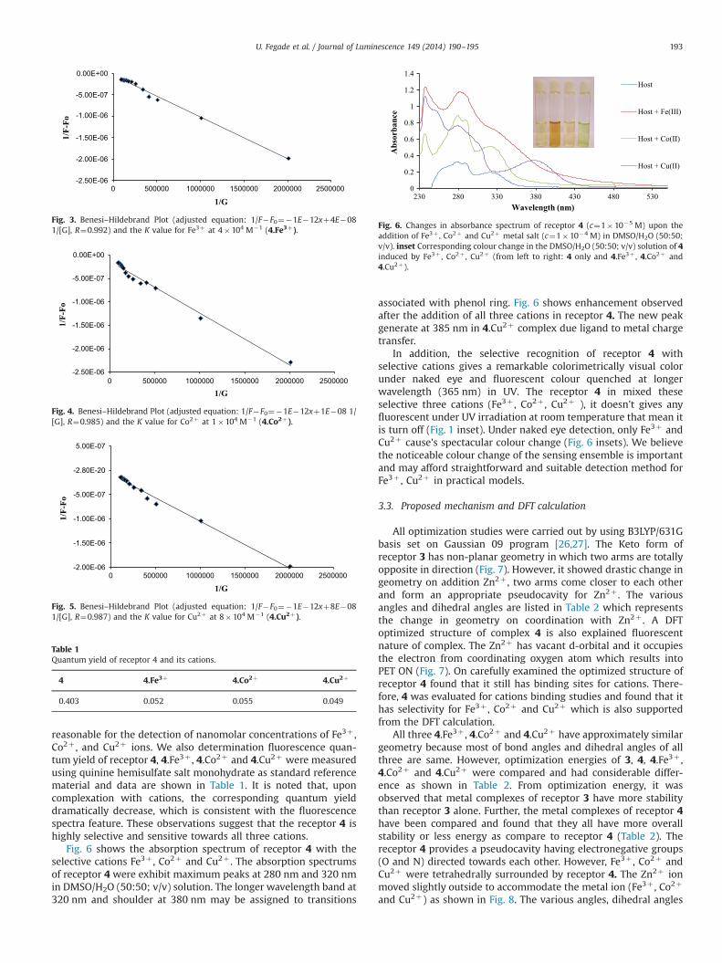
reasonable for the detection of nanomolar concentrations of Fe3þ ,Co2þ , and Cu2þ ions. We also determination fluorescence quan-tum yield of receptor 4, 4.Fe3þ , 4.Co2þ and 4.Cu2þ were measuredusing quinine hemisulfate salt monohydrate as standard referencematerial and data are shown in Table 1. It is noted that, uponcomplexation with cations, the corresponding quantum yielddramatically decrease, which is consistent with the fluorescencespectra feature. These observations suggest that the receptor 4 ishighly selective and sensitive towards all three cations.
Fig. 6 shows the absorption spectrum of receptor 4 with theselective cations Fe3þ , Co2þ and Cu2þ . The absorption spectrumsof receptor 4 were exhibit maximum peaks at 280 nm and 320 nmin DMSO/H2O (50:50; v/v) solution. The longer wavelength band at320 nm and shoulder at 380 nm may be assigned to transitions
associated with phenol ring. Fig. 6 shows enhancement observedafter the addition of all three cations in receptor 4. The new peakgenerate at 385 nm in 4.Cu2þ complex due ligand to metal chargetransfer.
In addition, the selective recognition of receptor 4 withselective cations gives a remarkable colorimetrically visual colorunder naked eye and fluorescent colour quenched at longerwavelength (365 nm) in UV. The receptor 4 in mixed theseselective three cations (Fe3þ , Co2þ , Cu2þ ), it doesn't gives anyfluorescent under UV irradiation at room temperature that mean itis turn off (Fig. 1 inset). Under naked eye detection, only Fe3þ andCu2þ cause's spectacular colour change (Fig. 6 insets). We believethe noticeable colour change of the sensing ensemble is importantand may afford straightforward and suitable detection method forFe3þ , Cu2þ in practical models.
3.3. Proposed mechanism and DFT calculation
All optimization studies were carried out by using B3LYP/631Gbasis set on Gaussian 09 program [26,27]. The Keto form ofreceptor 3 has non-planar geometry in which two arms are totallyopposite in direction (Fig. 7). However, it showed drastic change ingeometry on addition Zn2þ , two arms come closer to each otherand form an appropriate pseudocavity for Zn2þ . The variousangles and dihedral angles are listed in Table 2 which representsthe change in geometry on coordination with Zn2þ . A DFToptimized structure of complex 4 is also explained fluorescentnature of complex. The Zn2þ has vacant d-orbital and it occupiesthe electron from coordinating oxygen atom which results intoPET ON (Fig. 7). On carefully examined the optimized structure ofreceptor 4 found that it still has binding sites for cations. There-fore, 4 was evaluated for cations binding studies and found that ithas selectivity for Fe3þ , Co2þ and Cu2þ which is also supportedfrom the DFT calculation.
All three 4.Fe3þ , 4.Co2þ and 4.Cu2þ have approximately similargeometry because most of bond angles and dihedral angles of allthree are same. However, optimization energies of 3, 4, 4.Fe3þ ,4.Co2þ and 4.Cu2þ were compared and had considerable differ-ence as shown in Table 2. From optimization energy, it wasobserved that metal complexes of receptor 3 have more stabilitythan receptor 3 alone. Further, the metal complexes of receptor 4have been compared and found that they all have more overallstability or less energy as compare to receptor 4 (Table 2). Thereceptor 4 provides a pseudocavity having electronegative groups(O and N) directed towards each other. However, Fe3þ , Co2þ andCu2þ were tetrahedrally surrounded by receptor 4. The Zn2þ ionmoved slightly outside to accommodate the metal ion (Fe3þ , Co2þ
and Cu2þ) as shown in Fig. 8. The various angles, dihedral angles
-2.50E-06
-2.00E-06
-1.50E-06
-1.00E-06
-5.00E-07
0.00E+00
0 500000 1000000 1500000 2000000 2500000
1/F-
Fo
1/G
Fig. 3. Benesi–Hildebrand Plot (adjusted equation: 1/F�F0¼�1E�12xþ4E�081/[G], R¼0.992) and the K value for Fe3þ at 4�104 M�1 (4.Fe3þ).
-2.50E-06
-2.00E-06
-1.50E-06
-1.00E-06
-5.00E-07
0.00E+00
0 500000 1000000 1500000 2000000 2500000
1/F-
Fo
1/G
Fig. 4. Benesi–Hildebrand Plot (adjusted equation: 1/F�F0¼�1E�12xþ1E�08 1/[G], R¼0.985) and the K value for Co2þ at 1�104 M�1 (4.Co2þ).
-2.00E-06
-1.50E-06
-1.00E-06
-5.00E-07
-2.80E-20
5.00E-07
0 500000 1000000 1500000 2000000 2500000
1/F-
Fo
1/G
Fig. 5. Benesi–Hildebrand Plot (adjusted equation: 1/F�F0¼�1E�12xþ8E�081/[G], R¼0.987) and the K value for Cu2þ at 8�104 M�1 (4.Cu2þ).
Table 1Quantum yield of receptor 4 and its cations.
4 4.Fe3þ 4.Co2þ 4.Cu2þ
0.403 0.052 0.055 0.049
0
0.2
0.4
0.6
0.8
1
1.2
1.4
230 280 330 380 430 480 530
Abs
orba
nce
Wavelength (nm)
Host
Host + Fe(III)
Host + Co(II)
Host + Cu(II)
Fig. 6. Changes in absorbance spectrum of receptor 4 (c¼1�10�5 M) upon theaddition of Fe3þ , Co2þ and Cu2þ metal salt (c¼1�10�4 M) in DMSO/H2O (50:50;v/v). inset Corresponding colour change in the DMSO/H2O (50:50; v/v) solution of 4induced by Fe3þ , Co2þ , Cu2þ (from left to right: 4 only and 4.Fe3þ , 4.Co2þ and4.Cu2þ).
U. Fegade et al. / Journal of Luminescence 149 (2014) 190–195 193
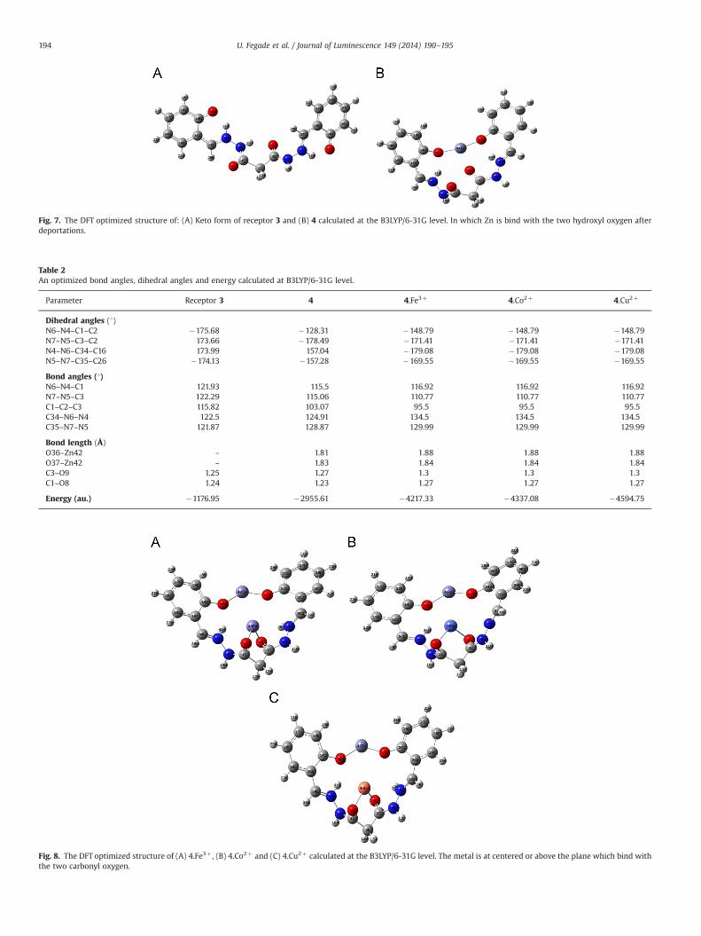
Fig. 7. The DFT optimized structure of: (A) Keto form of receptor 3 and (B) 4 calculated at the B3LYP/6-31G level. In which Zn is bind with the two hydroxyl oxygen afterdeportations.
Table 2An optimized bond angles, dihedral angles and energy calculated at B3LYP/6-31G level.
Parameter Receptor 3 4 4.Fe3þ 4.Co2þ 4.Cu2þ
Dihedral angles (1)N6–N4–C1–C2 �175.68 �128.31 �148.79 �148.79 �148.79N7–N5–C3–C2 173.66 �178.49 �171.41 �171.41 �171.41N4–N6–C34–C16 173.99 157.04 �179.08 �179.08 �179.08N5–N7–C35–C26 �174.13 �157.28 �169.55 �169.55 �169.55
Bond angles (1)N6–N4–C1 121.93 115.5 116.92 116.92 116.92N7–N5–C3 122.29 115.06 110.77 110.77 110.77C1–C2–C3 115.82 103.07 95.5 95.5 95.5C34–N6–N4 122.5 124.91 134.5 134.5 134.5C35–N7–N5 121.87 128.87 129.99 129.99 129.99
Bond length (Å)O36–Zn42 – 1.81 1.88 1.88 1.88O37–Zn42 – 1.83 1.84 1.84 1.84C3–O9 1.25 1.27 1.3 1.3 1.3C1–O8 1.24 1.23 1.27 1.27 1.27
Energy (au.) �1176.95 �2955.61 �4217.33 �4337.08 �4594.75
Fig. 8. The DFT optimized structure of (A) 4.Fe3þ , (B) 4.Co2þ and (C) 4.Cu2þ calculated at the B3LYP/6-31G level. The metal is at centered or above the plane which bind withthe two carbonyl oxygen.
U. Fegade et al. / Journal of Luminescence 149 (2014) 190–195194

and value of optimization energy are shown in Table 2 forcomparison. The photochemical behaviours of 3.Fe3þ , 3.Co2þ
and 3.Cu2þ dose not gave any considerable change in the formof fluorescence on or off as shown in Fig. S13, Supportinginformation (SI). But on the addition of Zn in 3 an appreciablechange in the form of fluorescence on and 3.Zn2þ causes changein its photochemical behaviour. But 4 gave the fluorescence off onaddition of Fe3þ , Co2þ and Cu2þ which is shown in Fig. 1.
4. Conclusion
In conclusion, we have synthesised Zn2þ complex act as anexcellent sensory system for detecting Fe3þ , Co2þ and Cu2þ , in semiaqueous solution. The sensing has been suggested to proceed via astanding emission quenching process. The various studies (fluores-cence and DFT calculations) are redolent of the formation of acoordination bonded between the carbonyl sites of the receptor 4and Fe2þ , Co2þ , Cu2þ ions thereby supporting the standing quenchingcourse of action. The work toward this end is in progress in our lab.
Appendix A. Supporting information
Supplementary data associated with this article can be found in theonline version at http://dx.doi.org/10.1016/j.jlumin.2014.01.035.
References
[1] R.D. Hancock, Chem. Soc. Rev. 42 (2013) 1500.[2] A.P. de Silva, H.Q.N. Gunaratne, T. Gunnlaugsson, A.J.M. Huxley, C.P. McCoy,
J.T. Rademacher, T.E. Rice, Chem. Rev. 97 (1997) 1515;K.J. Wallace, Supramol. Chem. 21 (2009) 89.
[3] J. de Jong, V. Schoemann, D. Lannuzel, J.L. Tison, N. Mattielli, Anal. Chim. Acta623 (2008) 126;A.K. Sharma, I. Singh, Food Anal. Methods 2 (2009) 221.
[4] P. Giamarchi, J.Y. Cabon, A. Le Bihan, Anal. Chim. Acta 664 (2010) 114;S. Saracoglu, M. Soylak, D.S. Peker, L. Elci, W.N. dos Santos, V.A. Lemos,S.L. Ferreira, Anal. Chim. Acta 575 (2006) 133.
[5] Z.Q. Liang, C.X. Wang, J.X. Yang, H.W. Gao, Y.P. Tian, X.T. Tao, M.H. Jiang, NewJ. Chem. 31 (2007) 906;S.P. Wu, Y.P. Chen, Y.M. Sung, Analyst 136 (2011) 1887.
[6] K. Attiq-ur-Rehman, M. Yaqoob, A. Waseem, A. Nabi, Int. J. Environ. Anal.Chem. 89 (2009) 1071;S.J. Ussher, A. Milne, W.M. Landing, K. Attiq-ur-Rehman, M.J. Seguret,T. Holland, E.P. Achterberg, A. Nabi, P.J. Worsfold, Anal. Chim. Acta 652(2009) 259.
[7] C.M.G. van den Berg, H. Obata, Anal. Chem. 73 (2001) 2522;R.K. Shervedani, A. Hatefi-Mehrjardi, A. Asadi-Farsani, Anal. Chim. Acta 601(2007) 164.
[8] U. Fegade, J. Marek, R. Patil, S. Attarde, A. Kuwar, Journal of Luminescence 146(2014) 234.
[9] A.M. Thomson, J.T. Rogers, P.J. Leedman, Int. J. Biochem. Cell Biol. 31 (1999)1139.
[10] J. Umbreit, Am. J. Hematol. 78 (2005) 225.[11] M. Gerlach, K.L. Double, M.B.H. Youdim, P. Riederer, J. Neural Transm. Suppl. 70
(2006) 133;S.L. Rhodes, B. Ritz, Neurobiology 32 (2008) 183.
[12] V. Cracan, R. Banerjee, Chapter 10 cobalt and corrinoid transport andbiochemistry, in: Ed.,in: Banci Lucia (Ed.), Metallomics and the Cell. MetalIons in Life Sciences, 12, Springer, 2013.
[13] U. Fegade, S. Attarde, A. Kuwar, Chem. Phys. Lett. 584 (2013) 165.[14] E.S. Mucklow, S.J. Griffin, D.H. Trevor, B. Suchak, Lancet 335 (1990) 981.[15] R.W Hay, Bioinorganic Chemistry, Ellis Harword Chichester, New York, 1984.[16] J.A. Cowan, Inorganic Biochemistry: An Introduction, Wiley-VCH, New York,
NY (1997) 133.[17] K.J. Barnham, C.L. Masters, A.I. Bush, Nat. Rev. Drug Discovery 3 (2004) 205.[18] S.H. Hahn, M.S. Tanner, D.M. Danke, W.A. Gahl, Biochem. Mol. Med 54 (1995)
142.[19] D.R. Brown, Brain Res. Bull. 55 (2001) 165.[20] D.J. Waggoner, T.B. Bartnikas, J.D. Gitlin, Neurobiol. Dis. 6 (1999) 221.[21] U. Fegade, A. Singh, G.K. Chaitanya, N. Singh S. Attarde, A. Kuwar, Spectrochim.
Acta, Part A, http://dx.doi.org/10.1016/j.saa.2013.11.007.[22] U. Fegade, H. Sharma, S. Attarde, N. Singh, A. Kuwar, J. Fluoresc. (2013), http:
//dx.doi.org/10.1007/s10895-013-1297-4.[23] H.A. Benesi, J.H. Hildebrand, J. Am. Chem. Soc. 71 (1949) 2703.[24] O. Stern, M. Volmer, Phys. Z 20 (1919) 183.[25] P. Job, Ann. Chim 9 (1928) 113.[26] A.D. Becke, J. Chem. Phys. 98 (1993) 5648.[27] C. Lee, W. Yang, R.G. Parr, Phys. Rev. B: Condens. Matter 37 (1988) 785.
U. Fegade et al. / Journal of Luminescence 149 (2014) 190–195 195