Mechanism of chemoresistance mediated by miR140 in human osteosarcoma and colon cancer cells
17
Mechanism of chemoresistance mediated by miR-140 in human osteosarcoma and colon cancer cells Bo Song 1,7 , Yuan Wang 1,5,7 , Yaguang Xi 2 , Kenji Kudo 2 , Skjalg Bruheim 3 , Galina I. Botchkina 4 , Elaine Gavin 2 , Yu Wan 5 , Andrea Formentini 6 , Marko Kornmann 6 , Oystein Fodstad 3 , and Jingfang Ju 1 1 Translational Research Laboratory, Department of Pathology, Stony Brook University, Stony Brook, NY 2 Mitchell Cancer Institute, University of South Alabama, Mobile, AL 3 Department of Tumor Biology, Institute for Cancer Research, The Norwegian Radium Hospital, Rikshospitalet University Hospital, and Faculty Division, The Norwegian Radium Hospital, University of Oslo, Oslo, Norway 4 Department of Surgery, Stony Brook University, Stony Brook, NY 5 Department of Physiology, Wuhan University, Wuhan, P. R. China 6 Department of Visceral and Transplantation Surgery, University of Ulm, Ulm, Germany Abstract In this study, our high throughput microRNA (miRNA) expression analysis revealed that the expression of miR-140 was associated with chemosensitivity in osteosarcoma tumor xenografts. Tumor cells ectopically transfected with miR-140 were more resistant to methotrexate (MTX) and 5-fluorouracil (5-FU). Overexpression of miR-140 inhibited cell proliferation in both osteosarcoma U-2 OS (wt-p53) and colon cancer HCT 116 (wt-p53) cell lines, but less so in osteosarcoma MG63 (mut-p53) and colon cancer HCT 116 (null-p53) cell lines. miR-140 induced p53 and p21 expression accompanied with G1 and G2 phase arrest only in cell lines containing wild type of p53. Histone deacetylase 4 (HDAC4) was confirmed to be one of the important targets of miR-140. The expression of endogenous miR-140 was significantly elevated in CD133 +hi CD44 +hi colon cancer stem-like cells which exhibit slow proliferating rate and chemoresistance. Blocking endogenous miR-140 by locked nucleic acid (LNA) modified anti-miR partially sensitized resistant colon cancer stem-like cells to 5-FU treatment. Taken together, our findings indicate that miR-140 is involved in the chemoresistance by reduced cell proliferation via G1 and G2 phase arrest mediated in part, through the suppression of HDAC4. miR-140 might be a candidate target to develop novel therapeutic strategy to overcome drug resistance. Keywords miR-140; chemosensitivity; histone deacetylase 4; cancer stem cells Users may view, print, copy, download and text and data- mine the content in such documents, for the purposes of academic research, subject always to the full Conditions of use: http://www.nature.com/authors/editorial_policies/license.html#terms Correspondence: Dr. Jingfang Ju, Translational Research Laboratory, Department of Pathology, Stony, Brook University, Stony Brook, NY 11794-8691, Office phone: (631)-444-3598, Office fax: (631)-444-3424, [email protected]. 7 These authors contributed equally to this work. Conflict of interest: The authors declare no conflict of interest. NIH Public Access Author Manuscript Oncogene. Author manuscript; available in PMC 2010 May 19. Published in final edited form as: Oncogene. 2009 November 19; 28(46): 4065–4074. doi:10.1038/onc.2009.274. NIH-PA Author Manuscript NIH-PA Author Manuscript NIH-PA Author Manuscript
Transcript of Mechanism of chemoresistance mediated by miR140 in human osteosarcoma and colon cancer cells
Mechanism of chemoresistance mediated by miR-140 in humanosteosarcoma and colon cancer cells
Bo Song1,7, Yuan Wang1,5,7, Yaguang Xi2, Kenji Kudo2, Skjalg Bruheim3, Galina I.Botchkina4, Elaine Gavin2, Yu Wan5, Andrea Formentini6, Marko Kornmann6, OysteinFodstad3, and Jingfang Ju1
1Translational Research Laboratory, Department of Pathology, Stony Brook University, StonyBrook, NY2Mitchell Cancer Institute, University of South Alabama, Mobile, AL3Department of Tumor Biology, Institute for Cancer Research, The Norwegian Radium Hospital,Rikshospitalet University Hospital, and Faculty Division, The Norwegian Radium Hospital,University of Oslo, Oslo, Norway4Department of Surgery, Stony Brook University, Stony Brook, NY5Department of Physiology, Wuhan University, Wuhan, P. R. China6Department of Visceral and Transplantation Surgery, University of Ulm, Ulm, Germany
AbstractIn this study, our high throughput microRNA (miRNA) expression analysis revealed that theexpression of miR-140 was associated with chemosensitivity in osteosarcoma tumor xenografts.Tumor cells ectopically transfected with miR-140 were more resistant to methotrexate (MTX) and5-fluorouracil (5-FU). Overexpression of miR-140 inhibited cell proliferation in bothosteosarcoma U-2 OS (wt-p53) and colon cancer HCT 116 (wt-p53) cell lines, but less so inosteosarcoma MG63 (mut-p53) and colon cancer HCT 116 (null-p53) cell lines. miR-140 inducedp53 and p21 expression accompanied with G1 and G2 phase arrest only in cell lines containingwild type of p53. Histone deacetylase 4 (HDAC4) was confirmed to be one of the importanttargets of miR-140. The expression of endogenous miR-140 was significantly elevated inCD133+hiCD44+hi colon cancer stem-like cells which exhibit slow proliferating rate andchemoresistance. Blocking endogenous miR-140 by locked nucleic acid (LNA) modified anti-miRpartially sensitized resistant colon cancer stem-like cells to 5-FU treatment. Taken together, ourfindings indicate that miR-140 is involved in the chemoresistance by reduced cell proliferation viaG1 and G2 phase arrest mediated in part, through the suppression of HDAC4. miR-140 might be acandidate target to develop novel therapeutic strategy to overcome drug resistance.
KeywordsmiR-140; chemosensitivity; histone deacetylase 4; cancer stem cells
Users may view, print, copy, download and text and data- mine the content in such documents, for the purposes of academic research,subject always to the full Conditions of use: http://www.nature.com/authors/editorial_policies/license.html#terms
Correspondence: Dr. Jingfang Ju, Translational Research Laboratory, Department of Pathology, Stony, Brook University, StonyBrook, NY 11794-8691, Office phone: (631)-444-3598, Office fax: (631)-444-3424, [email protected] authors contributed equally to this work.
Conflict of interest: The authors declare no conflict of interest.
NIH Public AccessAuthor ManuscriptOncogene. Author manuscript; available in PMC 2010 May 19.
Published in final edited form as:Oncogene. 2009 November 19; 28(46): 4065–4074. doi:10.1038/onc.2009.274.
NIH
-PA Author Manuscript
NIH
-PA Author Manuscript
NIH
-PA Author Manuscript
IntroductionmiRNAs are a class of small non-coding regulatory RNA molecules that recently have beenshown to play important roles in a wide array of biological processes (Ambros and Lee,2004; Bartel, 2004; Pillai et al., 2007; Plasterk, 2006; Zhang et al., 2007). miRNAs regulatemRNA translation mainly by binding to the 3′-UTR of their target mRNA transcripts toinhibit translation by sequence-specific mRNA cleavage or translational repression,although the detailed molecular mechanism of miRNAs in regulating target mRNAtranslation is still not being fully elucidated (Kozak, 2008). Precise chronological andtopological regulation of posttranscriptional gene silencing by miRNAs is essential foranimal development and tissue differentiation (Alvarez-Garcia and Miska, 2005). Abnormalexpression of miRNAs is suggested to be associated with various human disorders,including cancer (Calin and Croce, 2006; Lu et al., 2005; Volinia et al., 2006).
In this study, we utilized high throughput miRNA expression analysis to reveal miRNAsassociated with chemosensitivity using osteosarcoma tumor xenografts treated withchemotherapeutic agents doxorubicin, cisplatin, and or ifosfamide. Clustering and Venndiagram analysis revealed three miRNAs (miR-140, miR-217 and miR-431) overlapped withthese compounds. Among these, miR-140 showed consistent high expression levels acrossall the three drugs, suggesting that miR-140 may contribute to a broad spectrum ofchemoresistance mechanism. miR-140 was recently reported to target histone deacetylase 4(HDAC4) based on a luciferase reporter assay in mouse cells (Tuddenham et al., 2006).Histone deacetylases (HDACs) mediate changes in nucleosome conformation and areimportant in the regulation of gene expression (Finnin et al., 1999). HDACs are alsoinvolved in cell-cycle progression and differentiation, and their deregulation is associatedwith several cancers (Yang and Grégoire, 2005). Histone acetylation is important forregulating DNA chromatin structure and transcriptional control (Eberharter and Becker,2002; Grozinger and Schreiber, 2002; Sengupta and Seto, 2004). HDAC isozyme can becategorized into three classes and HDAC4 belongs to class II, which can be regulated andshuttled between the cytoplasm and the nucleus in response to various signal transductionstimuli. In addition, class II HDACs exert their transcriptional co-repressor functions byinteraction with other co-repressors or direct binding to (and sequestering) sequence-specifictranscriptional factors such as MEF2, Runx3, and nuclear factor κB (NF-κB) (Grozingerand Schreiber, 2002; Yang and Grégoire, 2005). Recent studies have shown that HDAC4promotes growth of colon cancer cells via repression of p21 (Wilson et al., 2008). Wereasoned that miR-140 have a broad impact on cell proliferation and chemosensitivitythrough the regulation of HDAC4 expression.
We directly investigated the functional significance of miR-140 in cancer biology and itspotential role in chemoresistance in both human osteosarcoma and colon cancer cell lines.HCT 116 (wt-p53) colon cancer cells transfected with miR-140 were more resistant tochemotherapeutic agents such as methotrexate (MTX) and 5-fluorouracil (5-FU).Overexpression of miR-140 significantly inhibited cell proliferation in cancer cell linescontaining wild type of p53, and this maybe the major contributing factor to thechemoresistance phenotype. It was achieved, at least in part, by the induction of both G1 andG2 cell cycle arrest along with induction of p21. This effect, however, was largely absent incell lines with either mutant or null p53. These results indicated that the impact of miR-140on cell proliferation and cell cycle control was dependant on the presence of functional wildtype of p53. We confirmed that one of the important targets of miR-140 was HDAC4. Theexpression of endogenous miR-140 was highly elevated in CD133+hiCD44+hi colon cancerstem-like cells compared to control colon cancer cells, further suggesting that tumor stem-like cells may be avoiding cellular and DNA damage caused by chemotherapy with areduced proliferating phenotype mediated, at least in part, by miR-140. The resistance can
Song et al. Page 2
Oncogene. Author manuscript; available in PMC 2010 May 19.
NIH
-PA Author Manuscript
NIH
-PA Author Manuscript
NIH
-PA Author Manuscript
be partially reversed by treating cells with Locked Nucleic Acid (LNA) modified anti-miR140. Furthermore, miR-140 expression level was decreased in clinical colon cancerspecimens compared to adjacent normal tissues of the same patients, suggesting the loweredlevels of miR-140 in tumors may be contributing to proliferating phenotype in differentiatedcolon cancer cells. miR-140 might be a candidate target to develop novel therapeuticstrategy to overcome drug resistance.
ResultsmiR-140 expression is associated with chemosensitivity
Based on the important roles of miRNAs in cancer, we reasoned that certain miRNAs maycontribute to the mechanisms of cancer chemoresistance. To identify miRNAs associatedwith chemosensitivity, miRNA expression was screened using a panel of osteosarcomatumor xenografts treated with doxorubicin, cisplatin and ifosfamide. Supplementary Table 1showed the classification of human osteosarcoma xenografts according to their response (++++/+++/++ good, or -/+ poor) to doxorubicin, cisplatin, and ifosfamide. Using Luminexbead based high throughput miRNA expression analysis, we analyzed miRNA expressionfrom control osteosarcoma xenografts and samples treated with doxorubicin, cisplatin, andifosfamide. Normalized miRNA expression results were analyzed by ANOVA usingGeneSpring expression analysis software. Differentially expressed miRNAs (average foldchange) affected by these drugs were listed in Supplementary Table 2. Venn diagramanalysis revealed that miR-140, miR-217 and miR-431 were overlapped among all threeagents (Figure 1). Among these, miR-140 showed consistent high expression levels acrossall three drugs (5.0-, 2.5- and 3.3-fold respectively), suggesting miR-140 may play importantroles in a broad spectrum of chemoresistance, and this has led us to focus on miR-140 in thisstudy.
Overexpression of miR-140 causes chemoresistance to MTX and 5-FUTo directly test the relationship of miR-140 with the chemoresistance, HCT 116 (wt-p53)cells were transfected with miR-140, non-specific miR (miR control), and siRNA againstHDAC4 to evaluate the impact of miR-140 on chemosensitivity to MTX and 5-FUtreatment. We found cells with elevated miR-140 were more resistant to MTX (Figure 2a)and 5-FU (Figure 2b) compared to miR control cells.
Effect of miR-140 on cell proliferation and cell cycle controlTo investigate the possible mechanisms of miR-140 in chemoresistance, we first evaluatedthe impact of miR-140 on cell proliferation using osteosarcoma cell lines U-2 OS (wt-p53)and MG63 (mut-p53), and colon cancer cell lines HCT 116 (wt-p53) and HCT 116 (null-p53). Non-specific miR was used as the negative control. Although previous report indicatedthat miR-140 may be cartilage specific based on in situ hybridization (Tuddenham et al.,2006), we did find miR-140 expressed in some of the colon cancer cell lines using real timeqRT-PCR analysis. This was consistent with other reports that miR-140 was expressed in anumber of different tumor types (Iorio et al., 2007; Izzotti et al., 2009). Our results showedthat overexpression of miR-140 can suppress cell proliferation in U-2 OS cells by64.05±4.01% (Figure 3a), in HCT 116 (wt-p53) by 81.4±3.75% (Figure 3b), with lessimpact on MG63 cells (31.3±4.96%) (Figure 3c) and HCT 116 (null-p53) cells(22.42±1.88%) (Figure 3d) on day 5. By contrast, the miR control had no effect on cellproliferation, indicating that this effect caused by miR-140 is highly specific.
To determine whether the impact of miR-140 on cell proliferation is related to cell cyclecontrol, we analyzed the effect of miR-140 on cell cycle by flow cytometry using U-2 OS,MG63, HCT 116 (wt-p53) and HCT 116 (null-p53) cells transfected with miR control or
Song et al. Page 3
Oncogene. Author manuscript; available in PMC 2010 May 19.
NIH
-PA Author Manuscript
NIH
-PA Author Manuscript
NIH
-PA Author Manuscript
miR-140. Our results showed that miR-140 induced both G1 (>3-fold) and G2 arrest (>2-fold) in HCT 116 (wt-p53) cells (Figure 4a). By contrast, this effect has not been observedin HCT 116 (null-p53) cells (Figure 4a). The results in U-2 OS and MG63 cells wereconsistent with HCT 116 (wt-p53) and HCT 116 (null-p53) cells respectively (data notshown). Thus, miR-140 inhibits cell proliferation and triggers cell cycle arrest possibly viap53 dependent manner.
To further determine whether the cell cycle regulating genes are related with miR-140overexpression, we analyzed the expression of cell cycle regulating genes p53 and p21.Figure 4b showed the results of p53 and p21 expression by Western immunoblot in HCT116 (wt-p53) and HCT 116 (null-p53) cells. Ectopic overexpression of miR-140 increasedthe expression of both p53 and p21 proteins (Figure 4b, left panel, lane 2) in HCT 116 (wt-p53) cells. There was no increase of p21 expression in HCT 116 (null-p53) cells (Figure 4b,right panel, lane 2). The expression of p53 and p21 results from osteosarcoma cell lines U2-OS and MG63 were consistent with the data from colon cancer cell lines (SupplementaryFigure 1).
Induction of endogenous miR-140 expression by MTX treatment in wild type p53 cell linesTo investigate whether endogenous miR-140 level may be associated with DNA damagingagent such as MTX treatment, the expression of miR-140 was quantified using real timeqRT-PCR analysis from RNAs isolated in control and miR-140 transfected colon cancercells. Our results showed that the induction of p53 protein by MTX treatment in HCT 116(wt-p53) cells caused a significant increase of miR-140, but not in HCT 116 (null-p53) cells(Supplementary Figure 2). These results indicate that endogenous miR-140 level isassociated with functional p53 after genotoxic stress by MTX treatment.
Translational regulation of HDAC4 expression by miR-140A recent report demonstrated that miR-140 targeted HDAC4 at the 3′-UTR in mouse cellsto regulate translation (Tuddenham et al., 2006). We then analyzed the human miR-140sequence and confirmed that mouse mmu-miR-140 has the same sequence as human hsa-miR-140 and it is highly conserved (Figure 5a). HDAC4 was reported to promote the growthof colon cancer cells (Wilson et al., 2008). To experimentally confirm that the expression ofHDAC4 is indeed regulated by miR-140 in human cell lines, we overexpressed miR-140 bytransient transfection in U-2 OS and HCT 116 (wt-p53) cells. First we accessed theexpression level of HDAC4 mRNA using real time qRT-PCR. Oligofectamine alone(vehicle control) and non-specific miR were used as the negative controls, siRNA againstHDAC4 was the positive control. There was only a slight reduction in HDAC4 mRNAexpression by miR-140 treatment (Figure 5b, column 4), by contrast, siHDAC4 clearlycaused mRNA degradation (Figure 5b, column 3). Next, we analyzed the protein level ofHDAC4 using Western immunoblot. Overexpression of miR-140 significantly decreased theexpression of HDAC4 protein without considerable amount of mRNA degradation (Figure5c, lane 3), which was consistent with the results of Tuddenham et al. and Nicolas et al.(Nicolas et al., 2008; Tuddenham et al., 2006). To further confirm that the expression ofHDAC4 is regulated by miR-140, we performed the loss-of-function analysis by knockingdown the endogenous miR-140 with locked nucleic acid (LNA) modified anti-miR140 inHCT 116 (wt-p53) and HCT 116 (null-p53) cells, scramble-miR (LNA-control) was used asthe negative control. Our results showed that knocking down endogenous miR-140 by LNAanti-miR140 can restore the expression of HDAC4 (Figure 5d, lane 2). We also confirmedthe knock down of HDAC4 protein expression by siRNA in HCT 116 (wt-p53) cells(Supplementary Figure 3a, lane 3). However, compared with the remarkable suppressingeffect on cell proliferation by miR-140, the HDAC4 specific siRNA treated cells showedonly a slight reduction in cell proliferation and the inhibitory effort was diminished after 4
Song et al. Page 4
Oncogene. Author manuscript; available in PMC 2010 May 19.
NIH
-PA Author Manuscript
NIH
-PA Author Manuscript
NIH
-PA Author Manuscript
days (Supplementary Figure 3b). Moreover, HCT 116 (wt-p53) cells transfected with siRNAspecific for HDAC4 showed sensitivity to MTX treatment compared to miR-140 (Figure2a), however, it did increase resistance to 5-FU (Figure 2b).
Elevated expression of miR-140 in human colon cancer stem-like cells contributes tochemoresistance
To determine that colon cancer stem-like cells may have higher levels of miR-140expression to process slow proliferating phenotype thereby avoiding damage caused bychemotherapeutic agents, the colon cancer stem-like cells were isolated using both CD133and CD44 as selection markers from HCT 116 (wt-p53) cells (Figure 6a). Both CD133 andCD44 have been used by several groups to isolate colon cancer stem-like cells (Dalerba etal., 2007; Du et al., 2008; O'Brien et al., 2007; Ricci-Vitiani et al., 2007). Thecharacterization of CD133+hiCD44+hi cancer stem-like cells has been described in details inour previous study (Botchkina et al., 2009). The expression of miR-140 in colon cancerstem-like cells was found to be nearly 4-fold higher than that in the control bulk cancer cells(Figure 6b). The results suggest that colon cancer stem-like cells may utilize miR-140 toslow down cell proliferation and avoid damage caused by chemotherapy until receiving aproliferation and differentiation signal, further verifying the impact of miR-140 on cellproliferation and chemotherapy resistance. CD133+hiCD44+hi cells were far more resistant(about 20% cell death) to high dose 5-FU treatment than non-sorted control HCT 116 (wt-p53) cells (>80% cell death) (Figure 6c, top panel). To directly demonstrate that we canreverse the chemoresistance to 5-FU treatment in CD133+hiCD44+hi cells, the expression ofmiR-140 was knocked down by LNA modified anti-miR140. Our results showed thatCD133+hiCD44+hi cells were more sensitive to 5-FU treatment compared to LNA-controltreated cells (Figure 6c, lower panel).
Expression of miR-140 is decreased in colorectal cancer specimensDue to the impact of miR-140 on cell proliferation, we reasoned that differentiatedproliferating tumor cells may have reduced miR-140 expression. To evaluate miR-140expression level in colon cancer patients, we compared the miR-140 levels in 24 freshfrozen colon cancer specimens and their paired adjacent normal mucosa using real timeqRT-PCR analysis (Supplementary Table 3). Our results showed that the expression levelsof miR-140 were significantly reduced compared to adjacent normal mucosa (P<0.05)(Supplementary Figure 4).
DiscussionOur laboratory was focusing on elucidating the impact of non-coding miRNAs inchemoresistance, in particular, the resistance mechanism to antifolates (e.g. MTX) andfluoropyrimidine based compounds (e.g. 5-FU). Our interest on miR-140 was based on ourhigh throughput miRNA expression profiling results using a panel of osteosarcoma tumorxenografts treated with high dose doxorubicin, cisplatin, and ifosfamide. The expressionanalysis showed that miR-140 was associated with chemoresistance in osteosarcoma (Figure1). We reasoned that miR-140 may influence chemosensitivity to a broad spectrum of anti-cancer agents. We directly demonstrated that overexpression of miR-140 rendered coloncancer cells more resistant to both MTX and 5-FU treatment (Figure 2). This could be due toseveral possible reasons.
One major reason of the resistant phenotype is that miR-140 reduced the cell proliferationrate (Figure 3a and 3b) through decreased S phase and increased cell cycle arrest (Figure4a). This was achieved in part, by the induction of p21 and p53 (Figure 4b). In general, slowproliferating or quiescent cells are more resistant to DNA damaging agent treatment as both
Song et al. Page 5
Oncogene. Author manuscript; available in PMC 2010 May 19.
NIH
-PA Author Manuscript
NIH
-PA Author Manuscript
NIH
-PA Author Manuscript

MTX and 5-FU acting in S phase of the cell cycle to cause DNA damage. Elevated level ofmiR-140 in the colon cancer stem-like cells further supported this notion (Figure 6b).Cancer stem-like cells, exhibit low rate of division and proliferation in their niche that helpthem to avoid chemotherapy and radiation (Zou, 2008). This is the major difference betweencancer stem-like cells and relative rapid proliferating differentiated cancer cells which canbe eliminated effectively by chemotherapy treatment. Our results showed that colon cancerstem-like cells had higher miR-140 level than control bulk cancer cells (Figure 6b),suggesting that colon cancer stem-like cells may utilize miR-140 to slow down cellproliferation and avoid damage caused by chemotherapy. This may be an important novelmechanism in that tumor stem-like cells acquire slow proliferative or quiescent phenotypeby certain miRNAs such as miR-140 to avoid damage caused by chemotherapy such asMTX and 5-FU. Results obtained from LNA modified anti-miRNA experimentsdemonstrated that we can partially sensitize colon cancer stem-like cells to 5-FU treatment(Figure 6c, lower panel). We also realized that it was difficult to achieve high transfectionefficiency in floating colon cancer stem-like cell spheres. It is quite conceivable that thischemo-sensitization effect can be further improved by optimizing transfection efficiency.Colon cancer specimens had reduced miR-140 expression levels (Supplementary Figure 4),which supports our hypothesis that only the small fraction of tumor stem-like cells with aslow proliferating phenotype are mediated at least in part, by elevated miR-140, whiledifferentiated tumor cells acquire relative rapid proliferation phenotype by reducing some ofthese miRNAs (e.g. miR-140) expression.
The other possibility is miR-140 causes chemoresistance through suppressing some keytargets such as HDAC4. HDAC4 was highly expressed in the proliferative compartment innormal colonic and small intestinal epithelium (Wilson et al., 2008). We experimentallyconfirmed that one of the important targets of miR-140 was HDAC4, miR-140 reduced theexpression level of HDAC4 protein without degradation of the target mRNA (Figure 5). Werealized that the role of HDAC4 in the chemoresistance to MTX and 5-FU was quitedifferent in our study. Although HDAC4 contributed to the slight reduction of proliferationof HCT 116 (wt-p53) cells, no direct relationship between HDAC4 and chemoresistance toMTX treatment was found (Figure 2a). Most likely that the overall impact of miR-140 onpathways is the important factor. Other efforts for discovering miR-140 mediated targetshave been reported by Nicolas et al. recently that miR-140 mediated 21 potential targetsusing mouse cell lines such as NIH-3T3 (Nicolas et al., 2008). However, HDAC4 was noton their reported target list, this is partly due to the fact that miR-140 reduced the proteinexpression level of HDAC4 without altering the level of mRNA. Therefore, total mRNAbased expression profiling will likely miss these targets. It's also worth noticing that one oftheir confirmed targets of miR-140 was Cxcl12 (Nicolas et al., 2008). Cxcl12/CXCR4interaction is important for chemotaxis, adhesion, tumor invasion, and T cell response(Koishi et al., 2006). In fact, it has been reported that p53 attenuates cancer cell migrationand invasion through suppression of Cxcl12 expression in stromal fibroblasts (Moskovits etal., 2006). Lowered expression levels of miR-140 in colorectal cancer specimens(Supplementary Figure 4) might result an elevated level of Cxcl12, which likely contributesto tumor progression and metastasis.
In contrast to MTX, there was a direct relationship between 5-FU resistance and HDAC4suppression by miR-140. Cells transfected with siRNA against HDAC4 were resistant to 5-FU (Figure 2b), elevated p21 protein may contribute to such resistance (Figure 4b, leftpanel). This was consistent with a previous report that p21 induction was associated with 5-FU resistance (Bunz et al., 1999). The induction of p21 expression by miR-140 may becaused by the down-regulation of HDAC4. Wilson et al. investigated the detailed molecularmechanism of HDAC4 in regulating p21 expression (Wilson et al., 2008). HDAC4 directlyinteracted with Sp1 to the proximal p21 promoter. Although HDAC4 is just one of the many
Song et al. Page 6
Oncogene. Author manuscript; available in PMC 2010 May 19.
NIH
-PA Author Manuscript
NIH
-PA Author Manuscript
NIH
-PA Author Manuscript
targets of miR-140, we reasoned that reduced expression of HDAC4 by miR-140 doesrelease the suppressive control for p21 expression to allow cell cycle control. The inductionof p21 was absent in cells lacking wild type p53 suggests that miR-140 is dependent on thepresence of p53 to exert its function. The exact mechanism of regulatory relationshipbetween p53 and miR-140 is important and remains to be determined in our future study.These findings also raise the notion that miR-140, like miR-192/215 and miR-34s, is one ofthe miRNAs that either directly or indirectly mediated by p53 to control cell cycle and cellproliferation (Braun et al., 2008; Georges et al., 2008; He et al., 2007; Raver-Shapira et al.,2007; Song et al., 2008). We are just at the beginning of understanding these complicatedcontrols. Future studies are clearly needed to fully understand the underline molecularregulatory mechanisms.
In conclusion, in this study, we provide direct evidence that miR-140 influenceschemosensitivity to MTX and 5-FU treatment. The function of miR-140 in cell proliferationand cell cycle control is clearly dependent on the presence of wild type p53 gene. Furtherstudies are needed to further define additional molecular pathways mediated by miR-140.
Materials and methodsCell culture and reagents
Human osteosarcoma cell lines U-2 OS (wt-p53) and MG63 (mut-p53) were purchased fromthe American Type Culture Collection (ATCC). The human colon cancer cell lines HCT 116(wt-p53) and HCT 116 (null-p53) were gifts from Professor Bert Vogelstein (The JohnsHopkins University). U-2 OS, HCT 116 (wt-p53) and HCT 116 (null-p53) cells weremaintained in McCoy's 5A medium (Invitrogen), and MG63 cells were maintained inEagle's Minimum Essential Medium (ATCC), supplemented with 10% fetal bovine serum.MG63 cells have a rearrangement mutation of the p53 gene, which results in disruption ofp53 protein function (Chandar et al., 1992).
Osteosarcoma tumor xenograftsTen human osteosarcoma xenografts were used, eight of which have been describedpreviously (Bruheim et al., 2004) and two newly established xenografts were included. Thedetails of drug treatment and anti-tumor activity evaluation were also described (details seeResults). The chemosensitivity results of each tumor xenograft to doxorubicin, cisplatin andifosfamide were summarized in Supplementary Table 1.
Clinical samplesA total of 48 snap frozen colorectal patient specimens were selected (24 paired colon normalmucosa and tumor samples). These patients had undergone surgical resection of primarycolorectal adenocarcinoma at the Department of Visceral and Transplantation Surgery,University of Ulm, Germany. Patient consent forms were obtained from every patientaccording to the institutional regulations. The characteristics of these patients were shown inSupplementary Table 3.
Transfection of miRNA and siRNA specific to HDAC4Cells were plated in six-well plates at a density of 2×10⁵ cells/well. The next day, cells weretransfected with 100 nM of miR-140 precursor or non-specific miR control (Ambion) withOligofectamine (Invitrogen) based on the manufacturer's instructions. Positive controlsiRNA specific against HDAC4 (ON-TARGET plus SMARTpool L-008799-00-0010,human HDAC4, NM_000791) was purchased from Dharmacon and transfected withOligofectamine according to the manufacturer's protocols at a final concentration of 100 nM.
Song et al. Page 7
Oncogene. Author manuscript; available in PMC 2010 May 19.
NIH
-PA Author Manuscript
NIH
-PA Author Manuscript
NIH
-PA Author Manuscript
RNA isolation and real time qRT-PCR analysis of miRNATotal RNA, including miRNAs, was isolated from cell lines, tumor xenografts or clinicalspecimens using TRIzol reagent (Invitrogen) according to the manufacturer's instructions.cDNA synthesis was carried out with the High Capacity cDNA synthesis kit (AppliedBiosystems) using 5 ng of total RNA as template. The miRNA sequence-specific RT-PCRprimers for miR-140 and endogenous control RNU6B were purchased from Ambion. Real-time qRT-PCR analysis was carried out using Applied Biosystems 7500 Real-Time PCRSystem (details see ref. Song et al., 2008). The gene expression threshold cycle (CT) valuesof miRNAs from each sample were calculated by normalizing with internal control RNU6Band relative quantitation values were plotted.
Real time qRT-PCR analysis of mRNA expressioncDNA was synthesized with the High Capacity cDNA synthesis kit (Applied Biosystems)using 2 μg of total RNA as template. The PCR primers and probes for HDAC4 and theinternal control gene GAPDH were purchased from Applied Biosystems. Real-time qRT-PCR analysis was performed on an ABI 7500HT instrument (details see ref. Song et al.,2008).
Cell proliferation analysisCells were plated in 96-well plates at 2×10³ cells/well after transfection with miR-140 ormiR control. Cells were cultured for 24, 48, 72, 96 and 120 h. The absorbance at 450 and630 nm was measured after incubation with 10 μl of WST-1 (Roche Applied Science) for 1h.
Cell cycle analysisCells were transfected with miR-140 and miR control described as above. At 36 h aftertransfection, cells were harvested and resuspended at 0.5-1×10⁵ cells/ml in modifiedKrishan buffer (Dressler et al., 1988; Krishan, 1975). Before being analyzed by flowcytometry, cells were treated with 0.02mg/ml RNase H and stained with 0.05mg/mlpropidium iodide (Sigma).
Western immunoblot analysisCells were lysed in 1×RIPA buffer (Sigma) supplied with 100 μM PMSF (Sigma) andproteinase inhibitor cocktail (Sigma) at 48 h after transfection. Equal amount of proteinswere resolved by 8% SDS-PAGE gels (Laemmli, 1970). The primary antibodies used for theanalysis included goat anti-HDAC4 polyclonal Ab (1:1000, N-18), mouse anti-p53 mAb(1:1000, DO-1), mouse anti-p21 mAb (1:1000, F-5), and mouse anti-α-tubulin mAb(1:1000, TU-02), all from Santa Cruz Biotechnology.
miR-140 knock downHCT 116 (wt-p53) and HCT 116 (null-p53) cells were transfected with 100 nM of scramble-miR or LNA anti-miR140 oligonucleotides (Exiqon) in the six-well plates (2×10⁵ cells/well)by Lipofectamine 2000 (Invitrogen). Cells were harvested at 72 h after transfection andcellular proteins were extracted. HDAC4 protein was detected by Western immunoblotanalysis.
MTX treatmentHCT 116 (wt-p53) and HCT 116 (null-p53) cells were seeded in 6-well plates at 2×10⁵ perwell and incubated with or without MTX (100 nM) for 24 h. Then total RNA and proteins
Song et al. Page 8
Oncogene. Author manuscript; available in PMC 2010 May 19.
NIH
-PA Author Manuscript
NIH
-PA Author Manuscript
NIH
-PA Author Manuscript
were extracted, respectively. The subsequent real time qRT-PCR for miR-140 and Westernimmunoblot analysis were performed as described above.
Isolation of colon cancer stem-like cells by FACSHCT 116 (wt-p53) cells were sorted with multiparametric flow cytometry with BD FACSAria cell sorter (Becton Dickinson) at sterile conditions. Cells were prepared and labeledwith conjugated anti-human CD133-PE (clone 105902, R&D Systems) and CD44-FITC(clone F10-44-2, R&D Systems). Antibodies were diluted in buffer containing 5% BSA,1mM EDTA and 15-20% blocking reagent (Miltenyi Biotec) to inhibit non-specific bindingto non-target cells. After 15 min incubation at 4°C, stained cells were washed, resuspendedin 500 μl of MACS buffer and sorted.
MTX and 5-FU cytotoxicityHCT 116 (wt-p53) cells were replated in 96-well plates at 2×10³ cells/well in triplicate aftertransfected with miR-140, miR control, or siRNA against HDAC4 in 100 μl of medium.Twenty-four hours later, MTX (ranged from 10-1000 nM) or 5-FU (ranged from 2 to 100μM) was added and incubated for 72 h. Ten μl of WST-1 was added to each well. After 1 hincubation, absorbance was measured at 450 and 630 nm respectively. Non-specific miRwas used as the negative control.
Colon cancer stem-like cells were transfected with 100 nM of LNA anti-miR140 usingLipofectamine 2000 described as above. Twenty-four hours later, cells were washed by FBSand then incubated with lethal dose 5-FU (100 μM) for 48 h. The dead cells weredetermined by the FITC Annexin V and PI detection kit (BD Biosciences, Pharmingen).Briefly, cells were harvested and re-suspended in 1×Annexin V binding and stained withAnnexin V (5 μl) and PI (5 μl) for 15 min at room temperature in the dark. After additional400 μl of binding buffer, cells were analyzed by flow cytometry. For the sensitivity of 5-FUin the colon cancer stem-like cells and control bulk cancer cells, cells were incubated with100 μM of 5-FU for 48 h before flow cytometery analysis.
Statistic analysisAll experiments were repeated at least twice. Statistical significance was evaluated byStudent's t test (two-tailed) comparison between two groups of data using GraphPad Prismsoftware 5 (GraphPad). Expression levels of miR-140 in each clinical sample werenormalized by the internal control RNU6B using SDS software v1.2 (Applied Biosystems).Sample with the lowest expression levels of miR-140 was set as 1 to generate relativeexpression values. Statistical difference in the expression level between tumor and normaltissues was calculated by two-tailed paired Wilcoxon test using MedCalc® 10.0.2 (MedCalcsoftware, Belgium). Statistical significance was set as a P<0.05.
Supplementary MaterialRefer to Web version on PubMed Central for supplementary material.
AcknowledgmentsWe appreciate the critical reading of our manuscript by Stephanie Burke (Stony Brook University). This work wassupported by Stony-Brook Translational Research Laboratory Start-up fund and NIH CA114043 (J. Ju) andMH075020 (J. Ju).
Song et al. Page 9
Oncogene. Author manuscript; available in PMC 2010 May 19.
NIH
-PA Author Manuscript
NIH
-PA Author Manuscript
NIH
-PA Author Manuscript
ReferencesAlvarez-Garcia I, Miska EA. MicroRNA functions in animal development and human disease.
Development. 2005; 132:4653–4662. [PubMed: 16224045]
Ambros V, Lee RC. Identification of microRNAs and other tiny noncoding RNAs by cDNA cloning.Methods Mol Biol. 2004; 265:131–158. [PubMed: 15103073]
Bartel DP. MicroRNAs: genomics, biogenesis, mechanism and function. Cell. 2004; 116:281–297.[PubMed: 14744438]
Botchkina IL, Rowehl RA, Rivadeneira DE, Karpeh MS Jr, Crawford H, Dufour A, et al. Phenotypicsubpopulations of metastatic colon cancer stem cells: genomic analysis. Cancer GenomicsProteomics. 2009; 6:19–29. [PubMed: 19451087]
Braun CJ, Zhang X, Savelyeva I, Wolff S, Moll UM, Schepeler T, et al. p53-responsive microRNAs192 and 215 are capable of inducing cell cycle arrest. Cancer Res. 2008; 68:10094–10104.[PubMed: 19074875]
Bruheim S, Bruland OS, Breistol K, Maelandsmo GM, Fodstad O. Human osteosarcoma xenograftsand their sensitivity to chemotherapy. Pathol Oncol Res. 2004; 10:133–141. [PubMed: 15448748]
Bunz F, Hwang PM, Torrance C, Waldman T, Zhang Y, Dillehay L, et al. Disruption of p53 in humancancer cells alters the responses to therapeutic agents. J Clin Invest. 1999; 104:263–269. [PubMed:10430607]
Calin G, Croce CM. MicroRNA signatures in human cancers. Nature Reviews Cancer. 2006; 6:857–866.
Chandar N, Billig B, McMaster J, Novak J. Inactivation of p53 gene in human and murineosteosarcoma cells. Br J Cancer. 1992; 65:208–214. [PubMed: 1739619]
Dalerba P, Dylla SJ, Park IK, Liu R, Wang X, Cho RW, et al. Phenotypic characterization of humancolorectal cancer stem cells. Proc Natl Acad Sci U S A. 2007; 104:10158–10163. [PubMed:17548814]
Dressler LG, Seamer LC, Owens MA, Clark GM, McGuire WL. DNA flow cytometry and prognosticfactors in 1331 frozen breast cancer specimens. Cancer. 1988; 61:420–427. [PubMed: 3338012]
Du L, Wang H, He L, Zhang J, Ni B, Wang X, et al. CD44 is of functional importance for colorectalcancer stem cells. Clin Cancer Res. 2008; 14:6751–6760. [PubMed: 18980968]
Eberharter A, Becker PB. Histone acetylation: a switch between repressive and permissive chromatin.Second in review series on chromatin dynamics. EMBO Rep. 2002; 3:224–229. [PubMed:11882541]
Finnin MS, Donigian JR, Cohen A, Richon VM, Rifkind RA, Marks PA, et al. Structures of a histonedeacetylase homologue bound to the TSA and SAHA inhibitors. Nature. 1999; 401:188–193.[PubMed: 10490031]
Georges SA, Biery MC, Kim SY, Schelter JM, Guo J, Chang AN, et al. Coordinated regulation of cellcycle transcripts by p53-Inducible microRNAs, miR-192 and miR-215. Cancer Res. 2008;68:10105–10112. [PubMed: 19074876]
Grozinger C, Schreiber SL. Deacetylase enzymes: biological functions and the use of small-moleculeinhibitors. Chem Biol. 2002; 9:3–16. [PubMed: 11841934]
He L, He X, Lim LP, de Stanchina E, Xuan Z, Liang Y, et al. A microRNA component of the p53tumour suppressor network. Nature. 2007; 447:1130–1134. [PubMed: 17554337]
Iorio MV, Visone R, Di Leva G, Donati V, Petrocca F, Casalini P, et al. MicroRNA signatures inhuman ovarian cancer. Cancer Res. 2007; 67:8699–8707. [PubMed: 17875710]
Izzotti A, Calin GA, Arrigo P, Steele VE, Croce CM, De Flora S. Downregulation of microRNAexpression in the lungs of rats exposed to cigarette smoke. FASEB J. 2009; 23:806–812. [PubMed:18952709]
Koishi K, Yoshikawa R, Tsujimura T, Hashimoto-Tamaoki T, Kojima S, Yanagi H, et al. PersistentCXCR4 expression after preoperative chemoradiotherapy predicts early recurrence and poorprognosis in esophageal cancer. World J Gastroenterol. 2006; 12:7585–7590. [PubMed:17171785]
Kozak M. Faulty old ideas about translational regulation paved the way for current confusion abouthow microRNAs function. Gene. 2008; 423:108–115. [PubMed: 18692553]
Song et al. Page 10
Oncogene. Author manuscript; available in PMC 2010 May 19.
NIH
-PA Author Manuscript
NIH
-PA Author Manuscript
NIH
-PA Author Manuscript
Krishan A. Rapid flow cytofluorometric analysis of mammalian cell cycle by propidium iodidestaining. J Cell Biol. 1975; 66:188–193. [PubMed: 49354]
Laemmli UK. Cleavage of structural proteins during the assembly of the head of bacteriophage T4.Nature. 1970; 227:680–685. [PubMed: 5432063]
Lu J, Getz G, Miska EA, Alvarez-Saavedra E, Lamb J, Peck D, et al. MicroRNA expression profilesclassify human cancers. Nature. 2005; 435:834–838. [PubMed: 15944708]
Moskovits N, Kalinkovich A, Bar J, Lapidot T, Oren M. p53 Attenuates cancer cell migration andinvasion through repression of SDF-1/CXCL12 expression in stromal fibroblasts. Cancer Res.2006; 66:10671–10676. [PubMed: 17108103]
Nicolas FE, Pais H, Schwach F, Lindow M, Kauppinen S, Moulton V, et al. Experimentalidentification of microRNA-140 targets by silencing and overexpressing miR-140. RNA. 2008;14:2513–2520. [PubMed: 18945805]
O'Brien CA, Pollett A, Gallinger S, Dick JE. A human colon cancer cell capable of initiating tumourgrowth in immunodeficient mice. Nature. 2007; 445:106–110. [PubMed: 17122772]
Pillai RS, Bhattacharyya SN, Fillipowicz W. Repression of protein synthesis by miRNAs: how manymechanisms? Trends cell Biol. 2007; 17:118–126. [PubMed: 17197185]
Plasterk RH. MicroRNAs in animal development. Cell. 2006; 124:877–881. [PubMed: 16530032]
Raver-Shapira N, Meiri E, Spector Y, Rosenfeld N, Moskovits N, Bentwich Z, et al. Transcriptionalactivation of miR-34a contributes to p53-mediated apoptosis. Mol Cell. 2007; 26:731–743.[PubMed: 17540598]
Ricci-Vitiani L, Lombardi DG, Pilozzi E, Biffoni M, Todaro M, Peschle C, et al. Identification andexpansion of human colon-cancer-initiating cells. Nature. 2007; 445:111–115. [PubMed:17122771]
Sengupta N, Seto E. Regulation of histone deacetylase activities. J Cell Biochem. 2004; 93:57–67.[PubMed: 15352162]
Song B, Wang Y, Kudo K, Gavin EJ, Xi Y, Ju J. miR-192 Regulates dihydrofolate reductase andcellular proliferation through the p53-microRNA circuit. Clin Cancer Res. 2008; 14:8080–8086.[PubMed: 19088023]
Tarasov V, Jung P, Verdoodt B, Lodygin D, Epanchintsev A, Menssen A, et al. Differential regulationof microRNAs by p53 revealed by massively parallel sequencing: miR-34a is a p53 target thatinduces apoptosis and G1-arrest. Cell Cycle. 2007; 6:1586–1593. [PubMed: 17554199]
Tuddenham L, Wheeler G, Ntounia-Fousara S, Waters J, Hajihosseini MK, Clark I, et al. The cartilagespecific microRNA-140 targets histone deacetylase 4 in mouse cells. FEBS Lett. 2006; 580:4214–4217. [PubMed: 16828749]
Volinia S, Calin GA, Liu CG, Ambs S, Cimmino A, Petrocca F, et al. A microRNA expressionsignature of human solid tumors defines cancer gene targets. Proc Natl Acad Sci U S A. 2006;103:2257–2261. [PubMed: 16461460]
Wilson AJ, Byun DS, Nasser S, Murray LB, Ayyanar K, Arango D, et al. HDAC4 promotes growth ofcolon cancer cells via repression of p21. Mol Biol Cell. 2008; 19:4062–4075. [PubMed:18632985]
Yang XJ, Grégoire S. Class II histone deacetylases: from sequence to function, regulation, and clinicalimplication. Mol Cell Biol. 2005; 25:2873–2874. [PubMed: 15798178]
Zhang B, Wang Q, Pan X. MicroRNAs and their regulatory roles in animals and plants. J Cell Physiol.2007; 210:279–289. [PubMed: 17096367]
Zou GM. Cancer initiating cells or cancer stem cells in the gastrointestinal tract and liver. J CellPhysiol. 2008; 217:598–604. [PubMed: 18651561]
Song et al. Page 11
Oncogene. Author manuscript; available in PMC 2010 May 19.
NIH
-PA Author Manuscript
NIH
-PA Author Manuscript
NIH
-PA Author Manuscript
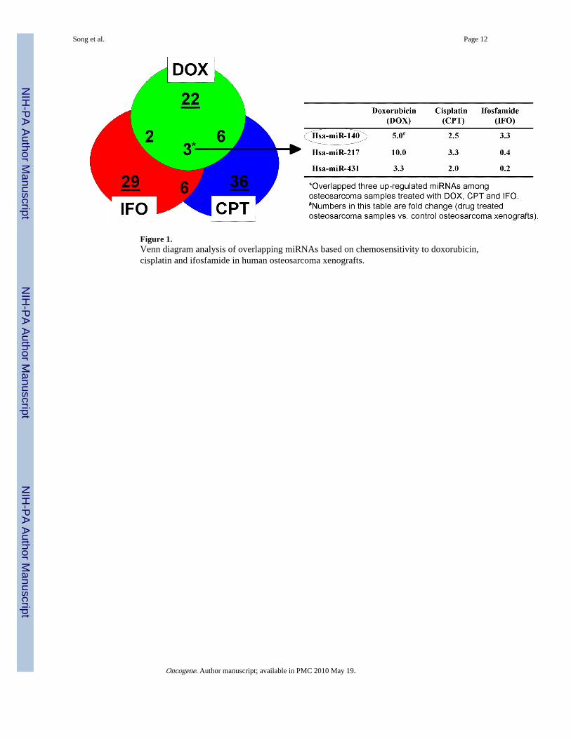
Figure 1.Venn diagram analysis of overlapping miRNAs based on chemosensitivity to doxorubicin,cisplatin and ifosfamide in human osteosarcoma xenografts.
Song et al. Page 12
Oncogene. Author manuscript; available in PMC 2010 May 19.
NIH
-PA Author Manuscript
NIH
-PA Author Manuscript
NIH
-PA Author Manuscript
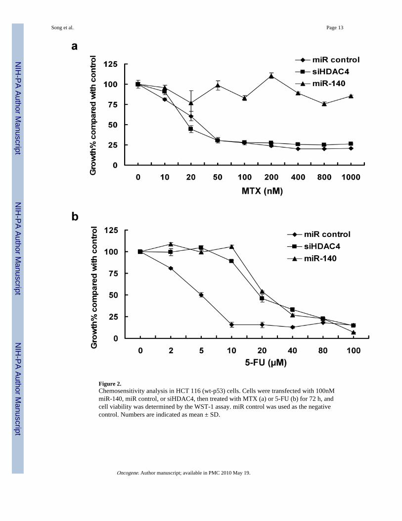
Figure 2.Chemosensitivity analysis in HCT 116 (wt-p53) cells. Cells were transfected with 100nMmiR-140, miR control, or siHDAC4, then treated with MTX (a) or 5-FU (b) for 72 h, andcell viability was determined by the WST-1 assay. miR control was used as the negativecontrol. Numbers are indicated as mean ± SD.
Song et al. Page 13
Oncogene. Author manuscript; available in PMC 2010 May 19.
NIH
-PA Author Manuscript
NIH
-PA Author Manuscript
NIH
-PA Author Manuscript
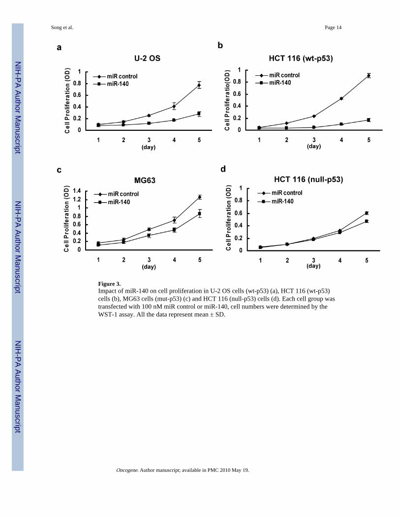
Figure 3.Impact of miR-140 on cell proliferation in U-2 OS cells (wt-p53) (a), HCT 116 (wt-p53)cells (b), MG63 cells (mut-p53) (c) and HCT 116 (null-p53) cells (d). Each cell group wastransfected with 100 nM miR control or miR-140, cell numbers were determined by theWST-1 assay. All the data represent mean ± SD.
Song et al. Page 14
Oncogene. Author manuscript; available in PMC 2010 May 19.
NIH
-PA Author Manuscript
NIH
-PA Author Manuscript
NIH
-PA Author Manuscript
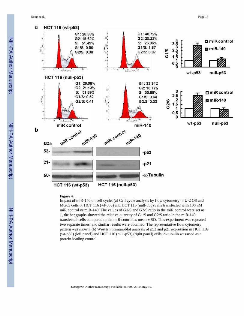
Figure 4.Impact of miR-140 on cell cycle. (a) Cell cycle analysis by flow cytometry in U-2 OS andMG63 cells or HCT 116 (wt-p53) and HCT 116 (null-p53) cells transfected with 100 nMmiR control or miR-140. The values of G1/S and G2/S ratio in the miR control were set as1, the bar graphs showed the relative quantity of G1/S and G2/S ratio in the miR-140transfected cells compared to the miR control as mean ± SD. This experiment was repeatedtwo separate times, and similar results were obtained. The representative flow cytometrypattern was shown. (b) Western immunoblot analysis of p53 and p21 expression in HCT 116(wt-p53) (left panel) and HCT 116 (null-p53) (right panel) cells, α-tubulin was used as aprotein loading control.
Song et al. Page 15
Oncogene. Author manuscript; available in PMC 2010 May 19.
NIH
-PA Author Manuscript
NIH
-PA Author Manuscript
NIH
-PA Author Manuscript
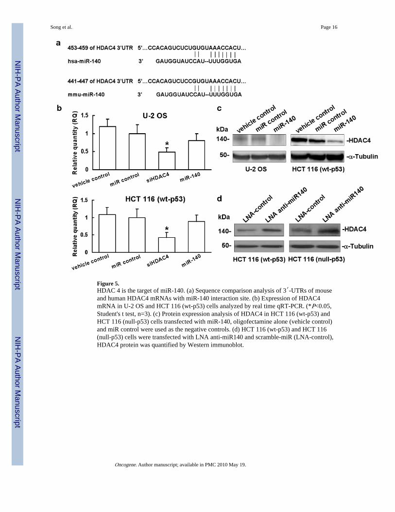
Figure 5.HDAC 4 is the target of miR-140. (a) Sequence comparison analysis of 3′-UTRs of mouseand human HDAC4 mRNAs with miR-140 interaction site. (b) Expression of HDAC4mRNA in U-2 OS and HCT 116 (wt-p53) cells analyzed by real time qRT-PCR. (*P<0.05,Student's t test, n=3). (c) Protein expression analysis of HDAC4 in HCT 116 (wt-p53) andHCT 116 (null-p53) cells transfected with miR-140, oligofectamine alone (vehicle control)and miR control were used as the negative controls. (d) HCT 116 (wt-p53) and HCT 116(null-p53) cells were transfected with LNA anti-miR140 and scramble-miR (LNA-control),HDAC4 protein was quantified by Western immunoblot.
Song et al. Page 16
Oncogene. Author manuscript; available in PMC 2010 May 19.
NIH
-PA Author Manuscript
NIH
-PA Author Manuscript
NIH
-PA Author Manuscript
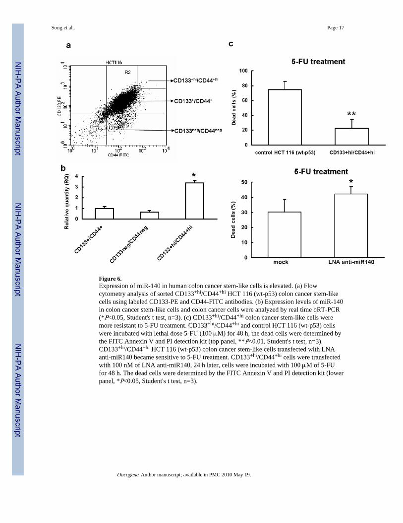
Figure 6.Expression of miR-140 in human colon cancer stem-like cells is elevated. (a) Flowcytometry analysis of sorted CD133+hi/CD44+hi HCT 116 (wt-p53) colon cancer stem-likecells using labeled CD133-PE and CD44-FITC antibodies. (b) Expression levels of miR-140in colon cancer stem-like cells and colon cancer cells were analyzed by real time qRT-PCR(*P<0.05, Student's t test, n=3). (c) CD133+hi/CD44+hi colon cancer stem-like cells weremore resistant to 5-FU treatment. CD133+hi/CD44+hi and control HCT 116 (wt-p53) cellswere incubated with lethal dose 5-FU (100 μM) for 48 h, the dead cells were determined bythe FITC Annexin V and PI detection kit (top panel, **P<0.01, Student's t test, n=3).CD133+hi/CD44+hi HCT 116 (wt-p53) colon cancer stem-like cells transfected with LNAanti-miR140 became sensitive to 5-FU treatment. CD133+hi/CD44+hi cells were transfectedwith 100 nM of LNA anti-miR140, 24 h later, cells were incubated with 100 μM of 5-FUfor 48 h. The dead cells were determined by the FITC Annexin V and PI detection kit (lowerpanel, *P<0.05, Student's t test, n=3).
Song et al. Page 17
Oncogene. Author manuscript; available in PMC 2010 May 19.
NIH
-PA Author Manuscript
NIH
-PA Author Manuscript
NIH
-PA Author Manuscript