Functional characterization of osteosarcoma cell lines provides representative models to study the...
11
Functional characterization of osteosarcoma cell lines provides representative models to study the human disease Alexander B Mohseny 1 , Isidro Machado 2 , Yongping Cai 1,3 , Karl-Ludwig Schaefer 4 , Massimo Serra 5 , Pancras CW Hogendoorn 1 , Antonio Llombart-Bosch 2 and Anne-Marie Cleton-Jansen 1 Cancer cell lines represent in vitro models for studying malignancies, general cell biology, drug discovery and more. Whether they can be considered as exact representative models of the parental tumors remains uncertain given the acquisition of additional ex vivo changes of the cells and the lack of tissue architecture and stroma. Previously, within the EuroBoNeT consortium, we characterized a collection of bone sarcoma cell lines on genomic and proteomic level. Here, we address the phenotypical and functional characterization of the unique set of osteosarcoma cell lines (n ¼ 19) in vitro and in vivo. For functional analysis of differentiation capacity, cells were stimulated towards osteoblasts, adipocytes and chondrocytes. Furthermore, all cell lines were injected subcutaneously and intramuscularly into nude mice to assay their in vivo tumor formation capacity as well as for phenotypical analysis of the tumors. All formed tumors were further characterized histologically and immunohistochemically. Out of 19 cell lines, 17 (89%) showed adipogenic differentiation, 13/19 (68%) could differentiate towards osteoblasts and in 6/19 (32%) cell lines chondrogenic differentiation was evident. About half of the cell lines (8/19, 42%) produced tumors in vivo after subcutaneous and intramuscular injections. Several cell lines showed invasion into adjacent tissues and one tumor developed several lung metastases. The use of cell lines, especially in cancer research, is of paramount importance. Here, we identify comprehensively characterized osteosarcoma cell lines, which robustly represent clinical osteosarcoma providing researchers useful in vitro and in vivo models to study the genetics and functional characteristics of this highly malignant neoplasm. Laboratory Investigation (2011) 91, 1195–1205; doi:10.1038/labinvest.2011.72; published online 25 April 2011 KEYWORDS: contamination; HOS; misidentification; MNNG; origin; tumorigenesis; U2OS Tumor cell lines have been considered as instrumental entries into tumor cell biology and often used for studying the me- chanisms of carcinogenesis, the functional characteristics of genes and drug discovery, screening and response. 1–8 Never- theless, concerns have been raised about the use of cell lines that can be divided into two categories of criticism. First, mainly due to poor experimental conduct, false (derived from another cell population within the cell culture than the one intended), cross-contaminated (with other human cells or cells from other species) and/or pathogen- (mycoplasm) con- taminated cell lines have been discovered repeatedly. 9–12 Sec- ond, the representativeness of cultured cell lines as compared with the original tumors is being questioned as these cells have been cultured in the absence of stroma, 13 hence lacking the proper microenvironment and the original tissue architecture. Moreover during culturing, specific cells are continuously se- lected based on the in vitro conditions and cells might undergo additional ex vivo mutations. On the other hand, it has been shown that cell lines adequately represent the tumors they are originating from, especially at the genetic level. 14–19 All in all cell lines appear to be adequate models as long as there are controlled culturing conditions and a good selection process to identify the appropriate ones. 10,11,20 Accordingly, to select osteosarcoma cell lines representative of human osteosarcoma, here we characterize 19 cell lines in vitro and in vivo by using robust methods subjected to regular quality control. Received 29 October 2010; revised 24 February 2011; accepted 6 March 2011 1 Department of Pathology, Leiden University Medical Center, Leiden, The Netherlands; 2 Department of Pathology, University of Valencia, Valencia, Spain; 3 Department of Pathology, School of Medicine, Shandong University, Jinan, China; 4 Institute of Pathology, University Medical Center Duesseldorf, Duesseldorf, Germany and 5 Laboratorio di Oncologia Sperimentale, Istituto Ortopedico Rizzoli, Bologna, Italy Correspondence: Dr A-M Cleton-Jansen, Department of Pathology, Leiden University Medical Center, PO Box 9600, L1-Q, Leiden 2300 RC, The Netherlands. E-mail: [email protected] Laboratory Investigation (2011) 91, 1195–1205 & 2011 USCAP, Inc All rights reserved 0023-6837/11 $32.00 www.laboratoryinvestigation.org | Laboratory Investigation | Volume 91 August 2011 1195
Transcript of Functional characterization of osteosarcoma cell lines provides representative models to study the...

Functional characterization of osteosarcoma cell linesprovides representative models to study the humandiseaseAlexander B Mohseny1, Isidro Machado2, Yongping Cai1,3, Karl-Ludwig Schaefer4, Massimo Serra5,Pancras CW Hogendoorn1, Antonio Llombart-Bosch2 and Anne-Marie Cleton-Jansen1
Cancer cell lines represent in vitro models for studying malignancies, general cell biology, drug discovery and more.Whether they can be considered as exact representative models of the parental tumors remains uncertain given theacquisition of additional ex vivo changes of the cells and the lack of tissue architecture and stroma. Previously, within theEuroBoNeT consortium, we characterized a collection of bone sarcoma cell lines on genomic and proteomic level. Here,we address the phenotypical and functional characterization of the unique set of osteosarcoma cell lines (n¼ 19) in vitroand in vivo. For functional analysis of differentiation capacity, cells were stimulated towards osteoblasts, adipocytes andchondrocytes. Furthermore, all cell lines were injected subcutaneously and intramuscularly into nude mice to assay theirin vivo tumor formation capacity as well as for phenotypical analysis of the tumors. All formed tumors were furthercharacterized histologically and immunohistochemically. Out of 19 cell lines, 17 (89%) showed adipogenic differentiation,13/19 (68%) could differentiate towards osteoblasts and in 6/19 (32%) cell lines chondrogenic differentiation was evident.About half of the cell lines (8/19, 42%) produced tumors in vivo after subcutaneous and intramuscular injections. Severalcell lines showed invasion into adjacent tissues and one tumor developed several lung metastases. The use of cell lines,especially in cancer research, is of paramount importance. Here, we identify comprehensively characterized osteosarcomacell lines, which robustly represent clinical osteosarcoma providing researchers useful in vitro and in vivo models to studythe genetics and functional characteristics of this highly malignant neoplasm.Laboratory Investigation (2011) 91, 1195–1205; doi:10.1038/labinvest.2011.72; published online 25 April 2011
KEYWORDS: contamination; HOS; misidentification; MNNG; origin; tumorigenesis; U2OS
Tumor cell lines have been considered as instrumental entriesinto tumor cell biology and often used for studying the me-chanisms of carcinogenesis, the functional characteristics ofgenes and drug discovery, screening and response.1–8 Never-theless, concerns have been raised about the use of cell linesthat can be divided into two categories of criticism. First,mainly due to poor experimental conduct, false (derived fromanother cell population within the cell culture than the oneintended), cross-contaminated (with other human cells or cellsfrom other species) and/or pathogen- (mycoplasm) con-taminated cell lines have been discovered repeatedly.9–12 Sec-ond, the representativeness of cultured cell lines as comparedwith the original tumors is being questioned as these cells have
been cultured in the absence of stroma,13 hence lacking theproper microenvironment and the original tissue architecture.Moreover during culturing, specific cells are continuously se-lected based on the in vitro conditions and cells might undergoadditional ex vivo mutations. On the other hand, it has beenshown that cell lines adequately represent the tumors they areoriginating from, especially at the genetic level.14–19 All in allcell lines appear to be adequate models as long as there arecontrolled culturing conditions and a good selection processto identify the appropriate ones.10,11,20 Accordingly, to selectosteosarcoma cell lines representative of human osteosarcoma,here we characterize 19 cell lines in vitro and in vivo by usingrobust methods subjected to regular quality control.
Received 29 October 2010; revised 24 February 2011; accepted 6 March 2011
1Department of Pathology, Leiden University Medical Center, Leiden, The Netherlands; 2Department of Pathology, University of Valencia, Valencia, Spain; 3Departmentof Pathology, School of Medicine, Shandong University, Jinan, China; 4Institute of Pathology, University Medical Center Duesseldorf, Duesseldorf, Germany and5Laboratorio di Oncologia Sperimentale, Istituto Ortopedico Rizzoli, Bologna, ItalyCorrespondence: Dr A-M Cleton-Jansen, Department of Pathology, Leiden University Medical Center, PO Box 9600, L1-Q, Leiden 2300 RC, The Netherlands.E-mail: [email protected]
Laboratory Investigation (2011) 91, 1195–1205
& 2011 USCAP, Inc All rights reserved 0023-6837/11 $32.00
www.laboratoryinvestigation.org | Laboratory Investigation | Volume 91 August 2011 1195
Osteosarcoma is a highly malignant tumor, fatal for aboutone-third of the patients who do not respond to che-motherapy and alternative therapies are still missing. This ismainly due to the rarity and the high genetic heterogeneity ofthese tumors, which makes it difficult to have patient cohortsthat are large enough to compensate for the high geneticvariability. One way to bypass this problem is to study os-teosarcoma-derived cell lines, which are abundantly used asin vitro models because they are highly proliferative and re-ceptive for genetic manipulation by transfection. Recently,through the effort of the EuroBoNet network, a large panel ofosteosarcoma cell lines, among other bone sarcoma cell lines,was described genetically.18 To select the cell lines that aremost representative of primary osteosarcoma, here we ana-lyzed their in vitro differentiation capacity and their in vivotumorigenicity in nude mice. Furthermore, generated tumorswere histologically and immunohistochemically classified.
By this comprehensive study, we were able to identify atleast eight cell lines, which convincingly represent primaryhuman osteosarcoma. HOS-143B was discovered to be highlymetastatic to the lungs of the animals after subcutaneous andintramuscular xenotransplantations. The cell lines char-acterized here represent excellent in vitro and especiallyin vivo models to obtain a better understanding of osteo-sarcoma biology, and subsequently the identification of noveltargets for specific therapies.
MATERIALS AND METHODSCell LinesCells were provided by the different partner institutes of theEuroBoNet network21–25 or derived from ATCC and weregrown in RPMI 1640 (Invitrogen, Karlsruhe, Germany)supplemented with 2% L-glutamine (Invitrogen) and 10%fetal calf serum under standard conditions. Control tests formycoplasm contamination were carried out routinely (twotimes a month), using a PCR-based commercially availabledetection kit according to the manufacturer’s protocol(VenorGem, Minerva Biolabs, Berlin, Germany). Further-more, all cell lines and xenografts were genotyped using thePowerplex 1.2 system (Promega, Leiden, the Netherlands), asdescribed previously,26 to match the cells to their previouslypublished identities and to detect any cross-contamina-tions.18 Table 1 summarizes the characteristics and theculturing conditions of the cell lines.
In Vitro DifferentiationBefore differentiation, alkaline phosphatase (ALP) activitywas measured for all cell lines to determine their basal ALPactivity. Differentiation studies were performed as describedpreviously:27 cells were induced to differentiate into maturemineralizing osteoblasts over a 3-week culture period byplating 104 cells in 500 ml medium per well of a 24-well plate(for ALP measurement) and by plating 2� 104 cells in 1 mlmedium per well of a 12-well plate (for mineralization).Ascorbic acid (50 mg/ml) was added to the medium starting
on day 4 and b-glycerolphosphate (5 mM) on day 11.Osteoblastic differentiation was quantified as described pre-viously,28 with some modification. In short, cells were lysedin ALP lysis buffer (10 mmol/l glycine, 0.1 mmol/l MgCl2,10 mmol/l ZnCl2, 0.1% Triton X-100) and 25 ml was used todetermine ALP activity using 6 mmol/l p-nitrophenylpho-sphate as a substrate and measuring absorbance at 405 nm onan ELISA reader. For comparisons, ALP activity was calcu-lated per ml per minute until saturation. The presence ofmineralization was assessed by staining with Alizarin Red S(20 mg/ml, pH 5.5). Adipogenic differentiation was inducedby culturing 104 cells in 500 ml medium per well of a 24-wellplate for 2 weeks. FBS was replaced by 10% charcoal-strippedFBS in the basal medium and indomethacin (50 mM) wasadded starting on day 4. Adipocytes containing lipid dropletswere stained with Oil Red O (3 mg/ml). For chondrogenicdifferentiation, cells were cultured as pellets (2� 105 cells per
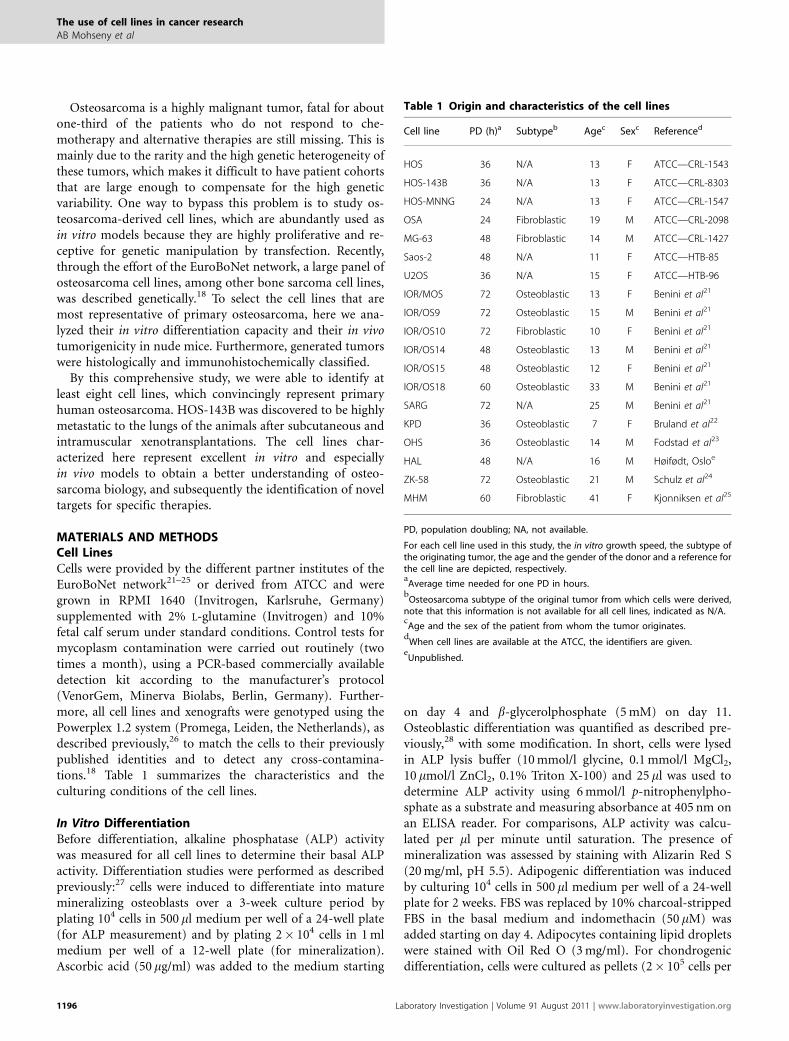
Table 1 Origin and characteristics of the cell lines
Cell line PD (h)a Subtypeb Agec Sexc Referenced
HOS 36 N/A 13 F ATCC—CRL-1543
HOS-143B 36 N/A 13 F ATCC—CRL-8303
HOS-MNNG 24 N/A 13 F ATCC—CRL-1547
OSA 24 Fibroblastic 19 M ATCC—CRL-2098
MG-63 48 Fibroblastic 14 M ATCC—CRL-1427
Saos-2 48 N/A 11 F ATCC—HTB-85
U2OS 36 N/A 15 F ATCC—HTB-96
IOR/MOS 72 Osteoblastic 13 F Benini et al21
IOR/OS9 72 Osteoblastic 15 M Benini et al21
IOR/OS10 72 Fibroblastic 10 F Benini et al21
IOR/OS14 48 Osteoblastic 13 M Benini et al21
IOR/OS15 48 Osteoblastic 12 F Benini et al21
IOR/OS18 60 Osteoblastic 33 M Benini et al21
SARG 72 N/A 25 M Benini et al21
KPD 36 Osteoblastic 7 F Bruland et al22
OHS 36 Osteoblastic 14 M Fodstad et al23
HAL 48 N/A 16 M Høifødt, Osloe
ZK-58 72 Osteoblastic 21 M Schulz et al24
MHM 60 Fibroblastic 41 F Kjonniksen et al25
PD, population doubling; NA, not available.
For each cell line used in this study, the in vitro growth speed, the subtype ofthe originating tumor, the age and the gender of the donor and a reference forthe cell line are depicted, respectively.a
Average time needed for one PD in hours.b
Osteosarcoma subtype of the original tumor from which cells were derived,note that this information is not available for all cell lines, indicated as N/A.c
Age and the sex of the patient from whom the tumor originates.d
When cell lines are available at the ATCC, the identifiers are given.e
Unpublished.
The use of cell lines in cancer research
AB Mohseny et al
1196 Laboratory Investigation | Volume 91 August 2011 | www.laboratoryinvestigation.org
pellet) in U-shaped 96-well plates containing 200 ml DMEMsupplemented with P/S (1%), pyruvate (100 mg/ml), trans-ferrin selenite (10 ml/ml) and proline (40 mg/ml). During thefirst 2 weeks, ascorbic acid (50 mg/ml), TGF-b3 (10 ng/ml)and dexamethasone (10�7 M) were added to the medium.Starting at the third week, ascorbic acid (50 mg/ml), BMP6(500 ng/ml) and b-glycerolphosphate (5 nM) were added.After 5 weeks, pellets were fixed in formalin and embedded inparaffin and sections were stained with toluidine blue toidentify chondrogenic matrix.
In Vivo Tumor FormationAfter trypsinization, cells were counted and dilutions of2� 106 cells in 10 ml phosphate-buffered saline were pre-pared. Each cell line was injected into one nude mouse atthree locations: two subcutaneous injections on the back(upper left and lower right corner) and one intramuscularinjection (upper part of the hind left paw). After the injec-tions, tumor growth was screened twice a week by observa-tion and palpation. Animals were killed when tumors reachedapproximately 1 cm in diameter or no sign of tumor for-mation was detected after 6 months or when any kind ofanimal suffering was detected. All cell lines that did notproduce tumors were re-injected into a second mouse (againat three locations) to validate this observation. After beingkilled, all tumors were surgically removed and archived byfreezing as well as by fixing in formalin and embedding inparaffin. Furthermore, a complete autopsy was performed todetect invasion, angiogenesis and/or metastases of the tu-mors. Growth into adjacent tissues was labeled as ‘invasion’only when after subcutaneous injection cells invaded un-derlying tissues (mostly muscle) and only when this wasconfirmed not to be caused by direct injection into thosetissues. (Neo)-angiogenesis was considered when blood ves-sels were identified inside the tumor’s mass at locationswhere they were not anatomically expected and/or whenvessel subcutaneously grew towards the tumors. All animalexperiments were performed according to the Dutch andSpanish animal experiments guidelines, and approved by theValencia University Animal Experiments Committee.
Tissue Array ConstructionTissue cores from formalin-fixed and paraffin-embedded(FFPE) tumor areas selected by two pathologists (IM andALLB) on the basis of a hematoxylin and eosin (H&E)-stained slide were taken from each specimen (BeecherInstruments, Silver Springs, MD, USA). The cores (2 mmdiameter) were arrayed on a recipient paraffin block usinga tissue arrayer from Beecher Instruments.29 At least threecores from each tumor were sampled to outweigh in-tratumoral heterogeneity.30 Each tissue array containsadditional cores from other tissue types both as internalcontrols for immunohistochemistry (IHC) as well as for orien-tation purposes.
StainingIHC was performed on FFPE tissue array sections as de-scribed previously.31 Next to the H&E, Periodic acid-Schiffand Masson’s trichrome stainings to assay the histology of thexenografts, the slides were stained with all antibodies listed inTable 3, which also summarizes the IHC conditions. Allstainings in this study were independently evaluated by twopathologists (IM and ALLB) as previously described in de-tail.32,33 Intensity and percentage of positive neoplastic cellswere evaluated. Cellular localization of immunopositivity(nuclear, cytoplasmic or membranous) was also recorded.Conflicting assessments were reviewed until final agreementwas achieved. A final immunopositivity score was indicatedper sample as negative (�), weakly positive (þ ), moderatelypositive (þ þ ) or strongly positive (þ þ þ ).
HOS-143BTo study the progression of the only metastatic cell line,HOS-143B, cells were injected two more rounds in 5 and 10animals, respectively. In addition from one of the primarytumors, a piece was subcutaneously transplanted to anotherrecipient nude mouse, and this was repeated three times.Both from subcutaneous and intramuscular tumors, lungmetastases were analyzed for p53 protein expression andmutation state as described previously.18,34,35
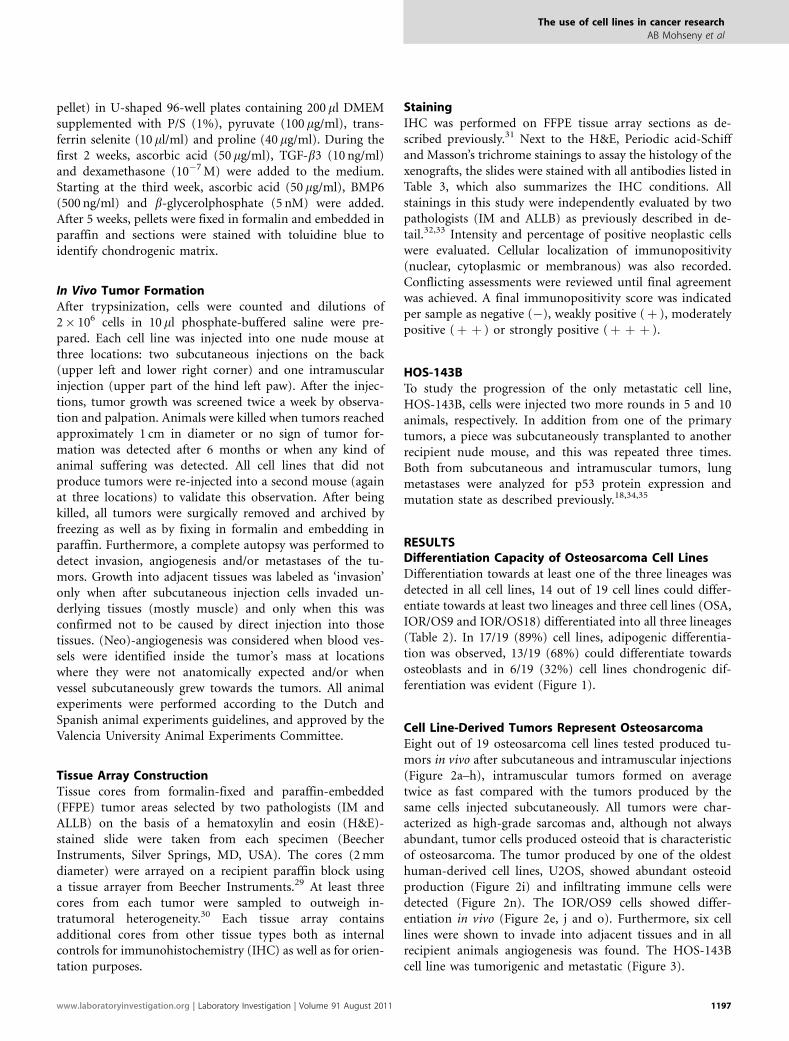
RESULTSDifferentiation Capacity of Osteosarcoma Cell LinesDifferentiation towards at least one of the three lineages wasdetected in all cell lines, 14 out of 19 cell lines could differ-entiate towards at least two lineages and three cell lines (OSA,IOR/OS9 and IOR/OS18) differentiated into all three lineages(Table 2). In 17/19 (89%) cell lines, adipogenic differentia-tion was observed, 13/19 (68%) could differentiate towardsosteoblasts and in 6/19 (32%) cell lines chondrogenic dif-ferentiation was evident (Figure 1).
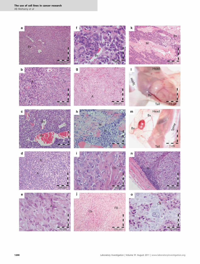
Cell Line-Derived Tumors Represent OsteosarcomaEight out of 19 osteosarcoma cell lines tested produced tu-mors in vivo after subcutaneous and intramuscular injections(Figure 2a–h), intramuscular tumors formed on averagetwice as fast compared with the tumors produced by thesame cells injected subcutaneously. All tumors were char-acterized as high-grade sarcomas and, although not alwaysabundant, tumor cells produced osteoid that is characteristicof osteosarcoma. The tumor produced by one of the oldesthuman-derived cell lines, U2OS, showed abundant osteoidproduction (Figure 2i) and infiltrating immune cells weredetected (Figure 2n). The IOR/OS9 cells showed differ-entiation in vivo (Figure 2e, j and o). Furthermore, six celllines were shown to invade into adjacent tissues and in allrecipient animals angiogenesis was found. The HOS-143Bcell line was tumorigenic and metastatic (Figure 3).
The use of cell lines in cancer research
AB Mohseny et al
www.laboratoryinvestigation.org | Laboratory Investigation | Volume 91 August 2011 1197
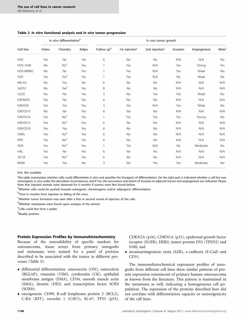
Protein Expression Profiles by ImmunohistochemistryBecause of the unavailability of specific markers forosteosarcoma, tissue arrays from primary xenograftsand metastases were stained for a panel of proteinsdescribed to be associated with the tumor in different pro-cesses (Table 3):
� differential differentiation: osteonectin (ON), osteocalcin(BGLAP), vimentin (VIM), cytokeratin (CK), epithelialmembrane antigen (EMA), CD34, smooth muscle actin(SMA), desmin (DES) and transcription factor SOX9(SOX9);
� oncogenesis: CD99, B-cell lymphoma protein 2 (BCL2),C-Kit (KIT), caveolin 1 (CAV1), Ki-67, TP53 (p53),
CDKN2A (p16), CDKN1A (p21), epidermal growth factorreceptor (EGFR), HER2, tumor protein D52 (TPD52) andS100; and
� invasion/migration: ezrin (EZR), e-cadherin (E-Cad) andCD31.
The immunohistochemical expression profiles of xeno-grafts from different cell lines show similar patterns of pro-tein expression reminiscent of primary human osteosarcomaas known from the literature. This pattern is maintained inthe metastases as well, indicating a homogeneous cell po-pulation. The expression of the proteins described here didnot correlate with differentiation capacity or tumorigenicityof the cell lines.
Table 2 In vitro functional analysis and in vivo tumor progression
In vitro differentiationa In vivo tumor growth
Cell line Osteo Chondro Adipo Follow upb 1st injectionc 2nd injectionc Invasion Angiogenesis Metat
HOS Yes No Yes 6 No No N/A N/A No
HOS-143B No Noe Yes 1 Yes N/A Yes Strong Yes
HOS-MNNG No No Yes 1 Yes N/A Yes Weak No
OSA Yes Yesf Yes 1 Yes N/A No Weak No
MG-63 No Yes No 6 No No N/A N/A N/A
SaOS2 No Yesf Yes 6 No No N/A N/A N/A
U2OS No No Yes 3 No Yes Yes Weak No
IOR/MOS Yes No Yes 6 No No N/A N/A N/A
IOR/OS9 Yes Yes Yes 3 Yes N/A Yes Weak No
IOR/OS10 No No Yes 6 No No N/A N/A N/A
IOR/OS14 Yes Noe Yes 1 Yes Yes Yes Strong No
IOR/OS15 Yes Noe Yes 6 No No N/A N/A N/A
IOR/OS18 Yes Yes Yes 6 No No N/A N/A N/A
SARG Yes Noe Yes 6 No No N/A N/A N/A
KPD Yes Noe Yes 6 No No N/A N/A N/A
OHS Yes Noe Yes 1 Yes N/A No Moderate No
HAL Yes No Yes 6 No No N/A N/A N/A
ZK-58 Yes Noe Yes 6 No No N/A N/A N/A
MHM Yes Yes No 3 No Yes Yes Moderate No
N/A, Not available.
This table summarizes whether cells could differentiate in vitro and specifies the lineage(s) of differentiation. On the right part is indicated whether a cell line wastumorigenic in vivo under the described circumstances, and if Yes, the occurrence and extent of invasion to adjacent tissues and angiogenesis are indicated. PleaseNote that injected animals were observed for 6 months if tumors were Not found before.a
Whether cells could be pushed towards osteogenic, chondrogenic and/or adipogenic differentiation.b
Time in months from injection to killing of the mice.c
Whether tumor formation was seen after a first or second round of injection of the cells.d
Whether metastases were found upon autopsy of the animal.e
Cells could Not form a pellet.fWeakly positive.
The use of cell lines in cancer research
AB Mohseny et al
1198 Laboratory Investigation | Volume 91 August 2011 | www.laboratoryinvestigation.org
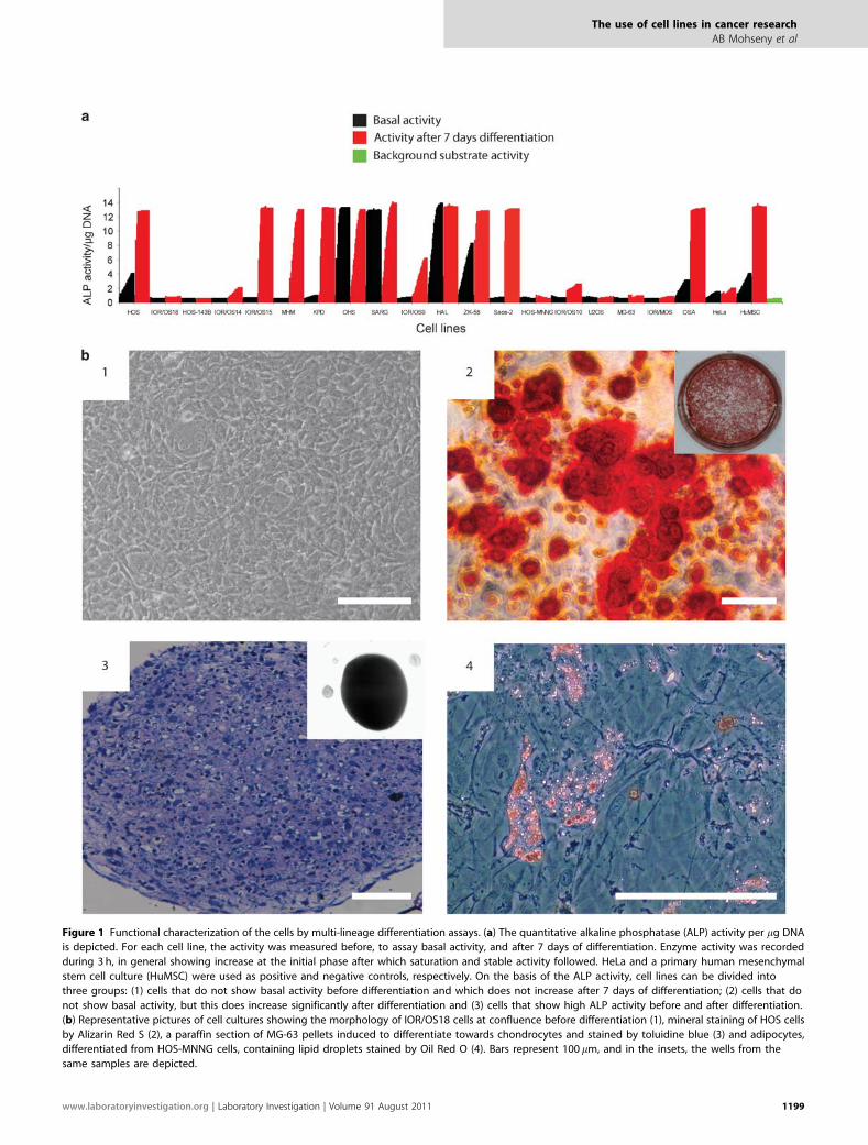
Figure 1 Functional characterization of the cells by multi-lineage differentiation assays. (a) The quantitative alkaline phosphatase (ALP) activity per mg DNA
is depicted. For each cell line, the activity was measured before, to assay basal activity, and after 7 days of differentiation. Enzyme activity was recorded
during 3 h, in general showing increase at the initial phase after which saturation and stable activity followed. HeLa and a primary human mesenchymal
stem cell culture (HuMSC) were used as positive and negative controls, respectively. On the basis of the ALP activity, cell lines can be divided into
three groups: (1) cells that do not show basal activity before differentiation and which does not increase after 7 days of differentiation; (2) cells that do
not show basal activity, but this does increase significantly after differentiation and (3) cells that show high ALP activity before and after differentiation.
(b) Representative pictures of cell cultures showing the morphology of IOR/OS18 cells at confluence before differentiation (1), mineral staining of HOS cells
by Alizarin Red S (2), a paraffin section of MG-63 pellets induced to differentiate towards chondrocytes and stained by toluidine blue (3) and adipocytes,
differentiated from HOS-MNNG cells, containing lipid droplets stained by Oil Red O (4). Bars represent 100 mm, and in the insets, the wells from the
same samples are depicted.
The use of cell lines in cancer research
AB Mohseny et al
www.laboratoryinvestigation.org | Laboratory Investigation | Volume 91 August 2011 1199
The use of cell lines in cancer research
AB Mohseny et al
1200 Laboratory Investigation | Volume 91 August 2011 | www.laboratoryinvestigation.org
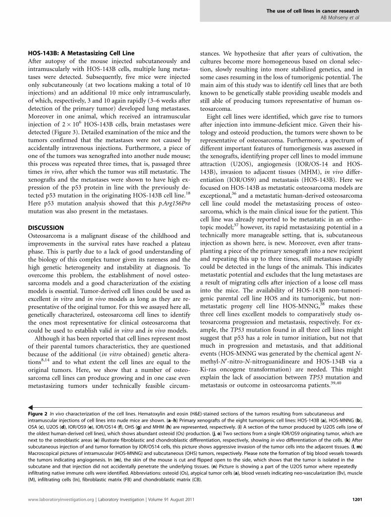
HOS-143B: A Metastasizing Cell LineAfter autopsy of the mouse injected subcutaneously andintramuscularly with HOS-143B cells, multiple lung metas-tases were detected. Subsequently, five mice were injectedonly subcutaneously (at two locations making a total of 10injections) and an additional 10 mice only intramuscularly,of which, respectively, 3 and 10 again rapidly (3–6 weeks afterdetection of the primary tumor) developed lung metastases.Moreover in one animal, which received an intramuscularinjection of 2� 106 HOS-143B cells, brain metastases weredetected (Figure 3). Detailed examination of the mice and thetumors confirmed that the metastases were not caused byaccidentally intravenous injections. Furthermore, a piece ofone of the tumors was xenografted into another nude mouse;this process was repeated three times, that is, passaged threetimes in vivo, after which the tumor was still metastatic. Thexenografts and the metastases were shown to have high ex-pression of the p53 protein in line with the previously de-tected p53 mutation in the originating HOS-143B cell line.18
Here p53 mutation analysis showed that this p.Arg156Promutation was also present in the metastases.
DISCUSSIONOsteosarcoma is a malignant disease of the childhood andimprovements in the survival rates have reached a plateauphase. This is partly due to a lack of good understanding ofthe biology of this complex tumor given its rareness and thehigh genetic heterogeneity and instability at diagnosis. Toovercome this problem, the establishment of novel osteo-sarcoma models and a good characterization of the existingmodels is essential. Tumor-derived cell lines could be used asexcellent in vitro and in vivo models as long as they are re-presentative of the original tumor. For this we assayed here all,genetically characterized, osteosarcoma cell lines to identifythe ones most representative for clinical osteosarcoma thatcould be used to establish valid in vitro and in vivo models.
Although it has been reported that cell lines represent mostof their parental tumors characteristics, they are questionedbecause of the additional (in vitro obtained) genetic altera-tions8,14 and to what extent the cell lines are equal to theoriginal tumors. Here, we show that a number of osteo-sarcoma cell lines can produce growing and in one case evenmetastasizing tumors under technically feasible circum-
stances. We hypothesize that after years of cultivation, thecultures become more homogeneous based on clonal selec-tion, slowly resulting into more stabilized genetics, and insome cases resuming in the loss of tumorigenic potential. Themain aim of this study was to identify cell lines that are bothknown to be genetically stable providing useable models andstill able of producing tumors representative of human os-teosarcoma.
Eight cell lines were identified, which gave rise to tumorsafter injection into immune-deficient mice. Given their his-tology and osteoid production, the tumors were shown to berepresentative of osteosarcoma. Furthermore, a spectrum ofdifferent important features of tumorigenesis was assessed inthe xenografts, identifying proper cell lines to model immuneattraction (U2OS), angiogenesis (IOR/OS-14 and HOS-143B), invasion to adjacent tissues (MHM), in vivo differ-entiation (IOR/OS9) and metastasis (HOS-143B). Here wefocused on HOS-143B as metastatic osteosarcoma models areexceptional,36 and a metastatic human-derived osteosarcomacell line could model the metastasizing process of osteo-sarcoma, which is the main clinical issue for the patient. Thiscell line was already reported to be metastatic in an ortho-topic model;37 however, its rapid metastasizing potential in atechnically more manageable setting, that is, subcutaneousinjection as shown here, is new. Moreover, even after trans-planting a piece of the primary xenograft into a new recipientand repeating this up to three times, still metastases rapidlycould be detected in the lungs of the animals. This indicatesmetastatic potential and excludes that the lung metastases area result of migrating cells after injection of a loose cell massinto the mice. The availability of HOS-143B non-tumori-genic parental cell line HOS and its tumorigenic, but non-metastatic progeny cell line HOS-MNNG,38 makes thesethree cell lines excellent models to comparatively study os-teosarcoma progression and metastasis, respectively. For ex-ample, the TP53 mutation found in all three cell lines mightsuggest that p53 has a role in tumor initiation, but not thatmuch in progression and metastasis, and that additionalevents (HOS-MNNG was generated by the chemical agent N-methyl-N0-nitro-N-nitroguanidineare and HOS-134B via aKi-ras oncogene transformation) are needed. This mightexplain the lack of association between TP53 mutation andmetastasis or outcome in osteosarcoma patients.39,40
Figure 2 In vivo characterization of the cell lines. Hematoxylin and eosin (H&E)-stained sections of the tumors resulting from subcutaneous and
intramuscular injections of cell lines into nude mice are shown. (a–h) Primary xenografts of the eight tumorigenic cell lines: HOS-143B (a), HOS-MNNG (b),
OSA (c), U2OS (d), IOR/OS9 (e), IOR/OS14 (f), OHS (g) and MHM (h) are represented, respectively. (i) A section of the tumor produced by U2OS cells (one of
the oldest human-derived cell lines), which shows abundant osteoid (Os) production. (j, o) Two sections from a single IOR/OS9 originating tumor, which are
next to the osteoblastic areas (e) illustrate fibroblastic and chondroblastic differentiation, respectively, showing in vivo differentiation of the cells. (k) After
subcutaneous injection of and tumor formation by IOR/OS14 cells, this picture shows aggressive invasion of the tumor cells into the adjacent tissues. (l, m)
Macroscopical pictures of intramuscular (HOS-MNNG) and subcutaneous (OHS) tumors, respectively. Please note the formation of big blood vessels towards
the tumors indicating angiogenesis. In (m), the skin of the mouse is cut and flipped open to the side, which shows that the tumor is isolated in the
subcutane and that injection did not accidentally penetrate the underlying tissues. (n) Picture is showing a part of the U2OS tumor where repeatedly
infiltrating native immune cells were identified. Abbreviations: osteoid (Os), atypical tumor cells (a), blood vessels indicating neo-vascularization (Bv), muscle
(M), infiltrating cells (In), fibroblastic matrix (FB) and chondroblastic matrix (CB).
The use of cell lines in cancer research
AB Mohseny et al
www.laboratoryinvestigation.org | Laboratory Investigation | Volume 91 August 2011 1201
The system used here, subcutaneous and intramuscularinjections of human osteosarcoma cell lines into immune-deficient mice, did not result in tumor formation in 11 out of19 cell lines. A second attempt of injecting these 11 cell linesin new recipient mice confirmed their inability of generatingtumors under these circumstances. This might indicate thatthese cell lines have lost their tumorigenicity because of long-
term selection for other characteristics important to survivethe in vitro culturing conditions or that the lines areoriginating from other cell populations in the initialheterogeneous cell culture than the osteosarcoma cells.Alternatively, this might reflect the dissimilar microenviron-ment used here (under the skin and inside the muscle)compared with the intramedullar locations where mostly
Figure 3 Metastatic HOS-143B cell line. (a) After subcutaneous (s.c.) and intramuscular (i.m.) injection of HOS-143B cells into one nude mouse, lung
metastases were detected. To determine whether i.m. or s.c. tumors or both were metastatic, respectively, 10 and 5 animals were solely i.m. and
solely s.m. injected. The primary tumors after these injections in the animals again led to the formation of multiple lung metastases and in one case a
brain metastasis was found as indicated by *. (b) Sections 1–8 show representative images of the immunohistochemical staining of HOS-143B lung
metastases. Pictures, respectively, depict: osteonectin, ezrin, Ki-67, p53, smooth muscle actin, vimentin, caveolin 1 and CD99. (c) To confirm that the
HOS-143B-derived tumors are actively metastasizing after the primary tumor is formed and that lung and brain metastases are not only a result of
migrating cells just after injection, a small piece of one of the tumors was subcutaneously transplanted into a new recipient mouse. After the tumor again
grew up to 1 cm, the procedure was repeated. Even after three subsequent repetitions, metastases were still detected in the lungs of the animal.
The use of cell lines in cancer research
AB Mohseny et al
1202 Laboratory Investigation | Volume 91 August 2011 | www.laboratoryinvestigation.org

osteosarcoma is found, indicating that we cannot exclude thetumorigenicity of these cell lines by other techniques as or-thotopic injections. Therefore, this study was especially usefulfor selecting cell lines that are tumorigenic under simplifiedconditions to identify technically practical models. Moreover,the limited in vivo lineage-specific differentiation of the celllines underlines the importance of (stromal or micro-environmental) stimulation for this process as most cell linesdo differentiate in vitro.
Recently, the use of cell lines has been questioned again asreports show high incidence of cross-contaminationsbetween cell lines with all disastrous consequences,41 forwhich the ATCC has developed a cell line identification stan-dard.42 One advantage of the cell lines series used here is thatthey all were previously characterized at genetic level,18
thereby allowing for checking cell identities and excludingcross-contaminations. In this study, all cell lines were geno-typed at the end of the experiments to match with their initialprofiles and all xenografts were genotyped to match withthe profiles of the originating cell lines. From our extensiveexperience, especially with these highly proliferative cells, andthe ongoing debate in literature, we strongly recommendregular-based DNA profiling of cell lines. Any cell line, as longas it is in culture, should regularly undergo quality checks toconfirm its identity and pathogen-free state, as cross-con-tamination with more aggressive cell lines, like HeLa andHOS, and animal cell lines can happen even in best hands.
For the past 30 years, study of osteosarcoma has widenedour knowledge about this aggressive malignancy. The highgenetic instability of the primary tumor, the rareness of the
Table 3 Immunohistochemical analysis of primary the xenografts and metastases
Antibodies Primary xenografts Metastases
Protein Company Dilution AR HOS-143B HOS-MNNG OSA U2OS IOR/OS9 IOR/OS14 OHS MHM 1 2 3 4
OSN Novocastra 1:50 Citrate +++ +++ +++ ++ +++ ++ +++ +++ +++ +++ +++ +++
VIM Novocastra 1:200 Citrate +++ +++ +++ +++ +++ +++ +++ +++ +++ +++ +++ +++
PLAP Dako 1:10 Citrate + + + � + � � + � ++ + +
CK Dako 1:50 Citrate +++ +++ � � � +++ � +++ ++ +++ +++ +++
EMA Dako 1:200 Citrate � � � � � � � ++ + + + �CD34 Novocastra 1:50 Citrate � � � � � � � � � � � �SMA Novocastra 1:200 Citrate � � � � +++ � � � � � � �DES Dako 1:100 Citrate � � � � � � � � � � � �CD 99 Dako 1:50 Citrate ++ ++ + � ++ ++ + +++ � ++ +++ +++
BCL-2 Dako 1:50 Citrate � � � � � + + � � ++ � �KIT Dako 1:50 Citrate � � � � � � � � � � � �CAV1 Santa Cruz 1:200 Citrate ++ +++ ++ ++ + +++ � +++ +++ +++ +++ +++
Ki67 Dako 1:50 Citrate +++ +++ +++ ++ + +++ +++ +++ +++ +++ +++ +++
P53 Novocastra 1:50 Citrate +++ +++ + � � +++ +++ +++ +++ +++ +++ +++
P16 Santa Cruz 1:100 Citrate � � +++ � � � +++ � � � � �P21 Dako 1:25 Citrate +++ + ++ + +++ ++ ++ ++ ++ ++ ++ ++
EGFR Dako 1:100 EDTA � � � � � � � + � + + +
HER2 Novocastra 1:50 Citrate � � � � � � � � � � � �TPD52 Australian 1:100 Citrate � � � ++ � � +++ + � � + �S100 Dako 1:200 Citrate � � � � ++ � � � � � � �SOX-9 Santa Cruz 1:100 Citrate � � � � � � � � � � � �EZR Santa Cruz 1:250 Citrate ++ +++ +++ + ++ ++ +++ +++ ++ +++ +++ +++
E-CAD Novocastra 1:20 Citrate � � � � � � � + � + + +
CD31 Dako 1:20 Citrate � � � � � � � � � � � �
�, negative; +, weakly positive; ++, moderate positive; +++, strongly positive.
The first four columns are depicting the antibodies’ characteristics and the immunohistochemical staining conditions. Furthermore, the staining patterns of thexenografts and four HOS-143B metastases are shown.
The use of cell lines in cancer research
AB Mohseny et al
www.laboratoryinvestigation.org | Laboratory Investigation | Volume 91 August 2011 1203
disease and poor access to primary patient material due tointensive treatment regimens hamper biological studies.Therefore, multiple representative models are needed to getmore insight into different processes involving osteosarcomainitiation, progression and treatment. Next to novel modelsthat could shed light to osteosarcoma initiation, we hy-pothesized that after extensive biological and geneticalcharacterization, osteosarcoma cell lines could provide goodmodels to study osteosarcoma progression and treatment. Wewere able to show (multi-lineage) differentiation capacity ofnearly all osteosarcoma cell lines. This might indicate thestemness of these tumors providing more knowledge abouttheir origin and its useful information to investigate the roleof differentiation in tumorigenesis. After identifying tu-morigenic cell lines under simplified conditions, we couldpinpoint a number of cell lines that could be used as modelsfor specific research questions.
ACKNOWLEDGEMENTS
We thank Elisa Alonso, Silvia Calabuig-Farinas, Laura Lopez, Marcel Winter,
Diane Kagabo, Brendy van den Akker, Maayke van Ruler, Pauline Wijers-
Koster and Inge Briaire-de Bruijn for technical assistance. Furthermore, we
thank Drs Ola Myklebost (Radiumhospitalet, Oslo) for providing the MHM
cell line and expression data, Jeniffer Byrne (The Children’s Hospital, Sydney)
for providing the TPD52 antibody and Marieke Kuijjer (Leiden University
Medical Center, Leiden) for sharing and analyzing unpublished expression
data. This work was supported by EuroBoNet, a European Commission
granted Network of Excellence for studying the pathology and genetics of
bone tumors (Grant LSHC-CT-2006-018814).
DISCLOSURE/CONFLICT OF INTEREST
The authors declare no conflict of interest.
1. Masters JR. HeLa cells 50 years on: the good, the bad and the ugly. NatRev Cancer 2002;24:315–319.
2. Chu D, Zheng J, Wang W, et al. Notch2 expression is decreased incolorectal cancer and related to tumor differentiation status. Ann SurgOncol 2009.
3. Zhong Y, Onuki J, Yamasaki T, et al. Genome-wide analysis identifies atumor suppressor role for aminoacylase 1 in iron-induced rat renal cellcarcinoma. Carcinogenesis 2009;301:158–164.
4. Xu L, Ding Y, Catalona WJ, et al. MEK4 function, genisteintreatment, and invasion of human prostate cancer cells. J NatlCancer Inst 2009.
5. Xu Z, Chen ZP, Malapetsa A, et al. DNA repair protein levels vis-a-visanticancer drug resistance in the human tumor cell lines of theNational Cancer Institute drug screening program. Anticancer Drugs2002;135:511–519.
6. Scheper MA, Shirtliff ME, Meiller TF, et al. Farnesol, a fungal quorum-sensing molecule triggers apoptosis in human oral squamouscarcinoma cells. Neoplasia 2008;109:954–963.
7. Vescio RA, Connors KM, Kubota T, et al. Correlation of histology anddrug response of human tumors grown in native-state three-dimensional histoculture and in nude mice. Proc Natl Acad Sci USA1991;8812:5163–5166.
8. Sharma SV, Haber DA, Settleman J. Cell line-based platforms toevaluate the therapeutic efficacy of candidate anticancer agents. NatRev Cancer 2010;104:241–253.
9. Arlett CF. The use of dubious cell lines in research: is trust enough?Lancet Oncol 2001;28:467.
10. Drexler HG, Dirks WG, Matsuo Y, et al. False leukemia–lymphoma celllines: an update on over 500 cell lines. Leukemia 2003;172:416–426.
11. Masters JR. Human cancer cell lines: fact and fantasy. Nat Rev Mol CellBiol 2000;13:233–236.
12. Drexler HG, Uphoff CC, Dirks WG, et al. Mix-ups and mycoplasma: theenemies within. Leuk Res 2002;264:329–333.
13. Webster JA, Beck AH, Sharma M, et al. Variations in stromal signaturesin breast and colorectal cancer metastases. J Pathol 2010;222:158–165.
14. Greshock J, Nathanson K, Martin AM, et al. Cancer cell lines as geneticmodels of their parent histology: analyses based on array comparativegenomic hybridization. Cancer Res 2007;678:3594–3600.
15. Ross DT, Scherf U, Eisen MB, et al. Systematic variation in geneexpression patterns in human cancer cell lines. Nat Genet2000;243:227–235.
16. Tinker AV, Boussioutas A, Bowtell DD. The challenges of geneexpression microarrays for the study of human cancer. Cancer Cell2006;95:333–339.
17. Douglas EJ, Fiegler H, Rowan A, et al. Array comparative genomichybridization analysis of colorectal cancer cell lines and primarycarcinomas. Cancer Res 2004;6414:4817–4825.
18. Ottaviano L, Schaefer KL, Gajewski M, et al. Molecular characterizationof commonly used cell lines for bone tumor research: a trans-EuropeanEuroBoNet effort. Genes Chromosomes Cancer 2010;491:40–51.
19. Stratton MR, Campbell PJ, Futreal PA. The cancer genome. Nature2009;4587239:719–724.
20. Sandberg R, Ernberg I. Assessment of tumor characteristic geneexpression in cell lines using a tissue similarity index (TSI). Proc NatlAcad Sci USA 2005;1026:2052–2057.
21. Benini S, Baldini N, Manara MC, et al. Redundancy of autocrine loops inhuman osteosarcoma cells. Int J Cancer 1999;804:581–588.
22. Bruland OS, Fodstad O, Stenwig AE, et al. Expression andcharacteristics of a novel human osteosarcoma-associated cellsurface antigen. Cancer Res 1988;4818:5302–5309.
23. Fodstad O, Brogger A, Bruland O, et al. Characteristics of a cell lineestablished from a patient with multiple osteosarcoma, appearing 13years after treatment for bilateral retinoblastoma. Int J Cancer1986;381:33–40.
24. Schulz A, Battmann A, Heinrichs CM, et al. Properties and reactivity of anew human osteosarcoma cell line (HOS 58). Calcif Tissue Int1993;52:30.
25. Kjonniksen I, Winderen M, Bruland O, et al. Validity and usefulness ofhuman tumor models established by intratibial cell inoculation in nuderats. Cancer Res 1994;547:1715–1719.
26. Lombaerts M, van WT, Philippo K, et al. E-cadherin transcriptionaldownregulation by promoter methylation but not mutation is relatedto epithelial-to-mesenchymal transition in breast cancer cell lines. Br JCancer 2006;945:661–671.
27. Mohseny AB, Szuhai K, Romeo S, et al. Osteosarcoma originates frommesenchymal stem cells in consequence of aneuploidization andgenomic loss of Cdkn2. J Pathol 2009;2193:294–305.
28. van der Horst G, van Bezooijen RL, Deckers MM, et al. Differentiation ofmurine preosteoblastic KS483 cells depends on autocrine bonemorphogenetic protein signaling during all phases of osteoblastformation. Bone 2002;316:661–669.
29. Kononen J, Bubendorf L, Kallioniemi A, et al. Tissue microarrays forhigh-throughput molecular profiling of tumor specimens. Nat Med1998;47:844–847.
30. Goethals L, Perneel C, Debucquoy A, et al. A new approach to thevalidation of tissue microarrays. J Pathol 2006;2085:607–614.
31. Bovee JVMG, Van den Broek LJCM, Cleton-Jansen AM, et al. Up-regulation of PTHrP and Bcl-2 expression characterizes theprogression of osteochondroma towards peripheral chondrosarcomaand is a late event in central chondrosarcoma. Lab Invest2000;80:1925–1933.
32. Romeo S, Bovee JVMG, Jadnanansing NAA, et al. Expression ofcartilage growth plate signalling molecules in chondroblastoma.J Pathol 2004;2021:113–120.
33. Detre S, Saccani Jotti G, Dowsett M. A ‘quickscore’ method forimmunohistochemical semiquantitation: validation for oestrogenreceptor in breast carcinomas. J Clin Pathol 1995;489:876–878.
34. Da Costa CE, Szuhai K, van Eijk R, et al. No genomicaberrations in Langerhans cell histiocytosis as assessed bydiverse molecular technologies. Genes Chromosomes Cancer2009;483:239–249.
35. van Eijk R, van Puijenbroek M, Chhatta AR, et al. Sensitive and specificKRAS somatic mutation analysis on whole-genome amplified DNAfrom archival tissues. J Mol Diagn 2010;12:27–34.
The use of cell lines in cancer research
AB Mohseny et al
1204 Laboratory Investigation | Volume 91 August 2011 | www.laboratoryinvestigation.org
36. Ek ET, Dass CR, Choong PF. Commonly used mouse models ofosteosarcoma. Crit Rev Oncol Hematol 2006;601:1–8.
37. Luu HH, Kang Q, Park JK, et al. An orthotopic model of humanosteosarcoma growth and spontaneous pulmonary metastasis. ClinExp Metast 2005;224:319–329.
38. Dass CR, Ek ET, Choong PF. Human xenograft osteosarcoma modelswith spontaneous metastasis in mice: clinical relevance and applicabilityfor drug testing. J Cancer Res Clin Oncol 2007;1333:193–198.
39. Wunder JS, Gokgoz N, Parkes R, et al. TP53 mutations and outcome inosteosarcoma: a prospective, multicenter study. J Clin Oncol 2005;237:1483–1490.
40. Gokgoz N, Wunder JS, Mousses S, et al. Comparison of p53 mutationsin patients with localized osteosarcoma and metastatic osteosarcoma.Cancer 2001;928:2181–2189.
41. Torsvik A, Rosland GV, Svendsen A, et al. Spontaneous malignanttransformation of human mesenchymal stem cells reflects cross-contamination: putting the research field on track—letter. Cancer Res2010;7015:6393–6396.
42. American Type Culture Collection Standards DevelopmentOrganization Workgroup ASN-0002Alston-Roberts C Barallon R, BauerSR, et al. Cell line misidentification: the beginning of the end. Nat RevCancer 2010;106:441–448.
The use of cell lines in cancer research
AB Mohseny et al
www.laboratoryinvestigation.org | Laboratory Investigation | Volume 91 August 2011 1205