a retrospective analysis of a series of forty orbital tumours - Via ...

Oncologic Trogocytosis of an Original Stromal CellsInduces Chemoresistance of Ovarian TumoursArash Rafii1,2,5, Pejman Mirshahi1, Mary Poupot4, Anne-Marie Faussat1, Anne Simon1, Elodie Ducros1,
Eliane Mery2, Bettina Couderc3, Raphael Lis3, Jerome Capdet3, Julie Bergalet3, Denis Querleu2, Francoise
Dagonnet1, Jean-Jacques Fournie4, Jean-Pierre Marie1, Eric Pujade-Lauraine1, Gilles Favre3, Jeanine
Soria1, Massoud Mirshahi1*
1 UMRS 872 INSERM, Universite Pierre et Marie Curie-Paris 6 and Universite Paris Descartes, Equipe 18, Centre de Recherche des Cordeliers, Paris, France, 2 LFR 44, IFR 31,
Institut Claudius Regaud, Toulouse, France, 3 INSERM U563, Department Innovations therapeutiques et Oncologie moleculaire, Institut Claudius Regaud & Faculte des
Sciences Pharmaceutiques, Toulouse, France, 4 INSERM U563, Centre de Physiopathologie de Toulouse Purpan, CHU Purpan, BP3028, Toulouse, France, 5 Department of
Genetic Medicine and Obstetrics and Gynecology, WCMC-Qatar, Qatar Foundation, Doha, Qatar
Abstract
Background: The microenvironment plays a major role in the onset and progression of metastasis. Epithelial ovarian cancer(EOC) tends to metastasize to the peritoneal cavity where interactions within the microenvironment might lead tochemoresistance. Mesothelial cells are important actors of the peritoneal homeostasis; we determined their role in theacquisition of chemoresistance of ovarian tumours.
Methodology/Principal Findings: We isolated an original type of stromal cells, referred to as ‘‘Hospicells’’ from ascitis ofpatients with ovarian carcinosis using limiting dilution. We studied their ability to confer chemoresistance throughheterocellular interactions. These stromal cells displayed a new phenotype with positive immunostaining for CD9, CD10,CD29, CD146, CD166 and Multi drug resistance protein. They preferentially interacted with epithelial ovarian cancer cells.This interaction induced chemoresistance to platin and taxans with the implication of multi-drug resistance proteins. Thiscontact enabled EOC cells to capture patches of the Hospicells membrane through oncologic trogocytosis, thereforeacquiring their functional P-gp proteins and thus developing chemoresistance. Presence of Hospicells on ovarian cancertissue micro-array from patients with neo-adjuvant chemotherapy was also significantly associated to chemoresistance.
Conclusions/Significance: This is the first report of trogocytosis occurring between a cancer cell and an original type ofstromal cell. This interaction induced autonomous acquisition of chemoresistance. The presence of stromal cells withinpatient’s tumour might be predictive of chemoresistance. The specific interaction between cancer cells and stromal cellsmight be targeted during chemotherapy.
Citation: Rafii A, Mirshahi P, Poupot M, Faussat A-M, Simon A, et al. (2008) Oncologic Trogocytosis of an Original Stromal Cells Induces Chemoresistance ofOvarian Tumours. PLoS ONE 3(12): e3894. doi:10.1371/journal.pone.0003894
Editor: Nils Cordes, Dresden University of Technology, Germany
Received May 5, 2008; Accepted November 4, 2008; Published December 16, 2008
Copyright: � 2008 Rafii Tabrizi et al. This is an open-access article distributed under the terms of the Creative Commons Attribution License, which permitsunrestricted use, distribution, and reproduction in any medium, provided the original author and source are credited.
Funding: The authors have no support or funding to report.
Competing Interests: The authors have declared that no competing interests exist.
* E-mail: [email protected]
Introduction
Epithelial Ovarian carcinoma (EOC) is the sixth most common
malignancy in woman and the leading cause of death from
gynaecological cancer in the world [1]. EOC has a predisposition
to metastatic involvement of the peritoneal cavity [2,3]. Late stage
EOC is characterized by widespread peritoneal dissemination,
ascites and a high rate of mortality with an overall survival ranging
from 20 to 30% at 5 years after surgery depending the studies [4].
Platinum associated to taxans chemotherapy, is a standard
treatment for ovarian cancer, and has achieved a high response
rate. The development of drug-resistant cancer cells exhibiting the
multidrug resistance phenotype is one of the major limitation of
efficacy that has been illustrated in the literature for platinum or
taxane chemotherapy [4,5].
A growing amount of studies are underlying the role of the
microenvironnement in EOC development. Indeed the peritoneal
sheath is composed by the mesothelium, a simple squamoid
epithelium lining also the pleural and pericardial cavities [6]. The
mesothelial cells play a major role in important physiologic
functions such as dialysis, localization of infections and formation
of abdominal adherences [7,8]. Their role in the dissemination of
gastric, pancreatic and ovarian carcinoma has also been reported
[9]. Several authors have demonstrated that ovarian cancer cells
were able to attach to peritoneal mesothelial cells through
activation of CD44 or beta-1 integrin [10–12]. Furthermore
Burleson et al. have demonstrated that ovarian carcinoma ascitis
spheroids (e.g. multi-cellular aggregates of ovarian cancer cells)
were able to adhere to live but non-fixed human mesothelial cells
[13,14]. Recently analysis of surgical specimens suggested that
mesothelial cells may nurture peritoneal metastases through the
production of growth factors such as vascular endothelial growth
factor (VEGF) and fibroblast growth factor 2 (FGF2). This
confirmed the findings of Wilson who demonstrated that
PLoS ONE | www.plosone.org 1 December 2008 | Volume 3 | Issue 12 | e3894

mesothelial cells from ovarian cancer patients were able to stimulate
the clonogenic growth of ovarian tumor cells [15,16]. Mesothelial
cells undergo morphologic changes in cancerous ascites, cirrhotic
ascites and peritonitis. Indeed, before tumoral peritoneal implan-
tation, mesothelial cells have been reported to become hemispheric
and exfoliate into the peritoneal cavity [17]. In particular it has been
demonstrated that the morphology of mesothelial cells obtained
from patients with EOC is different from those obtained from non–
cancer-bearing individuals. The mesothelial cells from patients with
EOC displayed also a growth advantage suggesting an activated
state [9]. Finally by using DNA microarray analysis, Wang et al.
demonstrated that peritoneal and subjacent stroma of patients with
EOC were transcriptionaly different from that of patients with
benign ovarian disorders [9]. The genes that were differentially
regulated were implicated in different important processes such as
cell adherence, growth and invasion.
As illustrated above there is a strong support that the interaction
between EOC and its surrounding microenvironment is a
primordial step in the development and progression of metastatic
EOC. Evidences in the literature are suggesting a ‘‘cross-talk’’
between cancer cells and peritoneal stromal cells. Beyond the
development of a peritoneal disease the occurrence of chemore-
sistant tumoral clones during chemotherapy remains a major issue
in ovarian cancer.
It has not yet been clearly illustrated whether mesothelial cells of
the peritoneum play an active role in the phenomenon of
chemoresistance. Therefore we hypothesized that in the peritoneal
cavity mesothelial cells could act as privileged partners of ovarian
cancer cells. We isolated non-previously described stromal cells
closely associated to cancer cells. These cells conferred chemore-
sistance to ovarian cancer cell. The molecular mechanism
underlying chemoresistance acquisition is described.
Results
Isolation and characterization of mesothelial cellsMesothelial cells are known to exfoliate at the beginning of the
metastatic process. This suggests that ovarian cancer-specific
mesothelial cells could interact with epithelial ovarian cancer cell
aggregates (EOCA). We thus isolated EOCA from ascitic fluid
from non-previously treated patients with stage IIIc ovarian cancer
(Figure 1A, B). Demographic characteristics of these patients are
described in Table 1. The EOCA, which were initially strongly
aggregated, gradually dissociated as from 10 to 15 days of culture
(Figure 1C), enabling us to clone the constitutive mesothelial cells
(Hospicells) by limiting dilution and enrichment. As demonstrated
in Figure 1C only some stromal cells had the ability to interact
with the ovarian cancer aggregates.
Cloned Hospicells are large cells with a unique morphology. They
have long, thin pseudopods forming a kind of net (Figure 1D) and an
active cytoskeleton (Figure 1E, F) able to capture bound cells.
Further analysis using electron microscopy studies have shown
that EOCA are aggregates of EOC cells and Hospicells
(Figure 2A). The Hospicells interact with the EOC cells via their
Figure 1. Isolation of Hospicells. A. Cellular aggregates from ascitic fluid of patients with ovarian carcinosis in suspension (640). B. Cellularaggregates on culture plates (640). C. Gradual disaggregation of strongly interacting stromal cells (‘‘Hospicells’’) (thick arrow) and cancer cells (thinarrow) at day 10 days of culture (620). D. Fluorescence microscopy (actin immunostaining) showing the ‘‘hammock-like’’ active cytoskeleton offreshly isolated Hospicells (660). E. Lamellipodal formation at the extremity of the Hospicells containing active actin fibers. F. Details of a Hospicell’sfillopode demonstrating numerous ‘‘comb-like filaments’’.doi:10.1371/journal.pone.0003894.g001
Oncologic Trogocytosis
PLoS ONE | www.plosone.org 2 December 2008 | Volume 3 | Issue 12 | e3894

pseudopods (Figure 2B), by membrane contact over a large surface
area (Figure 2C), and through tight junctions such as desmosomes
(Figure 2D). When cultured with EOC cells, Hospicells’ cytoplasm
rapidly expanded into several short pseudopods (Figure 2E),
producing small membrane fusion domains in the contact zones
(Figure 2F).
Phenotypic Characterization of the HospicellsAccording to immunochemistry studies (detailed in Table 2),
these original cells isolated for their specific adhesion to ovarian
cancer cells among the aggregates, did not express lineage-specific
cell surface markers such as cytokeratin and EMA (specific for
epithelial cell lines), vimentin (specific for mesenchymal cell lines),
CD45 (specific for lymphoid tissue), CD20 (specific for B
lymphocytes), CD3 (specific for T lymphocytes), CD68 (specific
for macrophages and histiocytes), CD34 (specific for stem cells),
S100 protein (specific for melanocytes), and myeloperoxidase
(specific for granulocytic lineage).
Specific adhesion of the HospicellsThe isolation of the Hospicells among the EOCA raised the
question about the specificity of the interaction between these cells
and ovarian cancer cells. Unlike cell-lines such as human bone
marrow endothelial cells (HBMEC) or fibroblasts, OVCAR3 cells
adhered strongly and specifically to freshly isolated Hospicells
(Figure 3A). It is noteworthy that OVCAR3 cells did not display a
significative adhesion profile to other OVCAR3 cells, increasing
the significance of Hospicells’ presence among the ECOA.
Table 1. Demographic characteristics of the initial patientsincluded in the study.
Patients Age Histologic Type Grade FIGO stage
1 58 Serous adenocarcinoma 3 IIIc
2 62 Serous adenocarcinoma 3 IIIc
3 67 Serous adenocarcinoma 2 IIIc
4 59 Papillary adenocarcinoma 3 IV
5 61 Papillary adenocarcinoma 2 IIIc
doi:10.1371/journal.pone.0003894.t001
Figure 2. Electronic microscopy analysis of ovarian canceraggregates and primocultures of Hospicells and OVCAR3. A.Electronic microscopy sections showing EOC cell and Hospicellsaggregates displaying numerous interactions between cancer cells(thin arrow) and Hospicells (thick arrow and (*)). B, C and D. Details ofdifferent interactions: through thin pseudopods (B and C), or throughlarge membranous contact (D). Hospicells are marked with an (*) sign. E.Co-cultures of freshly isolated Hospicells (*) and OVCAR3 displaying thenetwork of Hospicells’ pseudopods enhancing contact with EOC cells. F.Cell membrane fusion (arrows) between Hospicells (*) and cancer cellspseudopods in co-cultures.doi:10.1371/journal.pone.0003894.g002
Table 2. Molecular characterization of Hospicells.
Hospicells’ reactivity Negative Positive
Antigen specificity
Epithelial EMA, KL1, CD9, CD29
AE1, AE3,
CK5, CK6, CK7, CK8, CK18, CK19, CK20, E-cadherin, EGFR
Hematopoietic CD45, CD3, CD4, CD11a, CD15, CD16, CD19, CD23, CD26, CD34, CD68, CD41, LC, DBB42,DBA44, CD15, CD30, CD49d, VLA4, CD51, CD56, CD66, CD69, CD126, CD133
CD10, MHC1
Mesenchymal CD133, CD99, vimentine, CALB2, HBME1 (mesothelial cell surface protein) CD166
Endothelial, lymphatic D2-40, CD31, CD71, CD106 CD146
Neuro-epithelial NSE, Chromogranin, CD57, synaptophysin (p38), S100
Myo-epithelial alpha-Smooth actin
Ubiquitous CD44, CD47
Embryonic Stem-cells SSEA-1, SSEA-4
doi:10.1371/journal.pone.0003894.t002
Oncologic Trogocytosis
PLoS ONE | www.plosone.org 3 December 2008 | Volume 3 | Issue 12 | e3894
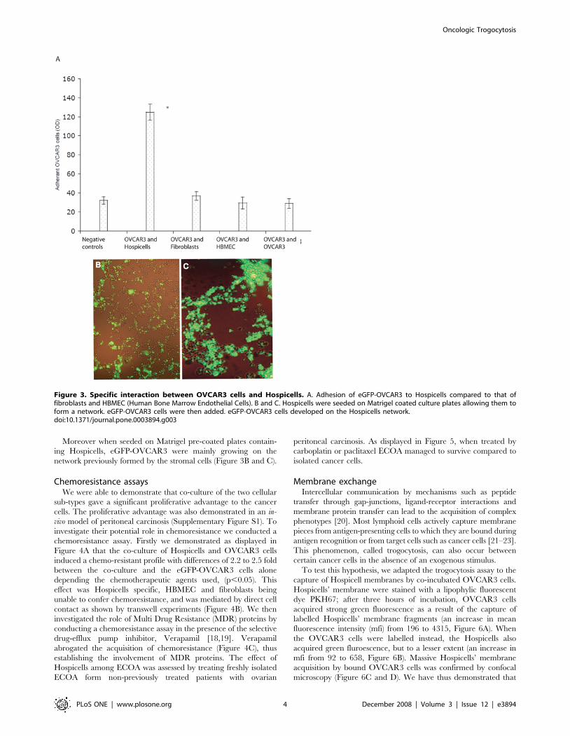
Moreover when seeded on Matrigel pre-coated plates contain-
ing Hospicells, eGFP-OVCAR3 were mainly growing on the
network previously formed by the stromal cells (Figure 3B and C).
Chemoresistance assaysWe were able to demonstrate that co-culture of the two cellular
sub-types gave a significant proliferative advantage to the cancer
cells. The proliferative advantage was also demonstrated in an in-
vivo model of peritoneal carcinosis (Supplementary Figure S1). To
investigate their potential role in chemoresistance we conducted a
chemoresistance assay. Firstly we demonstrated as displayed in
Figure 4A that the co-culture of Hospicells and OVCAR3 cells
induced a chemo-resistant profile with differences of 2.2 to 2.5 fold
between the co-culture and the eGFP-OVCAR3 cells alone
depending the chemotherapeutic agents used, (p,0.05). This
effect was Hospicells specific, HBMEC and fibroblasts being
unable to confer chemoresistance, and was mediated by direct cell
contact as shown by transwell experiments (Figure 4B). We then
investigated the role of Multi Drug Resistance (MDR) proteins by
conducting a chemoresistance assay in the presence of the selective
drug-efflux pump inhibitor, Verapamil [18,19]. Verapamil
abrogated the acquisition of chemoresistance (Figure 4C), thus
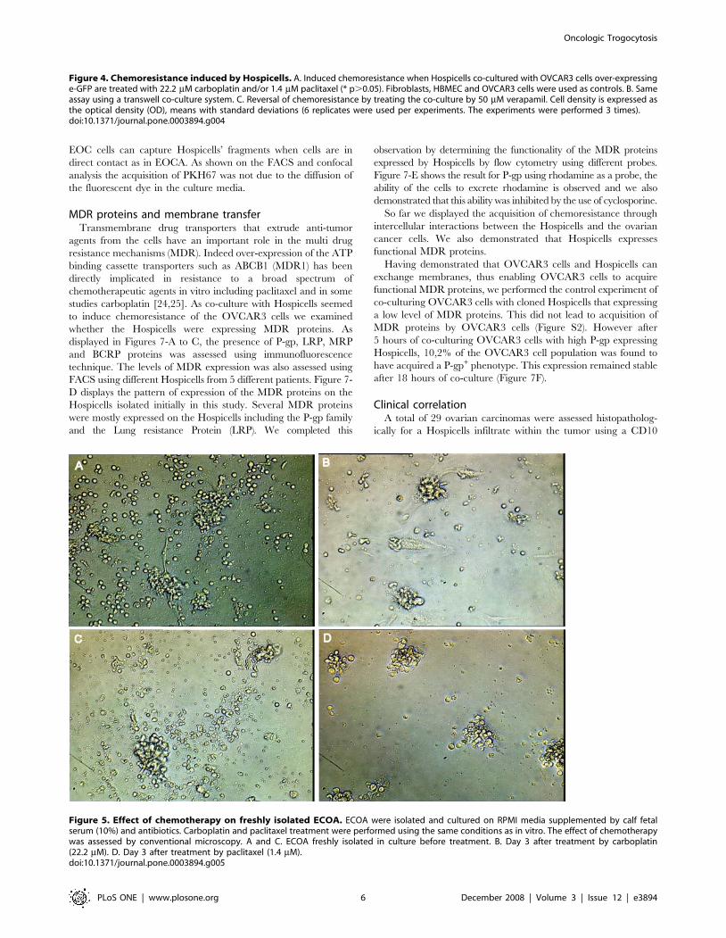
establishing the involvement of MDR proteins. The effect of
Hospicells among ECOA was assessed by treating freshly isolated
ECOA form non-previously treated patients with ovarian
peritoneal carcinosis. As displayed in Figure 5, when treated by
carboplatin or paclitaxel ECOA managed to survive compared to
isolated cancer cells.
Membrane exchangeIntercellular communication by mechanisms such as peptide
transfer through gap-junctions, ligand-receptor interactions and
membrane protein transfer can lead to the acquisition of complex
phenotypes [20]. Most lymphoid cells actively capture membrane
pieces from antigen-presenting cells to which they are bound during
antigen recognition or from target cells such as cancer cells [21–23].
This phenomenon, called trogocytosis, can also occur between
certain cancer cells in the absence of an exogenous stimulus.
To test this hypothesis, we adapted the trogocytosis assay to the
capture of Hospicell membranes by co-incubated OVCAR3 cells.
Hospicells’ membrane were stained with a lipophylic fluorescent
dye PKH67; after three hours of incubation, OVCAR3 cells
acquired strong green fluorescence as a result of the capture of
labelled Hospicells’ membrane fragments (an increase in mean
fluorescence intensity (mfi) from 196 to 4315, Figure 6A). When
the OVCAR3 cells were labelled instead, the Hospicells also
acquired green fluroescence, but to a lesser extent (an increase in
mfi from 92 to 658, Figure 6B). Massive Hospicells’ membrane
acquisition by bound OVCAR3 cells was confirmed by confocal
microscopy (Figure 6C and D). We have thus demonstrated that
Figure 3. Specific interaction between OVCAR3 cells and Hospicells. A. Adhesion of eGFP-OVCAR3 to Hospicells compared to that offibroblasts and HBMEC (Human Bone Marrow Endothelial Cells). B and C. Hospicells were seeded on Matrigel coated culture plates allowing them toform a network. eGFP-OVCAR3 cells were then added. eGFP-OVCAR3 cells developed on the Hospicells network.doi:10.1371/journal.pone.0003894.g003
Oncologic Trogocytosis
PLoS ONE | www.plosone.org 4 December 2008 | Volume 3 | Issue 12 | e3894

Oncologic Trogocytosis
PLoS ONE | www.plosone.org 5 December 2008 | Volume 3 | Issue 12 | e3894
EOC cells can capture Hospicells’ fragments when cells are in
direct contact as in EOCA. As shown on the FACS and confocal
analysis the acquisition of PKH67 was not due to the diffusion of
the fluorescent dye in the culture media.
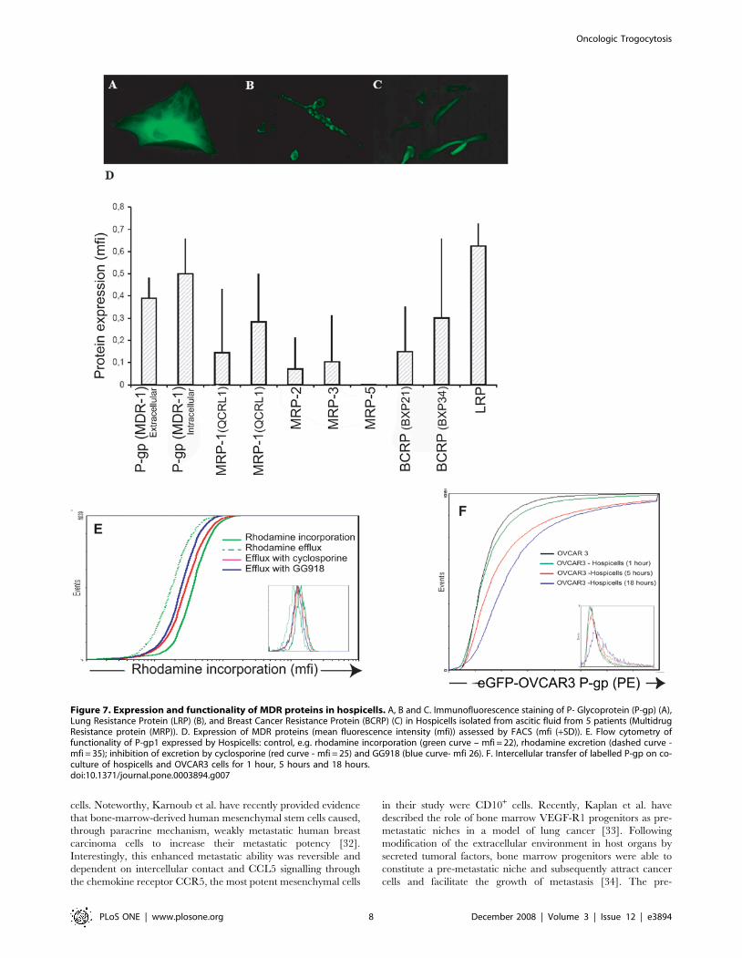
MDR proteins and membrane transferTransmembrane drug transporters that extrude anti-tumor
agents from the cells have an important role in the multi drug
resistance mechanisms (MDR). Indeed over-expression of the ATP
binding cassette transporters such as ABCB1 (MDR1) has been
directly implicated in resistance to a broad spectrum of
chemotherapeutic agents in vitro including paclitaxel and in some
studies carboplatin [24,25]. As co-culture with Hospicells seemed
to induce chemoresistance of the OVCAR3 cells we examined
whether the Hospicells were expressing MDR proteins. As
displayed in Figures 7-A to C, the presence of P-gp, LRP, MRP
and BCRP proteins was assessed using immunofluorescence
technique. The levels of MDR expression was also assessed using
FACS using different Hospicells from 5 different patients. Figure 7-
D displays the pattern of expression of the MDR proteins on the
Hospicells isolated initially in this study. Several MDR proteins
were mostly expressed on the Hospicells including the P-gp family
and the Lung resistance Protein (LRP). We completed this
observation by determining the functionality of the MDR proteins
expressed by Hospicells by flow cytometry using different probes.
Figure 7-E shows the result for P-gp using rhodamine as a probe, the
ability of the cells to excrete rhodamine is observed and we also
demonstrated that this ability was inhibited by the use of cyclosporine.
So far we displayed the acquisition of chemoresistance through
intercellular interactions between the Hospicells and the ovarian
cancer cells. We also demonstrated that Hospicells expresses
functional MDR proteins.
Having demonstrated that OVCAR3 cells and Hospicells can
exchange membranes, thus enabling OVCAR3 cells to acquire
functional MDR proteins, we performed the control experiment of
co-culturing OVCAR3 cells with cloned Hospicells that expressing
a low level of MDR proteins. This did not lead to acquisition of
MDR proteins by OVCAR3 cells (Figure S2). However after
5 hours of co-culturing OVCAR3 cells with high P-gp expressing
Hospicells, 10,2% of the OVCAR3 cell population was found to
have acquired a P-gp+ phenotype. This expression remained stable
after 18 hours of co-culture (Figure 7F).
Clinical correlationA total of 29 ovarian carcinomas were assessed histopatholog-
ically for a Hospicells infiltrate within the tumor using a CD10
Figure 4. Chemoresistance induced by Hospicells. A. Induced chemoresistance when Hospicells co-cultured with OVCAR3 cells over-expressinge-GFP are treated with 22.2 mM carboplatin and/or 1.4 mM paclitaxel (* p.0.05). Fibroblasts, HBMEC and OVCAR3 cells were used as controls. B. Sameassay using a transwell co-culture system. C. Reversal of chemoresistance by treating the co-culture by 50 mM verapamil. Cell density is expressed asthe optical density (OD), means with standard deviations (6 replicates were used per experiments. The experiments were performed 3 times).doi:10.1371/journal.pone.0003894.g004
Figure 5. Effect of chemotherapy on freshly isolated ECOA. ECOA were isolated and cultured on RPMI media supplemented by calf fetalserum (10%) and antibiotics. Carboplatin and paclitaxel treatment were performed using the same conditions as in vitro. The effect of chemotherapywas assessed by conventional microscopy. A and C. ECOA freshly isolated in culture before treatment. B. Day 3 after treatment by carboplatin(22.2 mM). D. Day 3 after treatment by paclitaxel (1.4 mM).doi:10.1371/journal.pone.0003894.g005
Oncologic Trogocytosis
PLoS ONE | www.plosone.org 6 December 2008 | Volume 3 | Issue 12 | e3894

staining. Figure 8A displays the different level of Hospicells
staining. As displayed in Figure 8B TMA from chemoresistant
patients had significantly higher density of Hospicells compared to
chemosensitive patients. Demographic characteristics of the
patients included in the study are displayed in Figure 8C.
Discussion
There are many evidences that preferential invasion of the
peritoneum by ovarian cancer is not only due to anatomic
proximity but also to an array of molecular signals secreted by
cancer cells and stromal cells predisposing the peritoneum to
invasion. Indeed the role of molecule-dependant cell-cell interac-
tions (integrin b-1, CD44), secreted factors (interleukin 6 and 8,
VEGF, FGF2, TGF-b) and matrix metalloproteinases, has been
illustrated in several reports [26–30]. However one of the most
important partners of cancer cells in the peritoneum are
mesothelial cells of the peritoneal surface. As described in the
introduction, few studies are addressing the role of these cells in
ovarian cancer. In our study, in order to avoid the bias associated
to the heterogeneity of peritoneal sampling, we isolated cancer cell
aggregates. The hypothesis of our work was that stromal cells
among these aggregates would display a high specificity regarding
their interactions with ovarian cancer cells.
In this study we have isolated stromal cells not previously
described that act as host cells. These cells represent a new subset
of stromal cells that do not express any of the common markers for
other cell types. The co-expression of the cell surface markers
CD9, CD10, CD29, CD146 and CD 166 has not been linked to
any specific lineage. However they have already been described as
part of the complex phenotype of the mesenchymal stem cells in
particular the adipose tissue derived stromal cells also called
stromal vascular fraction [31]. The Hospicells might therefore
represent a differentiated stromal subset of the mesenchymal stem
Figure 6. Intercellular transfer or ‘‘oncologic trogocytosis’’. A and B. Flow cytometry of OVCAR3 cells or Hospicells co-cultured with CellTrackerTM Orange CMTMR-labelled cells for 3 minutes or 3 hours. A. The Hospicells were labelled with the membranous lipophilic dye PKH67B. TheOVCAR3 cells were labelled with PKH67. The value given in the plot is the mean PKH67 fluorescence intensity. C. Confocal microscopy of PKH67-stained hospicells (green) and CMTMR-labelled OVCAR3 cells (red) after 3 minutes incubation. D. Uptake of PKH67 by OVCAR3 cells is observed at3 hours indicating membrane transfer without cytoplasmic transfer.doi:10.1371/journal.pone.0003894.g006
Oncologic Trogocytosis
PLoS ONE | www.plosone.org 7 December 2008 | Volume 3 | Issue 12 | e3894
cells. Noteworthy, Karnoub et al. have recently provided evidence
that bone-marrow-derived human mesenchymal stem cells caused,
through paracrine mechanism, weakly metastatic human breast
carcinoma cells to increase their metastatic potency [32].
Interestingly, this enhanced metastatic ability was reversible and
dependent on intercellular contact and CCL5 signalling through
the chemokine receptor CCR5, the most potent mesenchymal cells
in their study were CD10+ cells. Recently, Kaplan et al. have
described the role of bone marrow VEGF-R1 progenitors as pre-
metastatic niches in a model of lung cancer [33]. Following
modification of the extracellular environment in host organs by
secreted tumoral factors, bone marrow progenitors were able to
constitute a pre-metastatic niche and subsequently attract cancer
cells and facilitate the growth of metastasis [34]. The pre-
Figure 7. Expression and functionality of MDR proteins in hospicells. A, B and C. Immunofluorescence staining of P- Glycoprotein (P-gp) (A),Lung Resistance Protein (LRP) (B), and Breast Cancer Resistance Protein (BCRP) (C) in Hospicells isolated from ascitic fluid from 5 patients (MultidrugResistance protein (MRP)). D. Expression of MDR proteins (mean fluorescence intensity (mfi)) assessed by FACS (mfi (+SD)). E. Flow cytometry offunctionality of P-gp1 expressed by Hospicells: control, e.g. rhodamine incorporation (green curve – mfi = 22), rhodamine excretion (dashed curve -mfi = 35); inhibition of excretion by cyclosporine (red curve - mfi = 25) and GG918 (blue curve- mfi 26). F. Intercellular transfer of labelled P-gp on co-culture of hospicells and OVCAR3 cells for 1 hour, 5 hours and 18 hours.doi:10.1371/journal.pone.0003894.g007
Oncologic Trogocytosis
PLoS ONE | www.plosone.org 8 December 2008 | Volume 3 | Issue 12 | e3894

metastatic niche was constituted from CD34-positive progenitors,
however the exact composition of the niche is not illustrated in
their study. We can hypothesize that the pre-metastatic niche is a
complex microenvironment such as the stem cell niche. Different
cellular subtypes might then have different roles in promoting
metastasis. Our cell type may act as a ‘‘feeder’’ cell in the initiation
of the metastatic process. Moreover stem cells from a variety of
tissues express high levels of ABC transporting proteins that play a
role in cytoprotection through the excretion of genotoxic and
xenobiotic compounds out of the cells [35]. These latter properties
might therefore create a ‘‘drug-free’’ niche that will be more
suitable to cancer cells development.
Hospicells also displayed positivity for other markers. CD9 is a
member of the tetraspanin family. These proteins mediate signal
transduction events and play a role in the regulation of cell
development, activation, growth and motility and intercellular
interactions. CD9 in particular plays a role in fusion between the
spermatozoide and the ovocyte. In addition, it promotes muscle
cell fusion and support myotube maintenance [36–38]. CD9 down
regulation on ovarian cancer cells seem to be associated to higher
histological grade and metastatic progression. The role of CD9
expression on stromal cells in ovarian cancer has not been
established [39]. CD10 is a neutral endopeptidase (NEP) also
known as common acute lymphoblastic leukemia antigen
(CALLA). It is a zinc-dependent metalloprotease enzyme that
degrades a number of small-secreted peptides. Associations have
been observed between CD10 overexpression on cancer or
stromal cells and various types of cancer such as ovarian cancer
or advanced melanoma [40,41]. CD10 was specifically expressed
in the stroma of borderline and malignant ovarian tumors, but not
in adenomas. Furthermore, stromal CD10 was downregulated as
the histological grade advanced. These results suggest that CD10
may play a role in the regulation of neoplastic transformation and
tumor differentiation in epithelial ovarian carcinomas. Similarly
the presences of CD10 positive cells have not been assessed in
ovarian cancer so far. CD166 or ALCAM. Decreased/lost
ALCAM membrane expression is a marker of poorer outcome
in epithelial ovarian cancer. CD166 has recently been described as
a marker of the bone marrow stromal cells capable of supporting
hematopoiesis [42,43]. Its specific role in ovarian cancer has not
Figure 8. Clinical relevance of the presence of Hospicells among ovarian cancer tumors. Tissue Micro-arrays were prepared from tumorsoriginating from 29 patients who underwent a neo-adjuvant chemotherapy. A. Different densities of Hospicells as represented by CD10 staining.Morphological controls were used to rule out the staining of endothelial cells. B. Samples from chemoresistant patients had significantly higherdensity of Hospicells compared to chemosensitive patients (* p,0.05). Three different spots were used to characterize the Hospicells’ density for eachpatient.doi:10.1371/journal.pone.0003894.g008
Oncologic Trogocytosis
PLoS ONE | www.plosone.org 9 December 2008 | Volume 3 | Issue 12 | e3894

been studied. CD146, the melanoma cell adhesion molecule (M-
CAM), is a cell adhesion molecule currently used as a marker for
endothelial cell lineage. Its function is still poorly understood, but
evidence points to it being part of the endothelial junction
associated with the actin cytoskeleton. It is expressed, activated
human T cells, endothelial progenitors such as angioblasts and
mesenchymal stem cells, and strongly expressed on blood vessel
endothelium and smooth muscle [44]. In Ovarian cancer, M-
CAM is a marker of early relapse and poorer outcome in EOC. In
particular, M-CAM expression identifies a subgroup of front-line
therapy-responding patients who undergo dramatic relapses [45].
Mesenchymal stem cells expressing this marker seem to be
associated with perivascular cells surrounding the blood vessels
[46].
The morphology of these cells is perfectly adapted to their role
as they have many pseudopodia that allow large contacts with
cancer cells. As we observed a specific and intense adhesion
between the two cell types, we hypothesized that the Hospicells
would play a role in ovarian cancer cell physiology.
Intercellular communication can lead to acquisition of complex
phenotypes. Several mechanisms can occur during inter-cellular
communication such as molecules transferring through gap-
junctions, coupling through ligand receptor interactions and
finally transfer of membranes proteins leading to new-cell surface
proteins conferring new properties to the accepting cells [20,36–
38]. Levchenko et al. have described intercellular transfer of
functional P-glycoprotein among several tumor cell lines [47]. This
inter-cellular protein transfer was able to induce chemoresistance
in previously sensitive cells. As one of the major challenges in
ovarian cancer treatment remains chemo-resistance, we investi-
gated the role of Hospicells in the survival of cancer cells treated
with different chemotherapeutic agents. We were able to describe
the ability of a subset of normal cells to confer chemo-resistance to
ovarian cancer cells through inter-cellular contact. Two findings
were important in the chemoresistance assay performed: the
specificity of the Hospicells to confer chemoresistance compared to
other cell types used as controls and the importance of the inter-
cellular adherence. All together these two findings suggest that
Hospicells can be a crucial key in the chemoresistance phenom-
enon, and their presence might have consequences on patients’
prognoses. Moreover the necessity of the contact between the two
cells rules out the role of secreted factors and confirms the role of
the peritoneal cells ’’per se’’ in the occurrence of ovarian tumor
chemoresistance.
Several mechanisms can be responsible for chemoresistance
acquisition. The role of the MDR proteins in particular in ovarian
carcinoma has been widely illustrated in the literature [24,25]. We
were able to demonstrate the presence of MDR proteins on
Hospicells from several different patients with a variable
expression between the patients. However the expression of the
functional P-gp and LRP was constant. The ability of Verapamil
to revert the chemoresistance in the co-culture also confirmed the
role of the MDR proteins. The MDR proteins of interest in this
study were P-glycoprotein (P-gp, MDR1, ABC B1), a membrane
glycoprotein. Its resistance phenotype is similar and includes
anthracyclines (doxorubicin), vinca-alkaloids, epidophyloxins and
taxanes and it has also been related to carboplatin resistance
[18,19,24,25].
Several mechanisms can underlie the occurrence of chemore-
sistance after inter-cellular contact. However the specificity, the
crucial role of intercellular interaction, and the morphology of
these interactions displayed by electronic microscopic analysis
suggested the occurrence of ‘‘oncologic synapses’’ as compared to
‘‘immunological synapses’’. Indeed lymphoid cells (effectors) will
initiate their interactions with surveyed cells (targets) by setting
synapses that enable their surface receptors to interact with ligand,
to concentrate activation signals, and finally to deliver effectors
functions [48,49]. The lymphocyte cell surface in this contact area
makes small bridges with the target cell and captures patches of its
membrane on its own cell surface, an active process referred to as
trogocytosis [48]. Using the same experimental designs, we
describe ‘‘trogocytosis’’ between a cancer cell (as an effectors)
and a normal host cell (as a target) with a transfer of MDR
proteins from the host cells to the cancer cells. Trogocytosis
enables a better survey by the immune cells in the classical
‘‘immunological setting’’. The failure of such mechanisms can play
a role in immuno-evasion. Similarly in the ‘‘oncologic setting’’
trogocytosis might help cancer cells acquire new properties
spontaneously or under selective pressure (initiation of metastasis,
chemotherapy).
The correlation of Hospicells’ infiltrate with the patients’
response to neo-adjuvant chemotherapy displayed in the prelim-
inary clinical study might lead to the prediction of spontaneous
chemoresistance in patients and to optimisation of the chemo-
therapy regimen. One of the limitation of our TMA study is the
small number of patients included, however to avoid bias linked to
the heterogeneity of ovarian tumors and management protocols
we have selected only patients treated by neo-adjuvant chemo-
therapy. Moreover chemosensitivity was rigorously defined as the
absence of any active tumoral infiltrate at the time of final surgery.
These findings have to be confirmed in a larger independent set of
patients to raise clinical relevance.
The role of the host as major actor in controlling neoplasic
disease was illustrated in several reports underlying the role of the
immunologic activation in patients’ prognoses. Galon et al.
demonstrated that a sign of an immune response within colorectal
cancers was associated with the absence of pathological evidence
of early metastatic invasion and with prolonged survival [50].
These data, as well as our present data on acquired chemoresis-
tance of ovarian cancer cells by MDR protein transfer from
Hospicells, suggest that the host has a key role in the development
or control of neoplastic disease. The determination of cellular
mechanisms and pathways leading to intercellular recognition and
‘‘oncologic trogocytosis’’ with Hospicells could lead to the
identification of novel therapeutic approaches targeting intercel-
lular interactions.
Materials and Methods
Cell culturesMesothelial cells interacting with ovarian carcinoma cells were
isolated from ascitis of non-previously treated patients with stage
III ovarian cancer undergoing ascitis evacuation for clinical
discomfort following routine protocols approved by the IRB of the
Hospital Hotel-Dieu, Paris. As ascitis evacuation is part of the
routine management of patients in the medical oncology
department of Hotel-Dieu only oral consent was obtained from
the patients. The ascitis were then de-identified and addressed to
the research laboratory. As the laboratory and hospital have two
different locations there was no way to link the ascitis to the
patients’ file. Five different patients’ ascitis were used in this study;
ascitis fluids were centrifuged at 800 rpm for 1 minute. Lympho-
cytes and erythrocytes were separated from cancer cell aggregates
using a Ficoll procedure; dilution method was used to select
ovarian cancer cells aggregated on mesothelial cells. The
aggregates were separated using trypsin (Mediatech, Inc). Using
serial dilution and enrichment, mesothelial cells (Hospicells)
interacting specifically with the ovarian cancer aggregates were
Oncologic Trogocytosis
PLoS ONE | www.plosone.org 10 December 2008 | Volume 3 | Issue 12 | e3894

isolated and grown at 37uC in 5% CO2/95% air in RPMI
medium (Mediatech Inc.) supplemented with 10% fetal bovine
serum (FBS; Mediatech Inc.) and 5% glutamine. Human ovarian
cancer cells (OVCAR3), a fibroblast cell line (CCD-976SK) and
human bone marrow endothelial cells (HBMEC) were obtained
from American Type Culture Collection (ATCC) and were
maintained as instructed. The Tissue Micro Array were
constructed from surgical specimens obtained in the Institut
Claudius Regaud, the patients gave a general written informed
consent and the use of these specimens was authorized by the IRB
of the Institut Claudius Regaud.
ImmunohistochemistryStromal cells were pelleted and fixed in paraffin. Immunohis-
tochemistry was performed on 4 mm-thick routinely processed
paraffin sections. The following antibodies were used to charac-
terize stromal cells: cytokeratin (KL1, Beckman Coulter), vimentin
(V9, Beckman Coulter), CD45 (2b11 and PD7/26, Dako), CD20
(L26, Dako), CD3 (SP7, Neomarkers), CD68 (KP1 and PG-M1,
Dako), CD 34 (OBend10, Dako), CD-10 (clone 56C6, Novocastra,
Newcastle, UK), S100 protein (polyclonal, Dako), myeloperox-
ydase (polyclonal, Dako), CD166 (polyclonal, Dako), CD146
(polyclonal, Dako) and epithelial membrane antigen (E29, Dako).
The antibodies were used according to the manufacturers’
instructions. Antibodies against ABC proteins (QCRL1, QCRL3,
MRP2, MRP3, MRP5, LRP, BXP21, and BXP34) and the anti-P-
gp (CD243) antibody linked to phycoerythrin (PE) were provided
by Immunotech (Marseille). To assess clinical relevance of the
presence of Hospicells we retrospectively reviewed the correlation
between their presence and chemoresistance. Using a tissue micro-
array instrument (Beecher Instruments, Alphelys), we removed
two representative areas of the tumor from paraffin-embedded
tissue blocks that had been prepared at the time of resection.
Tissue micro-arrays containing the tissue cores were then cut into
5-um sections for staining with Harris’s hematoxylin and
immunohistochemical staining. 29 patients treated by neo-
adjuvant chemotherapy (e.g. six cycles of carboplatine and taxol)
and secondary surgery in our cancer center between January 2000
and January 2005 were included in this preliminary study. Staging
was assessed according to the International Federation of
Gynecology and Obstetrics (FIGO) classification. Chemosensitiv-
ity was defined as achieving complete clinical response after
completion of the first-line chemotherapy associated to a
pathologic complete response defined as no evidence of disease
after pathologic examination of all specimens at the time of
secondary surgery. Chemoresistant was defined as patients with
histologically confirmed residual disease at the time of secondary
surgery. This is a more rigorous definition of resistance as
compared with conventional clinical criteria. It has already been
used by others and recognizes that absence of a pathologic
complete response signifies the presence of a chemoresistant
cellular population. Each specimen was examined for the
Hospicells infiltrates within the tumor. The densities of these
stromal infiltrates were scored independently (EM, AR) on a 0 to
10 scale combining a CD10 immunoblotting and morphological
alaysis. All vascular-like structures were excluded from the
evaluation. The clinical data were independently retrieved by
another investigator (JC).
Electron microscopyOvarian cancer aggregates were cultured for 48 hours. Cells
were subsequently washed with the PBS and fixed for 45 min in
30% formaldehyde +5% glutaraldehyde. Thereafter, the cells were
centrifuged, treated with 50 mM ammonium chlorate, dehydrated
and enveloped in the Epoxy resin at low temperature at
polymerization conditions. The micro sections (600–800 Au) were
performed and colored with uranyl acetate and lead and visualized
on a Philips CM 10 electron microscope [51]. Primary Hospicells
and OVCAR3 cultures were performed and analyzed to establish
the microscopic characteristics of the two different cell types and
identify each one of them among the aggregates.
Adhesion assayCulture plates were coated with Hospicells, fibroblasts or
HBMEC (Human Bone Marrow Endothelial Cells) up to 70%
confluency. OVCAR3 cells overexpressing enhanced green
fluorescent protein (eGFP) were added. Gentle washing after
1 hour eliminated non-adherent cells. Optical density was
measured. The experiment is representative of three independent
experiences.
Matrigel culturesNinety six wells plates pre-coated with matrigel (Becton-
Dickinson, Le Pont de Claix, France) were used and matrigel
was allowed to polymerize for 1–2 hours at 37uC. Hospicells were
cultivated on Matrigel for 24 hours before eGFP-Ovcar3 were
seeded onto the matrigel-coated wells. Following a 24 hours
incubation periods at 37uC, intercellular interactions were assessed
using an inverted microscope fitted with a digital camera (Nikon-
Diafot).
Chemoresistance assayHospicells were first grown to 60% confluency in 96-well flat-
bottomed tissue-culture plates. OVCAR3 cells over-expressing e-
GFP were plated at a density of 20,000 cells/well and co-cultured
for 24 hours with the hospicells before addition of 22.2 mM
carboplatin or 1.4 mM paclitaxel [52]. The effect of these cytotoxic
agents was assessed using a quantitative sulphorhodamine B (SRB)
colorimetric assay, as described previously [53]. The same
experiment was also performed using a transwell co-culture
system in which Hospicells were cultured in the bottom chamber
and OVCAR3 cells were cultured in the upper membrane. Both
types of experiments were repeated after addition of 50 mM
verapamil. To confirm the potential role of Hospicells we isolated
aggregates from ascitis of non-previously treated patients with
ovarian peritoneal carcinosis. We cultured these aggregates on
RPMI media supplemented by calf fetal serum (10%) and
antibiotics. Carboplatin and paclitaxel treatment were performed
using the same conditions as in vitro. The effect of chemotherapy
was assessed by conventional microscopy. The experiment is
representative of three independent experiences.
Cell transfer (‘‘oncologic trogocytosis’’) assayThe assay was carried out as previously described [54]. Cancer
cells or Hospicells were stained with the lipophilic dye PKH67
(Sigma-Aldrich) or with the cytoplasmic Cell Tracker Orange
CMTMR (5-(and-6)-(4-chloromethyl-benzoyl-amino-tetramethyl-
rhodamine)) (Molecular Probes, Eugene, Oregon, USA) according
to the manufacturer’s instructions. The PKH67-labelled cells were
then co-cultured for 3 hours with CMTMR-labelled cells in 96-
well round-bottomed tissue-culture plates at a density of 66105
cells in 120 ml of complete RPMI 1640 medium supplemented
with 10% fetal calf serum (FCS). Culture plates were centrifuged
for 1 min at 700 rpm to promote cell contact and left for 1 hour at
37uC in 5% CO2. Cells were then washed twice with PBS
containing 0.5 mM EDTA and analysed at two time-points
(3 minutes and 3 hours) by flow cytometry using a LSRII
Oncologic Trogocytosis
PLoS ONE | www.plosone.org 11 December 2008 | Volume 3 | Issue 12 | e3894

cytometer and DIVA software (BD Biosciences, Mountain View,
CA). Live cells were gated on the basis of forward scatter/side
scatter parameters and 25,000 events were acquired in each
experiment with FL1 channel (log scale) for PKH67 and FL2
channel (log scale) for CMTMR. The synaptic transfers were
deducted from the PKH67 mean fluorescence of gated CMTMR-
labelled cells after 3 minutes and 3 hours of co-culture. The cell
transfer assay was repeated with labelled P-gp. The experiment is
representative of three independent experiences.
Confocal microscopyPKH67-stained hospicells and CMTMR-labelled OVCAR3
cells were processed as follows. After contact for 3 minutes or
3 hours, cells were gently resuspended and plated on poly-L-lysine
(Sigma-Aldrich, St Louis, MI) coated slides for 5 minutes at 37uC.
After fixation with PBS containing 4% p-formaldehyde, cells were
washed and directly mounted in PBS containing 90% glycerol and
2% 1-4-diazabicyclo (2.2.2) octane (DABCO, Sigma-Aldrich, St
Louis, MI). Samples were examined using an LSM 410 confocal
microscope (Carl Zeiss, Jena, Germany).
Fluorescence Activated Cell Sorting (FACS)To determine MDR protein expression in Hospicells, they were
seeded at a density of 86104 cells/well. Two days later, the cells
were serum-starved for 24 hours, fixed in 3% paraformaldehyde,
and permeabilized with 0.1% Triton X-100 in PBS. The pattern
of MDR protein expression was assessed by Fluorescence
Activated Cell Sorting (FACS) using the Intraprep permeabiliza-
tion reagent kit (Beckman-Coulter). Isotype controls were provided
by Chemicon International Inc. (Temecula, CA, USA). Protein
expression was analysed by flow cytometry (EPICS Altra, Beck-
man Coulter). The functionality of ABC proteins expressed by
Hospicells was determined by flow cytometry using rhodamine
(1026 mol/L) and cyclosporine (261026 mol/L) as probes. Their
effect was expressed as a shift in the mfi of dye accumulation. For
each sample, 5,000 events were collected. The experiment is
representative of three independent experiences.
Proliferation assay96 wells plate were seeded with 20000 Hospicells. 5000 eGFP-
OVCAR3 cells were then added. Culture were performed at 37uCin 5% CO2/95% air in RPMI medium (Mediatech Inc.)
supplemented with 10% fetal bovine serum (FBS; Mediatech
Inc.) and 5% glutamine. Proliferation was evaluated daily by
evaluating the fluorescence of each well using a fluorescent reader
(Wallac). Each condition was done in triplicate. The experiment is
representative of three independent experiences.
Statistical AnalysisDescriptive statistics were calculated for baseline demographic
and clinicopathologic characteristics. Associations between CD10
immunoreactivity and clinicopathologic features were assessed by
the chi-square test. For other experiments Student-t, Fisher exact
or chi-square test were performed as appropriate. All p-values are
two-sided with statistical significance evaluated at the 0.05 alpha
level. Ninety-five percent confidence intervals (95% CI) were
calculated to assess the precision of the obtained estimates. All
analyses were performed in SAS Version 9.1 (SAS Institute, Inc.,
Cary, North Carolina) and Stata Version 8.0 (Stata Corporation,
College Station, Texas). Mean6SEM are shown on the graphs.
Supporting Information
Figure S1 Proliferative effect of Hospicells on OVCAR3 cells.
In-vitro proliferation assay. Co-culture of Hospicells and eGFP-
OVCAR3 cells. 96 wells plate were seeded with 20000 Hospicells.
5000 eGFP-OVCAR3 cells were then added. Culture were
performed at 37uC in 5% CO2/95% air in RPMI medium
supplemented with 10% Fetal Calf Serum. Proliferation was
assessed daily using a fluorescent plate reader. (Representative of 3
different experiments).
Found at: doi:10.1371/journal.pone.0003894.s001 (0.32 MB TIF)
Figure S2 Acquisition of Pgp by e-GFP-OVCAR3 cells when
incubated with low PgP expressing Hospicells. Intercellular
transfer of labelled P-gp on co-culture of Hospicells with low
expression of Pgp and eGFP-OVCAR3 cells for 5 hours. As
displayed there was no acquisition of PgP by eGFP-OVCAR3
cells.
Found at: doi:10.1371/journal.pone.0003894.s002 (1.98 MB TIF)
Author Contributions
Conceived and designed the experiments: ART MP JJF JPM EPL JS.
Performed the experiments: ART MM MP AMF AS ED EM RL JC JB
FD MM. Analyzed the data: MP ED EM RL JC JB DQ FD JJF JS.
Contributed reagents/materials/analysis tools: BC EPL GF. Wrote the
paper: ART BC DQ JPM GF.
References
1. Jemal A, Siegel R, Ward E, Hao Y, Xu J, et al. (2008) Cancer statistics. CACancer J Clin 58: 71–96.
2. Bhoola S, Hoskins WJ (2006) Diagnosis and management of epithelial ovariancancer. Obstet Gynecol 107: 1399–1410.
3. Eisenkop SM, Spirtos NM, Lin WC (2006) ‘‘Optimal’’ cytoreduction for
advanced epithelial ovarian cancer: a commentary. Gynecol Oncol 103: 329–35.
4. Pfisterer J, Ledermann JA (2006) Management of platinum-sensitive recurrent
ovarian cancer. Semin Oncol 33: S12–6.
5. Di Nicolantonio F, Mercer SJ, Knight LA, Gabriel FG, Whitehouse PA, et al.
(2005) Cancer cell adaptation to chemotherapy. BMC Cancer 5: 78.
6. Wang E, Ngalame Y, Panelli MC, Nguyen-Jackson H, Deavers M, et al. (2005)
Peritoneal and subperitoneal stroma may facilitate regional spread of ovariancancer. Clin Cancer Res 111: 13–22.
7. Shostak A, Chakrabarti E, Hirszel P, Maher JF (1988) Effects of histamine andits receptor antagonists on peritoneal permeability. Kidney Int 34: 786–90.
8. Muijsken MA, Heezius HJ, Verhoef J, Verbrugh HA (1991) Role of mesothelial
cells in peritoneal antibacterial defence. J Clin Pathol 44: 600–4.
9. Zhang XY, Pettengell R, Nasiri N, Kalia V, Dalgleish AG, et al. (1999)
Characteristics and growth patterns of human peritoneal mesothelial cells:comparison between advanced epithelial ovarian cancer and non-ovarian cancer
sources. J Soc Gynecol Investig 6: 333–340.
10. Casey RC, Burleson KM, Skubitz KM, Pambuccian SE, Oegema TR, et al.
(2001) Beta1-integrins regulate the formation and adhesion of ovariancarcinoma multicellular spheroids. Am J Pathol 159: 2071–80.
11. Gardner MJ, Jones LM, Catterall JB, Turner GA (1995) Expression of celladherence molecules on ovarian tumour cell lines and mesothelial cells, in
relation to ovarian cancer metastasis. Cancer Lett 91: 229–34.
12. Jones LM, Gardner MJ, Catterall JB, Turner GA (1995) Hyaluronic acidsecreted by mesothelial cells: a natural barrier to ovarian cancer cell adherence.
Clin Exp Metastasis 13: 373–80.
13. Burleson KM, Hansen LK, Skubitz AP (2004) Ovarian carcinoma spheroidsdisaggregate on type I collagen and invade live human mesothelial cell
monolayers. Clin Exp Metastasis 21: 685–697.
14. Burleson KM, Casey RC, Skubitz KM, Pambuccian SE, Oegema TR, et al.(2004) Ovarian carcinoma ascites spheroids adhere to extracellular matrix
components and mesothelial cell monolayers. Gynecol Oncol 93: 170–181.
15. Sako A, Kitayama J, Yamaguchi H, Kaisaki S, Suzuki H, et al. (2003)Vascular endothelial growth factor synthesis by human omental mesothe-
lial cells is augmented by fibroblast growth factor-2: possible role ofmesothelial cell on the development of peritoneal metastasis. J Surg Res
115: 113–120.
16. Wilson A (1989) Mesothelial cells stimulate the anchorage-independent growthof human ovarian tumour cells. Br J Cancer 59: 876–82.
17. Kishikawa T, Sakamoto M, Ino Y, Kubushiro K, Nozawa S, et al. (1995) Two
distinct patterns of peritoneal involvement shown by in vitro and in vivo ovariancancer dissemination models. Invasion Metastasis 15: 11–21.
18. Trimble EL, Wright J, Christian MC (2001) Treatment of platinum-resistant
ovarian cancer. Expert Opin Pharmacother 2: 1299–306.
Oncologic Trogocytosis
PLoS ONE | www.plosone.org 12 December 2008 | Volume 3 | Issue 12 | e3894

19. Mechetner EB, Roninson IB (1992) Efficient inhibition of P-glycoprotein-
mediated multidrug resistance with a monoclonal antibody. Proc Natl Acad Sci
USA 89: 5824–5828.
20. Darland DC, D’Amore PA (2001) Cell–cell interactions in vascular develop-
ment. Curr Top Dev Biol 52: 107–149.
21. Batista FD, Iber D, Neuberger MS (2001) B cells acquire antigen from target
cells after synapse formation. Nature 411: 489–494.
22. Espinosa E, Tabiasco J, Hudrisier D, Fournie JJ (2001) Synaptic transfer by
human gamma delta T cells stimulated with soluble or cellular antigens.
J Immunol 166: 3645–3649.
23. Poupot M, Pont F, Fournie JJ (2005) Profiling blood lymphocyte interactions
with cancer cells uncovers the innate reactivity of human gamma delta T cells to
anaplastic large cell lymphoma. J Immunol 174: 1717–1722.
24. Naniwa J, Kigawa J, Kanamori Y, Itamochi H, Oishi T, et al. (2007) Genetic
diagnosis for chemosensitivity with drug-resistance genes in epithelial ovarian
cancer. Int J Gynecol Cancer 7: 76–82.
25. Yakirevich E, Sabo E, Naroditsky I, Sova Y, Lavie O, et al. (2006) Multidrug
resistance-related phenotype and apoptosis-related protein expression in ovarian
serous carcinomas. Gynecol Oncol 100: 152–159.
26. Sako A, Kitayama J, Shida D, Suzuki R, Sakai T, et al. (2006) Lysophosphatidic
acid (LPA)-induced vascular endothelial growth factor (VEGF) by mesothelial
cells and quantification of host-derived VEGF in malignant ascites. J Surg Res
130: 94–101.
27. Rodriguez-Rodriguez L, Sancho-Torres I, Mesonero C, Gibbon DG, Shih WJ,
et al. (2003) The CD44 receptor is a molecular predictor of survival in ovarian
cancer. Med Oncol 20: 255–63.
28. Rodriguez GC, Haisley C, Hurteau J, Moser TL, Whitaker R, Bast, et al. (2001)
Regulation of invasion of epithelial ovarian cancer by transforming growth
factor-beta. Gynecol Oncol 80: 245–53.
29. Jayne DG, Perry SL, Morrison E, Farmery SM, Guillou PJ (2000) Activated
mesothelial cells produce heparin-binding growth factors: implications for
tumour metastases. Br J Cancer 82: 1233–8.
30. Betjes MG, Tuk CW, Struijk DG, Krediet RT, Arisz L, et al. (1993) Interleukin-
8 production by human peritoneal mesothelial cells in response to tumor necrosis
factor-alpha, interleukin-1, and medium conditioned by macrophages cocul-
tured with Staphylococcus epidermidis. J Infect Dis 168: 1202–10.
31. Schaffler A, Buchler C (2007) Concise review: adipose tissue-derived stromal
cells–basic and clinical implications for novel cell-based therapies. Stem Cells 25:
818–27.
32. Karnoub AE, Dash AB, Vo AP, Sullivan A, Brooks MW, et al. (2007)
Mesenchymal stem cells within tumour stroma promote breast cancer
metastasis. Nature 449: 557–563.
33. Kaplan RN, Riba RD, Zacharoulis S, Bramley AH, Vincent L, et al. (2005)
VEGFR1-positive haematopoietic bone marrow progenitors initiate the pre-
metastatic niche. Nature 438: 820–827.
34. Wels J, Kaplan RN, Rafii S, Lyden D (2008) Migratory neighbors and distant
invaders: tumor-associated niche cells. Genes Dev 22: 559–74.
35. Zhou S, Schuetz JD, Bunting KD, Colapietro AM, Sampath J, et al. (2001) The
ABC transporter Bcrp1/ABCG2 is expressed in a wide variety of stem cells and
is a molecular determinant of the side-population phenotype. Nat Med 7:
1028–34.
36. Boucheix C, Rubinstein E (2001) Tetraspanins. Cell Mol Life Sci 58(9):
1189–205.37. Hemler ME (2005) Tetraspanin functions and associated microdomains. Nat
Rev Mol Cell Biol 6(10): 801–11.
38. Levy S, Shoham T (2005) The tetraspanin web modulates immune-signallingcomplexes. Nat Rev Immunol 5(2): 136–48.
39. Houle CD, Ding XY, Foley JF, Afshari CA, Barrett JC, Davis BJ (2002) Loss ofexpression and altered localization of KAI1 and CD9 protein are associated with
epithelial ovarian cancer progression. Gynecol Oncol 86(1): 69–7.
40. Velasquez EF, Yancovitz M, Pavlick A, Berman R, Shapiro R, et al. (2007)Clinical relevance of Neutral Endopeptidase (NEP/CD10) in melanoma. Journal
of Translational Medicine 5(2): 2.41. Khin EE, Kikkawa F, Ino K, Suzuki T, Shibata K, et al. (2003) Neutral
endopeptidase/CD10 expression in the stroma of epithelial ovarian carcinoma.Int J Gynecol Pathol 22(2): 175–80.
42. Mezzanzanica D, Fabbi M, Bagnoli M, Staurengo S, Losa M, et al. (2008)
Subcellular localization of activated leukocyte cell adhesion molecule is amolecular predictor of survival in ovarian carcinoma patients. Clin Cancer Res
14(6): 1726–33.43. Seshi B, Kumar S, Sellers D (2000) Human bone marrow stromal cell:
coexpression of markers specific for multiplemesenchymal cell lineages. Blood
Cells Mol Dis 26(3): 234–46.44. Guezguez B, Vigneron P, Lamerant N, Kieda C, Jaffredo T, Dunon D (2007)
Dual Role of Melanoma Cell Adhesion Molecule (MCAM)/CD146 inLymphocyte Endothelium Interaction: MCAM/CD146 Promotes Rolling via
Microvilli Induction in Lymphocyte and Is an Endothelial Adhesion Receptor.J Immunol 179(10): 6673–85.
45. Shih IM, Wang TL, Westra WH (1996) Diagnostic and biological implications
of mel-CAM expression in mesenchymal neoplasms. Clin Cancer Res 2(3):569–75.
46. Zannettino AC, Paton S, Arthur A, Khor F, Itescu S, et al. (2008) Multipotentialhuman adipose-derived stromal stem cells exhibit a perivascular phenotype in
vitro and in vivo. J Cell Physiol 214(2): 413–21.
47. Levchenko A, Mehta BM, Niu X, Kang G, Villafania L (2005) Intercellulartransfer of P-glycoprotein mediates acquired multidrug resistance in tumor cells.
Proc Natl Acad Sci USA 102: 1933–1938.48. Joly E, Hudrisier D (2003) What is trogocytosis and what is its purpose? Nat
Immunol 4: 815.49. Davis DM (2007) Intercellular transfer of cell-surface proteins is common and
can affect many stages of an immune response. Nat Rev Immunol 7: 238–243.
50. Galon J, Fridman WH, Pages F (2007) The adaptive immunologic microenvi-ronment in colorectal cancer: a novel perspective. Cancer Res 67: 1883–1886.
51. Bendayan M, Nanci A, Kan FW (1987) Effect of tissue processing on colloidalgold cytochemistry. J Histochem Cytochem 35: 983–996.
52. Smith JA, Ngo H, Martin MC, Wolf JK (2005) An evaluation of cytotoxicity of
the taxane and platinum agents combination treatment in a panel of humanovarian carcinoma cell lines. Gynecol Oncol 98: 141–145.
53. Skehan P, Storeng R, Scudiero D, Monks A, McMahon J, Vistica D, Warren JT,Bokesch H, Kenney S, Boyd MR (1990) New colorimetric cytotoxicity assay for
anticancer-drug screening. J Natl Cancer Inst 82: 1107–1112.54. Poupot M, Fournie JJ (2003) Membrane transfer through homotypic synapses
between lymphoma cells. J Immunol 171: 2517–2523.
Oncologic Trogocytosis
PLoS ONE | www.plosone.org 13 December 2008 | Volume 3 | Issue 12 | e3894
Copyright © 2022 FDOKUMEN