Chlorpyrifos modifies the expression of genes involved in human placental function
Upload
independentCategory
view
1download
0
PATHOLOGY RESEARCH AND PRACTICE o Urban & Fischer Verlag http://www.urbanfischer.defjournaI5lprp
Animal and In vitro Models in Human Diseases
Urban Levels of Air Pollution Modifies the Progression of Urethane-induced Lung Tumours in Mice
Patricia M. Cury, Ana Julia F. C. Lichtenfels, Maria Suely F. Reymao, Gleice M. S. Concei<;ao, Vera L. Capelozzi and Paulo H. N. Saldiva
Laborat6rio de Polui~ao Atmosferica Experimental, Faculdade de Medicina da Universidade de Sao Paulo, Brasil
Summary
This paper investigates the effects of air pollution in urethane-induced lung tumours in mice by means of histological, morphometrieal, and DNA ploidy. The experimental exposure was done in locations with different a ir pollution profiles: a polluted area (downtown Sao Paulo) and a "clean" environment. Swiss mice were employed and urethane (3 g/kg) was uscd as a carcinogenic substance. All the animals, whether exposed or not to air pollution, were sacrificed after 6 months, and the lung lesions were analysed. The results showed a significant effect of air pollution on tumour progression, observed by changes in the phenotype of the tumour cells as demonstrated by morphometry and DNA ploidy. We observed more atypical adenomas in the air pollution-exposed group (p = 0.02). Coherently, morphometric differences were also detected between the two groups. Neoplasms of exposed mice exhibited an increase in the nuclear fraction (p = 0.002) and in the nucleus/cytoplasm ratio (p = 0.011), as a decrease in the stromal fraction (p < 0.00 I ). There was a higher risk of aneuploidy in the 6-months-of-air-pollution-exposure group (relative risk: 1.58; 95% of confidence interval: 1.007 to 2.403). These results indicate that urban air pollution accelerates the process of progression towards malignancy.
Key words: Air pollution - Lung cancer - Mice -Pathology - Experimental carcinogenesis
Introduction
The observation that the incidence of lung cancer is higher in urban areas than in the countryside is not re-
PathoL Res. Praet. 196: 627-633 (2000)
cent. There are several epidemiological studies associating urban air pollution and lung cancer, most of them with ecological design 18, 10,23]. Although the aforementioned reports have suggested that urban dwellers are at greater risk for developing lung cancer, they have somewhat failed to adequately control for the different lifestyle factors of urban residents such as early age of initiation of smoking, environmental tobacco smoke, indoor pollution and occupational exposures.
More recently. addi tional works have been designed anempting to compare urban-vs-rural cancer rates controlling for the mOSI plausible confounding factors other than urban pollution "per se·'. In some cases, a positive association was found [I. 9. 14, 22/. although some of them failed to do so {II , 29].
Another controversial point is, which pollutant is mainly responsible for any eventually observed increase in lung cancer? Many compounds detected in urban air, such as fine particulate pollution , polycyclic aromatic hydrocarbons and sulphates, are reported to present greater ri sks 19. 22, 24]. The foregoing statements clearly indicate that most of the difficulties of characteri sing the possible role of air pollution in lung cancer using conventional epidemiological studies arise mainly from two points: the difficulty of establishing adequate controls for other factors that urban dwellers are exposed to and, on the other hand, of isolating possibly carcinogenic agents amongst those that make up the complex pollution "fabric" of the urban environment.
In order to minimise at least one of the sources of "noise" - the population faelor - our laboratory devel-
Address ror correspondence: Patricia Maluf Cury, Departamento de Patologia. Faculdade de Medicina de Sao Jose do Rio Preto (FARMERP). Av. Faria Lima 5416. CEP 15090-000. Sao Jose do Rio Prcto-Sr. Brazil. Fax: 55- 17-210-5056. E~mail: pmcury @hotmail.com
0344·0338/2000/1 96/8-627 $15 .0010
628 . P. M. Cury et al.
oped a mouse experimental model to study the effects of urban air pollution, whereby lung tumours were induced with urethane in Swiss mice before or after having been submitted to air pollution. In a previous report [24J we showed that air pollution acts as a promoter in mice lung cancer, in a dose-dependent manner. It is well known that the natural history of urethane-induced lung tumours in mice encompasses a spectrum of differentiation ranging from foci of alveolar hyperplasia up to fully developed lung carcinomas. Thus, considering the possible role of air pollution as a promoter agent of such lesions, one may speculate that tumours developed in animals exposed to air pollution would present evidence of earlier progression towards malignancy in comparison with lesions of control animals. Therefore, the comparative determination of the degree of differentiation of exposed and control animals would provide an additional basis for confirming the possible role of air pollution in lung cancer.
In this study we have investigated from the histopathological point of view the possible influence of air pollution on these lesions. For this purpose, we compared the lesions from mice submitted both to and not to air pollution, involving 6 months of evolution, using morphometrical and DNA ploidy indicators.
Materials and Methods
In this experiment we analysed fully developed lesions, studied after 6 months of urethane injection and exposed either to clean or polluted air. For this purpose, we re-evaluated the six-month cases employed in a previous study of our group [24/.
Places of Study
The animals were kept in two locations with different air pollution profiles - downtown Sao Paulo and the rural region of Atibaia, both previously used in other studies of our laboratory dealing with the chronic effects of urban air pollution on rats [5, 20, 24, 25]. The animals were kept in cages, in safe open-air places (the tower of a church in downtown Sao Paulo and in a private house in Atibaia). Air pollution in Sao Paulo is
seriously high, caused mainly by the automotive emissions generated by ahout 5,000,000 vehicles using different fuels: ethanol, methanol, gasoline and diesel oil (6l. The control region was tbe rural area of Atibaia, witb iosignificant levels of air pollution. Table I shows the mean levels of pollution in Sao Paulo from the period between 1994 and 1997 according to the Air Pollution State Agency [7]. Sporadic measurements of air pollution (CO and No2) were made in Atibaia using a mobile station. The levels of these two pollutants there were always negligible.
Exposure Protocols
15-day-old Swiss mice were employed as experimental animals. Lung tumours were induced with urethane, an ethilic ester of carbamic acid classically used in studies on chemical carcinogenesis in mice [3, 21, 26-27]. In the two experiments, the animals were treated with a dose of 3 glkg of body weight of urethane administered by two intraperitoneal injections of 1.5 g/kg, with an interval of 48 hours. In all these experiments food and water were given ad libitllm and no replacement of animals was done in the case of death.
In our study, we analysed paraffin blocks of cases studied by Reimao et al. [24], trying to explore the effects of prolonged exposure to air pollution on tumour phenotype. After urethane injection, the mice were divided in two subgroups, one that stayed for 6 months in downtown Sao Paulo (n ~ 48) and the control group whicb stayed for 6 months in Atibaia (n ~ 43). After this period, all the animals were sacrificed.
Pathological alld Morphometrical Studies
After completing the exposure protocol, the animals were anaesthetised with ether and sacrificed by sectioning the caudal aorta. Thc lungs wcre carcfully dissected and fixed for 48 hours by intratracheal instillation of 2 ml of Carnoy's solution and embedding in the same solution. The abdominal organs were analysed macroscopically, and in case of any alteration, they were also studied histologically. After routine processing, 5 ~ m-thick sections were made with the paraffin-embedded tissue, each side containing the whole cross-sectional area of the lung and mediastinal lymph nodes of each mouse, and were stained with hematoxylin and eosin.
The lesions were submitted to qualitative and quantitative histopathological analysis. They were classified as hyperplasia, adenoma, and atypical adenoma, and subdivided in papillary, sol id-alveolar and mixed lesions, according to pathological criteria [17 , 28]. Two pathologists (PMC and VLC) per-
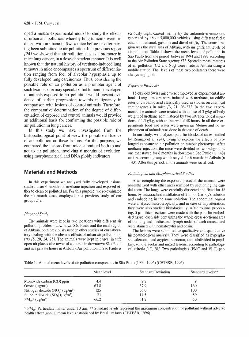
Table I. Annual mean levels of air pollution components in Sao Paulo (1994-1996) (CETESB, 1996)
Monoxide carbon (CO) ppm Ozone (~g/m ') Nitrogen dioxide (NO,) (~g1m J) Sulphur dioxide (SO,) (~g1m') PM,o' (~g1mJ)
Mean level
4.4 63.8 125 21
66.2
Standard Deviation
2.2 37.9 56.0 11.5 31.2
Standard leve ls **
9 160 100 80 50
* PM IO: Particulate matter under to 11m; ** Standard levels represent the maximum concentration of pollutant without adverse health effect (annual mean level) established by Brazilian laws (CETESB, 1996).

Fig. I. Histological aspect of a lung hyperplasia lesion (Hematoxylin-eosin. 20x).
Fig. 2. Histological aspect of a lung typical adenoma lesion (Hematoxylineosin,20x).
Fig. 3. Histological aspect of a lung atypical adenoma (Hematoxylin-eosin. 40x).
\ ' .. ~1
1 •
~I
•
• , 2
Erfects of Air Pollution in Urethane-induced Lung Tumours 629
l .... .
.' • '(
; ... . . " .. ..
.'
, ,
.. ~

630 ' P. M, Cury et al.
formed the classification. The criterion for classification as hyperplasia was alveolar cells proliferation without obliteration of more than four intra-alveolar spaces: adenoma was designated as proliferation with obliteration of more than four alveolar spaces, and a lesion was classified as atypical adenoma when presenting architectural and/or cellular atypia (Figs, 1, 2 and 3). We decided not to classify any case as carcinoma, as we did not find metastasis or invasion outside the lung, only cytological and architectural atypia, Thus, on behalf of prudence, we named lesions exhibiting a phenotype causing us to suspect malignancy as atypical adenomas, We defined a lesion as mixed when it presented both characteristics of solid and papillary patterns, with more than 20% of both types present.
Conventional morphometric procedures were done based on the point-counting technique [23J, with the aid of a coherent system of 100 points attached to the eyepiece of an optical microscope, The stromal fraction (STRO) was determined in 5 randomly selected fields at a magnification of X400 defined by the following relationship:
STRO = PstrolPtu (!)
where Pstro and Ptu were the number of points overlying stroma and tumour, respectively. The same method was used to calculate both nuclear fraction (FNUC) and cytoplasmic fraction (FCYT). The nucleus/cytoplasm ratio (N/C) was determined by the relationship:
N/C = Pnuc/Pcyt (2)
For all morphometric parameters, a coefficient of error (CE) was computed by the relationship:
CE = SEMlMean (3)
where SEM represents the standard error of the mean. Considering the number of points counted, CE was kept
under 5% in all morphometric measurements. For the morphomctrical analysis, 15 cases from each group
(both exposed and not to six moths of air pollution) were examined.
DNA and S-Phase analysis
Fifteen cases from each group (both exposed and not to 6 months of air pollution) were studied. The slides were stained by Feulgen's technique [I8J and analysed by a digital image system (Optimas 4,01 software) where DNA ploidy and Sphase were studied, Briefly, Feulgen's dye has a stoichiometric association with DNA histones. By measuring the colour intensity of the noclei, a histogram can be done, The inspection of this histogram allows the determination or the DNA index and the proliferation fraction of the tumour using standard criteria [19 J, In each case, at least 150 cells from the tumour were counted. and 50 cells from the control (normal alveolar cells).
Statistical analysis
Comparisons of the morphometric parameters among the different groups were done using the Mann Whitney test. For the DNA analysis. relative risk and the confidence interval were calculated.
The statistical analysis was done using the SPSS Version 6.0 statistical package and the level of significance was sel al5%.
Results
Histopathological analysis
The histological classification of the experiment is summarised in Figure 4. A higher number of atypical adenoma was observed in the air pollution-exposed group (p = 0,02), but no statistical difference was observed with the hyperplasias (p = 0,07) and adenomas (p = 0,6), There were no differences between the histological subgroups (data not shown), A slight trend was observed among pollution-exposed animals for presenting atypical adenomas with a more pronounced papillary pattern when compared with the non-exposed group (p = 0,09).
Significant morphometric differences were observed between lesions of animals exposed to air pollution when compared with those presented by control. We observed a statistically significant difference between the groups, in the FNUC (p = 0.002, greater in the exposed group), N/C ratio (p = 0.011, also greater in the exposed group) and FSTRO (p < 0,001, decreased in the exposed group) (Fig, 5), There was no statistically significant difference in the FCYT ratio (p = 0.075),
DNA and S-phase analysis
Nine of 15 cases from the control group were aneuploid, while in the air pollution exposure group 14 of 15 cases were aneuploid (Table 2). These results indicate a relative risk of aneuploidy with an air pollution exposure of 1.56 (95% of confidence interval: 1.007 to 2.403). There was no statistical difference in the Sphase analysis of either group (data not shown),
12.----------------.
o g 10
J 8 :. ~ '0 6
I ~ IT,ri ~~~ =::~ -2 i-:::--,,;--;::--:::--,,;--,::-'1JI8lYpicaI adellOOlla
N- 43 43 43 48 48 48 no yes
air pollution exposure
Fig, 4. Histological classification of the 6-month lesions suhmitted (right) or not (left) to air pollution exposure (nomber of lesions per animal). There arc more atypical adenomas in the exposed group (p = 0,002), Box Plot of the number of lesions per animal according the type of the lesion and pollution exposure status. N = number of animals studied; 0 (outlets) and * (extremes) represent. respectively. less than 1.5 and 3 lengths from the 25h
! pcn.:enlilc or more than 1.5 and 3 lengths from the 7Shl percentile.

Effects of Air Pollution in Urethane-induced Lung Tumours - 631
b ,7
1,6
~ ~ ~ I I • I ,4 L I
I ,3
N
'" ,eo
sir poUmaqxalt!
d
.6 I .' ,
8 .0
~
I ~
.' .
Fig. 5. Morphometrical analyses of the atypical adenomas submitted (right) or not (left) to 6 months of air pollution exposure, a) nuclear fraction (p = 0.002); b) cytoplasmic fraction (P = 0.075); c) stromal fraction (p < 0.00 1) and d) nucleus/cytoplasmic ratio (p = 0.0 II ).
Table 2. DNA ploidy analysis
Casc Exposure to air DNA index pollution (6 m)
atf29 no 1.20 (D) atf33 no 1.50 (A) atf36 no J.33 (A) atf37 no 1.33 (A) atf39 no 1.00 (0)
atf40 no 1.50 (AI atf41 no 1.00 (D) alf43 no 2.00 (A) atf44 no 1.1 7 (D) mm2 no 1.50 (A) atm6 no 1.00 (D) atm8 no 1.00 (D) atmll no 1.67 (A) atml4 no 1.33 (A) atml6 no 1.40 (A) Lpf5 yes 1.50 (A) LpfiO yes 1.67 (A)
Discussion
The results of the present study disc lose significant differences in the histopathological phenotype between tumours induced in mice exposed and those not ex-
Table 2. (Continued)
Case Exposure to air DNA indcx pollution (6 m)
Lpf14 yes 1.25 (A) Lpll0 yes 1.25 (A) Lpf22 yes 1.50 (A) Lpl14 yes 1.40 (A) Lpf30 yes 2.00 (A) Lpml5 yes 2.00 (A) Lpm20 yes 3.33 1A1 Lpm21 yes 2.00 (A) Lpm29 yes 2.00 (A) Lpm34 yes 2.50 (A) Lpm35 yes 1.00 (D) Lpm37 yes 1.25 (A) Lpm40 yes 1.25 (AI
D = diploid tumours (DNA index between 0.75 and 1.20). A = aneuploid tumours. R.R of aneuploidy with air pollution exposure: 1.56 (95% of confidence interval : 1.007 to 2.403).
posed to air pollution, rei nforcing the concept that agents present in the atmosphere of polluted areas may int1uence the progression andlor the development of lung tumours. Based on the assumption that histopathology, as evaluated by means of objective tools -

632 . P. M. Cury et al.
morphometry and DNA ploidy - can detect differences in tumour characteristics, our results imply that animals of the same species, with tumours induced by the same carcinogen injected at the same age may exhibit tumours with significant differences depending upon their exposure to urban levels of air pollution.
In a previous study [24} we showed that air pollution could act as a promoter agent, in a dose-dependent manner, in a mice lung cancer experiment. Considering the results of this study, based on the determination of the numerical density of the nodules, we decided to further explore the material collected from these animals to test the validity of the increased tumour progression proposed at that moment. However, most of the material we had was stored in paraffin blocks, and the exposure of the animals for 6 months in real pollution conditions was not minor. Therefore, we decided to perform new studies on the already existing material, conducting objective morphological studies and measurements of DNA ploidy.
The morphometrical findings indicated that tumours grown in a polluted environment displayed significantly greater nuclear fractions and nucleus/cytoplasm ratios. The biological significance of these findings cannot be expressed with absolute certainty, but the aforementioned parameters have been classically used by pathologists to routinely grade epithelial tumours of the lung [2, 4]. In addition, the diminished mount of supporting stroma is indicative of an increased degree of epithelial proliferation in epithelial neoplasms. More importantly, the risk for aneuploidy was augmented in pollution-exposed animals. Aneuploidy is a wel1-recognised marker of tumour progression towards malignancy. All the foregoing findings, which were measured independently of each other, depict a scenario supporting the concept that air pollution can enhance tumour progression.
There are several compounds in the atmospheres of large towns - primarily compounds or by-products of fossil fuel combustion - that may contribute to an increase in lung cancer. Polycyclic organic matter in urban atmospheres consists mostly of fine particles that can easily penetrate and become deposited in the lung, events that enhance their ability to induce neoplasms along the respiratory tract {l6}. In addition, other substances, such as oxidants, metals and aldehydes, also present in the urban environment, may also contribute to the carcinogenic process '13}. Relying on "real world" pollution exposure, it is not possible to identify which components are responsible for the observed findings. Clearly, controlled exposure protocols, perhaps using specific filtering devices, would have to be employed in order to clarify the responsibility of a given pollutant in causing tumour acceleration.
In conclusion, we performed analyses of material collected from mice injected with urethane and exposed to urban air pollution. Tumour phenotype and ploidy
analysis were significantly altered by pollution exposure, suggesting that tumour progrcssion is accelerated by airborne contaminants. These results support the conclusion that air pollution can influence the development of lung tumours, although they do not allow for the identification of the responsible agent(s).
References
I. Archer VE (1990) Air pollution and fatal lung disease in three Utah counties. Arch Environ Health 45: 325-334
2. BaUlehner CN, Saldiva PH, Carvalho CR, Takagaki TY, Montes GS. Younes RN. Capelozzi VL (1993) Nuclearl cytoplasmic ratio correlates strongly with survival in nondisseminated neuroendocrine carcinoma of the lung. Histopathol22: 31-34
3. Berenmblum I. Ben-Ishai D. Haran-Ghera N, Lapidot A. Simon E. Trainin N (1959) Skin initiating action and lung carcinogenesis by derivatives of urethane (ethyl carbamate) and related compounds. Biochemical Pharmacology 2: 168-176
4. Bernardi F del C, Capelozzi VL, Takagaki TY, Younes RN. Saldiva PH (! 995) Usefulness of morphometric evaluation or histopathologic slides in predicting longterm outcome of patients with squamous cell carcinoma of the lung. A preliminary report. Chest 107: 614-620
5. Bahm GM, Saldiva PHN, Pasqualucci CAG, Massad E, martins M, Zin WA, Cardoso WV. Criado PMP, Komatsuzaki M, Sakae RS, Negri EM, Lemos M, Capclozzi VD, Crestana C, and Silva R (1989) Biological effects of air pollution in Sao Paulo and Cubatao. Environ Res 49: 208-216
6. Bahm GM, Saldiva PHN, Wong A, and Szwarc A (1995) The methanol experience as automotive fuel in Brazil. Ecosystem Health I: 73-79
7. CETESB (1996) "Relatorio de quaJidade do ar no Estado de Sao Paolo-1996." (in Portuguesc)
8. Camow BW, Meier P (1973) Air pollution and polmonary cancer. Arch Environ Health 27: 207-218
9. Dockery DW, Pope III A, Xu X. Spenglcr JD, Ware JH, Fay ME, Ferris Jr BG. Speizer FE (1993) An association between air pollution and mortality in six US cities. New EngJ Med329: 1753- 1759
10. Doll R (1959) Occupational lung cancer: a review. Brit J Industr Med 16: 181-190
II. Engholm G. Palmgren F. Lynge E (1996) Lung cancer, smoking, and environment: a cohort study of the Danish population. BMI .112: 1259-1263
12. Environmental Pollution. (1996) Cancer Causes and Control 7: S37-38
13. Friberg L, Cederliif R (1978) Late effects of air pollution with special reference to lung cancer. Environ Health Perspec 22: 45-66
14. Gao YT, Blot WI, Zheng W, Ershow A, Hsu CW, Levin LI, Zhang R, Fraumeni Jr JF (1987) Lung cancer among Chinese women. Int I Canccr 40: 604-609
15. Gundersen HJG. Bendtsen TF, Korbo L, Marcussen N, Moller A, Nielsen K, Nyengaard JR. Pakkkenberg B, Sorensen FB, Vesterby A, and West MJ (1988) Some new, simple and efficienl stercological methods and their use

Effects of Air Pollution in Urethane-induced Lung Tumours . 633
in pathological research and diagnosi s. APMIS 96: 379-394
16. Health Effects Institute Diesel Working Group (1995) Diesel exhaust: a critical analysis of exposure and health effects. In "Health Effects Institute Special Report"
17. Kauffman SL, Alexander L, Sass L (1979) Histologic and ultrastruclural fealures of Clara Cell adenoma of lhe mouse lung. Lab Invest 40: 708-716
18. Lillie RD, Fullmer HM (1976) Histopathologic technic and histochemistry. New York, McGraw-Hili Book Company 165: 216
19. Lee S, TolmachoffT, Marchevsky AM (1994) DNA contenl analysis ("ploidy") by image analysis. In: Marchevsky AM, Bartels PH (ed) Image Analysis: A Primer for Pathologists. Raven Press, ltd. New York, 261-307
20. Lemos M, Lichtenfcls AJFC, Amaro Jr E, Maccione M, Martins MA, King M, Biihm GM, and Saldiva PHN (1994) Quantitative pathology of nasal passages in rats exposed to urban levels of air pollution. Environ Res 66: 87-95
21. Mirvish SS (1968) The carcinogenic action and metaholism of urethane and N-Hydroxyurethane. Advances Cancer Res 11: 1-36
22. Pope III CA, Thun MJ, Namboodiri MM, Dockery DW, Evans JS, Speizer FE, Health Jr CW (1995) Particulate air pollution as a predictor of mortality in a prospective study of US adults. Am J Respir Crit Care Med 151: 669-674
23. Report of a Task Group (1978) Air pollution and cancer: risk assessmcnL methodology and epidemiological evidcnce. Environmental Health Perspectives 2: 1-12
24. Reymiio MSF, Cury PM, Lichtenfels AJFC, Lemos M, Bathlener CN, Concei~iio GMS , Capelozzi VL, Montes GS, Junior MF, Martins MA, Bohn GM, Saldiva PHN (1997) Urban air pollution enhances the formation of urcthane-induced lung LUmours in mice. Environ Res 74: 150-158
25. Saldiva PHN, King M, Delmonte VLC, Macchione M, Parada MAC, Daliberto ML, Sakae RS , Criado PMP, Silveira PSP, Zin WA, and Bilhm GM (1992) Respiratory alterations due to urban air pollution: an experimental study in rats. Environ Res 57: 19-33
26. Shimkin MB (1955) Pulmonary turnouTS in experimental animals. Advances Cancer Res 3: 223-267
27. Trainin N, Precenutti A, Law LW (1964) Trends in carcinogenesis by urethane administered to newborn mice of different strains. Nature 202: 305-306
28. Ward JM, Singh G, Katyal S, Anderson LM, Kovatch RM (1985) Immunocytochemical localization of the surfactant apoprotein and Clara Cell antigen in chemically induced and naturally occurring pulmonary neoplasm of mice. Am J Pathol 118: 493-499
29. Xu X, Gao J, Dockery DW. Chen Y (1994) Air pollution and daily mortality in residential areas of Beijing, China. Arch Environ Health 49: 216-222
Received: September 24, 1999 Accepted in revised version: February 23, 2000
Copyright © 2022 FDOKUMEN